Article

In Silico Analysis of Shiga Toxin-Producing Escherichia coli O157:H7 Strains from Presumptive Super- and Low-Shedder Cattle

1
Agriculture and Agri-Food Canada, Lethbridge Research and Development Centre, Lethbridge, AB T1J 4B1, Canada
2
Faculty of Agronomy and Zootechnics, Federal University of Mato Grosso (UFMT), Cuiabá 78010-715, Brazil
3
Department of Biological Sciences, University of Lethbridge, Lethbridge, AB T1K 1M4, Canada
4
Department of Agricultural, Food and Nutritional Science, University of Alberta, Edmonton, AB T6G 2P9, Canada
*
Author to whom correspondence should be addressed.
Toxins 2024, 16(2), 86; https://doi.org/10.3390/toxins16020086
Submission received: 1 December 2023 / Revised: 19 January 2024 / Accepted: 3 February 2024 / Published: 5 February 2024
(This article belongs to the Collection Shiga Toxins)

Abstract

:
Cattle are the primary reservoir for STEC O157, with some shedding >104 CFU/g in feces, a phenomenon known as super-shedding (SS). The mechanism(s) responsible for SS are not understood but have been attributed to the environment, host, and pathogen. This study aimed to compare genetic characteristics of STEC O157 strains from cattle in the same commercial feedlot pens with SS or low-shedding (LS) status. Strains from SS (n = 35) and LS (n = 28) collected from 11 pens in three feedlots were analyzed for virulence genes, Shiga toxin-carrying bacteriophage insertion sites, and phylogenetic relationships. In silico analysis showed limited variation regarding virulence gene profiles. Stx-encoding prophage insertion sites mrlA and wrbA for stx1a and stx2a, respectively, were all occupied, but two isolates had fragments of the stx-carrying phage in mrlA and wrbA loci without stx1a and stx2a. All strains screened for lineage-specific polymorphism assay (LSPA-6) were 111111, lineage I. Of the isolates, 61 and 2 were clades 1 and 8, respectively. Phylogenetic analysis revealed that pens with more than one SS had multiple distantly related clusters of SS and LS isolates. Although virulence genes and lineage were largely similar within and across feedlots, multiple genetic origins of strains within a single feedlot pen illustrate challenges for on-farm control of STEC.
Key Contribution: Although STEC O157 from feedlot cattle may have a similar virulence profile, isolates show multiple genetic origins, which increases the difficulty of on-farm control.

Graphical Abstract

1. Introduction

Shiga toxin-producing Escherichia coli O157:H7 (STEC O157) are important foodborne pathogens of concern to public health. A recent example is the STEC O157 outbreak in six locations of a Canadian daycare which recorded 349 lab-confirmed and 37 hospitalized cases [1]. Disease varied from a self-limiting intestinal pain and bloody diarrhea to severe hemolytic uremic syndrome [1]. The main virulence factors of STEC O157 include Shiga toxins 1 and 2 (stx1, stx2), intimin (eae), and the translocated intimin receptor (tir) genes, which are necessary for STEC O157 colonization in the host [2]. Stx1 and stx2 possess variant subtypes, stx1a, stx1c, stx1d, and stx2a-stx2f [3,4], with additional subtypes being discovered [5]. Shiga toxin genes are associated with a specific lamboid bacteriophage (phage) in STEC O157 [6] and stx subtypes may differ in clinical outcomes, alone or combined [7,8]. Stx1a/2a subtypes are commonly identified in STEC O157 isolated from cattle [9,10], the principal reservoir for STEC O157. Fecal contamination of food and water is the most common source of STEC O157 transmission [11] and it is estimated that approximately 20% of cattle shed > 104 CFU/g in feces [12], a condition known as super-shedding (SS). A high prevalence of STEC O157 caused by SS events is thought to be a contributing factor to persistence, survival, and infection [13]. Although successful strategies have been implemented to control STEC O157 in food processing [14], on-farm control strategies have been inconsistent [15]. Accordingly, in order to further reduce cross-contamination along the food chain and human disease associated with the consumption of STEC-contaminated food products, effective strategies are required to control STEC O157 shedding by cattle, including SS.
A proper understanding of the mechanisms responsible for SS is necessary, although previous studies of SS have shown variable, and in some cases, conflicting results. The environment, the host, and the pathogen, independently or in combination, are considered to be risk factors responsible for SS [16]. As transmission of STEC O157 is of most concern in slaughter cattle, the role of farm management including stocking density has been evaluated as a risk factor for SS [17]. Others have proposed that the pen within the feedlot is a primary determinant of shedding STEC O157 [18,19], based on the source(s) of cattle within the pen. Alternatively, Robinson et al. [20] proposed that all cattle may become SS at different moments in time, a difficult hypothesis to verify as most studies of STEC O157 in feedlot cattle have cost and logistical restraints precluding frequent or concurrent collection of samples from multiple pens [18].
Further defining the factors that are the most important for SS is likely to be complex as these can vary over the year and sampling time [17], and in the same animal over time [20]. Additionally, these factors can be influenced by animal age [21], gut microbiota composition which is diet related, and by other management practices in the feedlot. It has also been suggested that friction in the intestinal walls caused by lower water content of feces can facilitate STEC biofilm detachment and trigger SS [22]. Increased SS has also been associated with higher rainfall and hide contamination [23], and during the first month of lactation, whereby shedding levels tend to decrease with increasing lactating months [17]. Wang et al. [24] suggest that T-cell responses and cholesterol metabolism in the intestinal tract may be associated with SS events, whereas some studies have associated phage type 21/28 with SS strains [25,26].
Genomic polymorphisms are non-synonymous mutations that contribute to the genetic diversity of STEC strains [27]. Genomic mutations range from a single-nucleotide polymorphism (SNP) to large segments of repeat sequences and insertions/deletions. Recently, whole genome sequencing has led to an increase in the number of comparative genomic analyses available to differentiate genetic traits in STEC isolates. For example, Katani et al. [28] revealed differences in SNPs within SS strains compared to other STEC O157 isolates. In the same study, a distinctive pattern of distribution of phage-associated genes was observed amongst the SS compared to other STEC strains. Additionally, Cote, et al. [29] found that an SS isolate differed in the number of SNPs from other STEC strains, but only a single SS isolate was evaluated. In contrast, Munns et al. [30] did not observe genetic differences between SS or low-shedder (LS) isolates and suggested that a cautious approach is required to differentiate SS and LS strains using genomics, as differences in the level of gene expression or genes of unknown function can contribute to STEC O157 shedding. Additionally, STEC strains have been differentiated using the lineage-specific polymorphism assay [31], stx prophage integration sites [32], and clade typing [33], which discriminates STEC subpopulations based on their pathogenicity and sources. Characterization of O157 strains by lineage classification and clade profile may also enhance our understanding of risk factors associated with pathogens from a common host.
Hybrid sequence analysis combines short- and long-read sequencing techniques, which produce high-quality assembly, capture most genes, and span repeat and mobile genetic elements in the genome. In this study, we analyzed the genetic profile of STEC O157 strains from cattle from commercial feedlot pens with shedding status determined from a single fecal sample using hybrid sequence analysis to generate an improved assembly, with improved bacteriophage insertion detection [34], and phylogenetics. The intent of this study was to use hybrid sequence analyses to provide a better understanding of SS in order to inform strategies for controlling STEC O157 in the food production system.

2. Results

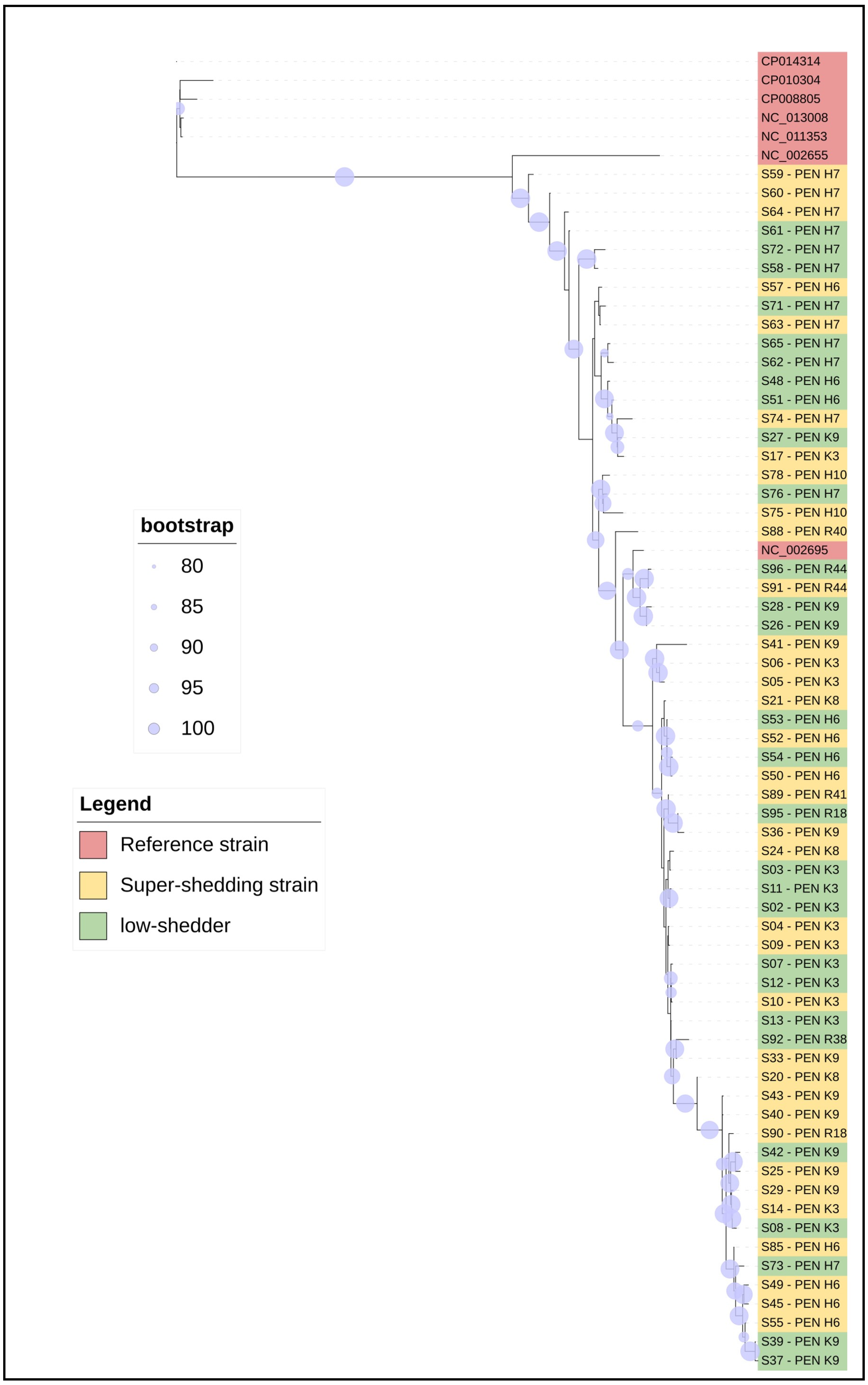
2.1. In Silico Detection of Serotype, Multi-Locus Sequence Typing (MLST), Virulence Genes, Insertion Sites, Lineage, and Clade Profile in STEC O157

All isolates were found to carry the H7 antigen determinant (Supplementary Table S1). All presumptive SS and LS O157 isolates were ST11 (Table 1). Genes encoding Shiga toxin (stx) were found in all STEC O157:H7 genomes. Isolates carried both (stx1/2a, n = 58) or either (stx1a, n = 4 or stx2a n = 1). The stx-encoding prophage insertion sites mrlA and wrbA for stx1a and stx2a, respectively, were occupied, although two isolates had fragments of stx-carrying phage sequences in the mrlA and wrbA loci without stx1a and stx2a. The plasmid-encoded hemolysin subunit (ehxA), the chromosomally encoded intimin (eae), the translocated intimin receptor (tir), and the anti-terminator Q933 genes were present in all isolates (Table 1). Additionally, fimH and fumC, genes involved in cellular adhesion, were also present and similar in all isolates (data not shown).
All the strains screened for lineage-specific polymorphism assay (LSPA-6) were 111111, lineage I (Table 1). Of the 63 isolates, 61 and 2 were identified as clades 1 and 8, respectively. The stx subtypes and phage insertion sites of STEC O157 were similar to those of reference strains Sakai and EDL933 (Table 2).

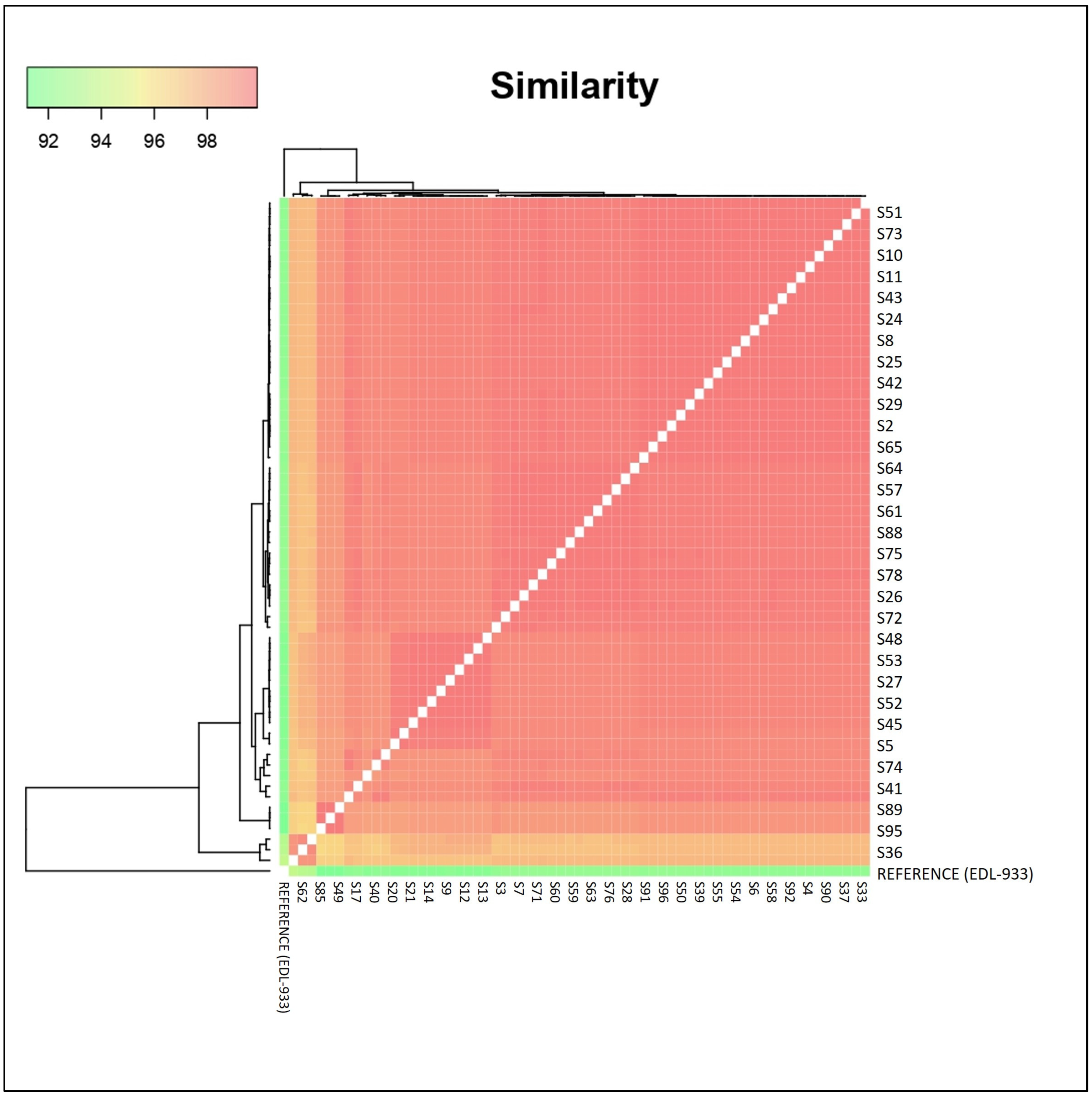
2.2. Cluster Analysis between Presumptive Super- and Low-Shedder O157 Strains

Phylogenetic analysis revealed that STEC O157 isolates were distinct from each other and distantly related to most reference strains, although the Sakai reference strain was closely related to strains from a single pen at two feedlots (Figure 1). In addition to the Sakai cluster, other closely related O157 isolates were found across feedlots, including a cluster that contained isolates from each of the three feedlots. Although there were some closely related strains across feedlots, pens within a feedlot were by no means homogenous and there was not a single closely related dominant strain shared by all SS and LS within feedlot pens sharing the same environment, management, and sampling date.
Additionally, we assembled a phylogenetic tree using SNPs of the isolates in order to see the single-nucleotide differences. The phylogenetic tree (Figure 2) identified 728 SNPs, 44 multiple nucleotide polymorphisms, 47 insertions, and 49 deletions across the evaluated genomes.
Finally, we plotted a graph depicting the pairwise distances between the isolates to facilitate their comparison (Figure 3). Genomic similarity of isolates ranged between 92 and 100%, with all > 96% with the exception of the EDL-933 reference strain.

3. Discussion

This study aimed to identify genomic characteristics of STEC O157 isolates from cattle based on shedding status determined at a single sampling to determine the phylogenetic relationships among reference outbreak, SS, and LS strains. STEC O157 isolates were collected from three different commercial feedlots at different times and years and the majority of isolates carried both stx1a and stx2a. Stx1a and stx2a are the most dominant stx subtypes from cattle and human clinical isolates, respectively, in Alberta [9,10], and stx2a is often associated with HUS [39,40]. This emphasizes the importance of these subtypes in bovine sources regarding cross-contamination in the food processing industry. The stx2a subtype, which is prophage-encoded, is mostly associated with PT21/28 [41], an important SS phenotype. This subtype was shared in all but four isolated colonies of STEC O157 in the present study. Additionally, all stx-encoding prophage insertion sites, mrlA and wrbA for stx1a and stx2a, respectively, were occupied, which corroborates other findings [10,42]. However, two isolates had fragments of an stx-carrying phage in mrlA and wrbA loci without stx1a and stx2a, suggesting that the stx gene has been lost, similar to another study [43]. Therefore, prophage regions are potential locations for bacterial genome evolution through loss or acquisition of new genetic material that can alter the virulence of the strain and result in rapid divergence among STEC O157.
Bacterial adherence genes (eae and tir), plasmid-encoded enterohaemolysin (ehxA), and the anti-terminator Q933 gene, which has been linked to increased toxin production [44], were present in all the isolates. Moreover, the carriage of eae indicates that these strains are not only STEC, but they belong to the enterohemorrhagic (EHEC) pathotype. Similarly, fumC and fimH were present and alleles were identical in all isolates, in contrast to another recent study [45] where these genes were useful for distinguishing clonal sources for on-farm transmission of E. coli. These observations, whereby STEC O157 isolates displayed limited variation regarding insertion sites, virulence, and housekeeping gene patterns, support the idea that SS may be an occasional event within cattle that helps to maintain the STEC pool in its bovine reservoir [46], but that it is not strain-dependent. Additionally, the molecular markers evaluated in the present study, some of which were important for virulence of STEC O157, were not sufficient to distinguish SS from LS strains. Similarly, a previous study comparing the genomics of two SS isolates also found them to be highly diverse [28]. Although the virulence repertoire of all SS and LS strains in the present study was similar, additional analyses revealed more genomic diversity.
Different subpopulations of STEC O157 have been grouped into two main lineages, I and II, with lineage I more prevalent among human clinical and bovine sources and lineage II more associated with cattle [31]. All STEC O157 strains in the present study were classified to lineage I, consistent with other studies indicating that lineage I is most prevalent in Alberta and is also associated with bovine sources [9,47], or both clinical and cattle sources [48]. It is thus plausible that persistent isolates from cattle and human origin may be circulating in Alberta, or that cattle are an important source of infection through SS and cross-contamination. Given that environmental conditions such as weather can contribute to SS and that the shedding status of a host can change, sometimes within a single day [16,17,19], associating genomic features with isolates may only assist in identifying closely related strains with similar virulence traits, not necessarily a SS genotype.
STEC O157 strains belonging to clade 8 have been more commonly associated with HUS patients than other clades [33]. Interestingly, 2 of 63 isolates we analyzed were identified as members of clade 8. Although the two isolates, each from a SS or LS source, had similar virulence features to clade 1 isolates, it is apparent that these two isolates are distinct at the genome level based on the clade profile and may offer fitness advantages associated with this clade [49]. The two clade 8 isolates had the stx2a-carrying prophage inserted within the wrbA insertion site. In contrast, a clade 8 SS strain JEONG-1266 [32] and other clade 8 strains, from a recent study [42], contained the stx2a-encoding prophages inserted at argW loci. Selection of prophage insertion sites may generate diversity that can be used to further segregate STEC O157 from different geographic regions or environmental sources [50]. However, in the present study, even though isolates were collected from multiple feedlots and over multiple years, all originated within 200 km of each other, demonstrating consistent prophage insertion sites for STEC O157.
Although two of the presumed LS isolates and a single SS isolate subclustered with the Sakai reference strain, phylogenetic analysis for the core genome (Figure 1) revealed that many of the STEC O157 isolates were distinct from each other and only distantly related to other reference strains. Furthermore, this observation is supported by Figure 2 and Figure 3. Despite the SNP phylogenetic tree displaying three major clusters, it did not show a distinct segregation of SS and LS strains. All feedlots sampled were within southern Alberta and the presence of closely related strains across feedlots is evidence of shared sources of cattle and common trucking firms used to transport cattle to and from the feedlots [51]. Nonetheless, Figure 2 illustrates that minor variations among feedlots can be discerned via SNP analysis, as the three clusters tend to group strains from comparable feedlots. This suggests a potential approach for tracing the origins of contaminated food products or pinpointing sources in foodborne outbreaks back to their production locations. Within commercial feedlot pens, which would share the same environment, management, and sampling date, results of this study demonstrated considerable phylogenetic diversity in SS and LS isolates, even though carriage of major virulence factors was consistent. The relative importance of the farm/feedlot and the pen within the feedlot in the transmission of STEC has varied across studies [17,18,19,52]. As the feedlot pens sampled within this study contained up to 350 cattle, genetic relationships among LS and SS isolates of STEC O157 were complex and indicate heterogeneity in the distribution of STEC O157 within the same animal reservoir. Closely related strains were present in pens and within feedlots, but pens with a single dominant strain were limited to those where only one SS was detected. Additionally, the heterogeneity of LS and SS isolated within a pen of cattle demonstrates a lack of influence of SS animals on the carriage of STEC O157 within the pen, in opposition to earlier theories that SS animals were responsible for transmission of 80% of the STEC O157 in the environment [12].

4. Conclusions

Conserved genetic trait(s) that can distinguish SS strains were not determined in the present study and may not exist due to the heterogeneity of STEC O157, even in the same feedlot pen. The number of STEC O157 shed may be less important than the ability of these strains to persist in the environment, something that was not possible to determine using the “snapshot in time” approach of the current study necessitated by logistical and budgetary constraints. The STEC O157 isolated within a pen and across feedlots shared the same virulence markers, lineage, and clade profiles as the reference outbreak strains, indicating the threat of these cattle-derived strains to food safety. However, strain differences at the phylogenetic level within the same feedlot pen may increase the difficulty of on-farm STEC control and help to explain the previous variable efficacy of on-farm control measures. Additional studies to evaluate host-related factors influencing SS are warranted.

5. Materials and Methods

5.1. Strains Selection, DNA Isolation, Genome Sequencing, and Quality Control

Sixty-three STEC O157 strains previously isolated [53,54] in our laboratory from cattle with shedding status based on a single fecal sample collected by digital retrieval (28 LS and 35 SS) were selected for analyses based on detection of SS and LS in the same pen (Supplementary Table S1). Isolates were collected from 11 pens in three commercial feedlots in southern Alberta over a seven-year period, from 2007 to 2013. Super-shedders were identified from fecal samples with >104 CFU/g of STEC O157 without enrichment, while LS were detected only after 18 h enrichment using immunomagnetic separation [53]. For resuscitation, strains on glycerol stock (TSB broth and glycerol) at −80 °C were defrosted and reactivated by streaking onto MacConkey agar. A single colony was inoculated into 9 mL of TSB broth over night with shaking. One milliliter of overnight culture was used to extract DNA using the Qiagen Blood and Tissue extraction kit (Qiagen Inc., Toronto, ON, Canada) according to the manufacturer’s instructions. The quality and quantity of the DNA was assessed using Nanodrop and Qubit 2.0 fluorometric systems (Invitrogen Inc., Burlington, ON, Canada), respectively. Samples were sent to Genome Quebec for short-read, 150-base-paired end sequencing (Illumina NovaSeq 6000 platform, Illumina Inc., San Diego, CA, USA; sequencing depth > 300 X.) and the University of Lethbridge for long-read sequencing (PromethION Nanopore system, Nanopore Inc., Montreal, QC, Canada; 8 kpb). The quality of the genomic sequence data was assessed using the FASTQC tool (Version 0.12.1).

5.2. Hybrid Genome Assembly

Short read raw data were trimmed using the Trimmomatic tool (version 0.38). Sliding window trimming was conducted with parameters specifying an average across 4 bases, and an average quality threshold set at 20 to remove the Illumina adapters. For generating hybrid assemblies using the Unicycler tool (Version 0.5.0), the pair-end reads from the sequencing of short reads were used along with the Flye-assembled [55] contigs from long-read sequencing. The parameters used were as follows: bridging mode set to normal (moderate contig size and mis-assembly rate), exclusion of contigs from the FASTA file shorter than 100 bp, the lowest k-mer size for SPAdes assembly set at 0.2 times the read length, the highest k-mer size for SPAdes assembly set at 0.95 times the read length, 10 k-mer steps utilized in SPAdes assembly, and filtering out contigs with a depth lower than 0.25 times the chromosomal depth.
The resulting hybrid-assembled contigs of genomes were obtained in FASTA format. The assembled genomes were annotated using Prokka (Version 1.14.6). The parameters employed included a minimum contig size set at 200 bp and a similarity e-value cut-off of 106. The FASTA files were also used for virulence (Abricate Version 1.0.1; E. coli_VF database), and Multi-locus Sequencing Typing analyses (bio.tools: mlst Version 2.22.0). All assemblies are available in BioProject PRJNA1038721 on the NCBI database.

5.3. In Silico Identification of Serogroup, Multilocus Sequence Typing (MLST), Insertion Sites, and Virulence Genes

The EcOH database was used for the determination of the E. coli serogroup [56], whereas the seven housekeeping gene loci (adk, fumC, gyrB, icd, mdh, purA, and recA) were used for MLST [57]. The O157 E. coli MLST database provided the sequence types (STs) for STEC isolates. Stx-encoding prophage insertion sites were determined as previously described [10]. The 1311 bp gene sequence of stx2-carrying prophage integrase inserted at the NADH quinone oxidoreductase (wrbA) loci (579 bp) in the Sakai strain (NC002695) was extracted and searched for against O157 strains using Geneious prime 2023 (https://www/geneious.com (accessed on 5 July 2023)). The flanking loci were identified as wrbA in O157 isolates. Similarly, a 1287 bp stx1-carrying prophage integrase site flanking the transcriptional activator (mlrA) (648 bp) insertion site in Sakai was obtained and verified against the O157 isolates. The presence of an integrase–insertion site combination was defined as an occupied site. The stx gene in the occupied prophage sequence was further verified by a subsequent sequence search using Geneious. The absence of a stx gene in the prophage sequence or in the entire genome was classified as loss of the stx gene. As mlrA and wrbA insertion sites were all occupied, other insertion sites such as argW, yecE, and sbcB were not further characterized. For virulence analysis, we used the Virulence Finder database with default settings: coverage ≥ 60% and identity ≥ 90% (https://cge.cbs.dtu.dk/services/VirulenceFinder, accessed on 28 August 2023) [58].

5.4. In Silico Detection of Anti-Terminator Q933 Gene, Lineage-Specific Polymorphism Assay (LSPA-6), and Clade Genotyping using Geneious Prime

The genome sequence of O157 isolates was screened for the anti-terminator Q933 gene using the primer sequence [44]. The anti-terminator Q genes located downstream and upstream of the stx1 and stx2 genes, respectively, were considered positive. LSPA-6 analyses were performed using six pairs of primers that target the genomic loci fold-sfmA, Z5935, yhcG, rtcB, rbsB, and arp-iclR [31]. A six-digit binary code was used to determine strains with LSPA-6 profile 111111 as lineage I. Clades 1 and 8 were identified using two primer combinations rhsA-C plus rhsA-Sakai, and rhsA-C plus rhsA-8, respectively [59,60]. A 578 bp gene sequence was considered positive for both clades. Reference strains Sakai (NC002695) and JEONG-1266 (CP014314; a super-shedder bovine strain) [37] were used as representative strains for clades 1 and 8, respectively.

5.5. Cluster Analysis between Presumptive Super- and Low-Shedder O157 Strains

After obtaining the annotated data through Prokka, we performed a core-genome alignment using gff3 format files. Thus, we selected all annotated hybrids and used the Roary tool (Version 3.13.0) to create the core genome. For this, we used a minimum percentage identity of 95% and a percentage of core-gene isolates of 10%. The parameters utilized were a maximum number of clusters set at 50,000. The core-genome alignment was obtained in FASTA format, and the file was used to build a phylogenetic tree to understand the genetic relationship between the isolates. Therefore, we used the IQ-Tree to build a genetic tree of the relationship between SS and LS with an ultrafast bootstrap value (n = 1000). The .nhx file was used for better visualization of the genetic tree using ITOL version 6.8.1. TW14359; NC013008.1 [36], EC4115.1; NC011353.1 [38], Sakai; NC002695.2 [6], EDL933; CP008957.1 [35] and SS17; CP008805.1 [29], SS52; CP010304.1; [28], JEONG-1266; CP014314 [37], which are outbreak and SS strains, were used as reference strains.

5.6. SNP Tree and Pairwise Comparison

We conducted a phylogenetic analysis using the SNIPPY software (Version 4.6.0) to pinpoint genomic variations. Our reference for mapping the isolates was E. coli O157:H7 strain EDL-933 (GCA_000732965.1). Parameters used were: Minimum mapping quality = 60; Minimum coverage = 10; Minimum proportion for variant evidence = 0.9. The SNIPPY direction file’s output was employed to create the SNIPPY-CORE, an alignment of the SNPs obtained across multiple sequences. This data was then utilized to construct the phylogenetic tree using the IQ-Tree software (Version 2.1.2), applying a bootstrap value of 1000 for robustness. The resulting tree visualization was refined using the ITOL version 6.8.1 software.
For the pairwise genomic comparison, we employed the SNP distance matrix pipeline (snp dists, Version 0.8.2), which enabled us to calculate distances between each SNP in the isolates and to generate a corresponding data matrix. We then analyzed this matrix using the ggplot2 tool.

Supplementary Materials

The following supporting information can be downloaded at: https://www.mdpi.com/article/10.3390/toxins16020086/s1, Table S1: List of STEC O157 isolates. Table S2: Quality data of hybrid sequences. Table S3: SPNs and INDELs presence in sequence data.

Author Contributions

Conceptualization, E.W.B., V.S.C., T.A., T.A.M., R.Z. and K.S.; methodology, E.W.B. and V.S.C.; software, E.W.B. and V.S.C.; validation, E.W.B., V.S.C., T.A., T.A.M., R.Z. and K.S.; formal analysis, E.W.B. and V.S.C.; investigation, E.W.B. and V.S.C.; data curation, E.W.B. and V.S.C.; writing—original draft preparation, E.W.B.; writing—review and editing, E.W.B., V.S.C., T.A., T.A.M., R.Z., L.L.G. and K.S.; supervision, T.A., T.A.M., R.Z. and K.S.; funding acquisition, K.S. and L.L.G. All authors have read and agreed to the published version of the manuscript.

Funding

This research was funded by the Results-Driven Agricultural Research (RDAR) project 2021R1014R and Beef Cattle Research Council FOS.07.17.

Institutional Review Board Statement

Not applicable.

Informed Consent Statement

Not applicable.

Data Availability Statement

Data is available in BioProject PRJNA1038721 on the NCBI database.

Acknowledgments

We express our gratitude to Allison McNaughton for her technical support in DNA extraction.

Conflicts of Interest

The authors declare no conflicts of interest.

References

  1. AHS. E. coli Outbreak-Latest Updates. Available online: https://www.albertahealthservices.ca/topics/Page18601.aspx#sept22 (accessed on 23 September 2023).
  2. Kenny, B.; DeVinney, R.; Stein, M.; Reinscheid, D.J.; Frey, E.A.; Finlay, B.B. Enteropathogenic E. coli (EPEC) transfers its receptor for intimate adherence into mammalian cells. Cell 1997, 91, 511–520. [Google Scholar] [CrossRef] [PubMed]
  3. Scheutz, F.; Teel, L.; Beutin, L.; Piérard, D.; Buvens, G.; Karch, H.; Mellmann, A.; Caprioli, A.; Tozzoli, R.; Morabito, S. Multicenter evaluation of a sequence-based protocol for subtyping Shiga toxins and standardizing Stx nomenclature. J. Clin. Microbiol. 2012, 50, 2951–2963. [Google Scholar] [CrossRef] [PubMed]
  4. He, X.; Quiñones, B.; McMahon, S.; Mandrell, R.E. A single-step purification and molecular characterization of functional Shiga toxin 2 variants from pathogenic Escherichia coli. Toxins 2012, 4, 487–504. [Google Scholar] [CrossRef] [PubMed]
  5. Hughes, A.C.; Zhang, Y.; Bai, X.; Xiong, Y.; Wang, Y.; Yang, X.; Xu, Q.; He, X. Structural and functional characterization of Stx2k, a New Subtype of Shiga Toxin 2. Microorganisms 2020, 8, 4. [Google Scholar] [CrossRef] [PubMed]
  6. Hayashi, T.; Makino, K.; Ohnishi, M.; Kurokawa, K.; Ishii, K.; Yokoyama, K.; Han, C.-G.; Ohtsubo, E.; Nakayama, K.; Murata, T. Complete genome sequence of enterohemorrhagic Eschelichia coli O157:H7 and genomic comparison with a laboratory strain K-12. DNA Res. 2001, 8, 11–22. [Google Scholar] [CrossRef] [PubMed]
  7. Friedrich, A.W.; Bielaszewska, M.; Zhang, W.-L.; Pulz, M.; Kuczius, T.; Ammon, A.; Karch, H. Escherichia coli harboring Shiga toxin 2 gene variants: Frequency and association with clinical symptoms. J. Infect. Dis. 2002, 185, 74–84. [Google Scholar] [CrossRef] [PubMed]
  8. Persson, S.; Olsen, K.E.; Ethelberg, S.; Scheutz, F. Subtyping method for Escherichia coli Shiga toxin (verocytotoxin) 2 variants and correlations to clinical manifestations. J. Clin. Microbiol. 2007, 45, 2020–2024. [Google Scholar] [CrossRef]
  9. Zhang, P.; Essendoubi, S.; Keenliside, J.; Reuter, T.; Stanford, K.; King, R.; Lu, P.; Yang, X. Genomic analysis of Shiga toxin-producing Escherichia coli O157:H7 from cattle and pork-production related environments. NPJ Sci. Food 2021, 5, 15. [Google Scholar] [CrossRef]
  10. Bumunang, E.W.; Zaheer, R.; Stanford, K.; Laing, C.; Niu, D.; Guan, L.L.; Chui, L.; Tarr, G.A.; McAllister, T.A. Genomic analysis of Shiga toxin-producing E. coli O157 cattle and clinical isolates from Alberta, Canada. Toxins 2022, 14, 603. [Google Scholar] [CrossRef]
  11. Heiman, K.E.; Mody, R.K.; Johnson, S.D.; Griffin, P.M.; Gould, L.H. Escherichia coli O157 outbreaks in the United States, 2003–2012. Emerg. Infect. Dis. 2015, 21, 1293. [Google Scholar] [CrossRef]
  12. Matthews, L.; Low, J.; Gally, D.; Pearce, M.; Mellor, D.; Heesterbeek, J.; Chase-Topping, M.; Naylor, S.; Shaw, D.; Reid, S. Heterogeneous shedding of Escherichia coli O157 in cattle and its implications for control. Proc. Natl. Acad. Sci. USA 2006, 103, 547–552. [Google Scholar] [CrossRef]
  13. Kempf, F.; La Ragione, R.; Chirullo, B.; Schouler, C.; Velge, P. Super shedding in enteric pathogens: A review. Microorganisms 2022, 10, 2101. [Google Scholar] [CrossRef]
  14. Brusa, V.; Restovich, V.; Galli, L.; Arias, R.; Linares, L.; Costa, M.; Ruiz Diaz, V.; Pugin, D.; Leotta, G. Reduction of Shiga toxin-producing Escherichia coli in a beef abattoir. Food Sci. Technol. Int. 2022, 28, 50–59. [Google Scholar] [CrossRef] [PubMed]
  15. Callaway, T.R.; Eddrington, T.S.; Loneragan, G.H.; Carrr, M.A.; Nisbet, D.J. Shiga toxin producing Escherichia coli (STEC) ecology in cattle and management-based options to reduce fecal shedding. Agric. Food Analy. Bacteriol. 2013, 3, 39–69. [Google Scholar]
  16. Munns, K.D.; Selinger, L.B.; Stanford, K.; Guan, L.; Callaway, T.R.; McAllister, T.A. Perspectives on super-shedding of Escherichia coli O157:H7 by cattle. Foodborne Pathog. Dis. 2015, 12, 89–103. [Google Scholar] [CrossRef]
  17. Spencer, S.E.; Besser, T.E.; Cobbold, R.N.; French, N.P. ‘Super’ or just ‘above average’? Supershedders and the transmission of Escherichia coli O157:H7 among feedlot cattle. J. R. Soc. Interface 2015, 6, 0446. [Google Scholar] [CrossRef]
  18. Beauvais, W.; Gart, E.V.; Bean, M.; Blanco, A.; Wilsey, J.; McWhinney, K.; Bryan, L.; Krath, M.; Yang, C.Y.; Manriquez Alvarez, D.; et al. The prevalence of Escherichia coli O157:H7 fecal shedding in feedlot pens is affected by the water-to-cattle ratio: A randomized controlled trial. PLoS ONE 2018, 7, e0192149. [Google Scholar] [CrossRef] [PubMed]
  19. Venegas-Vargas, C.; Henderson, S.; Khare, A.; Mosci, R.E.; Lehnert, J.D.; Singh, P.; Ouellette, L.M.; Norby, B.; Funk, J.A.; Rust, S. Factors associated with Shiga toxin-producing Escherichia coli shedding by dairy and beef cattle. Appl. Environ. Microbiol. 2016, 82, 5049–5056. [Google Scholar] [CrossRef]
  20. Robinson, S.; Brown, P.; Wright, E.; Hart, C.; French, N. Quantifying within-and between-animal variation and uncertainty associated with counts of Escherichia coli O157 occurring in naturally infected cattle faeces. J. R. Soc. Interface 2009, 6, 169–177. [Google Scholar] [CrossRef]
  21. Ballem, A.; Goncalves, S.; Garcia-Meniño, I.; Flament-Simon, S.C.; Blanco, J.E.; Fernandes, C.; Saavedra, M.J.; Pinto, C.; Oliveira, H.; Blanco, J. Prevalence and serotypes of Shiga toxin-producing Escherichia coli (STEC) in dairy cattle from Northern Portugal. PLoS ONE 2020, 15, e0244713. [Google Scholar] [CrossRef]
  22. Castro, V.S.; Figueiredo, E.; McAllister, T.; Stanford, K. Farm to fork impacts of super-shedders and high-event periods on food safety. Trends Food Sci. Technol. 2022, 127, 129–142. [Google Scholar] [CrossRef]
  23. Williams, K.; Ward, M.; Dhungyel, O.; Hall, E. Risk factors for Escherichia coli O157 shedding and super-shedding by dairy heifers at pasture. Epidemiol. Infect. 2015, 143, 1004–1015. [Google Scholar] [CrossRef] [PubMed]
  24. Wang, O.; McAllister, T.A.; Plastow, G.; Stanford, K.; Selinger, B.; Guan, L.L. Host mechanisms involved in cattle Escherichia coli O157 shedding: A fundamental understanding for reducing foodborne pathogen in food animal production. Sci. Rep. 2017, 7, 7630. [Google Scholar] [CrossRef] [PubMed]
  25. Chase-Topping, M.E.; McKendrick, I.J.; Pearce, M.C.; MacDonald, P.; Matthews, L.; Halliday, J.; Allison, L.; Fenlon, D.; Low, J.C.; Gunn, G. Risk factors for the presence of high-level shedders of Escherichia coli O157 on Scottish farms. J. Clin. Microbiol. 2007, 45, 1594–1603. [Google Scholar] [CrossRef]
  26. Chase-Topping, M.; Gally, D.; Low, C.; Matthews, L.; Woolhouse, M. Super-shedding and the link between human infection and livestock carriage of Escherichia coli O157. Nat. Rev. Microbiol. 2008, 6, 904–912. [Google Scholar] [CrossRef]
  27. Kaper, J.B.; Nataro, J.P.; Mobley, H.L. Pathogenic Escherichia coli. Nat. Rev. Microbiol. 2004, 2, 123–140. [Google Scholar] [CrossRef] [PubMed]
  28. Katani, R.; Cote, R.; Kudva, I.T.; DebRoy, C.; Arthur, T.M.; Kapur, V. Comparative genomics of two super-shedder isolates of Escherichia coli O157:H7. PLoS ONE 2017, 12, e0182940. [Google Scholar] [CrossRef]
  29. Cote, R.; Katani, R.; Moreau, M.R.; Kudva, I.T.; Arthur, T.M.; DebRoy, C.; Mwangi, M.M.; Albert, I.; Raygoza Garay, J.A.; Li, L. Comparative analysis of super-shedder strains of Escherichia coli O157:H7 reveals distinctive genomic features and a strongly aggregative adherent phenotype on bovine rectoanal junction squamous epithelial cells. PLoS ONE 2015, 10, e0116743. [Google Scholar] [CrossRef]
  30. Munns, K.D.; Zaheer, R.; Xu, Y.; Stanford, K.; Laing, C.R.; Gannon, V.P.; Selinger, L.B.; McAllister, T.A. Comparative genomic analysis of Escherichia coli O157:H7 isolated from super-shedder and low-shedder cattle. PLoS ONE 2016, 11, e0151673. [Google Scholar] [CrossRef]
  31. Yang, Z.; Kovar, J.; Kim, J.; Nietfeldt, J.; Smith, D.R.; Moxley, R.A.; Olson, M.E.; Fey, P.D.; Benson, A.K. Identification of common subpopulations of non-sorbitol-fermenting, β-glucuronidase-negative Escherichia coli O157:H7 from bovine production environments and human clinical samples. Appl. Environ. Microbiol. 2004, 70, 6846–6854. [Google Scholar] [CrossRef]
  32. Besser, T.E.; Shaikh, N.; Holt, N.J.; Tarr, P.I.; Konkel, M.E.; Malik-Kale, P.; Walsh, C.W.; Whittam, T.S.; Bono, J.L. Greater diversity of Shiga toxin-encoding bacteriophage insertion sites among Escherichia coli O157:H7 isolates from cattle than in those from humans. Appl. Environ. Microbiol. 2007, 73, 671–679. [Google Scholar] [CrossRef] [PubMed]
  33. Manning, S.D.; Motiwala, A.S.; Springman, A.C.; Qi, W.; Lacher, D.W.; Ouellette, L.M.; Mladonicky, J.M.; Somsel, P.; Rudrik, J.T.; Dietrich, S.E. Variation in virulence among clades of Escherichia coli O157:H7 associated with disease outbreaks. Proc. Natl. Acad. Sci. USA 2008, 105, 4868–4873. [Google Scholar] [CrossRef] [PubMed]
  34. Shaaban, S.; Cowley, L.A.; McAteer, S.P.; Jenkins, C.; Dallman, T.J.; Bono, J.L.; Gally, D.L. Evolution of a zoonotic pathogen: Investigating prophage diversity in enterohaemorrhagic Escherichia coli O157 by long-read sequencing. Microb. Genom. 2016, 2, e000096. [Google Scholar] [CrossRef]
  35. Perna, N.T.; Plunkett, G.; Burland, V.; Mau, B.; Glasner, J.D.; Rose, D.J.; Mayhew, G.F.; Evans, P.S.; Gregor, J.; Kirkpatrick, H.A. Genome sequence of enterohaemorrhagic Escherichia coli O157:H7. Nature 2001, 409, 529–533. [Google Scholar] [CrossRef] [PubMed]
  36. Kulasekara, B.R.; Jacobs, M.; Zhou, Y.; Wu, Z.; Sims, E.; Saenphimmachak, C.; Rohmer, L.; Ritchie, J.M.; Radey, M.; McKevitt, M. Analysis of the genome of the Escherichia coli O157:H7 2006 spinach-associated outbreak isolate indicates candidate genes that may enhance virulence. Infect. Immun. 2009, 77, 3713–3721. [Google Scholar] [CrossRef] [PubMed]
  37. Teng, L.; Lee, S.; Park, D.; Jeong, K.C. Genetic and functional analyses of virulence potential of an Escherichia coli O157:H7 strain isolated from super-shedder cattle. Front. Cell. Infect. Microbiol. 2020, 10, 271. [Google Scholar] [CrossRef]
  38. Eppinger, M.; Mammel, M.K.; Leclerc, J.E.; Ravel, J.; Cebula, T.A. Genomic anatomy of Escherichia coli O157:H7 outbreaks. Proc. Natl. Acad. Sci. USA 2011, 108, 20142–20147. [Google Scholar] [CrossRef]
  39. Naseer, U.; Løbersli, I.; Hindrum, M.; Bruvik, T.; Brandal, L.T. Virulence factors of Shiga toxin-producing Escherichia coli and the risk of developing haemolytic uraemic syndrome in Norway, 1992–2013. Eur. J. Clin. Microbiol. 2017, 36, 1613–1620. [Google Scholar] [CrossRef]
  40. Byrne, L.; Dallman, T.J.; Adams, N.; Mikhail, A.F.; McCarthy, N.; Jenkins, C. Highly pathogenic clone of Shiga toxin–producing Escherichia coli O157:H7, England and Wales. Emerg. Infect. Dis. 2018, 24, 2303. [Google Scholar] [CrossRef]
  41. Fitzgerald, S.F.; Beckett, A.E.; Palarea-Albaladejo, J.; McAteer, S.; Shaaban, S.; Morgan, J.; Ahmad, N.I.; Young, R.; Mabbott, N.A.; Morrison, L. Shiga toxin sub-type 2a increases the efficiency of Escherichia coli O157 transmission between animals and restricts epithelial regeneration in bovine enteroids. PLoS Pathog. 2019, 15, e1008003. [Google Scholar] [CrossRef]
  42. Henderson, S.T.; Singh, P.; Knupp, D.; Lacher, D.W.; Abu-Ali, G.S.; Rudrik, J.T.; Manning, S.D. Variability in the occupancy of Escherichia coli O157 integration sites by Shiga toxin-encoding prophages. Toxins 2021, 13, 433. [Google Scholar] [CrossRef]
  43. Bielaszewska, M.; Prager, R.; Köck, R.; Mellmann, A.; Zhang, W.; Tschäpe, H.; Tarr, P.I.; Karch, H. Shiga toxin gene loss and transfer in vitro and in vivo during enterohemorrhagic Escherichia coli O26 infection in humans. Appl. Environ. Microbiol. 2007, 73, 3144–31550. [Google Scholar] [CrossRef]
  44. Ahmad, A.; Zurek, L. Evaluation of the anti-terminator Q933 gene as a marker for Escherichia coli O157:H7 with high Shiga toxin production. Curr. Microbiol. 2006, 53, 324–328. [Google Scholar] [CrossRef]
  45. Oliveira, G.S.; Lentz, S.A.M.; Wink, P.L.; Martins, A.F. Molecular typing of mcr-1 Escherichia coli isolates from pigs and farm environment based on fumC and fimH alleles. Future Microbiol. 2023, 18. [Google Scholar] [CrossRef] [PubMed]
  46. Stanford, K.; Agopsowicz, C.A.; McAllister, T.A. Genetic diversity and antimicrobial resistance among isolates of Escherichia coli O157:H7 from feces and hides of super-shedders and low-shedding pen-mates in two commercial beef feedlots. BMC Vet. Res. 2012, 8, 178. [Google Scholar] [CrossRef]
  47. Pianciola, L.; Rivas, M. Genotypic features of clinical and bovine Escherichia coli O157 strains isolated in countries with different associated-disease incidences. Microorganisms 2018, 6, 36. [Google Scholar] [CrossRef]
  48. Sharma, R.; Stanford, K.; Louie, M.; Munns, K.; John, S.J.; Zhang, Y.; Gannon, V.; Chui, L.; Read, R.; Topp, E. Escherichia coli O157:H7 lineages in healthy beef and dairy cattle and clinical human cases in Alberta, Canada. J. Food Prot. 2009, 72, 601–607. [Google Scholar] [CrossRef]
  49. Ogura, Y.; Mondal, S.I.; Islam, M.R.; Mako, T.; Arisawa, K.; Katsura, K.; Ooka, T.; Gotoh, Y.; Murase, K.; Ohnishi, M. The Shiga toxin 2 production level in enterohemorrhagic Escherichia coli O157:H7 is correlated with the subtypes of toxin-encoding phage. Sci. Rep. 2015, 5, 16663. [Google Scholar] [CrossRef] [PubMed]
  50. Yara, D.A.; Greig, D.R.; Gally, D.L.; Dallman, T.J.; Jenkins, C. Comparison of Shiga toxin-encoding bacteriophages in highly pathogenic strains of Shiga toxin-producing Escherichia coli O157:H7 in the UK. Microb. Genom. 2020, 6, e000334. [Google Scholar] [CrossRef] [PubMed]
  51. Dewell, G.A.; Simpson, C.A.; Dewell, R.D.; Hyatt, D.R.; Belk, K.E.; Scanga, J.A.; Morley, P.S.; Grandin, T.; Smith, G.C.; Dargatz, D.A.; et al. Impact of transportation and lairage on hide contamination with Escherichia coli O157 in finished beef cattle. J. Food Protect. 2008, 71, 1114–1118. [Google Scholar] [CrossRef]
  52. Browne, A.S.; Midwinter, A.C.; Withers, H.; Cookson, A.L.; Biggs, P.J.; Marshall, J.C.; Benschop, J.; Hathaway, S.; Rogers, L.; Nisa, S. Transmission dynamics of Shiga toxin-producing Escherichia coli in New Zealand cattle from farm to slaughter. Appl. Environ. Microbiol. 2021, 87, e02907–e02920. [Google Scholar] [CrossRef] [PubMed]
  53. Stephens, T.; McAllister, T.; Stanford, K. Perineal swabs reveal effect of super shedders on the transmission of Escherichia coli O157:H7 in commercial feedlots. J. Anim. Sci. 2009, 87, 4151–4160. [Google Scholar] [CrossRef] [PubMed]
  54. Stanford, K.; Hannon, S.; Booker, C.W.; Jim, G.K. Variable efficacy of a vaccine and direct-fed microbial for controlling Escherichia coli O157:H7 in feces and on hides of feedlot cattle. Foodborne Pathog. Dis. 2014, 11, 379–387. [Google Scholar] [CrossRef] [PubMed]
  55. Kolmogorov, M.; Bickhart, D.M.; Behsaz, B.; Gurevich, A.; Rayko, M.; Shin, S.B.; Kuhn, K.; Yuan, J.; Polevikov, E.; Smith, T.P. metaFlye: Scalable long-read metagenome assembly using repeat graphs. Nat. Methods 2020, 17, 1103–1110. [Google Scholar] [CrossRef] [PubMed]
  56. Ingle, D.J.; Valcanis, M.; Kuzevski, A.; Tauschek, M.; Inouye, M.; Stinear, T.; Levine, M.M.; Robins-Browne, R.M.; Holt, K.E. In silico serotyping of E. coli from short read data identifies limited novel O-loci but extensive diversity of O:H serotype combinations within and between pathogenic lineages. Microb. Genom. 2016, 2, e000064. [Google Scholar] [CrossRef]
  57. Bessonov, K.; Laing, C.; Robertson, J.; Yong, I.; Ziebell, K.; Gannon, V.P.; Nichani, A.; Arya, G.; Nash, J.H.; Christianson, S. ECTyper: In silico Escherichia coli serotype and species prediction from raw and assembled whole-genome sequence data. Microb. Genom. 2021, 7, 000728. [Google Scholar] [CrossRef]
  58. Chen, L.; Yang, J.; Yu, J.; Yao, Z.; Sun, L.; Shen, Y.; Jin, Q. VFDB: A reference database for bacterial virulence factors. Nucleic Acids Res. 2005, 33, D325–D328. [Google Scholar] [CrossRef]
  59. Malberg Tetzschner, A.M.; Johnson, J.R.; Johnston, B.D.; Lund, O.; Scheutz, F. In silico genotyping of Escherichia coli isolates for extraintestinal virulence genes by use of whole-genome sequencing data. J. Clin. Microbiol. 2020, 58, e01269-20. [Google Scholar] [CrossRef]
  60. Hartzell, A.; Chen, C.; Lewis, C.; Liu, K.; Reynolds, S.; Dudley, E.G. Escherichia coli O157:H7 of genotype lineage-specific polymorphism assay 211111 and clade 8 are common clinical isolates within Pennsylvania. Foodborne Pathog. Dis. 2011, 8, 763–768. [Google Scholar] [CrossRef]
Figure 1. The core genome tree is shown. STEC O157 isolates belonging to super shedder, low shedder, and reference strains are listed in green, yellow, and red, respectively.
Toxins 16 00086 g001
Figure 2. SNP phylogenetic tree. Isolates of STEC O157 associated with super shedders, low shedders, and reference strains are color-coded in green, yellow, and red, respectively, for easy identification. Strains belonging to clade 8 are distinctly marked with a double asterisk. The scale provided illustrates the level of similarity among the isolates, quantifying it as a percentage to denote the extent of genetic resemblance. Additionally, the bootstrap values, which are above 80%, are indicated by circles at each leaf node of the phylogenetic tree, emphasizing the statistical confidence in the tree’s branching structure.
Toxins 16 00086 g002
Figure 3. A pairwise comparison of genomic similarity among the isolates. The intensity of the red color indicates a greater genomic closeness between the isolates.
Toxins 16 00086 g003
Table 1. Distribution of virulence, lineage, clade profile, insertion sites, and other genes among STEC O157:H7 isolates.
Number of IsolatesSourceMLSTLineageCladeInsertion Sitestx SubtypeeaetirehxAQ933
33SS11I1mlrAwrbAStx1a/2a++++
1SS8mlrAwrbAStx 1a/2a++++
1SS1mlrA-Stx 1a++++
24LS1mlrAwrbAStx 1a/2a++++
1LS8mlrAwrbAStx 1a/2a++++
1LS1mlrA-Stx 1a++++
1LS1mlrA *wrbAStx 2a++++
1LS1mlrAwrbA *Stx 1a++++
SS; super shedding, LS; low shedding, *; locus present but stx gene absent, +; gene present, -; gene absent.
Table 2. Distribution of lineage, clade profile, stx subtype, and insertion sites of reference O157 strains.
Strain IDSourceLineageCladestx SubtypeInsertion SiteAccession NumberYear
Isolated
References
SakaiClinicalI1Stx1a/2amlrAwrbANC002695.21996[6]
EDL933Ground beefI3Stx1a/2amlrAwrbACP008957.11982[35]
TW14359ClinicalI/II8Stx2a/2c-argWNC013008.12006[36]
JEONG-1266CattleI/II8Stx2a/2c-argWCP0143142011[37]
SS17CattleI/II8Stx2a/2c-argWCP008805.12009/10[29]
SS52CattleI/II----CP010304.12009/10[28]
EC4115.1SpinachI/II8Stx2a/2c-argWNC011353.12006[38]
Disclaimer/Publisher’s Note: The statements, opinions and data contained in all publications are solely those of the individual author(s) and contributor(s) and not of MDPI and/or the editor(s). MDPI and/or the editor(s) disclaim responsibility for any injury to people or property resulting from any ideas, methods, instructions or products referred to in the content.

