Article

Novel Reoviruses of Waterfowl Origin in Northern Vietnam: A Laboratory Investigation

by
Thi-Ngoc Vu
,
Van-Truong Le
,
Thi-Bich-Phuong Cao
,
Van-Giap Nguyen
* and
Thi-My-Le Huynh
*
Department of Veterinary Microbiology and Infectious Diseases, Faculty of Veterinary Medicine, Vietnam National University of Agriculture, Hanoi 100000, Vietnam
*
Authors to whom correspondence should be addressed.
Microbiol. Res. 2025, 16(8), 187; https://doi.org/10.3390/microbiolres16080187
Submission received: 3 July 2025 / Revised: 6 August 2025 / Accepted: 9 August 2025 / Published: 12 August 2025

Abstract

Novel waterfowl reoviruses (nWRVs) have been reported in several countries, but their circulation and genetic characteristics in Vietnam remain poorly understood. In this study, we investigated nWRVs in northern Vietnam through molecular detection, virus isolation, experimental infection in ducklings, and molecular analysis of the sigma C-encoding (sC) gene. We also applied immunoinformatic tools to explore the antigenic and structural features of the sC protein. nWRVs were detected in 15.6% of tested samples across ten provinces. Three isolates were successfully recovered, all showing a characteristic cytopathic effect—syncytium formation—in Vero cells. When tested in ducklings (n = 72), the isolates caused disease of varying severity, but all induced characteristic gross and microscopic lesions, particularly ecchymotic hemorrhages and large necrotic foci in the liver and spleen. Phylogenetic analysis based on sC sequences placed the Vietnamese isolates (n = 14) within the nWRV clade, with evidence of two genetically distinct groups. Our immunoinformatic analysis identified four predicted B-cell epitopes located in the head and body domains of the sC protein, with little variation.

1. Introduction

Waterfowl reoviruses (WRVs) have been recognized since the early 1980s, with their pathogenicity experimentally demonstrated and published in 1981 [1]. To date, outbreaks caused by WRVs have been reported in Europe and Asia [2,3,4]. For a long period, the disease in waterfowl was mainly described in Muscovy ducks and geese, with typical gross lesions such as focal necrosis in the liver and spleen [1,2,3]. It was not until 2006 that a novel reovirus was reported to cause disease in ducks [5,6]. This novel reovirus also possesses an expanded range of susceptible species, as the disease naturally occurred in Pekin ducks, Cherry Valley ducks, Muscovy ducks, Sheldrake ducks, wild mallards, geese, and others [5,7,8,9,10]. Affected animals had predominant gross lesions that were not only large necrotic foci but also hemorrhagic in the liver and spleen [4,11,12,13,14]. Recently, researchers in several countries have identified other novel waterfowl-associated reoviruses, proposed as a new species named Avian orthoreovirus B. The viruses have been isolated from Pekin ducks with clinical features totally different from those mentioned above [15,16,17]. These observed variations in clinical presentation and host range are now understood to correlate with distinct genomic and molecular characteristics of WRVs.
WRVs are classified within the species Avian orthoreovirus (ARV), genus Orthoreovirus, family Spinareoviridae [18]. Regardless of host origin, they are currently grouped into the classical genotype (ARV-Wa-1 or cWRV) and the novel genotype (ARV-Wa-2 or nWRV) [15,19,20,21]. All genotypes share a fundamental genomic feature: a segmented genome composed of 10 linear double-stranded RNA segments [22], categorized by size into Large (L1–L3), Medium (M1–M3), and Small (S1–S4) segments [2,17,19,21,22]. While most segments are monocistronic, segments S1 and S4 are polycistronic [22], representing key distinctions between the classical and novel genotypes. Specifically, the S4 segment (1124–1125 bp) of classical reoviruses encodes p10 and sigma C (sC) [19,23,24], whereas the S1 segment (1568 bp) of novel strains encodes p10, p18 and sC [13,21,25]. Among these, sC is a multifunctional protein that serves as a component of the viral outer capsid, mediates cell attachment [26], acts as the primary target of virus-neutralizing antibodies [27], and is capable of inducing apoptosis in vitro [28]. Notably, the gene encoding sC is the most variable [29], contributing to the genetic and antigenic diversity of reoviruses [30]. The other two, p10 and p18, are nonstructural proteins responsible for cell–cell fusion [31,32] and nucleocytoplasmic shuttling [33], respectively.
Despite being classified under a single species Avian orthoreovirus, WRVs exhibit substantial genetic diversity. Their genetic evolution involves mechanisms such as reassortment [12,16,20,34,35,36,37], intra-segmental recombination [13,20,38], as well as nucleotide mutation [39,40]. Interestingly, genetic reassortment events are diverse, occurring (i) between waterfowl-origin and chicken-origin reoviruses or even an unknown reovirus [16,37], (ii) between novel and classical WRVs [20,34,41], and (iii) across all classes of genomic segments (Large, Medium, and Small) that encode both structural and nonstructural proteins [12,20,35].
The aforementioned understanding of reovirus infection in waterfowl, and the virus diversity and evolution, however, is predominantly based on studies conducted in other parts of the world. Vietnam, though possessing a significant waterfowl population [42,43] and a thriving duck industry, has surprisingly few published reports detailing the characteristics of circulating WRV strains. Given the lack of data from Vietnam, this study was designed to investigate the field circulation of nWRV, evaluate its pathogenicity through experimental infection, and analyze the molecular features of endemic strains in Vietnam.

2. Materials and Methods

2.1. Sample Collection

This study collected samples from flocks suspected of reovirus infection, none of which had received the reovirus vaccine. Sampling was conducted between 2023 and 2024. A total of 282 pooled internal organ samples (liver, spleen, and heart) were collected from meat-type ducks and Muscovy ducks. The farms were located across ten northern provinces of Vietnam: Bac Giang, Bac Ninh, Ha Nam, Ha Noi, Hai Duong, Hung Yen, Nam Dinh, Phu Tho, Thai Nguyen, and Vinh Phuc. Metadata associated with each sample included the sampling date, farm code, flock size, species affected, clinical signs, age of animals, and vaccination history against reovirosis.

2.2. Detection of nWRVs by RT-PCR

Pooled organs from each sample were homogenized in 1× PBS to prepare a 10% (w/v) suspension and stored at −80 °C until analysis. Total nucleic acids were extracted using the WizPrep Viral DNA/RNA Mini Kit (W73052, Wizbiosolution, Republic of Korea), following the manufacturer’s instructions. Reverse transcription was performed using the Maxime RT PreMix (Random Primer) kit (25082, iNtRON Biotechnology, Seongnam-si, Gyeonggi-do, Republic of Korea) at 45 °C for 60 min. nWRVs were detected by RT-PCR using primers targeting the sigma C gene, as previously described [44] (Appendix A Table A1). PCR amplification was carried out using the GoTaq Master Mix (M7122, Promega, WI, USA). The thermal cycling conditions were as follows: initial denaturation at 95 °C for 5 min; 40 cycles of 95 °C for 30 s, 59 °C for 30 s, and 72 °C for 30 s; followed by a final extension at 72 °C for 5 min. PCR products were analyzed by 1% agarose gel electrophoresis containing RedSafe Nucleic Acid Staining Solution (21141, iNtRON Biotechnology, Seongnam-si, Gyeonggi-do, Republic of Korea). Positive detection results were visualized using a Sankey diagram (generated with an in-house Python 3.12.0 script) to illustrate the relationships among sampling date, affected species, and animal age.

2.3. Detection of Co-Infecting Viruses

Positive nWRV samples were further screened for the presence of four RNA/DNA viruses: Duck hepatitis A virus (DHAV) genotypes 1 and 3, Avian influenza virus (AIV), Duck Tembusu virus (DTMUV), and Duck circovirus (DuCV). The primer sets used for detecting these viruses followed those reported in previous studies and are listed in Appendix A Table A2. The same PCR kit as used for nWRV detection was applied. Annealing temperatures were based on the recommendations provided in the corresponding publications for each primer set.

2.4. Virus Isolation and Titration

Samples that were nWRV-positive but negative for co-infecting viruses were used for virus isolation. A 10% suspension of tissue homogenates was centrifuged at 4 °C and 12,000 rpm for 15 min, and the supernatant was filtered through a 0.22 µm membrane filter. The filtered supernatant was then inoculated onto duck embryo fibroblast (DEF) cells. These cells were prepared from 9–11-day-old embryonated duck eggs collected from a healthy parent flock with no history of reovirus vaccination. An aliquot of the cell suspension was tested to confirm the absence of WRV and the other co-infecting viruses mentioned above.
Inoculated DEF cells were cultured for 5–7 days at 37 °C with 5% CO2 in Dulbecco’s Modified Eagle Medium (DMEM), supplemented with 1% fetal bovine serum (FBS) (Gibco, 11965092, Grand Island, NY 14072, USA), and monitored daily for cytopathic effect (CPE), characterized by cell rounding and detachment. Cells exhibiting CPE were subjected to three freeze–thaw cycles and centrifuged at 4 °C and 4000 rpm for 15 min. The resulting supernatant was collected for the next passage. The DEF-adapted strains were subsequently passaged on Vero cells (ATCC CCL-81) using the same inoculation method and maintenance medium. Infected cells were monitored daily for CPE, characterized by syncytium formation.
The 50% tissue culture infectious dose (TCID50) of the isolated reovirus strains was determined using the limiting dilution method. The viral suspension was serially diluted 10-fold from 10−1 to 10−8. Each dilution (100 µL) was inoculated into five wells of confluent DEF cells. Inoculated cells were maintained in the same medium and conditions as described above. Final CPE in each inoculated well was recorded after 5 days. The TCID50 was calculated using the Spearman–Kärber method, implemented via an online tool [45] at the address: https://www.virosin.org/tcid50/TCID50.html (last accessed on 30 May 2025).
For determination of the 50% embryo lethal dose (ELD50), each dilution (200 µL) was inoculated into five 9–11-day-old embryonated duck eggs. Embryos were candled daily for 5 days. Embryos that died within 24 h post-inoculation were discarded. The numbers of live and dead embryos at 120 h post-inoculation were recorded for each dilution. The ELD50 was calculated using the Spearman–Kärber method, implemented via an online tool [45].

2.5. Pathogenicity Assessment of Isolated Strains in Ducklings

A total of 72 one-day-old ducklings were used to assess the pathogenicity and infectivity of the isolated reovirus strains. Conventional duck embryos, obtained from a parent flock free of reovirus infection and unvaccinated against reovirus, were incubated in a tabletop egg incubator until hatching. One-day-old ducklings were subcutaneously inoculated in the neck region with 0.5 mL of virus suspension (104 TCID50/mL), with six ducklings assigned to each isolate or control group. Each experimental infection was conducted in triplicate (in August, September, and November 2024). Control ducklings received 0.5 mL of 1× PBS via the same route. All ducklings were monitored daily for clinical signs and mortality over a four-week period. The number of live and dead ducklings was recorded daily and used to plot the survival curves. The log-rank test, implemented via an online tool (https://www.evanmiller.org/ab-testing/survival-curves.html, accessed on 30 May 2025), was applied to compare survival distributions between groups. A p-value ≤ 0.05 was considered statistically significant. Severely ill individuals were humanely euthanized via cervical dislocation. All deceased ducklings were necropsied to evaluate gross lesions. For histopathological examination, organs including the liver, spleen, heart, and lung were collected and fixed in 10% neutral-buffered formalin. After paraffin embedding, tissue sections of 2 µm thickness were prepared and stained with haematoxylin and eosin (H&E). The stained slides were examined under a light microscope using a 40× objective lens.
To evaluate viral distribution in experimentally infected ducklings, various internal organs (liver, spleen, lungs, intestine, kidney, heart, and bursa of Fabricius) were collected separately from ducks that died during post-inoculation observation periods to assess the presence of viral RNA in each organ. For each infected group, organs were pooled by tissue type. A 10% tissue suspension was prepared for total RNA extraction, cDNA synthesis, and subsequent nWRVs RT-PCR detection, as described in Section 2.2. All procedures related to experimental infection and sample collection were approved by the Animal Ethics Committee of the Vietnam National University of Agriculture (Approval No. VNUA-2023/06).

2.6. Genetic Classification Based on the Sigma C-Encoding Gene

The sigma C-encoding gene (sC gene) was chosen to study the genetic classification and variation in nWRVs detected in this study. This gene was amplified by the primer pairs shown in Appendix A Table A3, using the PCR kit mentioned in Section 2.2. Subsequently, PCR products were sequenced by Sanger’s method (Macrogen, Seoul, Republic of Korea). The sequences sC generated in this study were deposited in GenBank (PV696587, PV696589–PV696600).
The genetic relationships of nWRVs in northern Vietnam were inferred in a broad context that included other waterfowl-related reoviruses deposited in GenBank. A codon-based alignment strategy was applied to align the sC gene sequences. Briefly, the translated amino acid sequences of the sC gene were aligned using MAFFT [46] at https://mafft.cbrc.jp/alignment/server/index.html (last accessed 30 May 2025). The PAL2NAL tool [47] was then used to convert the multiple sequence alignment of proteins and their corresponding nucleotide sequences into a codon alignment. The genetic classification of WRVs followed previous studies [17,38]. The phylogenetic relationships between WRVs based on the sigma C gene sequences were reconstructed using IQ-TREE software version 2.4.0 [48]. The data best-fit model of nucleotide substitutions was chosen automatically by specifying the command “-m MFP”. Bootstrap values were calculated using 1000 ultrafast bootstrap replicates, specified by the “-B 1000” command. The dataset used for phylogenetic inference (n = 165) is provided in Supplementary File S1.

2.7. Immunoinformatic Analysis of the Sigma C Protein Amino Acid Sequence of nWRVs

Variations in the amino acid sequence of the sigma C (sC) protein were assessed using a dataset of 94 unique sequences (Supplementary File S2). The Pixel tool (https://www.hiv.lanl.gov/content/sequence/pixel/pixel.html, accessed on 30 May 2025) was used to visualize amino acid differences among the sC protein sequences.
B-cell epitope prediction was conducted using both sequence-based and structure-based approaches. Linear epitopes were identified with BepiPred-3.0 [49] (https://services.healthtech.dtu.dk/services/BepiPred-3.0, accessed on 30 May 2025), while conformational B-cell epitopes were predicted using ElliPro [50] (http://tools.iedb.org/ellipro, accessed on 30 May 2025). For ElliPro, the minimum score threshold was set at 0.7, and the maximum distance at 6 ångströms. Default settings were applied for all other tools. The 3D structure of the sC protein, used as input for ElliPro, was generated via the Phyre2.2 web server [51], in “intensive” modeling mode. WebLogo [52] (https://weblogo.threeplusone.com, accessed on 30 May 2025) was used to visualize amino acid substitution patterns at the predicted epitopes.
To explore the structural implications of sequence variation, the modeled 3D structure was further analyzed using ProteinTools (https://proteintools.uni-bayreuth.de/, accessed on 30 May 2025), focusing on hydrophobic clusters involved in protein folding and stability [53]. The potential impact of amino acid substitutions within these hydrophobic regions on protein stability was assessed using the DDGEmb predictor [54] (https://ddgemb.biocomp.unibo.it/, accessed on 30 May 2025). The tool predicts the ΔΔG value, which represents the change in Gibbs free energy between the wild-type and variant proteins upon both single- and multi-point variations. Five classes are identified: destabilizing variations (ΔΔG < −1), weakly destabilizing variations (−1 ≤ ΔΔG < −0.5), neutral variations (−0.5 ≤ ΔΔG ≤ 0.5), weakly stabilizing variations (0.5 < ΔΔG ≤ 1), and stabilizing variations (ΔΔG > 1) [54]. All web tools were last accessed in May 2025.

2.8. Evolutionary Analysis of the Sigma C-Encoding Gene

The Codeml program in the PAML 4.10.6 package [55] was used to investigate the evolutionary pattern of the complete sC gene of nWRVs (Supplementary File S5). As recommended [56], four site models (M1a, M2a, M7 and M8) were employed. Likelihood ratio tests were then performed for pair M2a vs. M1a and M8 vs. M7 to identify the best-fitting model for inferring codon-specific variation in synonymous (dS) and non-synonymous (dN) substitution rates. The input phylogenetic tree for Codeml was the one inferred using the IQ-TREE program [48].

3. Results

3.1. Detection of nWRVs and Co-Infection Status

Molecular screening of 282 pooled organ samples confirmed the presence of nWRV in 44 samples, yielding an overall prevalence of 15.6%. Positive samples were detected in all ten sampling provinces (Supplementary File S4). Figure 1 shows that, although the detection rate fluctuated, nWRV was consistently present in every month of the two investigation years. Regarding host origin, nWRV was detected in both ducks and Muscovy ducks. Excluding samples with missing age information (highlighted rows in Supplementary File S4), the remaining samples ranged from 4 to 68 days old (corresponding to the 1st to 10th week of age). Positive detections were limited to the 1st to 6th weeks of age, primarily during the brooding period, with a peak at two weeks of age (arrow, Figure 1).
Another notable epidemiological finding was the high rate of co-infection: 27 of the 44 (61.4%) nWRV-positive samples contained at least one additional waterfowl pathogen. Co-infection with DHAV and DuCV (Figure 1B) was particularly common, occurring in 11 and 10 samples, respectively. The presence of DHAV co-infection should be considered in field diagnoses, as this virus is known to cause hemorrhagic liver lesions that resemble those induced by nWRVs.

3.2. Isolation of nWRV Strains

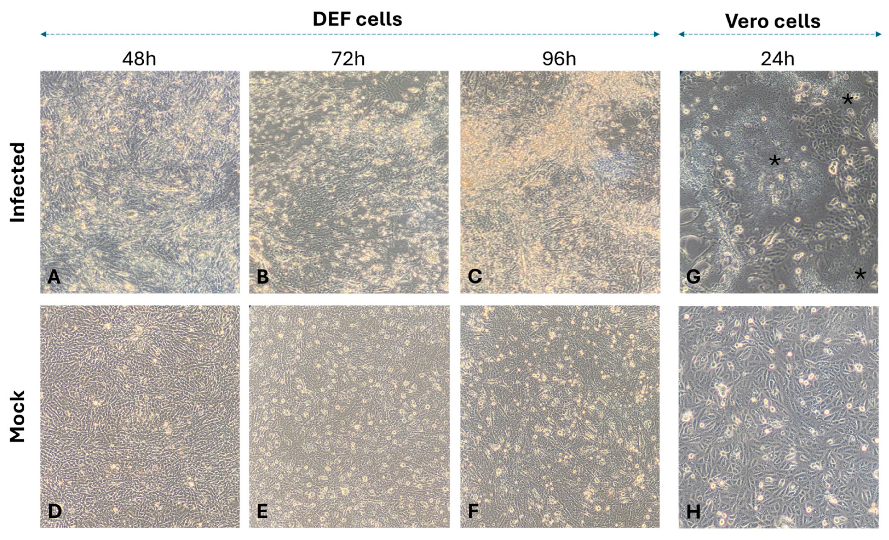
In this study, three reovirus strains (VNUA_07, VNUA_24, and VNUA_101) were successfully isolated in DEF cells. These isolates tested negative for a panel of co-infecting viruses listed in Section 2.3, as well as for duck enteritis virus. After five serial passages, the isolates exhibited adaptation to DEF cells and consistently induced CPE. The observed CPE included cell rounding and lysis, resulting in the detachment of the cell monolayer. CPE became evident at 24–48 h post-infection, intensified at 60–72 h, and peaked at 96–120 h post-infection. The DEF-adapted strains were subsequently passaged on Vero cells. After five passages, syncytium formation (indicated by a star in Figure 2G) was detected as early as 24 h post-infection. Representative images of CPE are shown in Figure 2.
At the fifth passage, the average viral titers of VNUA_101 and VNUA_07 were comparable, at 104.75 TCID50/mL and 104.50 TCID50/mL, respectively. In comparison, the titer of VNUA_24 was 105.30 TCID50/mL, differing by less than one log10 dilution from the other two strains.
The ELD50 titers of the three reovirus strains were also determined. All three DRV strains in this study caused embryo mortality when inoculated into 9-day-old duck embryos, with embryo death occurring within 48 h post-infection. All dead embryos exhibited characteristic lesions, including generalized hemorrhage or scattered petechial hemorrhages on the embryonic skin. Strains VNUA_101 and VNUA_07 exhibited comparable embryo lethality, with average ELD50 titers of 105.59 ELD50/0.2 mL and 105.72 ELD50/0.2 mL, respectively. Strain VNUA_24 displayed a higher average ELD50 titer of 106.40 ELD50/0.2 mL, differing by approximately one log10 unit from the other two strains. These results suggest that strain VNUA_24 exhibits the highest virulence in duck embryos among the three isolates.

3.3. Pathogenicity in One-Day-Old Ducklings

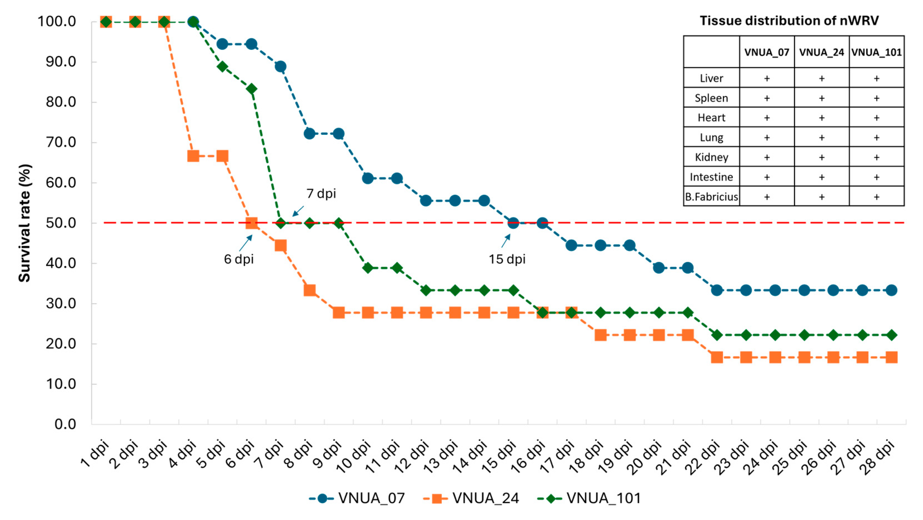
After inoculation with strains VNUA_07, VNUA_24, and VNUA_101, ducks exhibited signs of lethargy and anorexia at 2–3 days post-inoculation (dpi), and mortality began on day 4 post-inoculation. The time required for 50% mortality among inoculated ducklings varied between strains (Figure 3). The earliest 50% mortality was observed at 6 dpi for strain VNUA_24, followed by VNUA_101 at 7 dpi. The maximum time to death was 15 dpi for strain VNUA_07. By the end of the experiment, compared to the other strains, strain VNUA_24 was the most virulent, causing 83.3% cumulative mortality. In contrast, strain VNUA_101 caused less acute mortality, with a lower cumulative rate of 66.7%. The log-rank test showed that the survival curve of the group infected with VNUA_07 was significantly different from that of the VNUA_24-infected group (p = 0.00447). In contrast, no significant differences were observed between the VNUA_07- and VNUA_101-infected groups (p = 0.08), or between the VNUA_24- and VNUA_101-infected groups (p = 0.20). Although there were differences in disease progression, the distribution of the inoculated virus was consistent among the three inoculated groups, with a pan-tropic distribution across many internal organs (Figure 3, inserted panel).
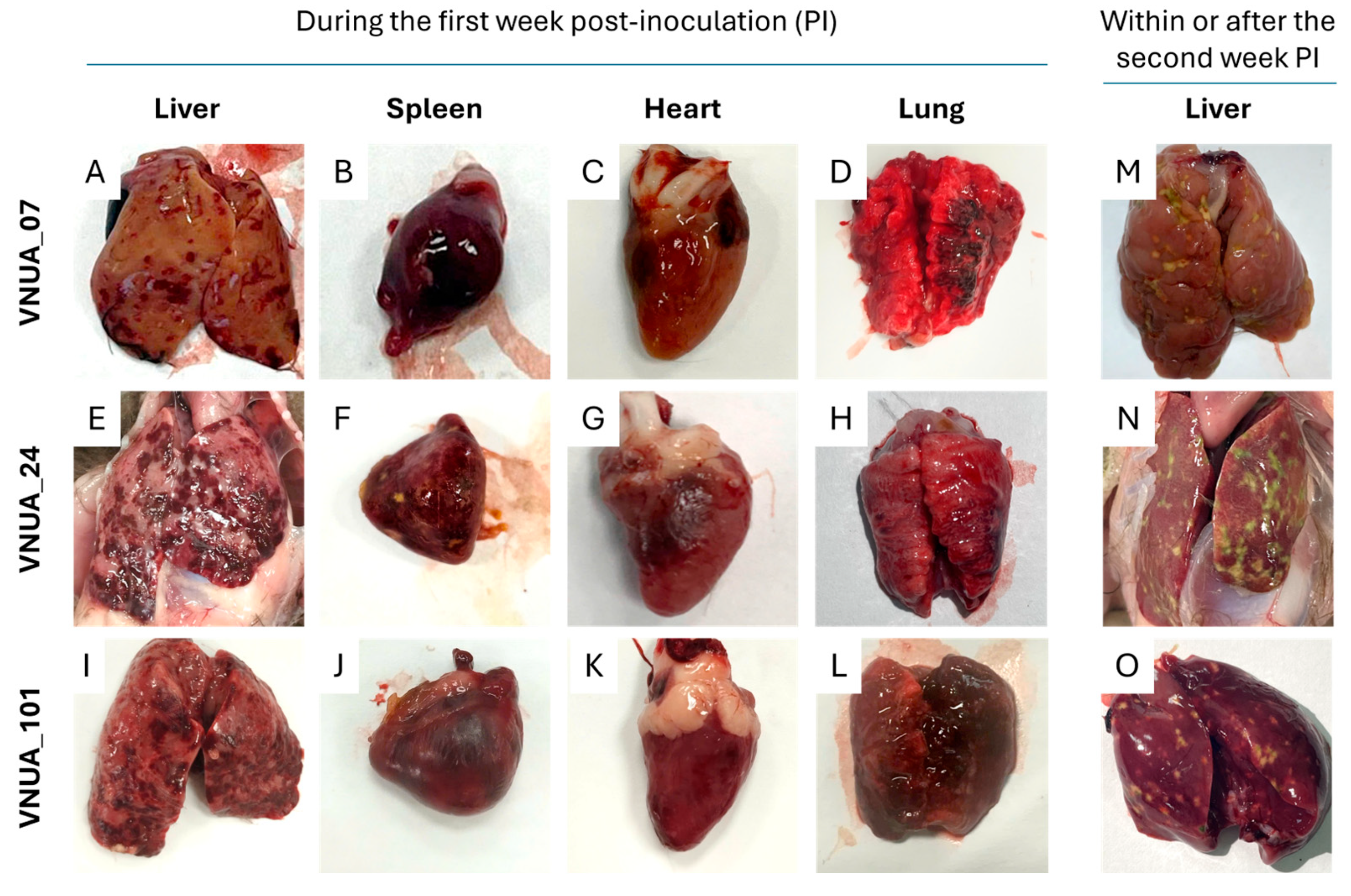
Among the pathological changes observed, animals that died within the first week post-inoculation exhibited prominent lesions characterized by generalized congestion and hemorrhages in multiple internal organs, particularly in the liver, spleen, heart, and lungs (Figure 4A–L). Rather than petechial hemorrhages, the liver parenchyma consistently showed ecchymotic hemorrhages. In contrast, animals that died during or after the second week post-inoculation showed the most distinct lesions in the liver, characterized by large necrotic areas scattered on its surface (Figure 4M–O). Other organs in the experimental group did not exhibit significant changes. No ducklings in the mock-inoculation groups died or showed gross lesions.
Microscopic lesions (Figure 5) were consistent with gross pathological changes observed at necropsy. The liver typically exhibited well-demarcated multifocal hepatocellular necrosis, accompanied by hemorrhage and occasional hemosiderin accumulation (Figure 5D,G,J). The spleen displayed large necrotic foci surrounded by inflammatory infiltrate (Figure 5E,H,K), while the lungs showed severe alveolar hemorrhage and edema (Figure 5F,I,L).

3.4. Genetic Classification Based on the sC Gene

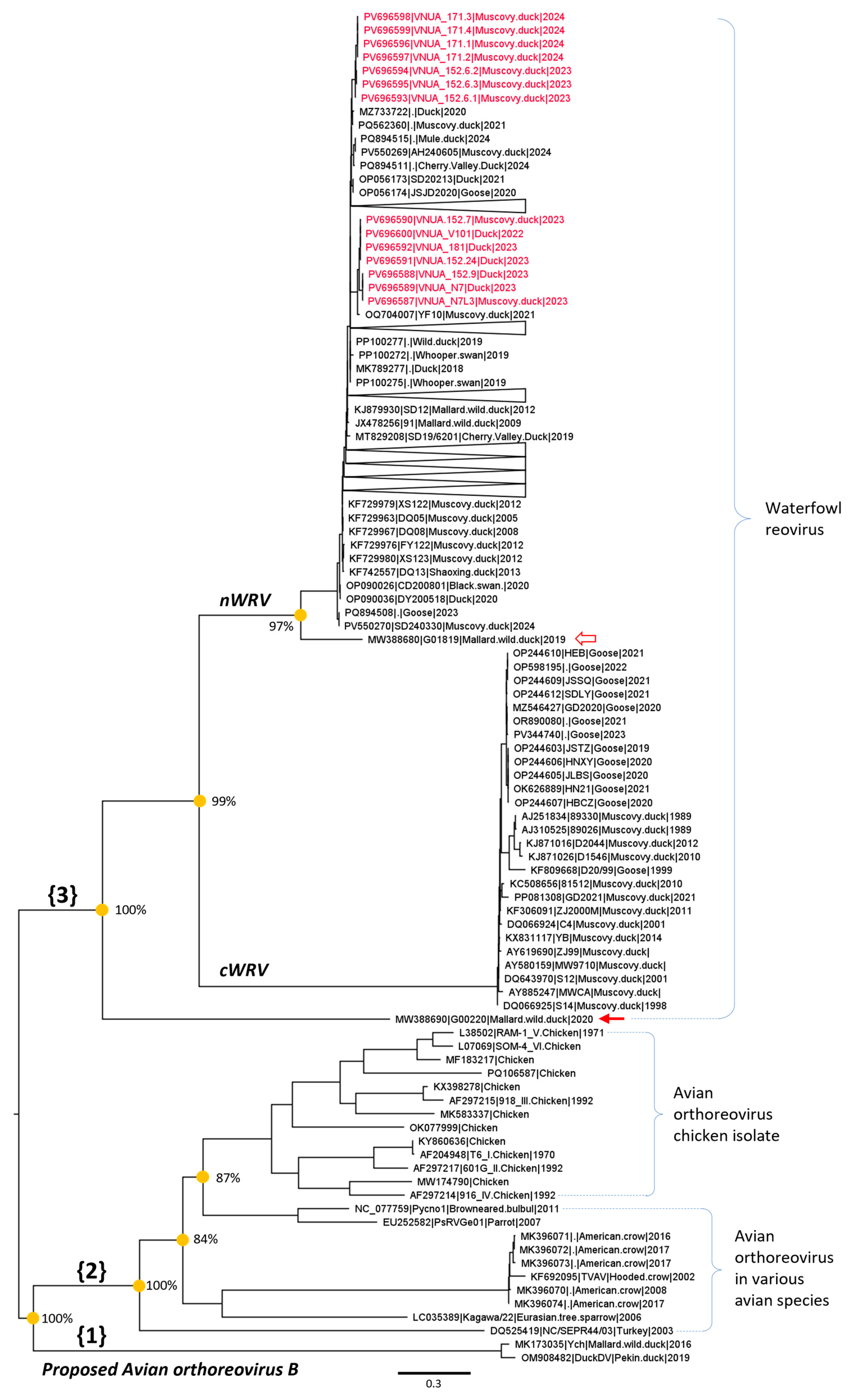
This study reconstructed the phylogeny of WRVs in the broad context with the other avian reoviruses deposited in GenBank (Figure 6, Supplementary File S3).
Based on the tree topology and bootstrap support values, three major branches (indicated as {1}, {2}, and {3} in Figure 6) were observed. The proposed avian orthoreovirus B was the deepest internal branch, followed by a complex branch comprising avian orthoreoviruses isolated from chickens and various other avian species. Waterfowl reoviruses constituted a distinct branch ({3}), separate from the two aforementioned branches. Within this branch, the majority of sequences fell into two major cWRV and nWRV genotypes. However, there were two orphan strains detected in wild mallards: one distantly related to nWRV and cWRV (filled arrow, Figure 6), and another distantly related to nWRV (empty arrow, Figure 6). Both cWRV and nWRV genotypes formed well-separated branches with a high bootstrap support value of 99%. All the Vietnamese WRVs detected in this study were clustered within the nWRV genotype but were separated by the strains detected in other countries.

3.5. Characterization of the Amino Acid Sequence of the nWRSs’ sC Protein

Amino acid mutations of the sC protein of nWRVs were scattered along the entire length of the sC protein. Insertion/deletion events (indicated in black in Figure 7A) were rare, occurring in only three out of 94 unique sC protein sequences. Amino acid mutations in Vietnamese WRVs mirrored the pattern observed in the global dataset. The amino acid sequence alignment divided the Vietnamese WRVs into two distinct zones (dashed boxes, Figure 7A).
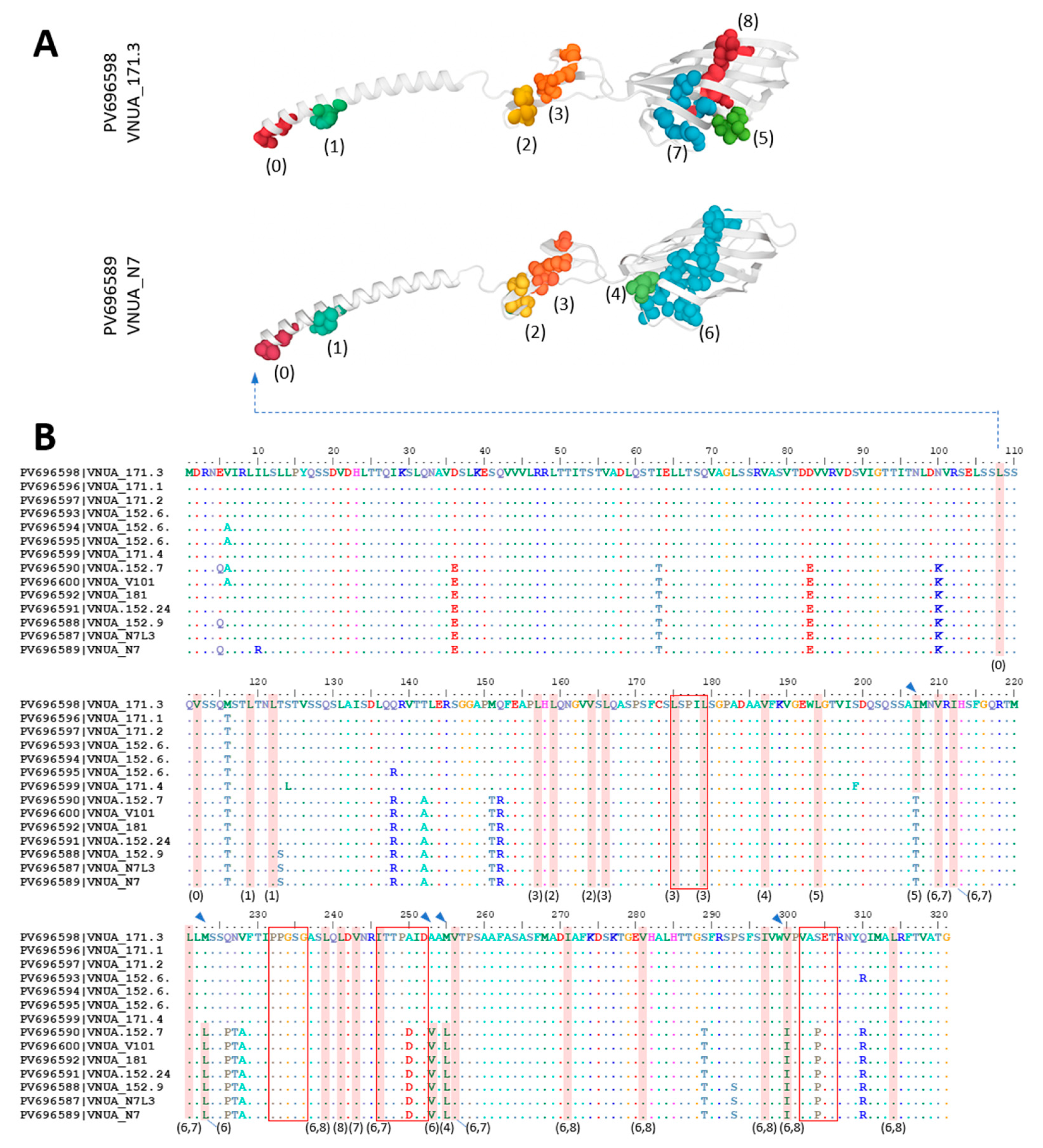
The prediction of antigenic regions by sequence-based and structure-based approaches consistently suggested that they predominantly located in the C-terminal of the sC protein (shaded area, Figure 7B,C). In 3D structure, the B-cell epitopes were located within the body and head domains (blue, Figure 7C). With the current dataset, most positions were conserved, and mutations were observed at only two sites (positions 250 and 304) within the predicted epitopes. In addition to the analysis of sequence diversity and antigenic properties, the hydrophobic clusters within the modeled 3D structure of the sC protein were characterized (Figure 8).
As shown in Figure 8, hydrophobic clusters are distributed throughout the sC protein structure, from the tail to the head; however, the largest cluster is located in the head region. Mutations were observed in cluster 4 (M255L), cluster 5 (I207T), cluster 6 (M223L, A253V, and V300I), and cluster 8 (V300I) (arrowheads, Figure 8). The combined multi-point variations (I207T, M223L, A253V, M255L, V300I) resulted in a ΔΔG value of −0.4, suggesting a neutral effect on protein stability.

3.6. Molecular Evolution of the nWRSs’ sC Gene

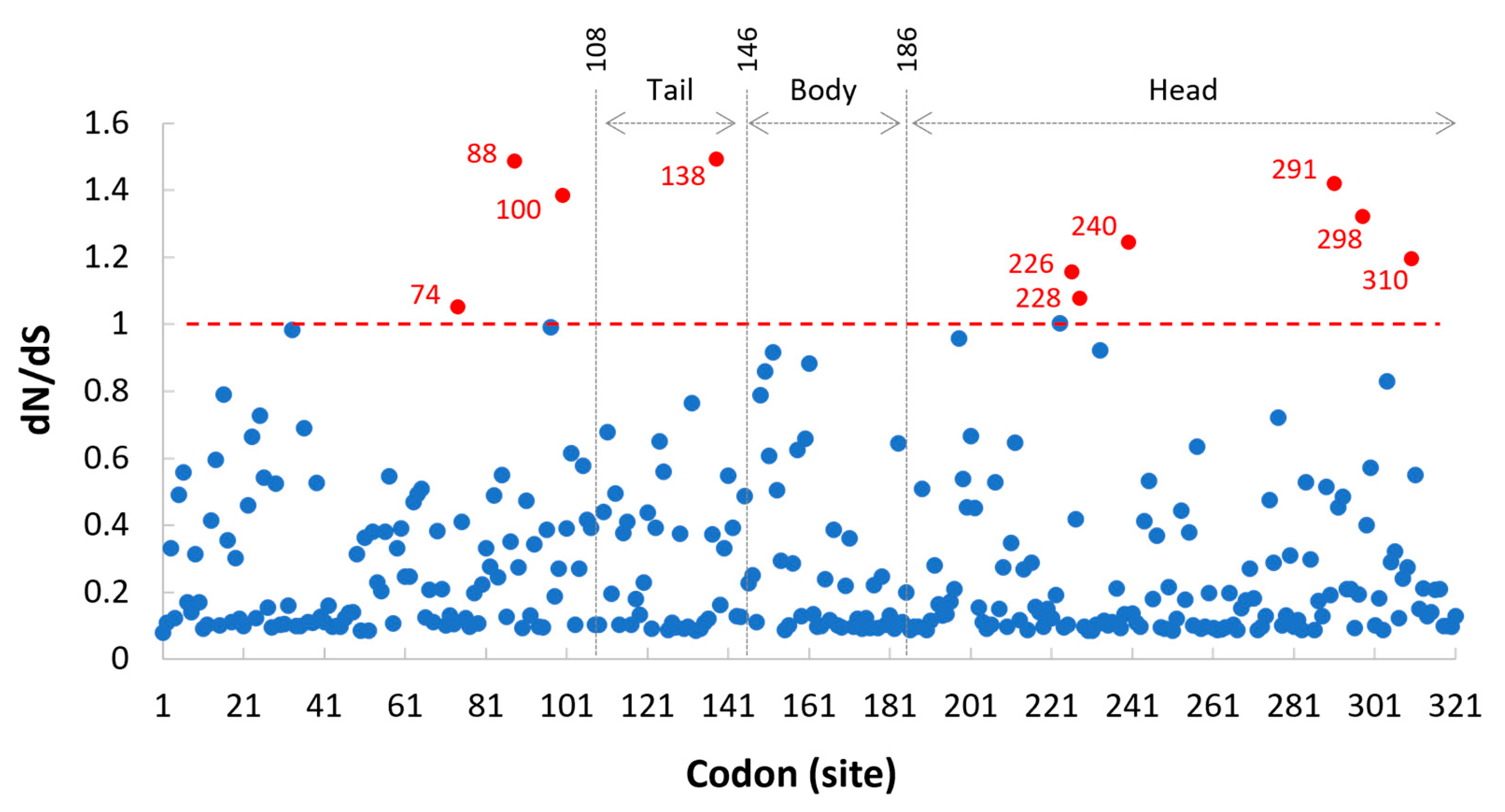
The pattern of molecular evolution of the sC gene was analyzed using the ratio of non-synonymous to synonymous substitution rates (dN/dS) across all codons (Figure 9). The overall dN/dS ratio for the complete sC gene was estimated to be 0.255. The likelihood ratio test (Appendix A Table A4) suggested that M8 was the best-fitting model for the data. At the codon level, most positions had dN/dS values < 1 (below the dashed line in Figure 9). Only 10 out of 321 codons (represented by red-filled dots in Figure 9) had dN/dS values > 1, but none were statistically predicted to be under diversifying selection at the 95% level. Six of the codons with dN/dS values > 1 were located in the head region of the sC protein, but they were outside the predicted conformational B-cell epitopes (as shown in Figure 7).

4. Discussion

To date, WRVs and their associated diseases have been described on different continents. Despite this, the majority of publications have focused on findings from China. Thus, this study aimed not only to reveal the situation in Vietnam, but also to contribute to public knowledge about the distribution and characterization of the virus. The detection rate of 15.6% across a wide geographic area, throughout the year, confirms that nWRV is not a sporadic occurrence but an endemic pathogen in northern Vietnam. It should be noted that a waterfowl reovirosis vaccine is not officially available in Vietnam, and thus all the positive samples reflect field infections. The field investigation result (Figure 1A) detected nWRV in a wide range of age groups (one to six weeks of age), but with a peak in the second week of age. This was congruent with previous findings that the disease was common in one- to two-week-old ducklings [11,58]. According to the literature, some publications have mentioned co-infecting viral pathogens such as Duck enteritis virus, AIV, DTMUV, DHAV type 1, waterfowl parvovirus, and DuCV [12,13,14,36,40,59]. To the best of the authors’ knowledge, no previous studies have investigated the co-infections in detail. The result presented in Figure 1B revealed co-infections with five viruses and suggested that co-infection with DHAV and DuCV was particularly common. However, due to the small number of positive samples (n = 44), no association tests were performed to determine whether co-infections affected detection rates. Co-infection with DHAV presents a diagnostic challenge for field veterinarians, as the susceptible age group and resulting hemorrhagic liver lesions are macroscopically similar to those of nWRV infection. nWRV infection is known to damage the intestinal barrier and induce intestinal mucosal immune suppression [60]. The co-infection with DuCV, a known immunosuppressive pathogen [61], raises concerns about potentially exacerbated clinical outcomes and increased complexity for disease management.
In previous studies, nWRV has been isolated using various cells, including Leghorn male hepatoma (LMH) cells [12], baby hamster kidney (BHK-21) cells [14], DEF cells [6] and Vero cells [58]. In this study, virus isolation was attempted in two commonly used cells: primary DEF and continuous Vero cells. The virus was found to readily adapt and replicate in both cell types (Figure 2). However, unlike some reports that described syncytium formation in DEF cells [6,62] and Vero cells [58], this study observed syncytium formation only after viral passages in Vero cells (Figure 2G). The absence of syncytium formation in DEF cells remains unexplained and warrants further investigation. Besides that, this study found that the Vietnamese isolates had similar properties to nWRV strains reported worldwide. Firstly, there was variation in virulence among WRV isolates [12]. By examining the gross lesions (Figure 4), three isolates (VNUA_07, VNUA_24, and VNUA_101) were pathogenic, as they induced lesions in the liver and spleen, and those lesions were typical of nWRV infections [10,63]. However, different levels of virulence among the three were revealed by comparing survival rates (Figure 3). Among them, VNUA_07 exhibited the lowest virulence, as indicated by a lower cumulative mortality rate and a longer time required to cause 50% mortality. Secondly, it was the pantropic nature of nWRVs [9] that was demonstrated in this study. The three isolates used for the challenge test exhibited a wide distribution in internal organs (Figure 3). This finding has also been reported in many publications [9,12,14,41].
From the literature, it is known that several genome segments of WRVs exhibit high divergence [19]. Among them, the sC gene is the most variable [29] and has been extensively used for phylogenetic studies [12,38,39]. Accordingly, this study employed the sC gene to infer the genetic relationships of the WRVs detected in northern Vietnam with those circulating in other countries. The analysis placed all Vietnamese isolates within the nWRV genotype (Figure 6), but at different positions on the phylogeny. This result, together with the amino acid alignment patterns (Figure 7A and Figure 8), suggested the presence of two genetic variant groups in northern Vietnam. However, it was not possible to associate differences in virulence with sC gene-based groupings. This is because a previous study showed that even closely related isolates (JN06, LC07, LC08, LC03, and LC05) caused variable survival rates in infected ducklings [12].
In many previous studies, the phylogenetic classification of WRVs relied on (i) a limited number of sC gene sequences (<100) and (ii) primarily used chicken-origin ARV as outgroups [10,14,20,34,37,38,39,40,62]. While this simplified approach successfully reconstructed the relationship between cWRV and nWRV, it overlooked the broader diversity within WRVs. To address this, the present study inferred the phylogeny from a larger dataset of completed sC gene sequences of ARV (n = 165) available from GenBank (as of May 2025) and contained orthoreoviruses originated from various avian hosts. This approach revealed that two orphan strains detected in wild mallards in Australia (empty and filled arrows, Figure 6) were distantly related to known cWRV and nWRV genotypes, providing further evidence of greater genetic divergence within the WRV branch than previously recognized.
Although the sC protein is known to be the primary immunogenic determinant of avian orthoreoviruses, antigenic characterization has only been performed for chicken-origin ARV [64]. As the functional regions of the sC protein of WRVs remain poorly defined, this study provides an immunoinformatic characterization of the sC protein of nWRVs. The consistent localization of predicted epitopes within the head and body domains by both sequence-based and structure-based methods, along with their high level of conservation (Figure 7D), suggests that these epitopes may represent important targets of the host immune response. This is further supported by the evolutionary pattern, which indicates that purifying selection predominantly acts on the sC gene (overall dN/dS value of 0.255), and that most positions in the gene had dN/dS values < 1 (below the dashed line in Figure 9), consistent with strong evolutionary constraint [65].
While this study provides some novel insights, it has certain limitations. The genetic analysis should be extended to whole-genome sequencing to better understand the genetic diversity of nWRVs in Vietnam. Furthermore, since this study focused solely on northern Vietnam and nWRVs, future surveillance is needed to determine the nationwide prevalence of both cWRV and nWRV.

5. Conclusions

This study successfully integrated classical virology with bioinformatic approaches to confirm the active circulation of nWRVs in northern Vietnam. Our findings expand the current understanding of WRV genetic diversity and provide an immunoinformatic profile of the sC of nWRV.

Supplementary Materials

The following supporting information can be downloaded at: https://www.mdpi.com/article/10.3390/microbiolres16080187/s1, Supplementary File S1: sigmaC-phylogenetic-inferencel.fas; Supplementary File S2: sigmaC-antigenic-characterizationl.fas; Supplementary File S3: phylogenetic-tree.pdf; Supplementary File S4: Details of nWRV detection results.pdf; Supplementary File S5: sigmaC-evolutionary-analysis.fas.

Author Contributions

Conceptualization, T.-N.V., V.-G.N. and T.-M.-L.H.; methodology, T.-N.V., V.-T.L. and V.-G.N.; validation, T.-N.V. and T.-B.-P.C.; formal analysis, T.-N.V. and V.-G.N.; investigation, T.-N.V., V.-T.L. and T.-B.-P.C.; data curation, T.-N.V., V.-T.L. and T.-B.-P.C.; writing—original draft preparation, T.-N.V. and V.-G.N.; writing—review and editing, T.-N.V., T.-M.-L.H. and V.-G.N.; supervision, T.-M.-L.H.; funding acquisition, T.-N.V. and T.-M.-L.H. All authors have read and agreed to the published version of the manuscript.

Funding

This research was funded by the Vietnam National University of Agriculture, grant number T2023-09-08TĐ and partially funded by Project 89 of 2023, Decision No. 2360/QĐ-BGDĐ, Ministry of Education and Training, Vietnam.

Institutional Review Board Statement

All protocols related to sample collection were approved by the Animal Ethics Committees, Vietnam National University of Agriculture. The Ethical Approval No. was VNUA-2023/06. The approval date was 28 August 2023 and the expiry date was 30 December 2025.

Informed Consent Statement

Not applicable.

Data Availability Statement

Data are contained within the article and Supplementary Materials.

Acknowledgments

We are grateful to all the farmers who granted us access to their farms. We also sincerely thank the field technicians for their support during sample collection. The authors would like to thank Nguyen Vu Son, Department of Veterinary Pathology, for his assistance in interpreting the microscopic lesions.

Conflicts of Interest

The authors declare no conflicts of interest.

Appendix A

Table A1. Primers used for detecting novel waterfowl-origin reoviruses.
Forward
/Reverse *
Sequences (5′-3′)Product Size (bp)References
nWRV-368FTGAGACGCCTGACTACGATT368[44]
/nWRV-368RATGCTTGGAGTGAGACGACT
* The primer used was renamed in this study.
Table A2. Primers used for detection of co-infecting viruses.
Forward
/Reverse *
Sequences (5′-3′)Product Size (bp)References
DHAV1.714FGCCCCACTCTATGGAAATTTG714[66]
/DHAV1.714RATTTGGTCAGATTCAATTTCC
DHAV3.720FATGCGAGTTGGTAAGGATTTTCAG720[66]
/DHAV3.720RGATCCTGATTTACCAACAACCAT
AIV.147FCTTCTAACCGAGGTCGAAACGTA147[67]
/AIV.147RGGATTGGTCTTGTCTTTAGCCA
DTMUV.350FTTTGGTACATGTGGCTCG350[68]
/DTMUV.350RACTGTTTTCCCATCACGTCC
DuCV.230FATGGCGAAGAGCGGCAACTAC230[69]
/DuCV.230RTCAGTAGTTTATTGGGAACGG
* The primer used in this study was renamed. Duck hepatitis A virus genotypes 1 (DHAV1) and 3 (DHAV3), Avian influenza virus (AIV), Duck Tembusu virus (DTMUV), and Duck circovirus (DuCV).
Table A3. Primers used for sequencing the sigma C gene of novel waterfowl-origin reoviruses.
Forward
/Reverse *
Sequences (5′-3′)Product Size (bp)References
nWRV-sC1001FACGATGGATCGCAACGAGGTG1001[21]
/nWRV-sC1001RGATGAATAGCTCTTCTCATYGC
* The primer used in this study was renamed and slightly modified (underlined). Degenerate base: Y = C or T.
Table A4. Summary of the likelihood ratio test (LRT).
ModelsC Gene
Number of codons 321
Omega (dN/dS) valueM1a0.255
Likelihood logarithm (lnL)M1a−6290.59
M2a−6232.39
M7−6230.07
M8−6225.15
LRTM2a vs. M1a116.40 **
M8 vs. M79.85 **
** The LRT values were then compared with the Chi-square distribution at 2 degrees of freedom for the model pairs M2a vs. M1a and M8 vs. M7 (** indicates significance at 99% critical value).

References

  1. Malkinson, M.; Perk, K.; Weisman, Y. Reovirus infection of young Muscovy ducks (Cairina moschata). Avian Pathol. 1981, 10, 433–440. [Google Scholar] [CrossRef]
  2. Heffels-Redmann, U.; Muller, H.; Kaleta, E.F. Structural and biological characteristics of reoviruses isolated from Muscovy ducks (Cairina moschata). Avian Pathol. 1992, 21, 481–491. [Google Scholar] [CrossRef]
  3. Palya, V.; Glavits, R.; Dobos-Kovacs, M.; Ivanics, E.; Nagy, E.; Banyai, K.; Reuter, G.; Szucs, G.; Dan, A.; Benko, M. Reovirus identified as cause of disease in young geese. Avian Pathol. 2003, 32, 129–138. [Google Scholar] [CrossRef] [PubMed]
  4. Yun, T.; Ye, W.; Ni, Z.; Chen, L.; Yu, B.; Hua, J.; Zhang, Y.; Zhang, C. Complete genomic sequence of goose-origin reovirus from China. J. Virol. 2012, 86, 10257. [Google Scholar] [CrossRef] [PubMed]
  5. Chen, Z.; Zhu, Y.; Li, C.; Liu, G. Outbreak-associated novel duck Reovirus, China, 2011. Emerg. Infect. Dis. 2012, 18, 1209–1211. [Google Scholar] [CrossRef]
  6. Liu, Q.; Zhang, G.; Huang, Y.; Ren, G.; Chen, L.; Gao, J.; Zhang, D.; Han, B.; Su, W.; Zhao, J.; et al. Isolation and characterization of a reovirus causing spleen necrosis in Pekin ducklings. Vet. Microbiol. 2011, 148, 200–206. [Google Scholar] [CrossRef]
  7. Yu, K.; Li, Y.; Han, H.; Song, M.; Ma, X.; Liu, C.; Huang, B.; Li, F. Complete genome sequence of an avian reovirus isolated from wild mallard ducks in china. Genome Announc. 2014, 2, e00813-14. [Google Scholar] [CrossRef]
  8. Wozniakowski, G.; Samorek-Salamonowicz, E.; Gawel, A. Occurrence of reovirus infection in Muscovy ducks (Cairina moschata) in south western Poland. Pol. J. Vet. Sci. 2014, 17, 299–305. [Google Scholar] [CrossRef]
  9. Li, N.; Hong, T.; Wang, Y.; Wang, Y.; Yu, K.; Cai, Y.; Liu, S.; Wei, L.; Chai, T. The pathogenicity of novel duck reovirus in Cherry Valley ducks. Vet. Microbiol. 2016, 192, 181–185. [Google Scholar] [CrossRef]
  10. Li, Y.; Yang, H.; Lu, Y.; Yin, Z.; Xu, H.; Mei, K.; Huang, S. Isolation and identification of a novel goose-origin reovirus GD218 and its pathogenicity experiments. Front. Vet. Sci. 2024, 11, 1423122. [Google Scholar] [CrossRef]
  11. Zheng, X.; Wang, D.; Ning, K.; Liang, T.; Wang, M.; Jiang, M.; Zhang, D. A duck reovirus variant with a unique deletion in the sigma C gene exhibiting high pathogenicity in Pekin ducklings. Virus Res. 2016, 215, 37–41. [Google Scholar] [CrossRef] [PubMed]
  12. Sun, X.; Guo, J.; Shen, J.; Guan, M.; Liu, L.; Xie, Y.; Xu, H.; Wang, M.; Ren, A.; Li, W.; et al. Genetics and biological characteristics of duck reoviruses isolated from ducks and geese in China. Vet. Res. 2025, 56, 30. [Google Scholar] [CrossRef] [PubMed]
  13. Kong, J.; Shao, G.; Zhang, Y.; Wang, J.; Xie, Z.; Feng, K.; Zhang, X.; Xie, Q. Molecular characterization, complete genome sequencing, and pathogenicity of Novel Duck Reovirus from South Coastal Area in China. Poult. Sci. 2023, 102, 102776. [Google Scholar] [CrossRef] [PubMed]
  14. Huang, C.; Huang, Y.; Liu, Z.; Li, J.; Han, J.; Liu, Y.; Liu, J.; Chen, H.; Chen, Z. Isolation and characterization of a duck reovirus strain from mature ducks in China. Poult. Sci. 2023, 102, 102345. [Google Scholar] [CrossRef]
  15. Cao, Y.; Sun, M.; Wang, J.; Hu, X.; He, W.; Su, J. Phenotypic and genetic characterisation of an emerging reovirus from Pekin ducks in China. Sci. Rep. 2019, 9, 7784. [Google Scholar] [CrossRef]
  16. Farkas, S.L.; Varga-Kugler, R.; Marton, S.; Lengyel, G.; Palya, V.; Banyai, K. Genomic sequence and phylogenetic analyses of two novel orthoreovirus strains isolated from Pekin ducks in 2014 in Germany. Virus Res. 2018, 257, 57–62. [Google Scholar] [CrossRef]
  17. Varga-Kugler, R.; Marton, S.; Thuma, A.; Szentpali-Gavaller, K.; Balint, A.; Banyai, K. Candidate ‘Avian orthoreovirus B’: An emerging waterfowl pathogen in Europe and Asia? Transbound. Emerg. Dis. 2022, 69, e3386–e3392. [Google Scholar] [CrossRef]
  18. Matthijnssens, J.; Attoui, H.; Banyai, K.; Brussaard, C.P.D.; Danthi, P.; Del Vas, M.; Dermody, T.S.; Duncan, R.; Fang, Q.; Johne, R.; et al. ICTV Virus Taxonomy Profile: Spinareoviridae 2022. J. Gen. Virol. 2022, 103, 001781. [Google Scholar] [CrossRef]
  19. Wang, D.; Shi, J.; Yuan, Y.; Zheng, L.; Zhang, D. Complete sequence of a reovirus associated with necrotic focus formation in the liver and spleen of Muscovy ducklings. Vet. Microbiol. 2013, 166, 109–122. [Google Scholar] [CrossRef]
  20. Zhang, X.L.; Shao, J.W.; Li, X.W.; Mei, M.M.; Guo, J.Y.; Li, W.F.; Huang, W.J.; Chi, S.H.; Yuan, S.; Li, Z.L.; et al. Molecular characterization of two novel reoviruses isolated from Muscovy ducklings in Guangdong, China. BMC Vet. Res. 2019, 15, 143. [Google Scholar] [CrossRef]
  21. Yun, T.; Yu, B.; Ni, Z.; Ye, W.; Chen, L.; Hua, J.; Zhang, C. Genomic characteristics of a novel reovirus from Muscovy duckling in China. Vet. Microbiol. 2014, 168, 261–271. [Google Scholar] [CrossRef] [PubMed]
  22. Day, J.M. The diversity of the orthoreoviruses: Molecular taxonomy and phylogentic divides. Infect. Genet. Evol. J. Mol. Epidemiol. Evol. Genet. Infect. Dis. 2009, 9, 390–400. [Google Scholar] [CrossRef] [PubMed]
  23. Banyai, K.; Palya, V.; Benko, M.; Bene, J.; Havasi, V.; Melegh, B.; Szucs, G. The goose reovirus genome segment encoding the minor outer capsid protein, sigma1/sigmaC, is bicistronic and shares structural similarities with its counterpart in Muscovy duck reovirus. Virus Genes 2005, 31, 285–291. [Google Scholar] [CrossRef]
  24. Kuntz-Simon, G.; Le Gall-Recule, G.; de Boisseson, C.; Jestin, V. Muscovy duck reovirus sigmaC protein is atypically encoded by the smallest genome segment. J. Gen. Virol. 2002, 83, 1189–1200. [Google Scholar] [CrossRef] [PubMed]
  25. Wang, D.; Xu, F.; Ma, G.; Zhang, C.; Huang, Y.; Li, H.; Zhang, D. Complete genomic sequence of a new Muscovy duck-origin reovirus from China. J. Virol. 2012, 86, 12445. [Google Scholar] [CrossRef]
  26. Martinez-Costas, J.; Grande, A.; Varela, R.; Garcia-Martinez, C.; Benavente, J. Protein architecture of avian reovirus S1133 and identification of the cell attachment protein. J. Virol. 1997, 71, 59–64. [Google Scholar] [CrossRef]
  27. Wickramasinghe, R.; Meanger, J.; Enriquez, C.E.; Wilcox, G.E. Avian reovirus proteins associated with neutralization of virus infectivity. Virology 1993, 194, 688–696. [Google Scholar] [CrossRef]
  28. Shih, W.L.; Hsu, H.W.; Liao, M.H.; Lee, L.H.; Liu, H.J. Avian reovirus σC protein induces apoptosis in cultured cells. Virology 2004, 321, 65–74. [Google Scholar] [CrossRef]
  29. Liu, H.J.; Lee, L.H.; Hsu, H.W.; Kuo, L.C.; Liao, M.H. Molecular evolution of avian reovirus: Evidence for genetic diversity and reassortment of the S-class genome segments and multiple cocirculating lineages. Virology 2003, 314, 336–349. [Google Scholar] [CrossRef]
  30. Vasserman, Y.; Eliahoo, D.; Hemsani, E.; Kass, N.; Ayali, G.; Pokamunski, S.; Pitcovskiad, J. The influence of reovirus sigma C protein diversity on vaccination efficiency. Avian Dis. 2004, 48, 271–278. [Google Scholar] [CrossRef]
  31. Bodelón, G.; Labrada, L.; Martínez-Costas, J.; Benavente, J. The Avian Reovirus Genome Segment S1 Is a Functionally Tricistronic Gene That Expresses One Structural and Two Nonstructural Proteins in Infected Cells. Virology 2001, 290, 181–191. [Google Scholar] [CrossRef]
  32. Yang, Y.; Gaspard, G.; McMullen, N.; Duncan, R. Polycistronic Genome Segment Evolution and Gain and Loss of FAST Protein Function during Fusogenic Orthoreovirus Speciation. Viruses 2020, 12, 702. [Google Scholar] [CrossRef] [PubMed]
  33. Du, X.; Ding, M.; Wu, Q.; Li, C.H.; Guo, H.; Liu, G.; Chen, Z. Characterization of a P18 protein in the S1 segment of the novel duck reovirus genome. Acta Virol. 2020, 64, 59–66. [Google Scholar] [CrossRef] [PubMed]
  34. Dandar, E.; Farkas, S.L.; Marton, S.; Oldal, M.; Jakab, F.; Mato, T.; Palya, V.; Banyai, K. The complete genome sequence of a European goose reovirus strain. Arch. Virol. 2014, 159, 2165–2169. [Google Scholar] [CrossRef] [PubMed]
  35. Farkas, S.L.; Dandár, E.; Marton, S.; Fehér, E.; Oldal, M.; Jakab, F.; Mató, T.; Palya, V.; Bányai, K. Detection of shared genes among Asian and European waterfowl reoviruses in the whole genome constellations. Infect. Genet. Evol. 2014, 28, 55–57. [Google Scholar] [CrossRef]
  36. Huang, Y.; Zhang, J.; Dong, J.; Li, L.; Kuang, R.; Sun, M.; Liao, M. Isolation and characterization of a new goose orthoreovirus causing liver and spleen focal necrosis in geese, China. Transbound. Emerg. Dis. 2022, 69, 3028–3034. [Google Scholar] [CrossRef]
  37. Zhang, X.; Chen, G.; Liu, R.; Guo, J.; Mei, K.; Qin, L.; Li, Z.; Yuan, S.; Huang, S.; Wen, F. Identification, pathological, and genomic characterization of novel goose reovirus associated with liver necrosis in geese, China. Poult. Sci. 2024, 103, 103269. [Google Scholar] [CrossRef]
  38. Wang, S.; Lin, F.; Cheng, X.; Wang, J.; Zhu, X.; Xiao, S.; Zheng, M.; Huang, M.; Chen, S.; Chen, S. The genomic constellation of a novel duck reovirus strain associated with hemorrhagic necrotizing hepatitis and splenitis in Muscovy ducklings in Fujian, China. Mol. Cell. Probes 2020, 53, 101604. [Google Scholar] [CrossRef]
  39. Luo, D.; Liu, R.; Weng, L.; Li, K.; Qi, X.; Gao, Y.; Liu, C.; Zhang, Y.; Cui, H.; Pan, Q.; et al. Genomic sequences and pathogenic characteristics of two variant duck reoviruses associated with spleen necrosis. Infect. Genet. Evol. 2021, 92, 104847. [Google Scholar] [CrossRef]
  40. Peng, Z.; Zhang, H.; Zhang, X.; Wang, H.; Liu, Z.; Qiao, H.; Lv, Y.; Bian, C. Identification and molecular characterization of novel duck reoviruses in Henan Province, China. Front. Vet. Sci. 2023, 10, 1137967. [Google Scholar] [CrossRef]
  41. Wang, H.; Gao, B.; Chen, H.; Diao, Y.; Tang, Y. Isolation and characterization of a variant duck orthoreovirus causing spleen necrosis in Peking ducks, China. Transbound. Emerg. Dis. 2019, 66, 2033–2044. [Google Scholar] [CrossRef] [PubMed]
  42. El Sabry, M.I.; Almasri, O. Global waterfowl production: Stocking rate is a key factor for improving productivity and well-being-a review. Trop. Anim. Health Prod. 2023, 55, 419. [Google Scholar] [CrossRef] [PubMed]
  43. Biswas, S.; Banerjee, R.; Bhattacharyya, D.; Patra, G.; Das, A.K.; Das, S.K. Technological investigation into duck meat and its products-A potential alternative to chicken. World’s Poult. Sci. J. 2019, 75, 609–620. [Google Scholar] [CrossRef]
  44. Niu, X.; Zhang, B.; Yu, X.; Zhang, X.; Dou, Y.; Tang, Y.; Diao, Y. Preparation and evaluation of goose reovirus inactivated vaccine. BMC Vet. Res. 2017, 13, 214. [Google Scholar] [CrossRef]
  45. Lei, C.; Yang, J.; Hu, J.; Sun, X. On the Calculation of TCID(50) for Quantitation of Virus Infectivity. Virol. Sin. 2021, 36, 141–144. [Google Scholar] [CrossRef]
  46. Katoh, K.; Rozewicki, J.; Yamada, K.D. MAFFT online service: Multiple sequence alignment, interactive sequence choice and visualization. Brief. Bioinform. 2017, 20, 1160–1166. [Google Scholar] [CrossRef]
  47. Suyama, M.; Torrents, D.; Bork, P. PAL2NAL: Robust conversion of protein sequence alignments into the corresponding codon alignments. Nucleic Acids Res. 2006, 34, W609–W612. [Google Scholar] [CrossRef]
  48. Minh, B.Q.; Schmidt, H.A.; Chernomor, O.; Schrempf, D.; Woodhams, M.D.; von Haeseler, A.; Lanfear, R. IQ-TREE 2: New Models and Efficient Methods for Phylogenetic Inference in the Genomic Era. Mol. Biol. Evol. 2020, 37, 1530–1534. [Google Scholar] [CrossRef]
  49. Clifford, J.N.; Høie, M.H.; Deleuran, S.; Peters, B.; Nielsen, M.; Marcatili, P. BepiPred-3.0: Improved B-cell epitope prediction using protein language models. Protein Sci. 2022, 31, e4497. [Google Scholar] [CrossRef]
  50. Ponomarenko, J.; Bui, H.H.; Li, W.; Fusseder, N.; Bourne, P.E.; Sette, A.; Peters, B. ElliPro: A new structure-based tool for the prediction of antibody epitopes. BMC Bioinform. 2008, 9, 514. [Google Scholar] [CrossRef]
  51. Powell, H.R.; Islam, S.A.; David, A.; Sternberg, M.J.E. Phyre2.2: A Community Resource for Template-based Protein Structure Prediction. J. Mol. Biol. 2025, 437, 168960. [Google Scholar] [CrossRef]
  52. Crooks, G.E.; Hon, G.; Chandonia, J.M.; Brenner, S.E. WebLogo: A sequence logo generator. Genome Res. 2004, 14, 1188–1190. [Google Scholar] [CrossRef]
  53. Ferruz, N.; Schmidt, S.; Höcker, B. ProteinTools: A toolkit to analyze protein structures. Nucleic Acids Res. 2021, 49, W559–W566. [Google Scholar] [CrossRef]
  54. Savojardo, C.; Manfredi, M.; Martelli, P.L.; Casadio, R. DDGemb: Predicting protein stability change upon single- and multi-point variations with embeddings and deep learning. Bioinformatics 2025, 41, btaf019. [Google Scholar] [CrossRef] [PubMed]
  55. Yang, Z. PAML 4: Phylogenetic analysis by maximum likelihood. Mol. Biol. Evol. 2007, 24, 1586–1591. [Google Scholar] [CrossRef] [PubMed]
  56. Álvarez-Carretero, S.; Kapli, P.; Yang, Z. Beginner’s Guide on the Use of PAML to Detect Positive Selection. Mol. Biol. Evol. 2023, 40, msad041. [Google Scholar] [CrossRef] [PubMed]
  57. Diller, J.R.; Halloran, S.R.; Koehler, M.; Natividade, R.d.S.; Alsteens, D.; Stehle, T.; Dermody, T.S.; Ogden, K.M. Reovirus σ1 Conformational Flexibility Modulates the Efficiency of Host Cell Attachment. J. Virol. 2020, 94, e01163-20. [Google Scholar] [CrossRef]
  58. Yang, H.; Zhang, W.; Wang, M.; Yuan, S.; Zhang, X.; Wen, F.; Guo, J.; Mei, K.; Huang, S.; Li, Z. Characterization and pathogenicity evaluation of recombinant novel duck reovirus isolated from Southeast China. Front. Veter. Sci. 2023, 10, 2023. [Google Scholar] [CrossRef]
  59. Nowak, T.; Kwiecinski, A.; Kwiecinski, P.; Tomczyk, G.; Wodz, K. Detection and Identification of Avian Reovirus in Young Geese (Anser anser domestica) in Poland. Animals 2022, 12, 3346. [Google Scholar] [CrossRef]
  60. Zhu, Z.; Liao, L.; Su, J.; Liu, Z.; Pan, S.; Huang, Y.; Wu, Y. Interactions of Muscovy duck reovirus, gut microbiota, and host innate immunity: Transcriptome and gut microbiota analysis. Vet. Microbiol. 2022, 264, 109286. [Google Scholar] [CrossRef]
  61. Lei, X.; Wang, A.; Zhu, S.; Wu, S. From obscurity to urgency: A comprehensive analysis of the rising threat of duck circovirus. Vet. Res. 2024, 55, 12. [Google Scholar] [CrossRef]
  62. Zhang, T.; Wang, X.; Jiang, W.; Fan, X.; Liu, N.; Miao, R.; Zhai, X.; Wei, L.; Jiao, P.; Jiang, S. Genetic characterization of novel duck reoviruses from Shandong Province, China in 2022. Poult. Sci. 2023, 102, 102969. [Google Scholar] [CrossRef]
  63. Li, B.; Jiang, X.; Zhang, S.; Wang, Q.; Cui, Y.; Wu, Q.; Jia, W.; Zhang, J.; Diao, Y.; Tang, Y. Isolation and Pathogenicity Analysis of a Novel Orthoreovirus Caused the Outbreak of Duck Viral Arthritis in China. Transbound. Emerg. Dis. 2023, 2023, 8179312. [Google Scholar] [CrossRef]
  64. Goldenberg, D.; Pasmanik-Chor, M.; Pirak, M.; Kass, N.; Lublin, A.; Yeheskel, A.; Heller, D.; Pitcovski, J. Genetic and antigenic characterization of sigma C protein from avian reovirus. Avian Pathol. 2010, 39, 189–199. [Google Scholar] [CrossRef] [PubMed]
  65. Yang, Z.; Nielsen, R.; Goldman, N.; Pedersen, A.-M.K. Codon-Substitution Models for Heterogeneous Selection Pressure at Amino Acid Sites. Genetics 2000, 155, 431–449. [Google Scholar] [CrossRef] [PubMed]
  66. Doan, H.T.; Le, X.T.; Do, R.T.; Hoang, C.T.; Nguyen, K.T.; Le, T.H. Molecular genotyping of duck hepatitis A viruses (DHAV) in Vietnam. J. Infect. Dev. Ctries. 2016, 10, 988–995. [Google Scholar] [CrossRef] [PubMed]
  67. Di Trani, L.; Bedini, B.; Donatelli, I.; Campitelli, L.; Chiappini, B.; De Marco, M.A.; Delogu, M.; Buonavoglia, C.; Vaccari, G. A sensitive one-step real-time PCR for detection of avian influenza viruses using a MGB probe and an internal positive control. BMC Infect. Dis. 2006, 6, 87. [Google Scholar] [CrossRef]
  68. Liu, M.; Chen, S.; Chen, Y.; Liu, C.; Chen, S.; Yin, X.; Li, G.; Zhang, Y. Adapted Tembusu-like virus in chickens and geese in China. J. Clin. Microbiol. 2012, 50, 2807–2809. [Google Scholar] [CrossRef]
  69. Fringuelli, E.; Scott, A.N.; Beckett, A.; McKillen, J.; Smyth, J.A.; Palya, V.; Glavits, R.; Ivanics, E.; Mankertz, A.; Franciosini, M.P.; et al. Diagnosis of duck circovirus infections by conventional and real-time polymerase chain reaction tests. Avian Pathol. 2005, 34, 495–500. [Google Scholar] [CrossRef]
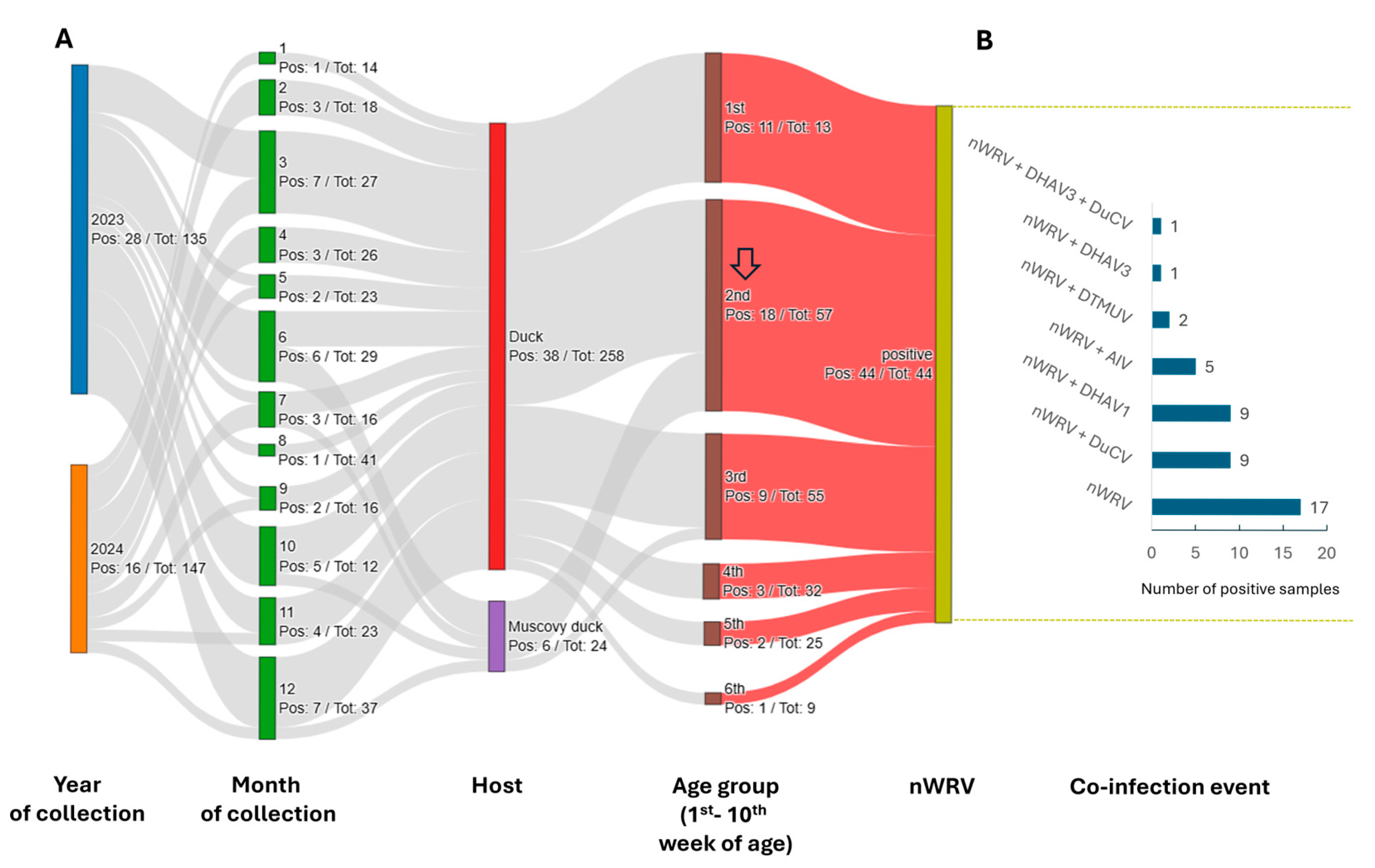
Figure 1. Summary of virology testing for nWRV nucleic acid. (A) Each node of the Sankey plot represents detection results categorized by year (2023, 2024), month (1–12), host origin, age group (by week). Node height is proportional to the number of positive samples. Labels adjacent to each node indicate the number of positive samples (Pos) relative to the total number tested (Tot). The arrow indicates the age at which the positive detection rate peaks. (B) The screening results for co-infecting viruses in nWRV-positive samples.
Microbiolres 16 00187 g001
Figure 2. Cytopathic effects induced by strain VNUA_24 in DEF and Vero cells. Panels (AC): infected cells at 48-, 72-, and 96-h post-inoculation. Panels (DF): mock-infected cells at corresponding time points. Panel (G): syncytium formation (*) within 24 h post-inoculation. Panel (H): mock-infected Vero cells. At all observed time points post-inoculation, mock-infected cells maintained an intact monolayer, while in infected flasks, partial cell detachment was observed, which gradually progressed to near-complete detachment. Magnification, 100×.
Microbiolres 16 00187 g002
Figure 3. Survival curve of ducklings inoculated with nWRVs. The graph shows the mean survival rate of triplicates for each virus strain. The dashed line indicates 50% survival. The inserted table shows the distribution of the inoculated virus across internal organs; the “+” sign indicates viral presence.
Microbiolres 16 00187 g003
Figure 4. Gross pathological changes in experimental ducks. Gross lesions observed during the first week post-inoculation with VNUA_07 (AD), VNUA_24 (EH), and VNUA_101 (IL). Gross liver lesions observed in the second week post-inoculation (MO). The sizes of the organs are not to scale.
Microbiolres 16 00187 g004
Figure 5. Histopathological findings in experimental ducks. (AC): Normal histological architecture is observed in the liver, spleen, and lung tissues. (D) Moderate multifocal hepatocellular swelling and necrosis (arrowheads) with mild mononuclear infiltration in the liver. (E) Severe hemorrhage and congestion in the red pulp of the spleen. (F) Severe hemorrhage within the alveolar spaces (arrowheads) in the lung. (G) A distinct necrotic focus is demarcated (dashed circle). (H) Large necrotic foci in the spleen, with a center of red blood cells and dead cells (asterisks), surrounded by inflammatory infiltrate (arrowheads) and fibroblast proliferation. (I) Interstitial pneumonia and marked alveolar edema (asterisks) in the lung. (J) Degeneration and necrosis of large patches of hepatocytes, with vacuolar changes (asterisks), severe hemorrhage, and accumulation of brown hemosiderin (arrows) in the liver. (K) Disruption of follicular structure by large necrotic foci (asterisk) and inflammatory cell infiltration (arrowheads) around the necrotic foci in the spleen. (L) Severe hemorrhage in the alveolar spaces (arrows) of the lung, with accumulated hemosiderin.
Microbiolres 16 00187 g005
Figure 6. The phylogenetic tree of Avian orthoreovirus was inferred based on the sC gene. For clarity, some branches are collapsed. Ultrafast bootstrap support values are shown for major nodes. A support value of 95% roughly corresponds to a 95% probability that a clade is correct [48]. Filled arrow: strain distantly related to both nWRV and cWRV; empty arrow: strain distantly related to nWRV. The tree is midpoint-rooted. The scale bar indicates the number of nucleotide substitutions per site.
Microbiolres 16 00187 g006
Figure 7. Immunoinformatic analysis of the sC protein of nWRV. (A) Amino acid alignment of sC protein sequences. The differences are relative to a reference sequence of GD218 strain (GenBank accession no. PQ391349). Dashed boxes indicate the sequences of nWRVs identified in this study. (B) Example output of BepiPred prediction, with scores plotted along the sC amino acid sequence; regions exceeding the threshold of 0.1512 (dashed line) are considered potential linear B-cell epitopes. (C) Predicted 3D structure of the sC protein (amino acids 108–320), with residues predicted to be part of conformational B-cell epitopes (blue), based on ElliPro analysis. Structural regions labeled as “tail,” “body,” and “head” follow the nomenclature adapted from a previous study [57]. (D) Sequence logo shows amino acid diversity at predicted B-cell epitopes. The height of each stack, measured in bits, reflects sequence conservation at that position. Only the most frequent residues are displayed, and their relative frequencies are represented by the symbol heights.
Microbiolres 16 00187 g007
Figure 8. Hydrophobic clusters in the sC protein (sites 108 to 321). Sites 1 to 107 were not modeled in 3D and thus no prediction is available. A hydrophobic cluster was formed by the sidechains of isoleucine (I), leucine (L), and valine (V). (A) Representative hydrophobic clusters mapped onto the protein structure. (B) Amino acid alignment highlighting hydrophobic clusters. The I, L, and V positions belonging to a hydrophobic cluster, as predicted by ProteinTools, are highlighted. Arrowheads indicate mutations within a hydrophobic cluster. The residues belonging to each hydrophobic cluster are numbered accordingly. The smallest clusters (0, 1, 2, and 5) contained two residues, while the largest cluster (6) contained 13 residues. Some residues shared between clusters are indicated in the format (cluster A number, cluster B number). Boxes indicate the predicted conformational B-cell epitopes.
Microbiolres 16 00187 g008
Figure 9. Plot of the dN/dS ratio across the full length of the sC gene. The dN/dS ratio for each codon was estimated using the M8 model. Codons with dN/dS > 1 are highlighted in red, whereas those with dN/dS ≤ 1 are represented by filled blue circles. Functional regions of the sC protein are annotated above the graph.
Microbiolres 16 00187 g009
Disclaimer/Publisher’s Note: The statements, opinions and data contained in all publications are solely those of the individual author(s) and contributor(s) and not of MDPI and/or the editor(s). MDPI and/or the editor(s) disclaim responsibility for any injury to people or property resulting from any ideas, methods, instructions or products referred to in the content.