Article

Protective Effects of Grape Seed Extract on Lipopolysaccharide Exposure and Radiation-Induced Intestinal Mucosal Damage: Insights from an In Vitro Study

by
Annamaria Altomare
1,2,
Michele Fiore
3,4,*,
Elena Imperia
1,
Gabriele D’Ercole
4,
Ludovica Spagnuolo
1,
Laura De Gara
1,
Gabriella Pasqua
5,
Michele Cicala
2,6,
Sara Ramella
3,4,† and
Michele Pier Luca Guarino
2,6,†
1
Department of Sciences and Technologies for Sustainable Development and One Health, Università Campus Bio-Medico di Roma, Via Alvaro del Portillo 21, 00128 Rome, Italy
2
Research Unit of Gastroenterology, Università Campus Bio-Medico di Roma, Via Alvaro del Portillo 21, 00128 Rome, Italy
3
Research Unit of Radiation Oncology, Department of Medicine and Surgery, Università Campus Bio-Medico di Roma, Via Alvaro del Portillo 200, 00128 Rome, Italy
4
Operative Research Unit of Radiation Oncology, Fondazione Policlinico Universitario Campus Bio-Medico di Roma, Via Alvaro del Portillo 21, 00128 Rome, Italy
5
Department of Environmental Biology, Sapienza Università di Roma, P.le Aldo Moro 5, 00185 Rome, Italy
6
Unit of Gastroenterology, Fondazione Policlinico Campus Bio-Medico di Roma, Via Alvaro del Portillo 200, 00128 Rome, Italy
*
Author to whom correspondence should be addressed.
These authors contributed equally to the work.
Microbiol. Res. 2025, 16(8), 176; https://doi.org/10.3390/microbiolres16080176
Submission received: 7 July 2025 / Revised: 23 July 2025 / Accepted: 25 July 2025 / Published: 1 August 2025

Abstract

Backgrounds and aim: Protective effects of natural compounds have been suggested in the prevention and treatment of radiation-induced mucositis or bacterial infections. In this study, the protective effects of proanthocyanidin-rich grape seed extract (GSE) on bacterial Lipopolysaccharide (LPS) and radiation-induced epithelial barrier damage and Reactive Oxygen Species (ROS) production were investigated in an in vitro model. Methods: Human intestinal epithelial cells Caco-2, previously treated with LPS, GSE, or LPS + GSE, were irradiated with 10 Gy divided into five daily treatments. Epithelial barrier integrity and ROS production were measured before and after each treatment. Results: Irradiation, at different doses, significantly increased intestinal permeability and ROS production; pretreatment with GSE was able to significantly prevent the increased intestinal permeability (4.63 ± 0.76 vs. 15.04 ± 1.5; p < 0.05) and ROS production (12.9 ± 1.08 vs. 1048 ± 0.5; p < 0.0001) induced by irradiation treatment. When the cells were pretreated with LPS, the same results were observed: GSE cotreatment was responsible for preventing permeability alterations (5.36 ± 0.16 vs. 49.26 ± 0.82; p < 0.05) and ROS production (349 ± 1 vs. 7897.67 ± 1.53; p < 0.0001) induced by LPS exposure when added to the irradiation treatment. Conclusions: The results of the present investigation demonstrated, in an in vitro model, that GSE prevents the damage to intestinal permeability and the production of ROS that are induced by LPS and ionizing radiation, suggesting a potential protective effect of this extract on the intestinal mucosa during irradiation treatment.

1. Introduction

Radiotherapy is a cornerstone in the treatment of cancer, not only as a primary treatment, but also as a complementary neoadjuvant or adjuvant therapy alongside chemotherapy and surgery [1]. Although antineoplastic treatments are becoming increasingly effective, they are still associated with several short- and long-term side effects due to their intrinsic tissue-killing nature [1]. The gastrointestinal tract is particularly affected by ionizing radiation, which is responsible for the disruption of the gut barrier integrity and inflammatory cell infiltration in the intestinal mucosa [2,3]. In recent years, radiotherapy techniques and equipment have been improved to reduce side effects [4]. Moreover, attempts have been made to identify novel, non-toxic, effective and affordable radio protective compounds that can shield healthy tissues from the harmful effects of radiation [5] protecting human tissues from oxidative stress processes caused by radiation exposure [5,6,7]. Several studies have been conducted, mostly in animal models, to identify natural compounds that are able to prevent radiation injury and/or reduce the negative effects on the gastrointestinal mucosa, thus preserving the barrier integrity and preventing local or systemic invasion by microorganisms [5,6,7,8,9]. Several extracts obtained from plant food, plant by-products, and dietary supplements can exert potent antioxidant activity to prevent radiation injury [5]; among them, it was demonstrated that polyphenols, obtained from Vitis vinifera L. seed extracts, protect against radiation-induced oxidative damage [10]. Moreover, grape seed extracts (GSEs) demonstrated a significant growth-inhibitory effect against several enteropathogens, with the potential, therefore, to exert possible beneficial actions at the level of organs and tissues [5,10,11,12,13,14]. In fact, GSE, containing proanthocyanidins (PCs)—characterized by high levels of hydrogen radical donor scavengers—could reduce oxidative stress by acting as a regulator of the inflammatory reaction caused by radiation exposure [10,15]. The current literature on radiation- and LPS-induced oxidative damage has predominantly focused on the mechanisms of injury rather than the identification of protective strategies [2,3]. Although GSE is well known for its antioxidant properties [5,6,10,15], its potential to mitigate the combined oxidative stress induced by radiation and LPS remains underexplored, particularly concerning the preservation of intestinal epithelial integrity. Saada et al. tested the effects of GSE against ionizing irradiation in a murine model: rats were treated with 100 mg GSE/kg/day by gastric tube 14 days before ionizing irradiation, and this administration significantly attenuated the oxidative stress induced by radiation in cardiac and pancreatic tissues [10]. Furthermore, GSE could reduce the clastogenic and cytotoxic effects of gamma irradiation in mouse bone marrow cells and demonstrated good radioprotective and immune modulatory properties in a rat model [16,17].
For this reason, the purpose of this study was to explore the protective effect of GSE on the preservation of mucosal integrity and the prevention of oxidative stress induced by ionizing radiation. To achieve this goal, a cellular model of Caco-2 was chosen, as these cells represent an interesting model of the human intestinal epithelium (including formation of TJ) after their post-confluent differentiation and polarization, showing similar functional and structural characteristics of the colonocytes [18,19]. There are currently few studies regarding a structured exposure model to ionizing radiation that can reproduce what happens in clinical practice [20,21,22]. In accordance with the available literature, the cells were irradiated with 2 Gy per day for 5 consecutive days up to a maximum of 10 Gy, mimicking the radiotherapy treatment to which patients are usually subjected. Moreover, in order to mimic the possible presence of Gram-negative components that are responsible for inflammatory activation [19], the cells were also exposed to bacterial Lipopolysaccharide (LPS) from a pathogenic strain of Escherichia coli before radiation treatment. In this experimental context, the potential beneficial effects of a well-characterized GSE [15] were explored.

2. Materials and Methods

2.1. Grape Seed Extract

Grape seed extract (GSE) was obtained from the unfermented pomace of V. vinifera L. (cv Bellone) according to the standardized method described in a recent study [6]. This method allows for obtaining an extract with a high concentration of polymeric procyanidins. GSE was obtained with a hydroalcoholic mixture and was chemically characterized by HPLC-DAD and NMR, as described in the Supplementary Materials, demonstrating that it is a very phenol-rich matrix (Supporting Informations Tables S1 and S2) [23].

2.2. Cell Culture

Human epithelial colorectal adenocarcinoma (Caco-2) cells, purchased from the American Type Culture Collection (ATCC, Manassas, VA, USA), were maintained under standard cell culture conditions: in a humidified 37 °C incubator; with 5% CO2; in Dulbecco’s modified Eagle’s Medium with 4.5 g/L glucose and L-glutamine without sodium pyruvate (high-glucose DMEM, Corning, Sigma-Aldrich, Milan, Italy); and supplemented with 10% (v/v) heat-inactivated fetal bovine serum (FBS, Euroclone, Pero (Ml) Italy), containing 2 mM L-glutamine (Aurogene, Rome, Italy), 100 U/mL penicillin, 100 µg/mL streptomycin (Merck, Sigma-Aldrich, Milan, Italy), and 1 mM HEPES (Dominique Dutscher, Bernolsheim, France). Caco-2 cells were periodically screened for contamination. Cells seeded at a density of 5.0 × 10⁴ per insert were grown on transwell chambers placed in a 12-well plate (12 mm with 0.4 μm pore polyester membrane inserts; Corning supplemented with Sigma-Aldrich, Milan, Italy); 500 μL of medium was placed in the apical compartments, and 1600 μL of medium was placed in the basolateral compartments. The cells were regularly monitored using a fully motorized epifluorescence inverted microscope (Eclipse Ti-E, Nikon, Tokyo, Japan) by measuring the epithelial resistance. Experiments were performed 21 days after seeding when the cells reached confluence and differentiation. Fresh media containing high-glucose DMEM (Corning, Sigma-Aldrich, Milan, Italy) supplemented with 10% (v/v) heat-inactivated fetal bovine serum containing (FBS, Euroclone, Pero (Ml) Italy), 2 mM L-glutamine (Aurogene, Rome, Italy), 100 U/mL penicillin, 100 µg/mL streptomycin (Merck, Sigma-Aldrich, Milan, Italy), and 1 mM HEPES (Dominique Dutscher, Bernolsheim, France), was changed every other day in the apical and basolateral compartments of the well, until the day of the experiment. We added 500 μL and 1500 μL on the apical and basolateral side, respectively.

2.3. Treatments with GSE and LPS

LPS (bacterial Lipopolysaccharide from a pathogenic strain of Escherichia coli 0111:B4—Sigma-Aldrich, Milan, Italy—at the concentration of 10 µg/mL w/v) and GSE (at the concentration of 6.25 µg/mL w/w) were dissolved in plain DMEM before cell treatment. The GSE dose (6.25 µg/mL) was chosen on the basis of preliminary experiments as the minimum effective dose [15].
Therefore, cells were challenged apically with LPS, GSE, and LPS + GSE for 24 h at 37 °C. LPS and GSE were dispensed only in the apical side as, once in the lumen, they are both absorbed by the microvilli covering the apical surface of the mucosa [19]. After the treatments, the cells were rinsed in phosphate-buffered saline w/o Ca2+ and Mg2+ (PBS, Euroclone, Milan, Italy) and used for the different subsequent assays.

2.4. MTT Assay

To determine the effect of GSE on Caco-2 cells and choose the correct concentration for the experiments, the viability was assessed using an MTT assay after 24 h of exposure of GSE to different concentrations (from 3.125 to 50 µg/mL, w/v). Then, the medium was removed, and each well was washed with 200 µL of PBS. After 3 h of incubation at 37 °C, the medium containing MTT was removed and washed in PBS. The absorbance was read at 570 nm using a microplate reader Tecan Infinite M200-Pro (Tecan, Männedorf, Switzerland). Cell proliferation values were expressed as percentages of relative absorbance measured in treated wells compared to control wells (untreated cells).

2.5. Irradiation of Cell Culture

Irradiation of Caco-2 cells was carried out at the Radiotherapy Department of the Fondazione Policlinico Universitario Campus Bio-Medico (Rome, Italy) using a 6 MV X-ray photon beam of a linear accelerator, which is routinely used to treat oncological patients. In preclinical studies, ionizing radiation is administered in fractional daily doses, mimicking the clinical administration method used to treat cancer patients [20,21,22,23]. This approach creates an experimental framework that replicates key aspects of clinical radiotherapy, including dose fractionation and precise beam delivery.
Twelve-well plates containing inserts with Caco-2 cells were placed on a 1.5 cm thick plexiglass sheet (corresponding to the build-up of the used radiation) at 100 cm from the radiation source. A 3 cm thick plexiglass sheet was placed on each plate before irradiation to ensure backscattered radiation components and charged particle equilibrium.
A treatment plan was created using the Eclipse 16.0 clinical treatment planning system (Varian Medical Systems, Palo Alto, CA, USA) (Figure 1 and Figure 2). Specifically, the dose distribution was computed using a dataset of a water-equivalent phantom. The resulting treatment plan showed an accurate dose uniformity in the plexiglass phantom that was irradiated with an antero-posterior beam. The use of 6 MV photon energy ensured precise equivalence between the plexiglass and water, as shown by the dosimetric evaluation. An antero-posterior beam arrangement was chosen to limit the set-up errors of the plate containing the cells.
Cells were irradiated using the TrueBeamTM linear accelerator by Varian Medical System (Palo Alto, CA, USA) (Figure 3). The cells were irradiated with a flat and symmetric (±2%) 40 × 40 cm2 radiation field and a dose rate of 3 Gy/min at room temperature. Cells were irradiated with 2 Gy per day for 5 consecutive days up to a maximum of 10 Gy. A total of 12 plates were irradiated in this study. The analysis of the irradiated material was performed at total doses of 0 Gy (control condition, sham, Ctrl w/o Gy), 2, 4, 6, 8, and 10 Gy. Every sham sample underwent the same environmental and mechanical stress, except for the radiation exposure. Control cells underwent the same procedural conditions as the irradiated ones, without entering the irradiation room (received dose: 0 Gy). The choice of a maximum dose of 10 Gy was based on its relevance to the biological effects and its consistency with similar studies in the field. A cumulative dose of 10 Gy, delivered in fractionated doses of 2 Gy per day over five consecutive days, reflects a clinically relevant irradiation regimen that is commonly used in radiation biology research to simulate therapeutic and subtherapeutic exposures [20,21,22,23]. This dosing regimen allows for observation of both immediate and cumulative biological responses to radiation, while avoiding excessive damage that might obscure measurable results or compromise experimental integrity. All the experiments were repeated three times, so the replicates are from three separate experiments.

2.6. Cell Permeability Assay

Epithelial barrier function was assessed by measuring the unidirectional paracellular flux of fluorescein isothiocyanate-dextran (FD-4, Sigma-Aldrich, Milan, Italy) from the apical to basolateral compartments. In brief, before transport studies, the culture medium was removed by aspiration (on both compartments) and replaced with prewarmed Krebs–Hensleit Buffer (KHB, pH 7.4 Sigma-Aldrich, Milan, Italy) for 1 h at 37 °C. Cells were then washed in PBS, and FD-4 was applied to the apical side of the Caco-2 monolayer at a final concentration of 1 mg/mL in prewarmed KHB [24]; the FD-4 paracellular permeability was calculated from the apical to basolateral direction by collecting undernatants. The concentration of FD-4 in the solution was measured every 20 min over 2 h by removing an aliquot from the receiver compartment (undernatant) and replacing it with an equal volume of fresh KHB. FD-4 is a large molecule with a molecular weight of 4 kDa after irradiation. Fluorescence readings were carried out at ex/em 490/520 nm by using a multi-plate reader (Tecan). Fluorescence values were converted to a concentration of fluorescein (pmol) using a standard curve. Each experiment was performed twice in triplicate, with independent controls among the three conditions.

2.7. Analysis of Oxidative Stress

Intracellular ROS production was assessed by adding 2′,7′-dichlorodihydrofluorescein diacetate (H2-DCF-DA, Sigma Aldrich, Burlington, MA, USA) according to Dinicola et al. [24], with slight modifications in the Caco-2 cells exposed to LPS, GSE, and LPS + GSE for 24 h in complete medium. On the day of the experiment, cells were irradiated and incubated for 3 h. Incubation with 400 μM H2O2 (Sigma-Aldrich, Milan, Italy) was used as a positive control for ROS. After incubation, the culture medium was replaced with PBS, and cells were loaded with 10 µM H2-DCF-DA for 30 min. After incubation, PBS and H2-DCF-DA were removed, and the cells were gently washed twice in PBS. Then, red-free medium was added. The increase in cell fluorescence was measured at excitation and emission wavelengths of 485 and 530 nm, respectively, using an Infinite F200 auto microplate reader (Tecan), at 25 °C. Each experiment was performed twice in triplicate, with independent controls among the three conditions.

2.8. Statistical Analysis

Data were presented as mean ± standard deviation, and experiments were carried out in triplicate under four different conditions. Data were analyzed using the GraphPad Prism v.9 (La Jolla, San Diego, CA, USA) software tool. Normally distributed data were analyzed for significance by unpaired t-test and one-way and two-way ANOVA, followed by post hoc test (Tukey’s Multiple Comparison Test and Bonferroni’s test). Significance was at the 0.05 level.

3. Results

3.1. MTT Assay

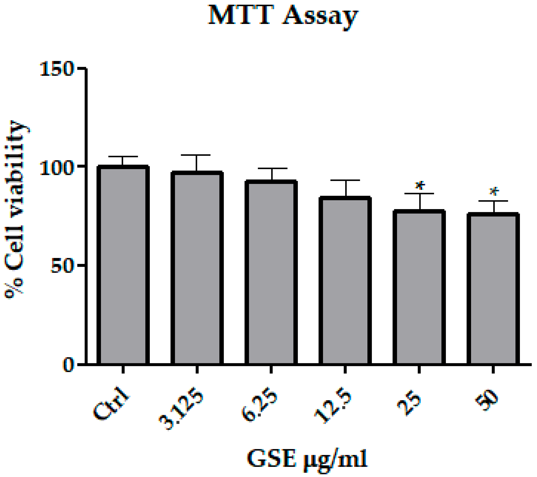
Following the exposure of Caco-2 cells to increasing concentration of GSE, the cell viability was significantly reduced compared to untreated cells (p < 0.05), at all time points when the concentration of GSE was 25 µg/mL or higher. On the other hand, no statistically significant reduction in cell viability was observed in Caco-2 cells treated with 3.125–12.5 µg/mL, as shown in Figure 4. For these reasons, a GSE concentration of 6.25 µg/mL was used in the following experiments, as previously reported [15].

3.2. FD-4 Permeability Analysis

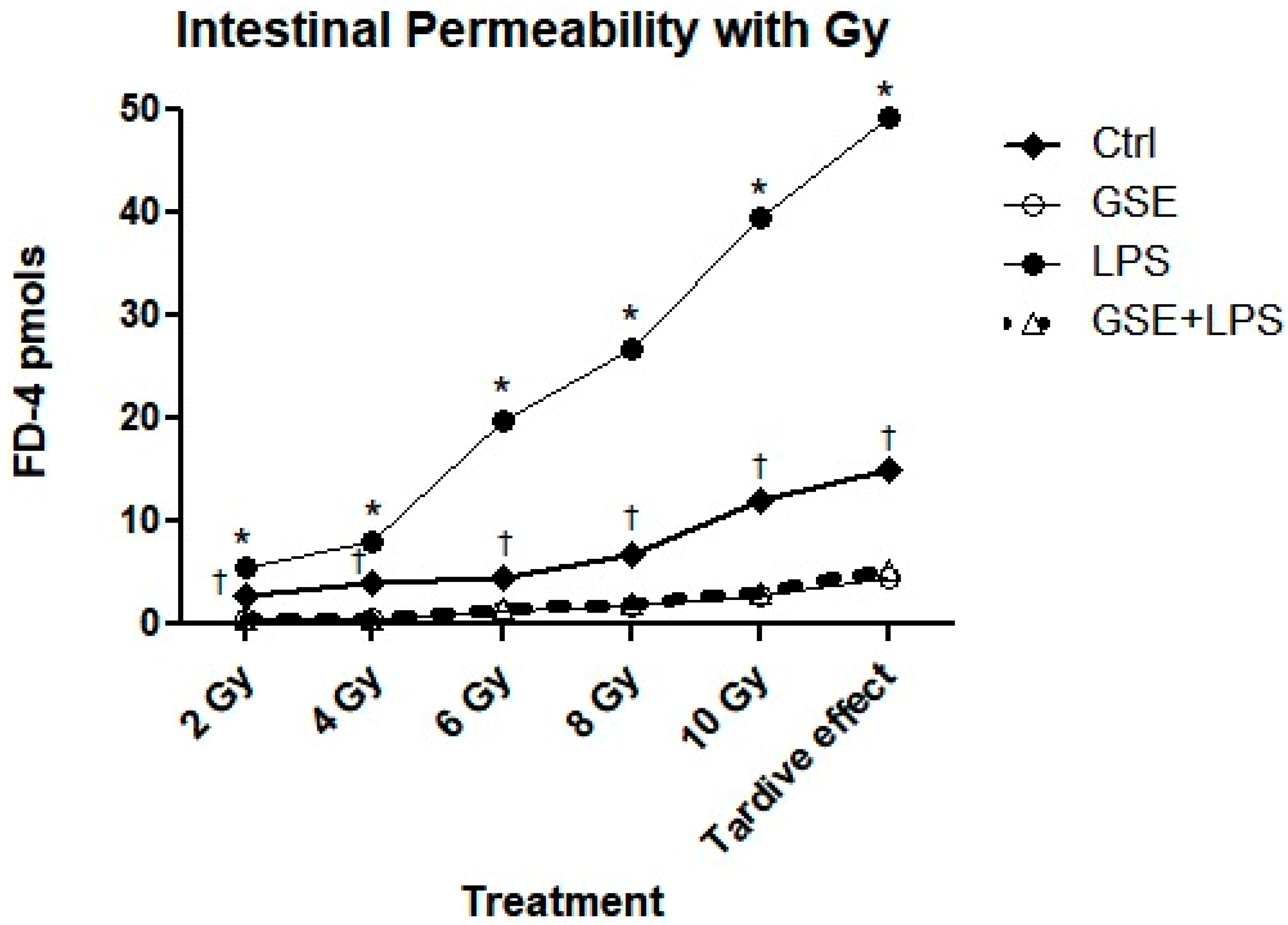
To evaluate the alteration of the Caco-2 cell monolayer integrity, the paracellular passage of FD-4 across Caco-2 monolayers was measured. Following LPS treatment (10 μg/mL for 24 h), the FD-4 permeability across the epithelial barrier was significantly increased compared to cells that were not treated with LPS (4.647 ± 0.306 vs. 0.65 ± 1.20; p < 0.05). Cotreatment with GSE was able to prevent the LPS-induced increase (0.457 ± 0.147 vs. 4.647 ± 0.306; p < 0.05), showing a similar value to the control. Interestingly, GSE alone did not alter the permeability values (p = ns). Radiation treatment was responsible for a significant increase in FD-4 permeability from 2 Gy to 10 Gy compared to the control (untreated cells) (Table S3, p < 0.05), maintaining a tardive effect after 10 days following irradiation (15.04 ± 1.5 vs. 0.65 ± 1.20 pmoles, respectively; p < 0.059. Pretreatment with GSE significantly reduced the increased FD-4 permeability due to irradiation at all treatment doses (Table S3, p < 0.05), maintaining a protective tardive effect after 10 days following irradiation (15.04 ± 1.5 vs. 4.63 ± 0.76; p < 0.05). When the cells were pretreated with LPS, a significantly increased permeability was observed at all doses of irradiation and 10 days after irradiation compared to cells without LPS exposure (p < 0.05). GSE treatment prior to LPS and radiation exposure significantly reduced this damage after 2 Gy (5.49 ± 0.31 vs. 0.45 ± 0.10; p < 0.05), 4 Gy (7.97 ± 0.22 vs. 0.62 ± 0.06; p < 0.05), 6 Gy (19.71 ± 0.38 vs. 1.46 ± 0.28; p < 0.05), 8 Gy (26.71 ± 1.36 vs. 1.96 ± 0.11; p < 0.05), and 10 Gy (39.49 ± 0.49 vs. 3.12 ± 0.10; p < 0.05), maintaining this beneficial effect 10 days after irradiation (49.26 ± 0.82 vs. 5.36 ± 0.16; p < 0.05), as shown in Figure 5 and in Supporting Informations, Table S3.

3.3. Analysis of ROS Production Following Treatments and Irradiation

To investigate the oxidative stress caused by irradiation with or without LPS co-exposure, intracellular ROS levels were measured in the LPS-, GSE-, and LPS + GSE-treated Caco-2 monolayers with and without irradiation, via the carboxy-H2 DCFDA fluorescent probe and according to previous works [25,26], and compared to untreated cells as the control. LPS treatment significantly increased ROS production compared to the control (p < 0.0001), while the cotreatment GSE-LPS seemed to completely prevent ROS production due to LPS exposure (p < 0.0001) (Supporting Informations, Table S4, Figure 3). GSE alone did not lead to a significant production of ROS compared to the control (p = ns). Radiation treatment was responsible for a significant increase in ROS production from 8 Gy to 10 Gy, maintaining this increase until 10 days after irradiation (tardive effect) compared to the cells before irradiation (used as the control) (Table S4, p < 0.05). No significant ROS production was observed for doses less than 8 Gy. Pretreatment with GSE significantly prevented the increase in ROS production due to irradiation at all treatment doses compared to the control (Figure 6, ††† p < 0.0001). When the cells were pretreated with LPS, a significant increase in ROS production was observed at all the treatment doses of irradiation and 10 days after irradiation compared to cells without LPS exposure (Figure 6, *** p < 0.0001). Cotreatment of irradiated cells with GSE-LPS significantly prevented ROS production compared to LPS-treated cells after 2 Gy (p < 0.0001), 4 Gy (p < 0.0001), 6 Gy (p < 0.0001), 8 Gy (p < 0.0001), and 10 Gy (p < 0.0001) and 10 days after irradiation (tardive effect) (p < 0.0001) (Supporting Informations, Table S4).

4. Discussion

In this study, the effect of fractional daily doses of ionizing radiation was explored on Caco-2 cell cultures, showing a significant and progressive increase in cell permeability and ROS production following the irradiation treatment. The cytotoxic effect of ionizing radiation on cells with a high proliferation rate, such as intestinal tissue, is well known [27,28,29]. Previous studies using the Caco-2 cell monolayer model have demonstrated an inverse relationship between intestinal epithelial resistance and paracellular permeability after exposure to various injuries [18,30,31]. Most of the cellular alterations induced by ionizing radiation are indirect and lead to the formation of free radicals [19]. These free radicals alter endogenous antioxidant systems and modify the genetic structure, leading to apoptosis and cell death [30]. Therefore, the first observation of the present investigation is in line with the current literature.
Conversely, the present study explored, for the first time, the additive effect of LPS exposure and irradiation on intestinal epithelial cells. Interestingly, the pre-exposure to LPS significantly increases the radiation effect on cellular permeability and ROS production, confirming the exacerbation of the radiation injury in the presence of concomitant Gram-negative contamination. No other study has previously investigated, on a cell culture model, the possible additive effect of these two harmful agents on mucosal permeability and oxidative stress production. The results confirmed a significantly increased FD-4 permeability across the epithelial barrier and ROS production after LPS exposure, as previously shown [15]. This bacterial endotoxin causes mucosal hyperpermeability, promoting gut barrier dysfunction through an oxidative mechanism [32] and an alteration of toll-like receptor 4 (TLR4) signaling [21,33,34,35]. Moreover, LPS stimulates epithelial cells to produce proinflammatory cytokines that alter tight junctions (TJs), leading to increased gut permeability [36].
Mucositis represents a serious complication of radio- and chemotherapy, significantly affecting the quality of life in cancer patients and often leading to treatment interruptions that can compromise the biological effects of the therapies [35,36,37,38]. It is also well known that oncological treatments can lead to the onset of bacterial mucositis, which worsens the patient’s clinical condition [30]. For this reason, the model used in the present study offers an interesting methodology for testing the potential negative effects of two different insults on the intestinal epithelium.
In recent years, several natural plant compounds have been studied for their protective effects on radiation-induced damage, showing emerging treatments for this debilitating side effect [5]. Plants possess different molecules that support the normal secondary metabolic processes in a biological system (i.e., the excessive production of ROS) [5,6]. Many natural sources are used both as anticancer drugs (e.g., vinblastine, vincristine, vinca alkaloids, and taxol) and as radioprotectants before or after radiation exposure, which can minimize or eliminate the effects of radiation-induced cellular damage [39].
In this study, the effect of a well-characterized GSE [15] was evaluated in irradiated Caco-2 cell cultures, as it was previously demonstrated to have anti-inflammatory properties in cells and animal models [40,41] when irradiated prior to and/or after bacterial LPS exposure. The data from the present study showed a significant reduction in the alterations in intestinal permeability and ROS production caused by the radiation treatment if the cells were pretreated with the GSE. Moreover, GSE was also able to reduce the epithelial damage induced by LPS plus irradiation exposure, with a significant reduction in epithelial permeability and ROS production. Previous studies have investigated cell co-culture systems and their response to irradiation, highlighting these systems as robust tools for advancing in vitro research and understanding the complexity of biological processes [20,21,22,42]. The methodology used in the present study builds on these papers, which used large datasets of colorectal adenocarcinoma Caco-2 cells that were exposed to X-ray doses of up to 10 Gy. They performed an integrated analysis including a spectrum of endpoints measured by different techniques to characterize the radiation response of this cell line [42]. This method was chosen to gain molecular insights into the underlying mechanisms of the impacts of radiation and the potential for translational applications in cancer therapy and radiation protection. The innovative aspect of this research consists of having used a model of fractional and progressive exposure to ionizing radiation to evaluate the possible beneficial effect of a natural substance. The results underlined how this experimental model is capable of showing the gradual progression of mucosal damage and, at the same time, allow for studying the gradual protective effect of a potentially beneficial substance. The preliminary in vitro results of the present investigation suggest that GSE could be able to protect the intestinal epithelium through its antibacterial [11] and antioxidant [10,43] properties, representing a potential radioprotector.
In conclusion, this study makes a unique contribution to the existing body of research by demonstrating that GSE can protect intestinal epithelial cells from oxidative damage in an in vitro Caco-2 model that has been exposed to both radiation and LPS. Furthermore, the findings highlight the ability of GSE to reduce ROS levels while maintaining the epithelial barrier, an essential component of gut health that has been underexplored in the context of radioprotective strategies. By addressing this critical gap in the literature, the study provides a valuable foundation for future translational research aimed at mitigating the combined effects of radiation and bacterial stress.
There are important limitations of the present investigation: first of all, an in vivo model could complete the analysis, and it is missing in this pilot study. Moreover, there is a potential variability in GSE quality based on the grape source, and the proposed methodology needs to be implemented to use GSE as a sustainable nutraceutical product obtained from the waste of viticulture [15].
Therefore, although our study did not include a comparison with established radioprotective agents, such as Amifostine or the herbal preparation STW5 (Iberogast), this choice was driven by the primary aim of evaluating the intrinsic protective potential of grape seed extract (GSE) in an in vitro model of radiation- and LPS-induced intestinal damage. Amifostine (WR-2721), the only FDA-approved synthetic radioprotector, exerts its effect mainly through free radical scavenging and hydrogen atom donation to stabilize DNA, mechanisms that partly overlap with the antioxidant properties of the polyphenols contained in GSE [44]. Similarly, STW5, a standardized multi-herbal formulation, has shown protective effects on intestinal barrier function, possibly via antioxidant and anti-inflammatory pathways [45,46]. Given the mechanistic similarities, future studies should aim to directly compare GSE with these compounds to better characterize its potential as a sustainable and nutraceutical alternative for mucosal protection during radiotherapy.

Supplementary Materials

The following supporting information can be downloaded at: https://www.mdpi.com/article/10.3390/microbiolres16080176/s1, S.1. Materials and Methods: a complete explanation of the measurement of Total Phenolic Content (S.1.1), analysis of High-Performance Liquid Chromatography (HPLC) (S.1.2) and analysis of Nuclear Magnetic Resonance (NMR) Analysis (S.1.3). S.2. Results: a specific explanation of the results about phytochemical analyses of GSE (S.2.1). In particular Table S1 summarizes the compounds determined by HPLC-DAD, their elution times and concentrations. Moreover, Figure S1 represents the 1H spectrum of procyanidins-rich grape seed extract. Table S2 illustrates the molecule amount measured by 1H NMR in procyanidins-rich grape seed extract. In the S.2.2 section the Table S3 shows in detail the results of FD-4 permeability. In the section S2.3 the Table S4 shows all the data produced by ROS analyses.

Author Contributions

Conceptualization, A.A., M.F., S.R. and M.P.L.G.; methodology, A.A., M.F., L.D.G. and M.P.L.G.; formal analysis, E.I., L.S. and G.D.; investigation, A.A., M.F., E.I., L.S. and G.D.; data curation, E.I., G.D. and L.S.; writing—original draft preparation, A.A., E.I., M.F. and G.D.; writing—review and editing, A.A., L.D.G., G.P., M.C., S.R. and M.P.L.G.; supervision, S.R., G.P., M.C. and M.P.L.G.; project administration, G.P., S.R. and L.D.G.; funding acquisition, G.P., S.R., M.P.L.G. and L.D.G. All authors have read and agreed to the published version of the manuscript.

Funding

This work was supported by POR FESR Lazio 2014–2020—Azione 1.2.1 VITEVITA project number A0375-2020-36590 “Valorizzazione di scarti delle aziende vitivinicole mediante recupero di principi attivi e sviluppo di formulazioni per prodotti antimicrobici, nutraceutici ed alimentari, in un’ottica di economia circolare”. Moreover, the authors would like to thank the Italian Ministry of Health for partially financially supporting this work under the National Recovery and Resilience Plan (NRRP) Trajectory 5 “Nutraceuticals, nutrigenomics and functional foods”—Action line 5.1 “Creation of an action program to fight malnutrition in all its forms and for the diffusion of the principles of the Mediterranean diet”.

Institutional Review Board Statement

Not applicable.

Informed Consent Statement

Not applicable.

Data Availability Statement

Research data are stored in an institutional repository and will be shared upon request to the corresponding author.

Conflicts of Interest

The authors declare no conflicts of interest.

References

  1. Baskar, R.; Lee, K.A.; Yeo, R.; Yeoh, K.W. Cancer and radiation therapy: Current advances and future directions. Int. J. Med. Sci. 2012, 9, 193–199. [Google Scholar] [CrossRef]
  2. Garg, S.; Zheng, J.; Wang, J.; Authier, S.; Pouliot, M.; Hauer-Jensen, M. Segmental Differences in Radiation-Induced Alterations of Tight Junction-Related Proteins in Non-Human Primate Jejunum, Ileum and Colon. Radiat. Res. 2015, 185, 50–59. [Google Scholar] [CrossRef]
  3. Zhou, Y.; Liu, J.; Li, X.; Wang, L.; Hu, L.; Li, A.; Zhou, J. JAC4 Protects from X-ray Radiation-Induced Intestinal Injury by JWA-Mediated Anti-Oxidation/Inflammation Signaling. Antioxidants 2022, 11, 1067. [Google Scholar] [CrossRef]
  4. Fiorino, C.; Guckemberger, M.; Schwarz, M.; van der Heide, U.A.; Heijmen, B. Technology-driven research for radiotherapy innovation. Mol. Oncol. 2020, 14, 1500–1513. [Google Scholar] [CrossRef]
  5. Altomare, A.; Fiore, M.; D’Ercole, G.; Imperia, E.; Nicolosi, R.M.; Della Posta, S.; Pasqua, G.; Cicala, M.; De Gara, L.; Ramella, S.; et al. Protective Role of Natural Compounds Under Radiation-Induced Injury. Nutrients 2022, 14, 5374. [Google Scholar] [CrossRef]
  6. Mulinacci, N.; Valletta, A.; Pasqualetti, V.; Innocenti, M.; Giuliani, C.; Bellumori, M.; De Angelis, G.; Carnevale, A.; Locato, V.; Di Venanzio, C.; et al. Effects of ionizing radiation on bioactive plant extracts useful for preventing oxidative damages. Nat. Prod. Res. 2019, 33, 1106–1114. [Google Scholar] [CrossRef]
  7. Qiu, X.; Dong, K.; Guan, J.; He, J. Hydrogen attenuates radiation-induced intestinal damage by reducing oxidative stress and inflammatory response. Int. Immunopharmacol. 2020, 84, 106517. [Google Scholar] [CrossRef]
  8. Azmoonfar, R.; Khosravi, H.; Rafieemehr, H.; Mirzaei, F.; Dastan, D.; Ghiasvand, M.R.; Khorshidi, L.; Pashaki, A.S. Radioprotective effect of Malva sylvestris L. against radiation-induced liver, kidney and intestine damages in rat: A histopathological study. Biochem. Biophys. Rep. 2023, 34, 101455. [Google Scholar] [CrossRef]
  9. Khan, S.A.; Wingard, J.R. Infection and mucosal injury in cancer treatment. J. Natl. Cancer Inst. Monogr. 2001, 29, 31–36. [Google Scholar] [CrossRef]
  10. Saada, H.N.; Said, U.Z.; Meky, N.H.; Azime, A.S.A. Grape seed extract Vitis vinifera protects against radiation-induced oxidative damage and metabolic disorders in rats. Phytother. Res. 2009, 23, 434–438. [Google Scholar] [CrossRef]
  11. Kitsiou, M.; Purk, L.; Gutierrez-Merino, J.; Karatzas, K.A.; Klymenko, O.V.; Velliou, E. A Systematic Quantitative Determination of the Antimicrobial Efficacy of Grape Seed Extract against Foodborne Bacterial Pathogens. Foods 2023, 12, 929. [Google Scholar] [CrossRef]
  12. Simonetti, G.; Santamaria, A.R.; D’Auria, F.D.; Mulinacci, N.; Innocenti, M.; Cecchini, F.; Pericolini, E.; Gabrielli, E.; Panella, S.; Antonacci, D.; et al. Evaluation of anti-Candida activity of Vitis vinifera L. seed extracts obtained from wine and table cultivars. Biomed. Res. Int. 2014, 2014, 127021. [Google Scholar] [CrossRef]
  13. Simonetti, G.; D’Auria, F.D.; Mulinacci, N.; Innocenti, M.; Antonacci, D.; Angiolella, L.; Santamaria, A.R.; Valletta, A.; Donati, L.; Pasqua, G. Anti-Dermatophyte and Anti-Malassezia Activity of Extracts Rich in Polymeric Flavan-3-ols Obtained from Vitis vinifera Seeds. Phytother. Res. 2017, 31, 124–131. [Google Scholar] [CrossRef]
  14. Simonetti, G.; Brasili, E.; Pasqua, G. Antifungal Activity of Phenolic and Polyphenolic Compounds from Different Matrices of Vitis vinifera L. against Human Pathogens. Molecules 2020, 25, 3748. [Google Scholar] [CrossRef] [PubMed]
  15. Nicolosi, R.M.; Bonincontro, G.; Imperia, E.; Badiali, C.; De Vita, D.; Sciubba, F.; Dugo, L.; Guarino, M.P.L.; Altomare, A.; Simonetti, G.; et al. Protective Effect of Procyanidin-Rich Grape Seed Extract against Gram-Negative Virulence Factors. Antibiotics 2023, 12, 1615. [Google Scholar] [CrossRef] [PubMed]
  16. Targhi, R.G.; Banaei, A.; Saba, V. Radioprotective effect of grape seed extract against gamma irradiation in mouse bone marrow cells. J. Cancer Res. Ther. 2019, 15, 512–516. [Google Scholar] [CrossRef]
  17. El-Desouky, W.; Hanafi, A.; Abbas, M.M. Radioprotective effect of green tea and grape seed extracts mixture on gamma irradiation induced immune suppression in male albino rats. Int. J. Radiat. Biol. 2017, 93, 433–439. [Google Scholar] [CrossRef]
  18. Lea, T. Caco-2 Cell Line. In The Impact of Food Bioactives on Health; Springer: Berlin/Heidelberg, Germany, 2015; pp. 103–111. [Google Scholar]
  19. Gori, M.; Altomare, A.; Cocca, S.; Solida, E.; Ribolsi, M.; Carotti, S.; Rainer, A.; Francesconi, M.; Morini, S.; Cicala, M.; et al. Palmitic Acid Affects Intestinal Epithelial Barrier Integrity and Permeability In Vitro. Antioxidants 2020, 9, 417. [Google Scholar] [CrossRef]
  20. McKelvey, K.J.; Hudson, A.L.; Donaghy, H.; Stoner, S.P.; Wheeler, H.R.; Diakos, C.I.; Howell, V.M. Differential effects of radiation fractionation regimens on glioblastoma. Radiat. Oncol. 2022, 25, 17. [Google Scholar] [CrossRef] [PubMed]
  21. Possenti, L.; Mecchi, L.; Rossoni, A.; Sangalli, V.; Bersini, S.; Cicchetti, A.; Costantino, M.L.; Candrian, C.; Arrigoni, C.; Rancati, T.; et al. Radiobiological Studies of Microvascular Damage through In Vitro Models: A Methodological Perspective. Cancers 2021, 13, 1182. [Google Scholar] [CrossRef]
  22. Gruber, S.; Dörr, W. Tissue reactions to ionizing radiation-Oral mucosa. Mutat. Res. Rev. Mutat. Res. 2016, 770, 292–298. [Google Scholar] [CrossRef]
  23. Muganu, M.; Paolocci, M.; Primiceri, S.; Tartaglia, R.; Benucci, I.; Cerreti, M.; D’Onofrio, C.; Paolacci, A.R.; Bignami, C. Intra-Varietal Variability of Romanesco Variety (Vitis vinifera L.). BIO Web Conf. 2019, 13, 01006. [Google Scholar] [CrossRef]
  24. Dinicola, S.; Cucina, A.; Pasqualato, A.; D’Anselmi, F.; Proietti, S.; Lisi, E.; Pasqua, G.; Antonacci, D.; Bizzarri, M. Antiproliferative and Apoptotic Effects Triggered by Grape Seed Extract (GSE) versus Epigallocatechin and Procyanidins on Colon Cancer Cell Lines. Int. J. Mol. Sci. 2012, 13, 651. [Google Scholar] [CrossRef]
  25. Fernández-Blanco, C.; Font, G.; Ruiz, M.J. Oxidative stress of alternariol in Caco-2 cells. Toxicol. Lett. 2014, 229, 458–464. [Google Scholar] [CrossRef]
  26. Araki, Y.; Sugihara, H.; Hattori, T. In vitro effects of dextran sulfate sodium on a Caco-2 cell line and plausible mechanisms for dextran sulfate sodium-induced colitis. Oncol. Rep. 2006, 16, 1357–1362. [Google Scholar] [CrossRef]
  27. Koch, J.; Mönch, D.; Maaß, A.; Gromoll, C.; Hehr, T.; Leibold, T.; Schlitt, H.J.; Dahlke, M.H.; Renner, P. Three-dimensional cultivation increases chemo- and radioresistance of colorectal cancer cell lines. PLoS ONE 2021, 16, e0244513. [Google Scholar] [CrossRef]
  28. Moyes, S.M.; Killick, E.M.; Morris, J.F.; Kadhim, M.A.; Hill, M.A.; Carr, K.E. Changes produced by external radiation in parameters influencing intestinal permeability and microparticle uptake in vitro. Int. J. Radiat. Biol. 2008, 84, 467–486. [Google Scholar] [CrossRef]
  29. Guardamagna, I.; Lonati, L.; Savio, M.; Stivala, L.A.; Ottolenghi, A.; Baiocco, G. An Integrated Analysis of the Response of Colorectal Adenocarcinoma Caco-2 Cells to X-Ray Exposure. Front. Oncol. 2021, 11, 688919. [Google Scholar] [CrossRef]
  30. Jeena, K.; Liju, V.B.; Ramanath, V.; Kuttan, R. Protection against Whole Body γ-Irradiation Induced Oxidative Stress and Clastogenic Damage in Mice by Ginger Essential Oil. Asian Pac. J. Cancer Prev. 2016, 17, 1325–1332. [Google Scholar] [CrossRef]
  31. Catalioto, R.M.; Festa, C.; Triolo, A.; Altamura, M.; Maggi, C.A.; Giuliani, S. Differential Effect of Ethanol and Hydrogen Peroxide on Barrier Function and Prostaglandin E2 Release in Differentiated Caco-2 Cells: Selective Prevention by Growth Factors. J. Pharm. Sci. 2009, 98, 713–727. [Google Scholar] [CrossRef]
  32. Safdari, B.; Sia, T.; Wattchow, D.; Smid, S. Effects of pro-inflammatory cytokines, lipopolysaccharide and COX-2 mediators on human colonic neuromuscular function and epithelial permeability. Cytokine 2016, 83, 231–238. [Google Scholar] [CrossRef]
  33. Hirotani, Y.; Ikeda, K.; Kato, R.; Myotoku, M.; Umeda, T.; Ijiri, Y.; Tanaka, K. Protective effects of lactoferrin against intestinal mucosal damage induced by lipopolysaccharide in human intestinal Caco-2 cells. Yakugaku Zasshi 2008, 128, 1363–1368. [Google Scholar] [CrossRef] [PubMed]
  34. Stephens, M.; von der Weid, P.Y. Lipopolysaccharides modulate intestinal epithelial permeability and inflammation in a species-specific manner. Gut Microbes 2020, 11, 421–432. [Google Scholar] [CrossRef] [PubMed]
  35. Yang, H.S.; Haj, F.G.; Lee, M.; Kang, I.; Zhang, G.; Lee, Y. Laminaria japonica Extract Enhances Intestinal Barrier Function by Altering Inflammatory Response and Tight Junction-Related Protein in Lipopolysaccharide-Stimulated Caco-2 Cells. Nutrients 2019, 11, 1001. [Google Scholar] [CrossRef]
  36. Sonis, S.T.; Eilers, J.P.; Epstein, J.B.; LeVeque, F.G.; Liggett, W.H.; Mulagha, M.T.; Peterson, D.E.; Rose, A.H.; Schubert, M.M.; Spijkervet, F.K.; et al. Validation of a new scoring system for the assessment of clinical trial research of oral mucositis induced by radiation or chemotherapy. Cancer 1999, 85, 2103–2113. [Google Scholar] [CrossRef]
  37. Keefe, D.M.; Sonis, S.T.; Bowen, J.M. Emerging drugs for chemotherapy-induced mucositis. Expert. Opin. Emerg. Drugs 2008, 13, 511–522. [Google Scholar] [CrossRef]
  38. Dowlath, M.J.H.; Karuppannan, S.K.; Sinha, P.; Dowlath, N.S.; Arunachalam, K.D.; Ravindran, B.; Chang, S.W.; Nguyen-Tri, P.; Nguyen, D.D. Effects of radiation and role of plants in radioprotection: A critical review. Sci. Total Environ. 2021, 779, 146431. [Google Scholar] [CrossRef]
  39. Alfouzan, A.F. Radiation therapy in head and neck cancer. Saudi Med. J. 2021, 42, 247–254. [Google Scholar] [CrossRef]
  40. Mantena, S.K.; Katiyar, S.K. Grape seed proanthocyanidins inhibit UV-radiation-induced oxidative stress and activation of MAPK and NF-κB signaling in human epidermal keratinocytes. Free Radic. Biol. Med. 2006, 40, 1603–1614. [Google Scholar] [CrossRef]
  41. Cheah, K.Y.; Howarth, G.S.; Bastian, S.E. Grape seed extract dose-responsively decreases disease severity in a rat model of mucositis; concomitantly enhancing chemotherapeutic effectiveness in colon cancer cells. PLoS ONE 2014, 9, e85184. [Google Scholar] [CrossRef] [PubMed]
  42. Babini, G.; Morini, J.; Barbieri, S.; Baiocco, G.; Ivaldi, G.B.; Liotta, M.; Tabarelli de Fatis, P.; Ottolenghi, A. A Co-culture Method to Investigate the Crosstalk Between X-ray Irradiated Caco-2 Cells and PBMC. J. Vis. Exp. 2018, 131, 56908. [Google Scholar]
  43. Calabriso, N.; Massaro, M.; Scoditti, E.; Verri, T.; Barca, A.; Gerardi, C.; Giovinazzo, G.; Carluccio, M.A. Grape Pomace Extract Attenuates Inflammatory Response in Intestinal Epithelial and Endothelial Cells: Potential Health-Promoting Properties in Bowel Inflammation. Nutrients 2022, 14, 1175. [Google Scholar] [CrossRef]
  44. Santini, V. Amifostine: Chemotherapeutic and radiotherapeutic protective effects. Expert. Opin. Pharmacother. 2001, 2, 479–489. [Google Scholar] [CrossRef] [PubMed]
  45. Elbadawi, M.; Ammar, R.M.; Aziz-Kalbhenn, E.; Rabini, S.; Klauck, S.M.; Dawood, M.; Saedd, M.E.M.; Kampf, C.J.; Efferth, T. Anti-inflammatory and tight junction protective activity of the herbal preparation STW 5-II on mouse intestinal organoids. Phytomedicine 2021, 88, 153589. [Google Scholar] [CrossRef] [PubMed]
  46. El-Ghazaly, M.; El Hazek, R.M.; Khayyal, M.T. Protective effect of the herbal preparation, STW 5, against intestinal damage induced by gamma radiation in rats. Int. J. Radiat. Biol. 2015, 91, 150–156. [Google Scholar] [CrossRef] [PubMed]
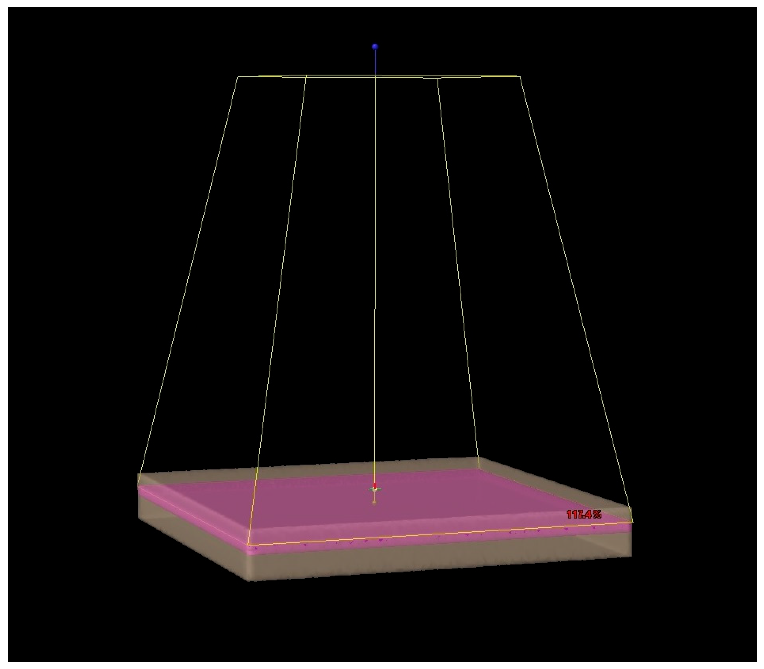
Figure 1. Three-dimensional view of the treatment plan.
Microbiolres 16 00176 g001
Figure 2. Axial view of the treatment plan.
Microbiolres 16 00176 g002
Figure 3. The 12-well plates containing cells are positioned as planned in the linear accelerator to be irradiated.
Microbiolres 16 00176 g003
Figure 4. Cell viability following exposure to different doses of GSE. Data are reported as mean ± SD. Statistical analysis was performed using a two-way ANOVA, followed by Bonferroni’s post hoc correction test. Experiments were independent and were carried out three times in triplicate under four different conditions (n = 9 for each condition, three times). * p < 0.05. Ctrl (control); GSE (grape seed extract).
Microbiolres 16 00176 g004
Figure 5. FD-4 permeability assay following the different irradiation doses (from 2 Gy to 10 Gy and 10 days after irradiation as tardive effect). Ctrl indicates non-irradiated cells. * LPS-treated cells vs. GSE + LPS, p < 0.05. † GSE-treated cells versus control cells, p < 0.05. Experiments were independent and carried out three times in triplicate under four different conditions (n = 9 for each condition, three times). Data is reported as mean ± SD. Statistical analysis was performed using a two-way ANOVA, followed by Bonferroni’s post hoc correction test. Ctrl (control); LPS (Lipopolysaccharide); GSE (grape seed extract); GSE + LPS (mix).
Microbiolres 16 00176 g005
Figure 6. Reactive Oxygen Species (ROS) production following the different irradiation doses (from 2 Gy to 10 Gy and 10 days after irradiation as a tardive effect). The analyses were performed by using carboxy-H2DCFDA staining and a fluorescence plate reader. Data are reported as fluorescence relatives. Ctrl indicates non-irradiated cells. *** LPS-treated cells vs. GSE + LPS, p < 0.0001. ††† irradiated cells (controls) compared to cells treated with GSE before irradiation, p < 0.0001. Experiments were independent and carried out three times in triplicate under four different conditions (n = 9 for each condition). Data are reported as mean ± SD. Statistical analysis was performed using a two-way ANOVA, followed by Bonferroni’s post hoc correction test. Ctrl (control); LPS (Lipopolysaccharide); GSE (grape seed extract); GSE + LPS (cotreatment of LPS and GSE).
Microbiolres 16 00176 g006
Disclaimer/Publisher’s Note: The statements, opinions and data contained in all publications are solely those of the individual author(s) and contributor(s) and not of MDPI and/or the editor(s). MDPI and/or the editor(s) disclaim responsibility for any injury to people or property resulting from any ideas, methods, instructions or products referred to in the content.