Article

Polysaccharides from Marine Bacteria and Their Anti-SARS-CoV-2 Activity

by
Tatyana A. Kuznetsova
1,*,
Natalia V. Krylova
1,
Maksim S. Kokoulin
2,
Elena V. Persiyanova
1,
Olga S. Maistrovskaya
1,
Pavel. G. Milovankin
1,
Yurii A. Belov
1 and
Mikhail Yu. Shchelkanov
1
1
Laboratory of Immunobiological Preparates, Laboratory of Respiratory Infections, Center of Molecular Diagnostics, G.P. Somov Institute of Epidemiology and Microbiology, Rospotrebnadzor, 1, Selskya St., Vladivostok 690087, Russia
2
Laboratory of Marine Glicobiology, G.B. Elyakov Pacific Institute of Bioorganic Chemistry, Far-Eastern Branch, Russian Academy of Science, 159, Prospect 100-let Vladivostok, Vladivostok 690022, Russia
*
Author to whom correspondence should be addressed.
Microbiol. Res. 2025, 16(5), 102; https://doi.org/10.3390/microbiolres16050102
Submission received: 10 April 2025 / Revised: 16 May 2025 / Accepted: 16 May 2025 / Published: 19 May 2025

Abstract

:
This study investigated the anti-SARS-CoV-2 activity of Polysaccharides (PSs) from three species of marine bacteria (Alteromonas nigrifaciens KMM 156, Cobetia amphilecti KMM 3890, and Idiomarina abyssalis KMM 227T). The chemical structure of PSs from marine bacteria is characterized using 1H and 13C NMR spectroscopy, including 2D NMR experiments. PS from A. nigrifaciens KMM 156 consists of tetrasaccharide repeating units containing two L-rhamnose residues and one residue each of 2-acetamido-2-deoxy-D-glucose and an ether of D-glucose with (R)-lactic acid, 3-O-[(R)-1-carboxyethyl]-D-glucose. PS from C. amphilecti KMM 3890 is constructed from branched trisaccharide repeating units consisting of D-glucose, D-mannose, and sulfated 3-deoxy-D-manno-oct-2-ulosonic acid. A unique PS from deep-sea marine bacterium I. abyssalis KMM 227T consists of branched pentasaccharide repeating units and is characterized by the presence of a rare bacterial polysaccharide component 2-O-sulfate-3-N-(4-hydroxybutanoyl)-3,6-dideoxy-D-glucose. The activity of PSs against SARS-CoV-2 was assessed by inhibition of the virus cytopathogenic effect (CI) in the methylthiazolyl tetrazolium (MTT) test and using a real-time reverse transcription polymerase chain reaction (RT-PCR-RV). Results of the study demonstrate that PSs, which differ in chemical structure, exhibited anti-SARS-CoV-2 activity differences. This is confirmed both in the test of inhibition of the virus CI and in the reduction in the SARS-CoV-2 virus RNA level. PSs from A. nigrifaciens KMM 156 exhibited the strongest anti-SARS-CoV-2 effect, effectively inhibiting the stages of attachment and penetration of SARS-CoV-2 into the cells.

1. Introduction

Severe acute respiratory syndrome coronavirus 2 (SARS-CoV-2) (Nidovirales: Coronaviridae, Betacoronavirus) is the etiologic agent of the deadly human coronavirus disease 2019 (COVID-19) [1,2,3]. For a long time, coronaviruses were considered a veterinary problem [4,5,6]. Only at the beginning of the 21st century did the significant epidemic potential of these viruses become apparent; and the COVID-19 pandemic that broke out in 2020–2023, confirmed the assumptions of experts about the existence of pandemic potential in coronaviruses [2,7,8,9].
Currently, intensive research is being conducted to find new drugs for the treatment of COVID-19, including the search for alternative antiviral treatments [10,11,12]. Promising sources of antivirus including anti-SARS-CoV-2 are marine organisms that produce unique chemical compounds, including polysaccharides, amino acids, glycosides, polyphenols, alkaloids, terpenoids, peptides, steroids, etc., exhibiting antiviral activity [11,13,14,15,16,17,18,19].
Marine microorganisms compare favorably with other marine sources, since they have high growth rates and biomass productivity [11,13,15,20]. Marine bacterial polysaccharides (PSs) are hydrocarbon biopolymers secreted by unicellular microorganisms, marine flora and fauna, which can be found on the outer surface of cell walls, in a mucous capsule or in the environment. Microbial PSs can exist as homo- or heteropolysaccharides with different properties, such as monosaccharide composition, structural conformation, molecular weight, and functional groups. Due to the polymeric repeating structure, these PSs are capable of polyvalent interactions that monomeric polysaccharides do not have. In this regard, PSs can be modified by attaching ligands that determine multiple binding to receptors on the surface of target cells or viruses [11,20,21].
PSs of marine bacteria are safe, biodegradable and biocompatible polymers with a wide range of biological activity, including anti-inflammatory, antioxidant, antitumor, immunomodulatory, antimicrobial, and the ability to exert antiviral effects. In this regard, PSs attract the attention of scientists as a promising source of antiviral medicinal substances. Currently, intensive research is being conducted around the world to find new drugs including PS substances from marine hydrobionts for the treatment of virus infections, especially COVID-19 [11,19,20,21,22,23,24].
This study aimed to evaluate the antiviral activity of PSs from three species of marine bacteria (Alteramonas nigrifaciens KMM 156, Cobetia amphilecti KMM 3890, Idiomarina abyssalis KMM 227T) against SARS-CoV-2 virus.

2. Materials and Methods

2.1. Isolation and Structural Characterization of Marine Bacteria Polysaccharides

The strains Alteramonas nigrifaciens KMM 156, Cobetia amphilecti KMM 3890, and Idiomarina abyssalis KMM 227T were cultivated in a liquid medium containing of (L−1): 5.0 g bactopeptone, 2.5 g yeast extract, 1.0 g glucose, 0.2 g K2HPO4, 0.05 g MgSO4, and 750 mL natural seawater/250 mL distilled water at an ambient temperature for 48 h under aerobic conditions and stirring. Briefly, dried cells were subjected to hot phenol/water extraction, followed by enzymatic treatment with DNase, RNase, and proteinase K. The resulting phenol/water extracts were dialyzed, freeze-dried, and further purified through sequential ultracentrifugation, anion-exchange chromatography, hydrophobic interaction chromatography, and size-exclusion chromatography.
Monosaccharides were analyzed as acetylated methyl glycosides using the method described earlier. Fatty acid analysis was performed by gas chromatography-mass spectrometry (GC-MS) of methyl derivatives. The PSs were subjected to methanolysis with 2 M acetylchloride in methanol at 80 °C for 16 h. All derivatives were analyzed using a Hewlett Packard 5890 chromatograph (USA) equipped with a Hewlett Packard 5973 mass spectrometer (USA) and a HP-5MS capillary column as described earlier [25,26,27].
The NMR spectra of the PSs were obtained using a Bruker Avance-III (700.13 MHz for 1H and 176.04 MHz for 13C) spectrometer (Bruker, Karlsruhe, Germany). The spectra were recorded in 99.95% D2O, and acetone (δC 31.45, δH 2.225) was used as the standard.
The identity of the PSs was confirmed through comparative analysis using GC-MS, and NMR spectroscopy, aligning with previous structural reports [25,26,27]. The structures of the polysaccharides isolated for this study are the same as those previously described for these strains.

2.2. SARS-CoV-2 Strain

The SARS-CoV-2/Vladivostok/5130/2020 strain (Wuhan-like genotype B1.1.397) was obtained from the collection of the Somov Research Institute of Epidemiology and Microbiology of Rospotrebnadzor (ID в VGARus prim000098, GISAID EPI_ISL_16756941, GenBank OQ363272). The strain was isolated from the nasopharyngeal lavage of a patient with a clinically and laboratory-confirmed diagnosis of COVID-19 by sequential passaging in an African green monkey kidney cell culture model (Vero E6) obtained from the State Research Center of Virology and Biotechnology (VECTOR, Novosibirsk, Russia). The strain was accumulated in Vero E6 using DMEM medium (Biolot, St. Petersburg, Russia) supplemented with 10% fetal bovine serum (FBS) (Biolot, St. Petersburg, Russia) and 100 U/mL gentamicin (Dalhimfarm, Khabarovsk, Russia) at 37 °C, 5% CO2. In the maintenance medium, the FBS concentration was reduced to 1%. The cell concentration in all experiments was 104 cells/mL. The infectious titer of SARS-CoV-2 in the Vero E6 cell culture after the fifth passage was 5.8 lg (TCID50/mL) (50% tissue cytopathic infectious dose per 1 mL).

2.3. Real-Time Reverse Transcription Polymerase Chain Reaction (RT-PCR-RV)

A real-time reverse transcription polymerase chain reaction (RT-PCR-RV) was carried out using a reagent kit for detecting SARS-CoV-2 coronavirus RNA using the RT-PCR method (Synthol, Moscow, Russia) on a Rotor-Gene Q analyzer (Qiagen, Hilden, Nordrhein-Westfalen, Germany) in accordance with the manufacturer’s instructions.

2.4. The Cytotoxic Activity of the PSs Assay

The cytotoxic activity of the PS assay on Vero E6 cells was evaluated using methylthiazolyl tetrazolium bromide (MTT) assay [28,29].
The test PSs were applied to a 24-h monolayer of cells grown in 96-well plates at various concentrations (5–2000 μg/mL) and were incubated at 37 °C in an atmosphere of 5% CO2 for 5 days. After incubation, 5 mg/mL MTT (Sigma-Aldrich, St. Louis, MO, USA) was added and the cells were incubated for 2 h at 37 °C. Cells with intracellular formazan crystals were dissolved with isopropanol. The optical density was measured at λ = 540 nm on a plate reader (LabSystems, Finland). Experiments were performed in triplicate and repeated three times. The cytotoxicity index ( C I C ) at a given concentration C of the test drug—polysaccharides (PSs) and Ribavirin® (reference drug)—was calculated using the formula:
C I C = 1 D s D 0 · 100 % ,
where Ds is the optical density in the MTT test of cells treated with the test PSs; D0 is the optical density in the MTT test of control cells (not treated with the tested PSs). The value of the 50% cytotoxic dose ( C D 50 ), which reduces cell viability by half compared to the control, was calculated using linear-logarithmic interpolation as the root of the equation
C I C D 50 = 50 % .
The maximum non-cytotoxic dose (MNCD) of the tested PSs was considered to be the concentration satisfying the equation
C I M N C D = 10 % .

2.5. The Study of Anti-SARS-CoV-2 Activity

The anti-SARS-CoV-2 activity of the tested PSs at the early stages of the SARS-CoV-2 life cycle was studied by the level of suppression of the cytopathogenic effect (CPE) of the virus (using the MTT test) and virus replication (using RT-PCR-RV). A monolayer of Vero E6 cells grown in 96-well plates was infected with the SARS-CoV-2/Vladivostok/5130/2020 strain at doses of 1.0 lg (TCID50/mL) and 2.0 lg (TCID50/mL); PSs and Ribavirin® were added at concentrations from 5 μg/mL to 500 μg/mL and at MNCD (RT-PCR-RV) according to schemes: cells were treated with PSs or Ribavirin® 1 h before infection (prophylactic effect); in another variant, cells were treated with the virus and PSs or Ribavirin® simultaneously (simultaneous effect). Treated cells were incubated for 5 days at 37 °C, 5% CO2.
The MTT test for assessing the anti-SARS-CoV-2 activity of the studied PSs at a specific concentration C was assessed based on the protection index (PI(C)—protection index):
P I C = D s i D i D 0 D i · 100 % ,
where D 0 —is the optical density in the MTT test of control cells (not treated with test PSs); D i —is the optical density in the MTT test of infected cells without the PSs; D s i —is the optical density in the MTT test of infected cells in the presence of PSs. The value of the 50% effective dose (ED50) was calculated using linear-logarithmic interpolation as the root of the equation
P I E D 50 = 50 % .
The final antiviral activity of the PSs was assessed using the selectivity index (SI):
S I = C D 50 E D 50 .
RT-PCR-RV for assessing the anti-SARS-CoV-2 activity of the PSs was performed as described above, based on the threshold amplification cycle ( C t ). The value C t 36 was taken as the absence of SARS-CoV-2 RNA in the samples. The based protection index on the results of RT-PCR-RV ( P C R P I C for the studied PSs at a specific concentration C was estimated using the formula of Shchelkanov M.Yu.:
P C R P I C = 2 C t s i C t i C t 0 C t i 1 · 100 % ,  
where C t 0 —is the C t value in real-time RT-PCR for control cells (not treated with test PSs); C t i —is the C t value in real-time RT-PCR for infected cells without PSs; C t s i —is the C t value in real-time RT-PCR for infected cells in the presence of PSs. All negative values of C t 36 were rounded to 36 (in particular, obviously C t 0 = 36 ). It is easy to see that P C R P I C = 0 % , if the tested drug has absolutely no ability to reduce viral reproduction (i.e.,   C t s i = C t i ), and P C R P I C = 100 % , if the tested PSs completely suppresses viral reproduction (i.e.,   C t s i = C t 0 ).

2.6. Statistical Analysis

Statistical analysis of the obtained data was performed using Statistica ver. 10.0 (StatSoft, Inc., Tulsa, OK, USA). Sample data were characterized using the mean value (M) and standard deviation of the mean (σ)Differences in experimental value groups were assessed using the Mann–Whitney-Wilcoxon test with a significance threshold p < 0.05.

3. Results

Antiviral Activity of PSs Against SARS-CoV-2

The PSs were isolated following previously published methods [25,26,27]. PS1 from A. nigrifaciens KMM 156 consists of tetrasaccharide repeating units containing two L-rhamnose residues and one residue each of 2-acetamido-2-deoxy-D-glucose and an ether of D-glucose with (R)-lactic acid, 3-O-[(R)-1-carboxyethyl]-D-glucose [25]:
→4)-β-D-GlcpNAc-(1→2)-α-L-Rhap-(1→3)-β-L-Rhap-(→
3
1
α-D-Glcp3(R-Lac)
PS2 from C. amphilecti KMM 3890 is constructed from branched trisaccharide repeating units consisting of D-glucose, D-mannose and sulfated 3-deoxy-D-manno-oct-2-ulosonic acid [26]:
→7)-β-Kdoр4Ac5S-(2→4)-β-D-Manр6Ac-(1→
2
1
β-D-Glcp
A unique PS3 from deep-sea marine bacterium I. abyssalis KMM 227T consists of branched pentasaccharide repeating units and is characterized by the presence of a rare bacterial polysaccharide component 2-O-sulfate-3-N-(4-hydroxybutanoyl)-3,6-dideoxy-D-glucose [27]:
→3)-β-D-QuipNAc4NAc-(1→3)-β-D-GlcpNAcA-(1→4)-β-D-GlcpNAcA-1→
4
1
β-D-Quip2S3N(4Hb)-(1→2)-α-L-Rhap
The results of the cytotoxicity assessment showed that tested PSs had low toxicity to Vero E6 cells: C D 50 > 2000 μg/mL; MNCD = 250 ± 23 μg/mL. For Ribavirin® as a reference drug C D 50 = 730 ± 88 μg/mL, MNCD = 150 ± 18 μg/mL, respectively.
A study of the effect of PSs on the early stages of the SARS-CoV-2 life cycle demonstrated that these PSs most effectively suppressed virus replication when Vero E6 cells were treated with the virus and PSs simultaneously. According to the MTT test, PS1 exhibited the highest antiviral activity: E D 50 = 96.0   μg/mL at an infectious dose of 2.0 lg(TCID50/mL), S I = 20.8 ; at an infectious dose of 1.0 lg (TCID50/mL), SI = 31.6 (Table 1, Figure 1). It should be noted that when pre-treating cells with PSs (prophylactic effect), higher inhibitory activity was noted for PS2 (Table 1, Figure 1). At the same time, Ribavirin® exhibited insignificant anti-SARS-CoV-2 activity only when exposed to cells simultaneously.
The anti-SARS-CoV-2 activity of PSs from marine bacteria was also studied using RT-PCR-RT. The most pronounced decrease in the SARS-CoV-2 RNA level was found with simultaneous exposure to PSs at MNCD = 250 μg/mL and an infectious dose of 2.0 lg (TCID50/mL) on Vero E6 (Table 2). It was found that under these conditions, PS1 most effectively suppresses virus reproduction (compared to the control), protecting an average of 42% of infected cells (p < 0.05). At the same time, the protection indices of PS2 and PS3 were 22% and 15%, respectively. Equally, with preliminary treatment of cells with the studied compounds at a concentration of 250 μg/mL and at an infectious dose of the virus of 2.0 lg (TCID50/mL), the decrease in viral reproduction was less pronounced, and the protection index on average for all tested PSs was about 9% (Table 2).
Thus, the results obtained both in the test of inhibition of the cytopathogenic effect of the virus (MTT test) and in the test of reducing the level of SARS-CoV-2 virus RNA (RT-PCR-RV) demonstrated the anticoronavirus activity of the studied PSs. The greatest effect was shown by PS1, which exhibits significant anticoronavirus activity, effectively inhibiting the early stages of SARS-CoV-2 interaction with the cell.

4. Discussion

The pandemic potential of coronaviruses necessitates the development of new antiviral drugs. Currently, research has focused on creating a novel antiviral drugs based on natural compounds due to their broad-spectrum biological activity and safety. PSs from three species of marine bacteria were chosen as the object of our research.
The results of the structural analysis reveal that each of the studied PSs possesses a unique composition, sequence, configuration, and substitution type of individual monosaccharide residues within a repeating structural unit. Additionally, PS2 and PS3 are sulfated, unlike PS1.
As the results of our studies have shown, PSs, which differ in chemical structure, exhibited anti-SARS-CoV-2 activity.. This is confirmed both in the test of inhibition of the virus CI and in the reduction in the SARS-CoV-2 virus RNA level. PS1 from A. nigrifaciens KMM 156 exhibited the strongest anti-SARS-CoV-2 effect.
Results of the MTT test show that PSs have low cytotoxic activity against Vero E6: C D 50 > 2000   μg/mL. As for the toxicity of microbial PSs, we have previously shown that these PSs were non-toxic as in parenteral administration to laboratory animals and in vitro experiments. PSs did not induce apoptosis of lymphocytes and neutrophilic leukocytes in human peripheral blood too. We also identified the immunomodulant and pronounced antiviral effect of PSs from A. nigrifaciens KMM 156 against the tick-borne encephalitis virus (TBEV) [30]. The maximal concentration of this PS (1000 μg/mL) caused a decrease in virus titers by 4.0 lg (TCID50/mL) and a suppression of virus reproduction by 66.7%. This PS restored the TBEV-virus-induced decreasing expression of cellular activation markers CD69, HLA-DR, and CD107a on the membrane of monocytes, NK and CD8+ T cells and the production of proinflammatory cytokines (IL-1, IL-6, IL-8, IFNα, IFNγ) [30]. It seems that the anti-TBEV-viral effect of the PS was indirectly associated with the ability to induce a Th1-type immune response at the early stage of TBEV infection through the activation of immunocompetent cells, increasing their cytotoxicity and production of Th1 cytokines. Taking into account the role of NK cells in the antiviral immune response, changes in the expression of adhesion molecules (increased density of CD11b and decreased CD62L) and increased expression of the activation marker (HLA-DR), determine the features of activation and implementation of effector functions of NK cells under PSs influence [30].
According to a number of authors, the mechanisms of antiviral activity of microbial PSs are universal. They are realized both systemically and with local action of PSs on the host’s cellular targets. The systemic action of PSs can be manifested by stimulating factors of innate and adaptive immunity, implementing anti-inflammatory, antioxidant and other types of activity [19,20,31]. Acceleration of virus elimination from the macroorganism can be associated with stimulation of innate immune cells’ functional activity—proliferation and cytotoxic activity of NK cells and the production of proinflammatory cytokines with antiviral activity (interferons type 1) [31,32,33]. NK cells are among the first cells to react against viral infections. The cytotoxicity of NK cells is associated with various mechanisms, including the participation of apoptosis receptors FasL (Fas ligand) and TRAIL (tumor necrosis factor-associated apoptosis-inducing ligand), as well as exocytosis of cytolytic granules containing perforins and granzymes [32,33,34].
The mechanism of antiviral activity of PSs can also be realized through a blocking attachment and penetration of the virus into cells, inhibiting either the virus binding to specific receptors of host cells or direct inactivation of viral particles [20,33,35]. Another possible mechanism is the suppression of viral replication by enhancing apoptotic reactions [24].
One of the possible mechanisms of anti-SARS-CoV-2 activity (both preventive and simultaneous) may be the ability of studied marine bacteria PSs to bind and/or inactivate viral particles. Therefore, the PSs exhibited an anti-coronavirus effect, effectively inhibiting the stages of attachment and penetration of SARS-CoV-2 into the cells.

5. Conclusions

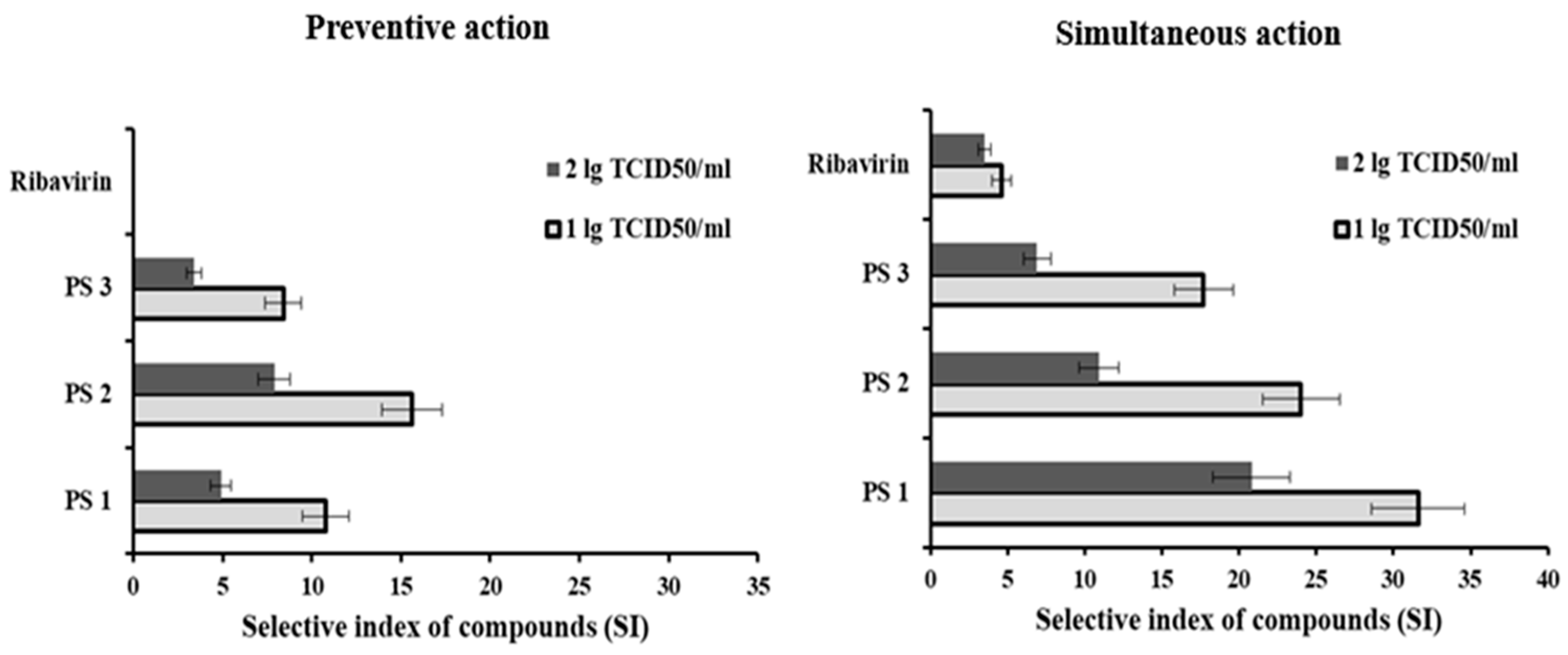
  • PSs from marine bacteria A. nigrifaciens KMM 156 (PS1), C. amphilecti KMM 3890 (PS2), I. abyssalis KMM 227T (PS3), differing in chemical structure, exhibit anti-coronavirus activity, effectively inhibiting the stages of attachment and penetration of SARS-CoV-2 into the cells.
  • This PSs can be considered a promising source of antiviral medicinal substances, including in the fight against the SARS-CoV-2 virus. However, further research is required to study the in-depth mechanisms of the antiviral activity of studied PSs.

Author Contributions

Conceptualization, T.A.K., N.V.K. and M.Y.S.; Methodology, T.A.K., N.V.K. and M.Y.S.; Validation, T.A.K., N.V.K. and M.Y.S.; Formal Analysis, T.A.K., N.V.K. and M.Y.S.; Investigation, T.A.K., N.V.K., E.V.P., M.S.K., O.S.M., P.G.M. and Y.A.B.; Data Curation, T.A.K., N.V.K. and M.Y.S.; Writing—Original Draft Preparation, T.A.K. and N.V.K.; Writing—Review and Editing, T.A.K., N.V.K., M.S.K. and M.Y.S.; Visualization, T.A.K., N.V.K., E.V.P., M.S.K., O.S.M., P.G.M. and Y.A.B.; Supervision, T.A.K., N.V.K., E.V.P., M.S.K., O.S.M., P.G.M. and Y.A.B.; Project Administration, M.Y.S. All authors have read and agreed to the published version of the manuscript.

Funding

This research was supported by the state tasks № 141-00089-21-00 on 2021–2025 of G.P. Somov Institute of Epidemiology and Microbiology, Rospotrebnadzor, Vladivostok, Russia and № 124042700016-1 on 2024–2026 of G.B. Elyakov Pacific Institute of Bioorganic Chemistry, Far-Eastern Branch, Russian Academy of Science, Vladivostok, Russia.

Institutional Review Board Statement

Not applicable.

Informed Consent Statement

Not applicable.

Data Availability Statement

The data used to support the findings of this study are included within this article. Any required further information can be provided by the corresponding author upon request.

Acknowledgments

A. nigrifaciens KMM 156, C. amphilecti KMM 3890, and I. abyssalis KMM 227T were obtained from the Collection of Marine Microorganisms (KMM) of the G.B. Elyakov Pacific Institute of Bioorganic Chemistry, Far Eastern Branch of Russian Academy of Sciences (Vladivostok, Russia).

Conflicts of Interest

The authors have no conflicts of interest to disclose.

Abbreviations

The following abbreviations are used in this manuscript:
CD5050% Cytotoxic Dose
CICytotoxicity Index
CPECytopathogenic Effect
ED5050% Effective Dose
MNCDMaximum Non-Cytotoxic Dose
MTTMethylthiazolyl Tetrazolium Bromide
PCRPIProtection Index on the Results of RT-PCR-RV
PIProtection Index
PSsPolysaccharides
TCID50Tissue Culture Infectious Dose
RT-PCR-RVReal-Time Reverse Transcription Polymerase Chain Reaction
SARS-CoV-2Severe Acute Respiratory Syndrome-2

References

  1. Sharma, A.; Ahmad, F.I.; Lal, S.K. COVID-19: A Review on the Novel Coronavirus Disease Evolution, Transmission, Detection, Control and Prevention. Viruses 2021, 13, 202. [Google Scholar] [CrossRef] [PubMed]
  2. Shchelkanov, M.Y.; Popova, A.Y.; Dedkov, V.G.; Akimkin, V.G.; Maleev, V.V. Research history of coronaviruses. Russ. J. Infect. Immun. 2020, 10, 221–246. (In Russian) [Google Scholar] [CrossRef]
  3. Yoshimoto, F.K. The Proteins of Severe Acute Respiratory Syndrome Coronavirus-2 (SARS CoV-2 or n-COV19), the Cause of COVID-19. Protein J. 2020, 39, 198–216. [Google Scholar] [CrossRef] [PubMed]
  4. Chakraborty, C.; Sharma, A.; Bhattacharya, M.; Sharma, G.; Lee, S.S. The 2019 novel coronavirus disease (COVID-19) pandemic: A zoonotic prospective. Asian Pac. J. Trop. Med. 2020, 13, 242–246. [Google Scholar] [CrossRef]
  5. Lorusso, A.; Calistri, P.; Petrini, A.; Savini, G.; Decaro, N. Novel coronavirus (SARS-CoV-2) epidemic: A veterinary perspective. Vet. Ital. 2020, 56, 5–10. [Google Scholar] [CrossRef]
  6. Munir, K.; Ashraf, S.; Munir, I.; Khalid, H.; Muneer, M.A.; Mukhtar, N.; Amin, S.; Ashraf, S.; Imran, M.A.; Chaudhry, U.; et al. Zoonotic and reverse zoonotic events of SARS-CoV-2 and their impact on global health. Emerg. Microbes Infect. 2020, 9, 2222–2235. [Google Scholar] [CrossRef]
  7. Benvenuto, D.; Giovanetti, M.; Ciccozzi, A.; Spoto, S.; Angeletti, S.; Ciccozzi, M. The 2019-new coronavirus epidemic: Evidence for virus evolution. J. Med. Virol. 2020, 92, 455–459. [Google Scholar] [CrossRef]
  8. Majumdar, A.; Malviya, N.; Alok, S. An overview on COVID-19 outbreak: Epidemic to pandemic. Int. J. Pharm. Sci. Res. 2020, 11, 1958–1968. [Google Scholar] [CrossRef]
  9. Wang, Y.T.; Landeras-Bueno, S.; Hsieh, L.E.; Terada, Y.; Kim, K.; Ley, K.; Shresta, S.; Saphire, E.O.; Regla-Nava, J.A. Spiking Pandemic Potential: Structural and Immunological Aspects of SARS-CoV-2. Trends Microbiol. 2020, 28, 605–618. [Google Scholar] [CrossRef]
  10. Lin, J.G.; Huang, G.J.; Su, Y.C. Efficacy analysis and research progress of complementary and alternative medicines in the adjuvant treatment of COVID-19. J. Biomed. Sci. 2023, 30, 30. [Google Scholar] [CrossRef]
  11. Mishra, N.; Gupta, E.; Walag, A.M.P.; Kharwar, R.N.; Singh, P.; Mishra, P. A Review of Marine Natural Product Resources with Potential Bioactivity Against SARS-COV-2. Trop. J. Nat. Prod. Res. 2023, 7, 2093–2103. [Google Scholar] [CrossRef]
  12. Wu, D.Y.; Hou, X.T.; Xia, Z.S.; Hao, E.W.; Xie, J.L.; Liang, J.Y.; Liang, Q.M.; Du, Z.C.; Deng, J.G. Analysis on oral medication rules of traditional Chinese medicine prescriptions for prevention of COVID-19. Chin. Herb. Med. 2021, 13, 502–517. [Google Scholar] [CrossRef]
  13. Chaisuwan, W.; Phimolsiripol, Y.; Chaiyaso, T.; Techapun, C.; Leksawasdi, N.; Jantanasakulwong, K.; Rachtanapun, P.; Wangtueai, S.; Sommano, S.R.; You, S.; et al. The Antiviral Activity of Bacterial, Fungal, and Algal Polysaccharides as Bioactive Ingredients: Potential Uses for Enhancing Immune Systems and Preventing Viruses. Front. Nutr. 2021, 8, 772033. [Google Scholar] [CrossRef]
  14. Chhetri, B.K.; Tedbury, P.R.; Sweeney-Jones, A.M.; Mani, L.; Soapi, K.; Manfredi, C.; Sorscher, E.; Sarafianos, S.G.; Kubanek, J. Marine Natural Products as Leads against SARS-CoV-2 Infection. J. Nat. Prod. 2022, 85, 657–665. [Google Scholar] [CrossRef]
  15. Kim, H.J.; Park, J.G.; Moon, K.-S.; Jung, S.-B.; Kwon, Y.M.; Kang, N.S.; Kim, J.-H.; Nam, S.-J.; Choi, G.; Baek, Y.-B. Identification and characterization of a marine bacterium extract from Mameliella sp. M20D2D8 with antiviral effects against influenza A and B viruses. Arch. Virol. 2024, 169, 41. [Google Scholar] [CrossRef] [PubMed]
  16. Kwon, P.S.; Oh, H.; Kwon, S.J.; Jin, W.; Zhang, F.; Fraser, K.; Hong, J.J.; Linhardt, R.J.; Dordick, J.S. Sulfated polysaccharides effectively inhibit SARS-CoV-2 in vitro. Cell Discov. 2020, 6, 50. [Google Scholar] [CrossRef] [PubMed]
  17. Osmanović, A.; Salihović, M.; Veljović, E.; Hindija, L.; Pazalja, M.; Malenica, M.; Selmanagić, A.; Špirtović-Halilović, S. Marine Origin vs. Synthesized Compounds: In Silico Screening for a Potential Drug Against SARS-CoV-2. Sci. Pharm. 2025, 93, 2. [Google Scholar] [CrossRef]
  18. Rahman, M.M.; Islam, M.R.; Shohag, S.; Hossain, M.E.; Shah, M.; Shuvo, S.K.; Khan, H.; Chowdhury, M.A.R.; Bulbul, I.J.; Hossain, M.S.; et al. Multifaceted role of natural sources for COVID-19 pandemic as marine drugs. Environ. Sci. Pollut. Res. Int. 2022, 29, 46551. [Google Scholar] [CrossRef]
  19. Salimi, F.; Farrokh, P. Recent advances in the biological activities of microbial exopolysaccharides. World J. Microbiol. Biotechnol. 2023, 39, 3. [Google Scholar] [CrossRef]
  20. Kuznetsova, T.A.; Besednova, N.N.; Zaporozhets, T.S.; Kokoulin, M.S.; Khotimchenko, Y.S.; Shchelkanov, M.Y. Antiviral Potential of Marine Bacteria Polysaccharides. Russ. J. Mar. Biol. 2024, 50, 107–115. [Google Scholar] [CrossRef]
  21. Bianculli, R.H.; Mase, J.D.; Schulz, M.D. Antiviral polymers: Past approaches and future possibilities. Macromolecules 2020, 53, 9158–9186. [Google Scholar] [CrossRef]
  22. Eichberg, J.; Maiworm, E.; Oberpaul, M.; Czudai-Matwich, V.; Lüddecke, T.; Vilcinskas, A.; Hardes, K. Antiviral Potential of Natural Resources against Influenza Virus Infections. Viruses 2022, 14, 2452. [Google Scholar] [CrossRef]
  23. Liu, Z.; Liu, Y.; Pan, B. Exploring the structural, functional, and biocompatibility aspects of marine bacterial extracellular polysaccharides for biopharmaceutical applications. Theor. Nat. Sci. 2024, 37, 277–282. [Google Scholar] [CrossRef]
  24. Moon, K.-S.; Choi, G.; Jung, S.-B.; Kim, H.-J.; Park, J.-G.; Kwon, Y.M.; Cho, E.-S.; Shin, M.; Yu, J.-Y.; Choi, J.; et al. Influenza Virus-Based Antiviral Strategy: A Broad-Spectrum Potential of a Marine Bacterium Targeting Future Pandemics. Preprints 2024. [Google Scholar] [CrossRef]
  25. Gorshkova, R.P.; Nazarenko, E.L.; Zubkov, V.A.; Ivanova, E.P.; Ovodov, I.S.; Shashkov, A.S.; Knirel’, I.A. Structure of the repeating link of the acid polysaccharide of Alteromonas haloplanktis KMM 156. Bioorg Khim 1993, 19, 327–336. (In Russian) [Google Scholar] [PubMed]
  26. Kokoulin, M.S.; Kuzmich, A.S.; Kalinovsky, A.I.; Tomshich, S.V.; Romanenko, L.A.; Mikhailov, V.V.; Komandrova, N.A. Structure and anticancer activity of sulfated O-polysaccharide from marine bacterium Cobetia litoralis KMM 3880T. Carbohydr. Polym. 2016, 154, 55–61. [Google Scholar] [CrossRef]
  27. Kokoulin, M.S.; Komandrova, N.A.; Kalinovskiy, A.I.; Tomshich, S.V.; Romanenko, L.A.; Vaskovsky, V.E. Structure of the O-specific polysaccharide from the deep-sea marine bacterium Idiomarina abyssalis KMM 227T containing a 2-O-sulfate-3-N-(4-hydroxybutanoyl)-3,6-dide-oxy-D-glucose. Carbohydr. Res. 2015, 413, 100–106. [Google Scholar] [CrossRef] [PubMed]
  28. Mosmann, T. Rapid colorimetric assay for cellular growth and survival: Application to proliferation and cytotoxicity assays. J. Immunol. Methods 1983, 65, 55–63. [Google Scholar] [CrossRef]
  29. Shchelkanov, M.Y.; Sakhuria, I.B.; Polyakova, E.B.; Baranova, V.V.; Pashkova, T.A.; Kornilaeva, G.V.; Karamov, E.V. Improving the quality of the MTT method using microdosing tips of a special design. Immunology 1998, 19, 57–59. (In Russian) [Google Scholar]
  30. Krylova, N.V.; Smolina, T.P.; Berlizova, M.V.; Leonova, G.N. Immunocorrective and Antiviral Activity of Polysaccharide from Marine Bacteria Against Tick-Borne Encephalitis Virus. Antibiot. Khimioter 2019, 64, 16–24. (In Russian) [Google Scholar]
  31. Saadat, Y.R.; Khosroushahi, A.Y.; Gargari, P. A comprehensive review of anticancer, immunomodulatory and health beneficial effects of the lactic acid bacteria exopolysaccharides. Carbohydr. Polym. 2019, 217, 79–89. [Google Scholar] [CrossRef] [PubMed]
  32. Hammer, Q.; Rückert, T.; Romagnani, C. Natural killer cell specificity for viral infections. Nat. Immunol. 2018, 19, 800–808. [Google Scholar] [CrossRef] [PubMed]
  33. van Erp, E.A.; van Kampen, M.R.; van Kasteren, P.B.; de Wit, J. Viral infection of human natural killer cells. Viruses 2019, 11, 243. [Google Scholar] [CrossRef] [PubMed]
  34. Letafati, A.; Ardekani, O.S.; Naderisemiromi, M.; Norouzi, M.; Shafiei, M.; Nik, S.; Mozhgani, S.H. Unraveling the dynamic mechanisms of natural killer cells in viral infections: Insights and implications. Virol. J. 2024, 21, 18. [Google Scholar] [CrossRef]
  35. Khan, M.T.; Ali, A.; Wang, Q.; Irfan, M.; Khan, A.; Zeb, M.T.; Zhang, Y.J.; Chinnasamy, S.; Wei, D.Q. Marine natural compounds as potents inhibitors against the main protease of SARS-CoV-2-a molecular dynamic study. J. Biomol. Struct. Dyn. 2021, 39, 3627–3637. [Google Scholar] [CrossRef]
Figure 1. The selective index (SI) of polysaccharides from marine bacteria in different schemes of their application.
Microbiolres 16 00102 g001
Table 1. Anti-SARS-CoV-2 activity of polysaccharides from marine bacteria (MTT-test).
PSsPreventive Action Simultaneous Action
1.0 lg(TCID50/мл)2.0 lg(TCID50/мл)1.0 lg(TCID50/мл)2.0 lg(TCID50/мл)
E D 50 ,
мкг/мл
S I E D 50 ,
мкг/мл
S I E D 50 ,
мкг/мл
S I E D 50 ,
мкг/мл
S I
PS1185 ± 2010.8 ± 1.3410 ± 454.9 ± 0.663 ± 731.6 ± 3.096 ± 1020.8 ± 2.5
PS2128 ± 1415.6 ± 1.7253 ± 287.9 ± 0.983 ± 924.0 ± 2.5182 ± 2010.9 ± 1.3
PS3238 ± 268.4 ± 0.9581 ± 643.4 ± 0.4113 ± 1217.7 ± 1.9289 ± 326.9 ± 0.9
Ribavirin®n/an/a160 ± 184.6 ± 0.6207 ± 233.5 ± 0.4
Note: The results were shown as M ± σ and include data of 3 experiments; ED5050% the virus inhibition concentration of the compounds; SI—selective index of compounds ( S I = C D 50 / E D 50 ); TCID50/mL—50% the tissue cytopathic infectious dose of the virus; n/a—no activity.
Table 2. Anti-SARS-CoV-2 activity of polysaccharide from marine bacteria (RT-PCR).
PSsPreventive Action Simultaneous Action
CtsiCtsi − Cti 2 C t s i C t i C t 0 C t i 1 · 100 , % CtsiCtsi − Cti 2 C t s i C t i C t 0 C t i 1 · 100 , %
PS118.8 ± 2.2 *2.4 ± 0.38.8 ± 1.126.3 ± 3.4 *9.9 ± 1.341.9 ± 5.0
PS219.9 ± 2.4 *3.5 ± 0.413.1 ± 1.722.0 ± 2.6 *5.6 ± 0.721.9 ± 2.8
PS317.9 ± 2.11.5 ± 0.25.4 ± 0.720.3 ± 2.43.9 ± 0.514.8 ± 1.8
Ribavirin®16.9 ± 1.90.5 ± 0.11.7 ± 0.219.8 ± 2.2 *3.4 ± 0.412.7 ± 1.5
Note: Ct—the threshold cycle; Cto—Ct value for control cells (not treated with tested compounds), (Cto 36); Cti—average Ct value for infected cells without the drug (virus control), (Cti = 16.4 ± 1.8); Ctsi—average Ct value for infected samples after treatment with polysaccharides. The results were shown as M ± m and include data of 3 experiments. *—marks confidence level p < 0.05 when comparing Ctsi values with Cti values.
Disclaimer/Publisher’s Note: The statements, opinions and data contained in all publications are solely those of the individual author(s) and contributor(s) and not of MDPI and/or the editor(s). MDPI and/or the editor(s) disclaim responsibility for any injury to people or property resulting from any ideas, methods, instructions or products referred to in the content.

