Article

Antimicrobial Resistance in Swine and Cattle Farms

by
Bruna F. Pinto
1,2,
Sara A. M. Silva
1,
Inês C. Rodrigues
1,3,
J. M. Lopes-Jorge
4,
J. Niza-Ribeiro
1,5,6,7,
Joana C. Prata
1,8,9 and
Paulo Martins da Costa
1,3,*
1
Aquatic Production Department, School of Medicine and Biomedical Sciences, University of Porto (ICBAS-UP), Rua de Jorge Viterbo Ferreira, 228, 4050-313 Porto, Portugal
2
School of Life and Environmental Sciences, University of Trás-os-Montes and Alto Douro (UTAD), Quinta de Prados, 5000-801 Vila Real, Portugal
3
Interdisciplinary Centre of Marine and Environmental Research (CIIMAR), Terminal de Cruzeiros do Porto, de Leixões, Av. General Norton de Matos s/n, 4450-208 Matosinhos, Portugal
4
Scientific Society of Swine Production, SCS, Rua Tavares Belo, 2A, 1750-279 Lisboa, Portugal
5
EPIUnit ITR, Institute of Public Health, University of Porto, Rua das Taipas, n° 135, 4050-600 Porto, Portugal
6
Department of Population Studies, Institute of Biomedical Sciences Abel Salazar, University of Porto, Rua de Jorge Viterbo Ferreira, 228, 4050-313 Porto, Portugal
7
EPIUnit, Institute of Public Health, ISPUP, University of Porto, 4050-091 Gandra, Portugal
8
Associate Laboratory i4HB, Institute for Health and Bioeconomy, CESPU, Avenida Central de Gandra, 1317, 4585-116 Gandra, Portugal
9
UCIBIO, Applied Molecular Biosciences Unit, Translational Toxicology Research Laboratory, University Institute of Health Sciences (1H-TOXRUN, IUCS-CESPU), Avenida Central de Gandra, 1317, 4585-116 Gandra, Portugal
*
Author to whom correspondence should be addressed.
Microbiol. Res. 2025, 16(4), 83; https://doi.org/10.3390/microbiolres16040083
Submission received: 17 February 2025 / Revised: 1 April 2025 / Accepted: 2 April 2025 / Published: 9 April 2025

Abstract

:
Antimicrobial resistance is increasingly becoming a serious public health issue. There is scientific evidence linking the use of antibiotics in livestock production to the emergence and spread of resistance in bacteria that are important for human health. To assess the prevalence of antimicrobial resistance in Escherichia coli and Enterococcus spp., fecal and slurry wastewater samples were collected from various cattle and swine farms, mainly located in the northern and central regions of Portugal. Samples from each farm were pooled for microbiological processing to isolate Escherichia coli and Enterococcus spp., followed by specific antibiotic susceptibility testing for each species using the disk diffusion method. The results of these analyses indicated a significant issue with tetracycline resistance in E. coli and Enterococcus spp. Furthermore, a notably higher frequency in resistant strains was observed in the majority of slurry samples compared to those derived from swine feces. This observation led to the hypothesis that slurry may provide a comprehensive historical perspective for studying the antibiotic resistance patterns present on a farm.

1. Introduction

Resistance to antimicrobials has been on the rise, as have infections that were easily treatable with drugs a few years ago [1] as a result of their overuse in medicine and livestock production [2]. In animal production, antimicrobials have been used excessively. They have been used as growth promoters (ban since 2006- in the European Union, EU; 2017 in the United States), prophylactically (only individual administration in EU since 2022) and metaphylactically [3,4]. However, these administered antibiotics are not fully metabolized by the animals and end up being excreted in their pharmacologically active form into the environment [5]. The constant contact of environmental bacteria with these excreted antibiotics causes them to become resistant [6]. Although some bacteria are naturally resistant, the widespread use of these compounds has created a selective pressure that has increased the frequency of resistant strains [7]. Even after wastewater treatment, antimicrobials may not be completely removed, and can reach the environment and contribute to an increase in the resistance of environmental bacteria [6,8]. Bacteria can acquire antibiotic resistance genes through spontaneous mutations in their genome or through horizontal gene transfer. After this gene exchange, microorganisms can continue to evolve and proliferate through natural selection [9]. In this process, bacteria have the capacity to acquire various types of resistance. These can be classified into three categories: multidrug-resistant (MDR) bacteria, extensively drug-resistant (XDR) bacteria and pan-drug-resistant (PDR) bacteria. MDR bacteria are resistant to at least one agent from three antimicrobial classes, XDR bacteria remain susceptible to only one or two antimicrobial classes and PDR bacteria are resistant to all antimicrobial categories [10].
Monitoring and surveillance studies have become essential to assess the impact of antimicrobial resistance, providing valuable information on its occurrence and the future of bacterial infection control. In the EU, Decision 2013/652/EU made surveillance mandatory in Member States for commensal indicator bacteria, such as Escherichia coli (E. coli) and Enterococcus spp., and for bacteria resistant to third-generation cephalosporins isolated from animals and food. An analysis of 19 isolates obtained from swine in Greece during the spring of 2020 detected a significant resistance to tetracyclines (84.2%), ampicillin (31.6%) and sulfamethoxazole (36.8%) [11]. In another study in dairy cattle in France (2006–2016), there were high proportions of resistance amoxicillin (28.1%) and tetracycline (23.1%) in E. coli, raising concerns about the use of these antibiotics as one of the last therapeutic alternatives to fight severe infectious diseases in humans. In addition, 7.6% of isolates exhibited a multidrug resistance profile [12]. Considering that resistance genes can be transferred to opportunistic and pathogen species, the high prevalence of resistant strains raises public health concerns.
The objective of this work was to determine the presence and antimicrobial resistance of two common indicators of fecal contamination bacteria (Escherichia coli and Enterococcus spp.) in cattle and pig farms in Portugal. These bacteria are commensals present in the intestines of mammals and are used as main indicators of fecal contamination in water and food, making them excellent indicators for antibiotic use [13]. Due to the health impacts, there is increased interest in E. coli that produce extended-spectrum beta-lactamases (ESBLs), thus being resistant to third and fourth generation cephalosporins [14], and in vancomycin-resistant Enterococcus spp. (VRE), which are often found in hospitals and induce infections difficult to treat [15]. We hypothesized that antimicrobial resistance in the studied bacteria would be highest in pig effluents, followed by pig feces, and is lowest in cattle feces.

2. Materials and Methods

2.1. Sample Collection and Processing

Samples were collected from 24 cattle farms and 14 swine farms in the northern and central regions of Portugal from October of 2023 to May of 2024. For all samples, farm identification, type of animal (cattle or swine) and date and time of collection were recorded. On each dairy farm, ten samples of feces were randomly collected through rectal palpation from different adult animals (1 from each animal) and stored into sterile plastic flasks. In swine farms, samples from ten fattening pigs in the final period of life were collected. Five samples of 200 mL of each slurry were collected at a depth of approximately 50 cm in the storage tanks from swine farms. The 5 subsamples were taken at different points (as far apart as possible) in tanks without agitation or in the same point with 20 min apart in tanks with agitation. All samples were refrigerated at 4 °C immediately after collection and transported to the laboratory for processing. Afterward, a pool of ten fecal samples from the same farm was produced by mixing 1 g of each animal’s sample. Samples were then precultured in Buffered Peptone Water (BPW, Biokar Diagnostics, Allonne, France) at a 1/10 (v/v) dilution for 1 h. The pooling approach was also used in the slurry samples.
Procedures were carried out to isolate and identify E. coli and Enterococcus spp. Culturomics were used to study the phenotype of bacteria. For the detection of E. coli, 100 μL of the initial suspension was aseptically inoculated on Tryptone Bile X-glucuronide (TBX, Biokar Diagnostics) plates with and without antibiotics, namely ampicillin (AMP-8 μg/mL), ciprofloxacin (CIP-4 μg/mL), cefotaxime (CTX-2 μg/mL), colistin (COL-3,5 μg/mL) and imipenem (IPM-1 μg/mL). For Enterococcus spp., 100 μL of the initial suspension was aseptically inoculated on Slanetz and Bartley (SB, Biokar Diagnostics) plates with and without antibiotics, AMP-8 μg/mL, CIP-4 μg/mL, vancomycin (VAN)-8 μg/mL). Plates were incubated at 37 °C for 24 h for TBX plates and at 37 °C for 48 h for SB plates. Tested antibiotics were selected based on their significance in medicine and public health, according to Clinical and Laboratory Standards Institute (CLSI) guidelines [16]. A confirmation step consisted in a culture of isolates from TBX on Hektoen enteric agar (HEA, Biokar Diagnostics) and those from SB on Kanamycin aesculin Azide (KAA, Biokar Diagnostics), both incubated for 24 h at 37 °C (Figure 1).

2.2. Antimicrobial Susceptibility Testing

The resistance patterns of the isolates were determined with the Kirby–Bauer method on Mueller–Hinton agar (MHA, Biokar Diagnostics) following the CLSI guidelines (Figure 2) [16]. The antimicrobial disks used for the antimicrobial susceptibility tests are described in Table 1. All antimicrobial disks were from Oxoid (Basingstoke, UK). After incubation at 37 °C for 24 h, all bacterial isolates were classified as susceptible, intermediate or resistant, using the current CLSI breakpoints [16].

2.3. Data and Statistical Analysis

Data were registered in Microsoft Excel 365, and statistical analysis was performed in IBM SPSS Statistics 29 using Fisher’s exact test to compare the frequencies of resistance obtained for each antibiotic with the expected frequencies, considering α = 0.05. The resistance profiles of isolates along with the results of statistical tests can be found in the Supplementary Materials.

3. Results

A total of 49 pooled samples were processed, with 24 (49.0%) from cattle feces (CF), 14 (28.6%) from swine feces (SF) and 11 (22.4%) from swine slurry (SS). Altogether, a total of 502 E. coli isolates and 250 Enterococcus spp. isolates were recovered (Table 2).
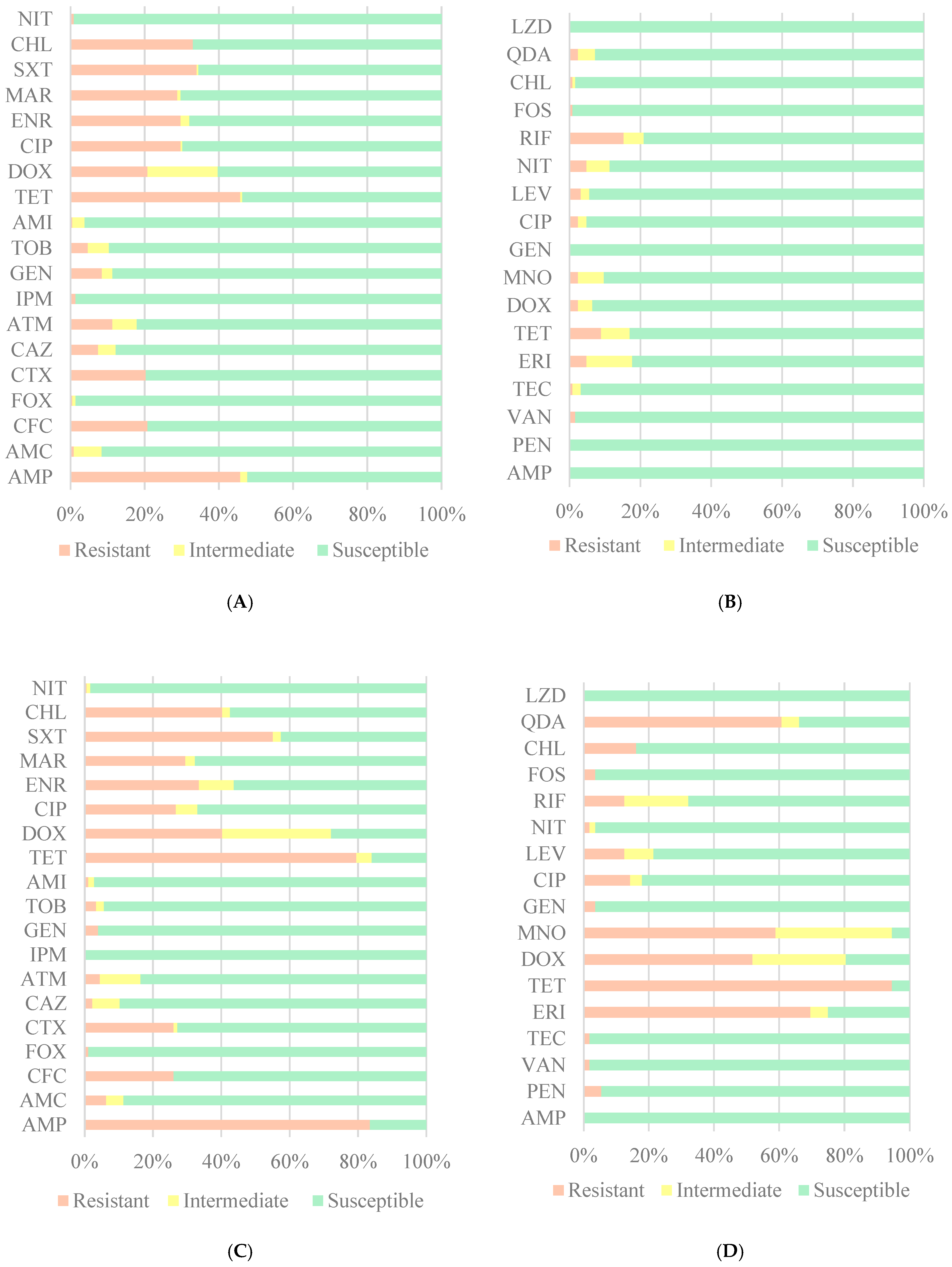
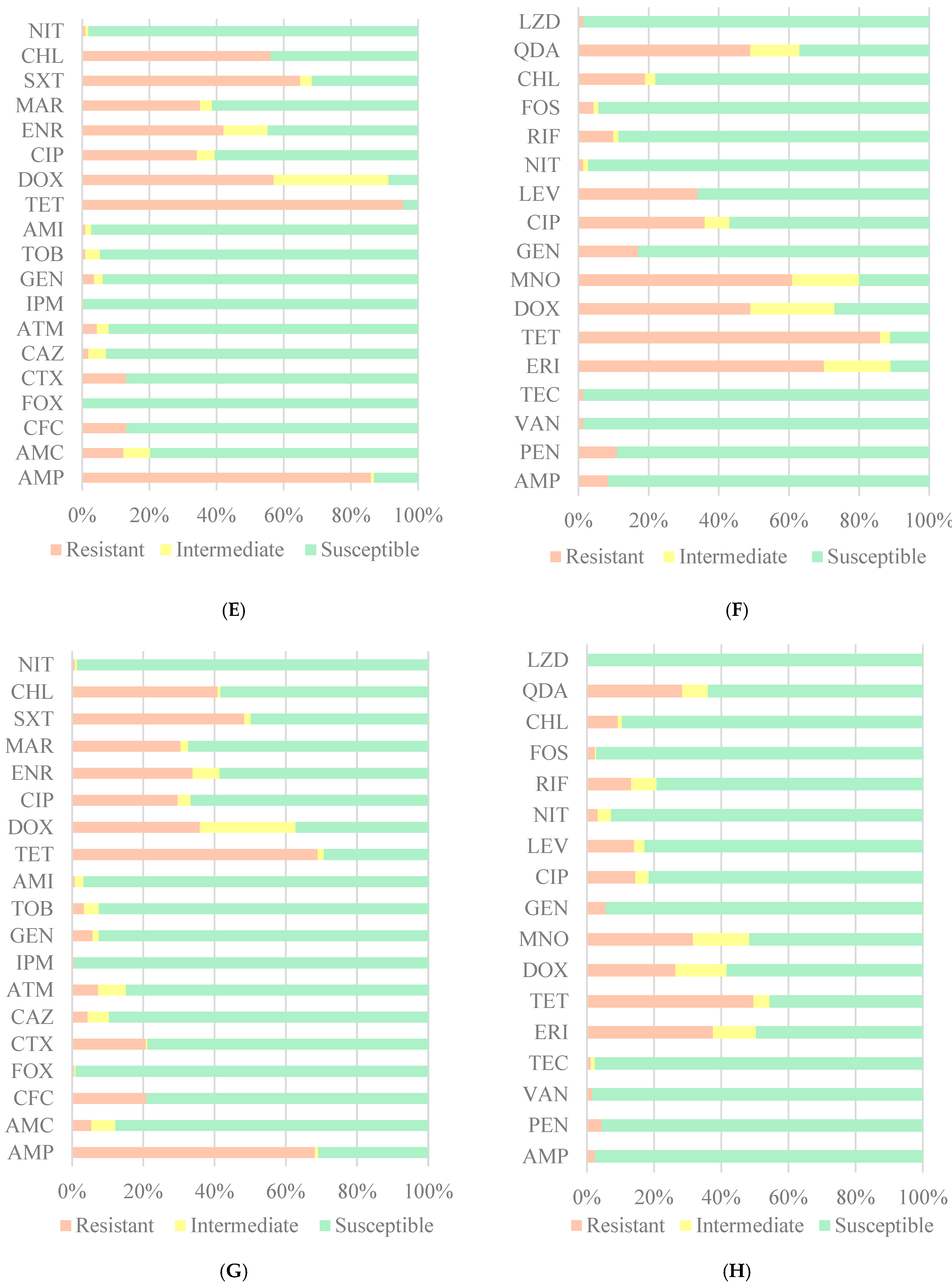
Resistance to at least one antimicrobial was present in 77.5% of E. coli isolates and 62.4% in Enterococcus spp. isolates. E. coli isolates were predominantly resistant to AMP (68.1%) and TET (68.9%) and susceptible to FOX, IPM and NIT in the three types of samples. Enterococcus spp. isolates were predominantly resistant to TET (49.6%) and susceptible to AMP and LZD (Figure 3).
Significant differences between sample types were observed for 13 of the 19 antibiotics in E. coli and 12 of the 17 antibiotics in Enterococcus spp. isolates. These differences mainly resulted from a lower prevalence of resistance strains in CF and a higher prevalence in SS samples.
In the case of TET, the antibiotic with highest resistance frequency, isolates from both swine feces and slurry presented a higher frequency of resistance than isolates from cattle feces. This was true of both E. coli and Enterococcus spp. isolates (Supplementary Materials, Tables S1 and S2, respectively).
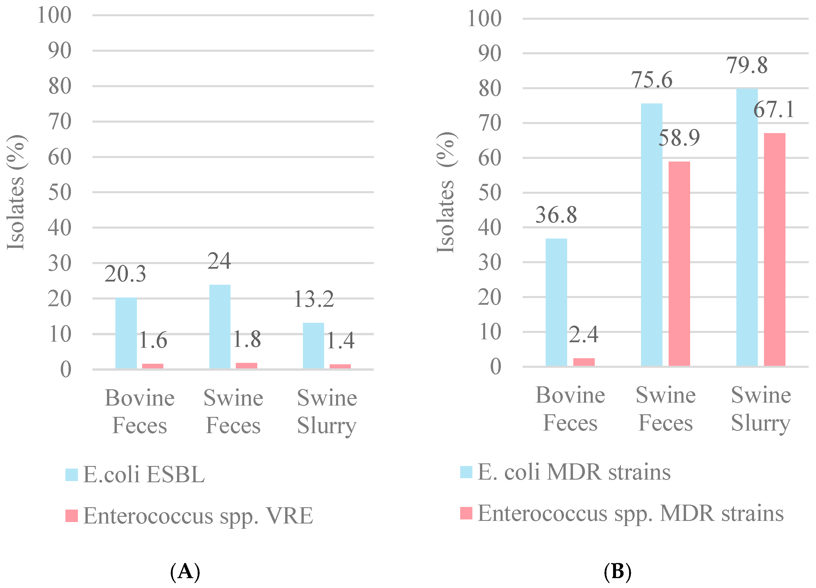
As shown in Figure 4, SF presented the highest percentage of E. coli ESBLs isolates (24.0%) and SS presented the highest percentage of E. coli and Enterococcus spp. MDR isolates (79.8% and 67.1%, respectively). While CF presented the lowest number of MDR isolates, it was the only sample type presenting 2.4% XDR isolates (i.e., those with more than eight classes of antibiotic resistance).
Antimicrobial resistant profiles were compared between sample types (Table 3). For E. coli isolates, twenty-two resistance profiles were common to two sample types and six were common to all three sample types. For Enterococcus spp. isolates, seven resistance profiles were common to two types of samples and two were common to all three types of samples.

4. Discussion

This work aimed at contributing to a better understanding of AMR (antimicrobial resistance) in animal production. A total of 512 E. coli and 250 Enterococcus spp. isolates were recovered from 49 pooled samples of cattle feces, swine feces and swine slurry. The frequency of resistant isolates amounted to 77.5% of E. coli and 62.4% in Enterococcus spp. isolates.
The findings pertaining to antibiotic resistance in E. coli to tetracycline and penicillin (67.6% and 67.0%, respectively) are consistent with those observed in Greece [11] and France [12], with tetracycline (84.2% and 23.1%, respectively) and penicillin (31.0% and 28.1%, respectively) identified as the two most problematic antibiotic resistances. A similar study conducted in Spain also found a prevalence of tetracycline resistances in E. coli (64.9%) and Enterococcus spp. (78.8%) recovered from pig farms [17]. On the other hand, the 2011 Regional Resistance Surveillance study, involving 21 countries in Europe, revealed a prevalence of 66.9% for tetracyclines resistance in E. coli isolated from hospital samples, obtained from the respiratory tract [18,19]. Resistance to tetracycline and penicillin likely arises from the widespread use of this antimicrobial in animal production [20], which contributes to the selection and propagation of resistance strains [8].
The highest percentage of ESBL-producing E. coli and VRE isolates was found in pig feces. It has been demonstrated that isolates from swine feces generally present more resistance to antimicrobials when compared to bovine feces. This can be attributed to the fact that pig production is more intensive than cattle production, and additionally, that their intestinal microbiome is more directly exposed than ruminal microbiota, which has a greater buffer effect [21,22]. A study in cattle has found that the diversity of rectal microbiota had a fastest and greatest decreased in diversity than the ruminal microbiota when exposed to antibiotics [23], suggesting larger susceptibility of the intestinal microbiota. On the other hand, the conditions of pig farms may be prone to antimicrobial challenges leading to the use of a greater quantity of oral antimicrobials [24]. According to the European Veterinary Antibiotic Sales Report, pigs represent a bigger animal mass than cattle (366 vs. 216 PUC, in 1000 tonnes) in Portugal in 2022 [25], which could lead to a higher use of antimicrobials by pig production. Antimicrobial resistance profiles also depend on the animal species and its specific microbiota, husbandry practices including hygiene and diet, as well as the historical presence of antibiotic resistance genes [26]. For instance, swine feed may be cross contaminated with antibiotics, causing a selective pressure in favor of the development of resistant strains [27]. Indeed, the development of bacterial resistance in livestock farms depends on the presence of selective pressures, either by antibiotics and other substances (e.g., heavy metals, biocides), or from reservoirs of resistance genes (e.g., wastewater), which may facilitate horizontal gene transfer [28]. The development of antimicrobial resistance is a multifactorial event.
The prevalence of E. coli was found to predominate over that of Enterococcus spp. across all sample types. However, a higher prevalence of E. coli was observed in fecal samples, while Enterococcus spp. exhibited higher adaptability, with the capacity to survive in both feces and slurry. Despite the higher level of antimicrobial resistance observed in E. coli, its adaptability is comparatively inferior to that of Enterococcus spp., which demonstrates a greater capacity to survive in environments with higher water concentrations, though it does not multiply in such conditions. Consequently, Enterococcus spp. is regarded as a more effective fecal indicator due to its superior longevity outside of fecal matter [29,30].
In swine samples, isolates from slurry generally presented more resistance to antimicrobials than feces. Slurry and farm environments present different profiles of antimicrobial resistance genes compared to pig feces [31]. The environment presents a unique microbiota that facilitates the spread and recirculation of AMR. Moreover, selective pressures may occur in the environment due to the excretion of antimicrobials that accumulate in slurry tanks [32]. For instance, up to 90% of tetracycline administered is excreted in the feces and urine of animals [33,34], being able to exert a selective pressure in the farm environment. Excretion of other antibiotics occurs in their original form varies from 5% to 94% (Supplementary Materials, Table S3). For instance, chlortetracycline has a one day half-life of one day in swine feces, with a corresponding decrease in the abundance of resistance genes (i.e., tet (X) and tet (Q)) [35]. This suggests that non-lethal concentrations of antimicrobial may still exert selective pressures when present in the environment.
The slurry tank can act as a source and/or reservoir for resistant bacteria [36,37], while slurry treatment may not remove all antimicrobials and microorganisms, acting as a source of environmental contamination. Since slurry contains excretions from all animals and presents a unique microbiota with a resistant profile that represent the historical use of antimicrobials in the farm [38], it is more realistic indicators of antimicrobial resistance. Indeed, by sampling slurry, it is possible to better characterize the antimicrobials used on the farm and thus control their use [39].
The release of slurry can contaminate the environment with antibiotics and resistant bacteria, including agricultural crops, leading to the contamination of the food chain [40]. The introduction of AMR bacteria and antimicrobials in agricultural soils can change the resident microbial communities, which are required to maintain soil health [38]. Slurry application accounts for approximately 70% of the total number of antimicrobial-resistant genes in soil, including those for minocycline, tetracycline, streptomycin, gentamicin, kanamycin, amikacin, chloramphenicol and rifampicin [41,42]. Moreover, antibiotics may exert toxic effect on soil microorganisms, possibly altering biochemical processes, and on plants, potentially reducing germination and growth [43].
Tetracycline, one of the most used antibiotics, is largely excreted in the urine and feces after administration [33,34]. It has been found in concentrations of 17.30–2495 mg/kg and 15.90–30.941 mg/kg in the feces of dairy cows and pigs, respectively [44]. Application of manure to farmlands can result in a surface runoff containing up to 35.97 µg/L of tetracyclines, contaminating surface and groundwaters [45]. Concentrations in soil can also reach up to 24.66 mg/kg [46]. Environmental concentrations may induce different responses from bacteria. For instance, low concentrations (0.015–0.03 μg/mL) induced a faster multiplication of E. coli [47], but aerobic sludge bacteria presented an EC50 (i.e., effect concentration of 50%) of 0.08 mg/L [48]. While toxic effects of tetracycline are expected in bacteria (by blocking the 30S subunit of the 16S rRNA) [49], other organisms may also be affected. Concentrations ≥100 mg/L affect the germination, shoot height, root length and mitotic index of wheat (Triticum aestivum L.) [50]. While soil invertebrates can be more resistant, such as Folsomia candida (EC50: 2560 mg/kg) and Eisenia fetida (EC50: 2735 mg/kg), freshwater organisms are more sensitive, such as Daphnia magna (EC50: 8.16 mg/L) and Pseudokirchneriella subcapitata (EC50: 1.82 mg/L) [51]. Indeed, tetracycline has presented a risk quotient of 1.95, representing a high risk, for concentrations found in Wangyang River, China [52]. Therefore, antibiotics can impact ecosystems beyond the development of antimicrobial resistance.
The present study followed CLSI guidelines [16], which focus on standards for antimicrobial susceptibility testing and clinical microbiology, since the objective was to assess the presence of antimicrobial resistance in livestock samples. Another approach, which could be explored in future studies, is the toxicity of antibiotics in different bacteria. In these cases, standardized methods from the International Organization for Standardization (e.g., ISO 10712:1995) [53] and Organization for Economic Co-operation and Development (e.g., OECD test no. 209) [54] should be followed. The complementary information from these tests could contribute to a more thorough understanding of the ecological impacts of antibiotic contamination, while also enabling more informed and effective management strategies [55].
The persistence of antibiotics in soil is dependent on their intrinsic properties and the soil properties, which may be estimated on the basis of the organic carbon–water sorption coefficient (KOC). The degradation of antibiotics with a high KOC is minimal, resulting in a high persistence in the soil. In contrast, antibiotics with a KOC value of less than 15 L/kg are subject to significant degradation in the soil. Tetracyclines are antibiotics with a high KOC, which results in high persistence and the formation of stable bonds with soil sediments [41]. The log Koc of the tested antibiotics vary from −8.78 to 4.24, meaning that some can be persistent (e.g., AMP, CIP, RIF; Supplementary Materials, Table S4).
Antimicrobial susceptibility profiles were common among different samples. While common profiles were expected between swine feces and slurry (as these samples may belong to the same farm), some were also common with cattle feces. Interestingly, these profiles often belong to geographically distant farms, since cattle farms were mainly concentrated in the North of Portugal and swine farms in the Center. These profiles could originate either from similar selective forces (i.e., the common use of antibiotics in both species; sales detailed in Table S5) or from circulating strains. Moreover, the highest percentage of MDR was observed in E. coli isolates across all sample types. This finding suggests that this bacterium exhibits a significantly higher level of AMR compared to Enterococcus spp.
As seen in this study, farm slurry is an environmental source of AMR contamination and/or selective pressure through the release of bioactive antimicrobials, posing a possible threat to public health. Other sources of human exposure include antibiotic residues in animal products [56,57]. In addition to the presence of AMR in feces and the persistence of antimicrobials in the environment, resistance genes can also persist in soil [58]. These may contribute to vertical and horizontal propagation of resistance genes. In 2019, it was estimated that 1.3 million deaths were caused by resistant bacteria [1]. The presence of antibiotics and resistant bacteria in the food chain, through agriculture, can also change the human gut microbiome [59].
The problem of antimicrobial resistance has seen improvements over recent years due to the implementation of various measures in the EU aimed at reducing antibiotic consumption both in human and veterinary medicine. However, the fact that it requires a significant period without antibiotic selective pressure for resistance frequencies to begin to regress [60] means that these measures have not yet had an immediate impact. Antimicrobial resistance should be regarded as a matter of urgent public concern. Humans are dependent on a number of these antibiotics to survive a variety of bacterial infections, and the emergence of multidrug-resistant bacteria will inevitably lead to treatment difficulties [1,61]. The One Health perspective, which states that the health of humans, animals and the environment are directly linked, is the principle behind the measures adopted by the EU [62].
In light of the findings of this study, it would be advantageous for public health to prioritize the treatment of all effluents, particularly those from livestock farms, given the routine administration of antibiotics in these settings to treat a range of infections [32,63,64]. The presence of antibiotics in slurry has the potential to contaminate the environment, thereby facilitating the development of antibiotic resistance. According to Feng Huang “If not effectively treated, they can threaten animal production, public health and the ecological safety of the surrounding environment” [65]. Therefore, it is crucial to develop an effective water treatment system that can fully remove these contaminants [39,65,66,67]. Developments in water treatment technology to remove antibiotics and resistant bacteria are already being put into practice in water treatment centers in some countries. These technologies include disinfection with ultraviolet radiation, water ozonation, solar photocatalysis and membrane bioreactors [68]. New strategies such as bioremediation of antibiotics in contaminated sites and phage therapy are, according to Ezzariai et al. and Ly-Chatain, good bets for controlling antimicrobial resistance [69,70].

5. Conclusions

This study provides new insights into antimicrobial resistance (AMR), including resistance to commonly used antibiotics (e.g., TET) in livestock farms, particularly swine and cattle farms. Our findings highlight the implications for animal, human and environmental health within a One Health framework. While resistance genes can persist for generations, reduced antibiotic exposure may eventually lead to their loss. The identification of resistance patterns and transmission pathways underscores the need for enhanced surveillance and antimicrobial stewardship. Additionally, the study highlights the environmental risks posed by farm slurry, which can disseminate antibiotics and promote bacterial resistance, reinforcing the necessity of improved water purification systems. The integration of microbiological and genomic approaches strengthens our understanding of AMR dynamics, contributing to more effective mitigation strategies. This work emphasizes the urgency of a multidisciplinary approach to combat AMR and protect public health.

Supplementary Materials

The following supporting information can be downloaded at: https://www.mdpi.com/article/10.3390/microbiolres16040083/s1, Table S1: Differences between achieved and expected frequencies of E. coli, by Fisher’s method; Table S2: Differences between achieved and expected frequencies of Enterococcus spp., by Fisher’s method; Table S3: Antibiotic excretion rate; Table S4: Water sorption coefficient (KOC) for ampicillin, ciprofloxacin and rifampicin. Table S5. Antibiotic sales in food-producing animals in weight per population correction unit (mg/PCU; i.e., per estimated weight of the animal) in Portugal in 2022.

Author Contributions

Conceptualization, B.F.P., S.A.M.S. and I.C.R.; methodology, B.F.P. and I.C.R.; software, B.F.P., S.A.M.S. and J.C.P.; formal analysis, B.F.P., S.A.M.S. and J.C.P.; investigation, B.F.P. and S.A.M.S.; resources, P.M.d.C.; data curation, B.F.P., S.A.M.S. and J.C.P.; writing—original draft preparation, B.F.P. and S.A.M.S.; writing—review and editing, J.M.L.-J., J.N.-R., J.C.P. and P.M.d.C.; visualization, B.F.P., J.C.P. and P.M.d.C.; supervision J.C.P. and P.M.d.C.; project administration, J.M.L.-J. and J.N.-R.; funding acquisition, J.M.L.-J. and J.N.-R. All authors have read and agreed to the published version of the manuscript.

Funding

This research was funded by RE-C05-i03 da Agenda de Investigação e Inovação para a Sustentabilidade da Agricultura, Alimentação e Agroindústria grant number 13/C05-i03/2021-PRR-C05-i03-I-000173-Projetos I&D + I Projetos de Investigação e Inovação-UMA SÓ SAÚDE.

Institutional Review Board Statement

Not applicable.

Informed Consent Statement

Not applicable.

Data Availability Statement

All relevant data can be found in the article.

Acknowledgments

We would like to thank all the members of the Abel Salazar Biomedical Sciences Institute Aquatic Production Department, especially Elizabete Lopes for her technical assistance.

Conflicts of Interest

The authors declare no conflicts of interest.

References

  1. WHO. Antimicrobial Resistance. Available online: https://www.who.int/health-topics/antimicrobial-resistance (accessed on 27 March 2024).
  2. Henriques, I.; Freitas, H.; Costa, J.; Silva, V.; Poeta, P.; Pereira, J. One Health: Um Planeta, Uma Saúde, Uma Ética; Avenida D. Carlos I, n° 134-5° andar 1200-651; CNECV: Lisbon, Portugal, 2023. [Google Scholar]
  3. Comissão Europeia. Ban on Antibiotics as Growth Promoters in Animal Feed Enters into Effect; European Commission: Brussels, Belgium, 2005; Available online: https://ec.europa.eu/commission/presscorner/detail/en/ip_05_1687 (accessed on 13 January 2025).
  4. Stockton, B.; Davies, M. Antibiotic Use Plummets on US Farms After Ban on Using Drugs to Make Livestock Grow Faster. TBIJ. Available online: https://www.thebureauinvestigates.com/stories/2018-12-19/antibiotic-use-falls-on-us-farms-after-ban-on-using-drugs-to-make-livestock-grow-faster (accessed on 13 January 2025).
  5. Gaskins, H.R.; Collier, C.T.; Anderson, D.B. Antibiotics as growth promotants: Mode of action. Anim. Biotechnol. 2002, 13, 29–42. [Google Scholar] [CrossRef]
  6. Beyene, T. Veterinary Drug Residues in Food-animal Products: Its Risk Factors and Potential Effects on Public Health. J. Vet. Sci. Technol. 2016, 7, 285. [Google Scholar] [CrossRef]
  7. Silva, V.; Igrejas, G.; Pereira, J.; Maltez, L.; Poeta, P. Resistências aos Antibióticos no Contexto “One Health”: Um Mundo, uma Luta, uma só Saúde; Avenida D. Carlos I, n° 134-5° andar 1200-651; CNECV: Lisbon, Portugal, 2023. [Google Scholar]
  8. INSA. Vigilância Epidemiológica das Resistências aos Antimicrobianos Categoria. Available online: https://www.dgs.pt/upload/membro.id/ficheiros/i018609.pdf (accessed on 19 April 2024).
  9. Burmeister, A.R. Horizontal Gene Transfer. Evol. Med. Public Health 2015, 2015, 193–194. [Google Scholar] [CrossRef]
  10. Magiorakos, A.-P.; Srinivasan, A.; Carey, R.B.; Carmeli, Y.; Falagas, M.E.; Giske, C.G.; Harbarth, S.; Hindler, J.F.; Kahlmeter, G.; Olsson-Liljequist, B.; et al. Multidrug-resistant, extensively drug-resistant and pandrug-resistant bacteria: An international expert proposal for interim standard definitions for acquired resistance. Clin. Microbiol. Infect. 2012, 18, 268–281. [Google Scholar] [CrossRef]
  11. Christodoulou, M.K. Comparison of Antimicrobial Resistance in Escherichia coli Strains Isolated From Swine, Poultry, and Farm Workers in the Respective Livestock Farming Units in Greece. Cureus 2023, 15, e51073. [Google Scholar] [CrossRef]
  12. Boireau, C.; Cazeau, G.; Jarrige, N.; Calavas, D.; Madec, J.-Y.; Leblond, A.; Haenni, M.; Gay, É. Antimicrobial resistance in bacteria isolated from mastitis in dairy cattle in France, 2006–2016. J. Dairy Sci. 2018, 101, 9451–9462. [Google Scholar] [CrossRef]
  13. EFSA; ECDC. The European Union One Health 2022 Zoonoses Report. 2023. Available online: https://onlinelibrary.wiley.com/doi/abs/10.2903/j.efsa.2023.8442 (accessed on 4 January 2024).
  14. Brito, T.; Portugal, C.; Sancho, L.; Carvalho, C.; Grima, B.; Alves, J.D. Sensibilidade das Enterobacteriáceas Produtoras de Beta-Lactamases de Espectro Alargado à Fosfomicina. Trimestral 2015, 22, 131–135. Available online: http://www.spmi.pt/revista/vol22/vol22_n3_2015_131_135.pdf (accessed on 31 March 2024).
  15. Levitus, M.; Rewane, A.; Perera, T.B. Vancomycin-Resistant Enterococci; StatPearls Publishing: Treasure Island, FL, USA, 2023. Available online: http://www.ncbi.nlm.nih.gov/books/NBK513233/ (accessed on 31 March 2024).
  16. CLSI. Performance Standards for Antimicrobial Susceptibility Testing, 28th ed.; Clinical and Laboratory Standards Institute: Wayne, PA, USA, 2018. [Google Scholar]
  17. Mencía-Ares, O.; Argüello, H.; Puente, H.; Gómez-García, M.; Manzanilla, E.G.; Álvarez-Ordóñez, A.; Carvajal, A.; Rubio, P. Antimicrobial resistance in commensal Escherichia coli and Enterococcus spp. is influenced by production system, antimicrobial use, and biosecurity measures on Spanish pig farms. Porc. Health Manag. 2021, 7, 27. [Google Scholar] [CrossRef]
  18. Grossman, T.H. Tetracycline Antibiotics and Resistance. Cold Spring Harb. Perspect. Med. 2016, 6, a025387. [Google Scholar] [CrossRef]
  19. Jones, R.N.; Flonta, M.; Gurler, N.; Cepparulo, M.; Mendes, R.E.; Castanheira, M. Resistance surveillance program report for selected European nations (2011). Diagn. Microbiol. Infect. Dis. 2014, 78, 429–436. [Google Scholar] [CrossRef]
  20. Ferreira, I. Caracterização da Utilização de Antimicrobianos em Produção Animal-Alimentos Medicamentosos em Suinicultura.pdf. Dissertação de Mestrado, Faculdade de Medicina Veterinária da Universidade de Lisboa, Lisbon, Portugal. 2014. Available online: https://www.repository.utl.pt/bitstream/10400.5/7093/1/Caracteriza%C3%A7%C3%A3o%20da%20Utiliza%C3%A7%C3%A3o%20de%20Antimicrobianos%20em%20Produ%C3%A7%C3%A3o%20Animal-Alimentos%20Medicamentosos%20em%20Suinicultura.pdf (accessed on 27 June 2024).
  21. Barton, M.D. Impact of antibiotic use in the swine industry. Curr. Opin. Microbiol. 2014, 19, 9–15. [Google Scholar] [CrossRef]
  22. Dinis, J. Estratégias Nutricionais na Prevençao de Doenças Digestivas dos Suínos. Dissertação de Mestrado; Faculdade de Medicina Veterinária da Universidade de Lisboa: Lisbon, Portugal, 2010. [Google Scholar]
  23. Ji, S.; Jiang, T.; Yan, H.; Guo, C.; Liu, J.; Su, H.; Alugongo, G.M.; Shi, H.; Wang, Y.; Cao, Z.; et al. Ecological Restoration of Antibiotic-Disturbed Gastrointestinal Microbiota in Foregut and Hindgut of Cows. Front. Cell. Infect. Microbiol. 2018, 8, 79. [Google Scholar] [CrossRef]
  24. Monger, X.C.; Gilbert, A.-A.; Saucier, L.; Vincent, A.T. Antibiotic Resistance: From Pig to Meat. Antibiotics 2021, 10, 1209. [Google Scholar] [CrossRef]
  25. EMA. Sales of Veterinary Antimicrobial Agents in 31 European Countries in 2022—Trends from 2010 to 2022—Thirteenth ESVAC Report; European Union: Brussels, Belgium, 2023. [Google Scholar] [CrossRef]
  26. Rothrock, M.J.; Min, B.R.; Castleberry, L.; Waldrip, H.; Parker, D.; Brauer, D.; Pitta, D.; Indugu, N. Antibiotic resistance, antimicrobial residues, and bacterial community diversity in pasture-raised poultry, swine, and beef cattle manures. J. Anim. Sci. 2021, 99, skab144. [Google Scholar] [CrossRef]
  27. Suárez, C.S.-S.; Sanders, P.; Gaugain, M.; Viel, A.; Paboeuf, F.; Taillandier, J.-F.; Houée, P.; Valentin, C.; Perrin-Guyomard, A. Selection of antibiotic resistance in pigs after exposure to feed cross-contaminated with oxytetracycline. Vet. Microbiol. 2023, 287, 109924. [Google Scholar] [CrossRef]
  28. Guo, K.; Zhao, Y.; Cui, L.; Cao, Z.; Zhang, F.; Wang, X.; Feng, J.; Dai, M. The Influencing Factors of Bacterial Resistance Related to Livestock Farm: Sources and Mechanisms. Front. Anim. Sci. 2021, 2, 650347. [Google Scholar] [CrossRef]
  29. Boehm, A.B.; Sassoubre, L.M. Enterococci as Indicators of Environmental Fecal Contamination. In Enterococci: From Commensals to Leading Causes of Drug Resistant Infection; Gilmore, M.S., Clewell, D.B., Ike, Y., Shankar, N., Eds.; Eye and Ear Infirmary: Boston, MA, USA, 2014. Available online: http://www.ncbi.nlm.nih.gov/books/NBK190421/ (accessed on 21 June 2024).
  30. WHO. Guidelines for Drinking-Water Quality: Fourth Edition Incorporating the First and Second Addenda; WHO Guidelines Approved by the Guidelines Review Committee; World Health Organization: Geneva, Switzerland, 2022. Available online: http://www.ncbi.nlm.nih.gov/books/NBK579461/ (accessed on 21 June 2024).
  31. Mencía-Ares, O.; Cabrera-Rubio, R.; Cobo-Díaz, J.F.; Álvarez-Ordóñez, A.; Gómez-García, M.; Puente, H.; Cotter, P.D.; Crispie, F.; Carvajal, A.; Rubio, P.; et al. Antimicrobial use and production system shape the fecal, environmental, and slurry resistomes of pig farms. Microbiome 2020, 8, 164. [Google Scholar] [CrossRef]
  32. Barathe, P.; Kaur, K.; Reddy, S.; Shriram, V.; Kumar, V. Antibiotic pollution and associated antimicrobial resistance in the environment. J. Hazard. Mater. Lett. 2024, 5, 100105. [Google Scholar] [CrossRef]
  33. Sarmah, A.K.; Meyer, M.T.; Boxall, A.B.A. A global perspective on the use, sales, exposure pathways, occurrence, fate and effects of veterinary antibiotics (VAs) in the environment. Chemosphere 2006, 65, 725–759. [Google Scholar] [CrossRef]
  34. Zhu, Y.-G.; Johnson, T.A.; Su, J.-Q.; Qiao, M.; Guo, G.-X.; Stedtfeld, R.D.; Hashsham, S.A.; Tiedje, J.M. Diverse and abundant antibiotic resistance genes in Chinese swine farms. Proc. Natl. Acad. Sci. USA 2013, 110, 3435–3440. [Google Scholar] [CrossRef]
  35. Joy, S.R.; Li, X.; Snow, D.D.; Gilley, J.E.; Woodbury, B.; Bartelt-Hunt, S.L. Fate of antimicrobials and antimicrobial resistance genes in simulated swine manure storage. Sci. Total Environ. 2014, 481, 69–74. [Google Scholar] [CrossRef]
  36. Hendriksen, R.S.; Munk, P.; Njage, P.; Van Bunnik, B.; McNally, L.; Lukjancenko, O.; Röder, T.; Nieuwenhuijse, D.; Pedersen, S.K.; Kjeldgaard, J.; et al. Global monitoring of antimicrobial resistance based on metagenomics analyses of urban sewage. Nat. Commun. 2019, 10, 1124. [Google Scholar] [CrossRef]
  37. Ibekwe, A.M.; Bhattacharjee, A.S.; Phan, D.; Ashworth, D.; Schmidt, M.P.; Murinda, S.E.; Obayiuwana, A.; Murry, M.A.; Schwartz, G.; Lundquist, T.; et al. Potential reservoirs of antimicrobial resistance in livestock waste and treated wastewater that can be disseminated to agricultural land. Sci. Total Environ. 2023, 872, 162194. [Google Scholar] [CrossRef]
  38. Chessa, L.; Jechalke, S.; Ding, G.-C.; Pusino, A.; Mangia, N.P.; Smalla, K. The presence of tetracycline in cow manure changes the impact of repeated manure application on soil bacterial communities. Biol. Fertil. Soils 2016, 52, 1121–1134. [Google Scholar] [CrossRef]
  39. Xu, C.; Kong, L.; Gao, H.; Cheng, X.; Wang, X. A Review of Current Bacterial Resistance to Antibiotics in Food Animals. Front. Microbiol. 2022, 13, 822689. [Google Scholar] [CrossRef]
  40. Samtiya, M.; Matthews, K.R.; Dhewa, T.; Puniya, A.K. Antimicrobial Resistance in the Food Chain: Trends, Mechanisms, Pathways, and Possible Regulation Strategies. Foods 2022, 11, 2966. [Google Scholar] [CrossRef]
  41. Cycoń, M.; Mrozik, A.; Piotrowska-Seget, Z. Antibiotics in the Soil Environment—Degradation and Their Impact on Microbial Activity and Diversity. Front. Microbiol. 2019, 10, 338. [Google Scholar] [CrossRef]
  42. Su, J.Q.; Wei, B.; Xu, C.Y.; Qiao, M.; Zhu, Y.G. Functional metagenomic characterization of antibiotic resistance genes in agricultural soils from China. Environ. Int. 2014, 65, 9–15. [Google Scholar] [CrossRef]
  43. Du, L.; Liu, W. Occurrence, fate, and ecotoxicity of antibiotics in agro-ecosystems. A review. Agron. Sustain. Dev. 2012, 32, 309–327. [Google Scholar] [CrossRef]
  44. Chang, D.; Mao, Y.; Qiu, W.; Wu, Y.; Cai, B. The Source and Distribution of Tetracycline Antibiotics in China: A Review. Toxics 2023, 11, 214. [Google Scholar] [CrossRef]
  45. Huang, X.; Chen, C.; Zeng, Q.; Ding, D.; Gu, J.; Mo, J. Field study on loss of tetracycline antibiotics from manure-applied soil and their risk assessment in regional water environment of Guangzhou, China. Sci. Total Environ. 2022, 827, 154273. [Google Scholar] [CrossRef]
  46. Ji, X.; Shen, Q.; Liu, F.; Ma, J.; Xu, G.; Wang, Y.; Wu, M. Antibiotic resistance gene abundances associated with antibiotics and heavy metals in animal manures and agricultural soils adjacent to feedlots in Shanghai; China. J. Hazard. Mater. 2012, 235–236, 178–185. [Google Scholar] [CrossRef]
  47. Migliore, L.; Rotini, A.; Thaller, M.C. Low Doses of Tetracycline Trigger the E. coli Growth: A Case of Hormetic Response. Dose-Response 2013, 11, 550–557. [Google Scholar] [CrossRef]
  48. Halling-Sørensen, B.; Sengeløv, G.; Tjørnelund, J. Toxicity of Tetracyclines and Tetracycline Degradation Products to Environmentally Relevant Bacteria, Including Selected Tetracycline-Resistant Bacteria. Arch. Environ. Contam. Toxicol. 2002, 42, 263–271. [Google Scholar] [CrossRef]
  49. EFSA. Maximum Levels of Cross-Contamination for 24 Antimicrobial Active Substances in Non-Target Feed. Part 12: Tetracyclines: Tetracycline, Chlortetracycline, Oxytetracycline, and Doxycycline; EFSA Journal; Wiley Online Library: Hoboken, NJ, USA, 2021; Available online: https://efsa.onlinelibrary.wiley.com/doi/full/10.2903/j.efsa.2021.6864 (accessed on 1 April 2025).
  50. Xie, X.; Zhou, Q.; Lin, D.; Guo, J.; Bao, Y. Toxic effect of tetracycline exposure on growth, antioxidative and genetic indices of wheat (Triticum aestivum L.). Environ. Sci. Pollut. Res. 2011, 18, 566–575. [Google Scholar] [CrossRef]
  51. Havelkova, B.; Beklova, M.; Kovacova, V.; Hlavkova, D.; Pikula, J. Ecotoxicity of selected antibiotics for organisms of aquatic and terrestrial ecosystems. Neuro Endocrinol. Lett. 2016, 37 (Suppl. 1), 38–44. [Google Scholar]
  52. Xu, L.; Zhang, H.; Xiong, P.; Zhu, Q.; Liao, C.; Jiang, G. Occurrence, fate, and risk assessment of typical tetracycline antibiotics in the aquatic environment: A review. Sci. Total Environ. 2021, 753, 141975. [Google Scholar] [CrossRef]
  53. ISO 10712:1995; Water Quality—Pseudomonas Putida Growth Inhibition Test (Pseudomonas Cell Multiplication Inhibition Test). ISO: Geneva, Switzerland, 1995. Available online: https://www.iso.org/standard/18800.html (accessed on 1 April 2025).
  54. OECD. Test No. 209: Activated Sludge, Respiration Inhibition Test (Carbon and Ammonium Oxidation); OECD: Paris, France, 2010; Available online: https://www.oecd.org/en/publications/test-no-209-activated-sludge-respiration-inhibition-test_9789264070080-en.html (accessed on 1 April 2025).
  55. Strotmann, U.; Durand, M.-J.; Thouand, G.; Eberlein, C.; Heipieper, H.J.; Gartiser, S.; Pagga, U. Microbiological toxicity tests using standardized ISO/OECD methods—Current state and outlook. Appl. Microbiol. Biotechnol. 2024, 108, 454. [Google Scholar] [CrossRef]
  56. Ascensão, F. Aproveitamento do Leite de Rejeição na Alimentação de Vitelos: Implicação nas Resistências aos Antimicrobianos. Dissertação de Mestrado, Instituto de Ciências Biomédicas Abel Salazar da Universidade do Porto, Porto, Portugal. 2022. Available online: https://repositorio-aberto.up.pt/bitstream/10216/141011/2/555309.pdf (accessed on 2 October 2023).
  57. Bacanlı, M.G. The two faces of antibiotics: An overview of the effects of antibiotic residues in foodstuffs. Arch. Toxicol. 2024, 98, 1717–1725. [Google Scholar] [CrossRef]
  58. Han, B.; Ma, L.; Yu, Q.; Yang, J.; Su, W.; Hilal, M.G.; Li, X.; Zhang, S.; Li, H. The source, fate and prospect of antibiotic resistance genes in soil: A review. Front. Microbiol. 2022, 13, 976657. [Google Scholar] [CrossRef]
  59. Kumar, S.B.; Arnipalli, S.R.; Ziouzenkova, O. Antibiotics in Food Chain: The Consequences for Antibiotic Resistance. Antibiotics 2020, 9, 688. [Google Scholar] [CrossRef]
  60. Uddin, T.M.; Chakraborty, A.J.; Khusro, A.; Zidan, B.R.M.; Mitra, S.; Bin Emran, T.; Dhama, K.; Ripon, K.H.; Gajdács, M.; Sahibzada, M.U.K.; et al. Antibiotic resistance in microbes: History, mechanisms, therapeutic strategies and future prospects. J. Infect. Public Health 2021, 14, 1750–1766. [Google Scholar] [CrossRef]
  61. UNEP—UN Environment Programme. Antimicrobial Resistance: A Global Threat. Available online: http://www.unep.org/topics/chemicals-and-pollution-action/pollution-and-health/antimicrobial-resistance-global-threat (accessed on 11 July 2024).
  62. Robinson, T.P.; Bu, D.P.; Carrique-Mas, J.; Fèvre, E.M.; Gilbert, M.; Grace, D.; Hay, S.I.; Jiwakanon, J.; Kakkar, M.; Kariuki, S.; et al. Antibiotic resistance is the quintessential One Health issue. Trans. R. Soc. Trop. Med. Hyg. 2016, 110, 377–380. [Google Scholar] [CrossRef]
  63. Ilurdoz, M.S.; Sadhwani, J.J.; Reboso, J.V. Antibiotic removal processes from water & wastewater for the protection of the aquatic environment—A review. J. Water Process Eng. 2022, 45, 102474. [Google Scholar] [CrossRef]
  64. Wu, J.; Wang, J.; Li, Z.; Guo, S.; Li, K.; Xu, P.; Ok, Y.S.; Jones, D.L.; Zou, J. Antibiotics and antibiotic resistance genes in agricultural soils: A systematic analysis. Crit. Rev. Environ. Sci. Technol. 2023, 53, 847–864. [Google Scholar] [CrossRef]
  65. Huang, F.; Hong, Y.; Mo, C.; Huang, P.; Liao, X.; Yang, Y. Removal of antibiotic resistance genes during livestock wastewater treatment processes: Review and prospects. Front. Vet. Sci. 2022, 9, 1054316. [Google Scholar] [CrossRef]
  66. Dunai, A.; Spohn, R.; Farkas, Z.; Lázár, V.; Györkei, Á.; Apjok, G.; Boross, G.; Szappanos, B.; Grézal, G.; Faragó, A.; et al. Rapid decline of bacterial drug-resistance in an antibiotic-free environment through phenotypic reversion. eLife 2019, 8, e47088. [Google Scholar] [CrossRef]
  67. Fang, P.; Elena, A.X.; Kunath, M.A.; Berendonk, T.U.; Klümper, U. Reduced selection for antibiotic resistance in community context is maintained despite pressure by additional antibiotics. ISME Commun. 2023, 3, 52. [Google Scholar] [CrossRef]
  68. Mutuku, C.; Gazdag, Z.; Melegh, S. Occurrence of antibiotics and bacterial resistance genes in wastewater: Resistance mechanisms and antimicrobial resistance control approaches. World J. Microbiol. Biotechnol. 2022, 38, 152. [Google Scholar] [CrossRef]
  69. Ezzariai, A.; Hafidi, M.; Khadra, A.; Aemig, Q.; El Fels, L.; Barret, M.; Merlina, G.; Patureau, D.; Pinelli, E. Human and veterinary antibiotics during composting of sludge or manure: Global perspectives on persistence, degradation, and resistance genes. J. Hazard. Mater. 2018, 359, 465–481. [Google Scholar] [CrossRef]
  70. Ly-Chatain, M.H. The factors affecting effectiveness of treatment in phages therapy. Front. Microbiol. 2014, 5, 51. [Google Scholar] [CrossRef] [PubMed]
Figure 1. Sample processing diagram.
Microbiolres 16 00083 g001
Figure 2. Example of antibiotics disk tests in E. coli (A) and Enterococcus spp. (B) antibiograms.
Microbiolres 16 00083 g002
Figure 3. Antimicrobial susceptibility of E. coli isolates for cow feces (A), swine feces (C), swine slurry (E) and for all samples (G) and of Enterococcus spp. for cow feces (B), swine feces (D), swine slurry (F) and for all samples (H), classified according to CLSI guidelines [16].
Microbiolres 16 00083 g003aMicrobiolres 16 00083 g003b
Figure 4. Percentage of (A) E. coli ESBL and Enterococcus VRE isolates and of (B) multidrug-resistant (MDR) isolates of E. coli and Enterococcus spp., found in the three types of samples: cow feces (CF), swine feces (SF) and swine slurry (SS).
Microbiolres 16 00083 g004
Table 1. Antibiotics used in Antimicrobial Susceptibility Testing in Escherichia coli and Enterococcus spp., based on antibiotics and concentrations set by the Clinical and Laboratory Standards Institute (CLSI) guidelines [16].
Escherichia coliEnterococcus spp.
AbbreviationsAntibioticsConcentrations (μg)AbbreviationsAntibioticsConcentrations (μg)
AMIAmikacin30AMPAmpicillin30
AMCAmoxicillin-clavulanate30CHLChloramphenicol30
AMPAmpicillin10CIPCiprofloxacin5
ATMAztreonam30GENGentamicin120
CHLChloramphenicol30DOXDoxycycline30
CAZCeftazidime30ERIErythromycin15
CIPCiprofloxacin5NITNitrofurantoin300
GENGentamicin30FOSFosfomycin200
CTXCefotaxime30LEVLevofloxacin5
DOXDoxycycline30LZDLinezolid30
ENREnrofloxacin5MNOMinocycline30
NITNitrofurantoin300PENPenicillin G30
FOXCefoxitin30QDAQuinupristin-dalfopristin15
IPMImipenem10RIFRifampicin30
CFCCefazolin30TETTetracycline30
MARMarbofloxacin5TECTeicoplanin30
SXTTrimethoprim-sulfamethoxazole25VANVancomycin30
TETTetracycline30
TOBTobramycin10
Table 2. Number of samples collected, and number of E. coli and Enterococcus spp. isolates obtained, as well as tetracycline (TET) resistant strains.
Samples (n)Isolates (n)TET Resistance (n)
E. coliEnterococcus spp.E. coliEnterococcus spp.
CF2421212497 (45.8%)11 (8.9%)
SF1417656140 (79.5%)53 (94.6%)
SS1111470109 (95.6%)60 (85.7%)
Total49502250346 (68.9%)124 (49.6%)
CF, cattle feces; SF, swine feces; SS, swine slurry; n, number.
Table 3. Number of common antimicrobial resistant profiles to different sample types.
E. coliEnterococcus spp.
Cow Feces/Swine Feces61
Cow Feces/Swine Slurry21
Swine Feces/Swine Slurry145
Cow Feces/Swine Feces/Swine Slurry62
Disclaimer/Publisher’s Note: The statements, opinions and data contained in all publications are solely those of the individual author(s) and contributor(s) and not of MDPI and/or the editor(s). MDPI and/or the editor(s) disclaim responsibility for any injury to people or property resulting from any ideas, methods, instructions or products referred to in the content.