Article

The Fungus Phlebiopsis flavidoalba’s Pathogenicity and Virulence Toward the Fluted Scale (Praelongorthezia acapulcoa) Pest of Rice and Sugarcane Crops

by
Silvia Hernández-Hernández
1,
Facundo Muñiz-Paredes
2,
Guadalupe Peña-Chora
3 and
Víctor Manuel Hernández-Velázquez
1,*
1
Laboratorio de Control Biológico, Centro de Investigación en Biotecnología (CEIB) Universidad Autónoma del Estado de Morelos (UAEM), Cuernavaca C.P.62000, Mexico
2
Laboratorio de Biología Molecular de Hongos, Centro Nacional de Referencia de Control Biológico, Colima C.P.28100, Mexico
3
Laboratorio de Parasitología Vegetal, Centro de Investigaciones Biológicas, Universidad Autónoma del Estado de Morelos (UAEM), Cuernavaca C.P.62000, Mexico
*
Author to whom correspondence should be addressed.
Microbiol. Res. 2024, 15(4), 2414-2424; https://doi.org/10.3390/microbiolres15040162
Submission received: 9 September 2024 / Revised: 11 November 2024 / Accepted: 14 November 2024 / Published: 26 November 2024

Abstract

:
Sugarcane is one of the main crops in Morelos State, Mexico. The presence of the insect pest Praelongorthezia acapulcoa (Morrison), commonly known as the fluted scale insect, was observed in sugarcane and rice crops, causing losses of up to 30% of production in both crops. In this work, a fungus isolated from the mycosic cadavers of P. acapulcoa was identified as the basidiomycete Phlebiopsis flavidoalba (Cooke) Hjortstam via morphological and molecular identification using the ITS, tef 1, and 28S regions. Its pathogenicity toward P. acapulcoa was verified in laboratory tests, causing a mortality rate higher than 60%. Its virulence toward P. acapulcoa, estimated as the mean lethal concentration (LC50), was 9.7 × 10⁶ conidia mL−1. This work constitutes the first report about a basidiomycete with direct entomopathogenic activity and biological control of the fluted scale insect, P. acapulcoa.

1. Introduction

Sugarcane (Saccharum officinarum L.), a Gramineae, is an economically important crop in the State of Morelos, Mexico. It is the third most important crop nationally in terms of area, with 20,419 hectares, and the first in terms of production value [1]. Since 2009, the presence of the insect Praelongorthezia acapulcoa (Hemiptera: Ortheziidae) (Morrison) has been observed in sugarcane crops in the State of Morelos. Before 2009, there were no reports of this insect as a pest of any crop in this state [2]. Furthermore, it was observed in fields of rice in Xochitepec in 2018, as well as in each year from 2009 to August 2024 in Morelos. Rice is another economically important crop in this state [2].
Praelongorthezia acapulcoa is a scale insect belonging to the family Ortheziidae. Members of the genus Praelongorthezia are commonly recognized as citrus pests; however, they are actually polyphagous insects [3]. They are sap-sucking insects that excrete honeydew, causing fumagine on leaves and preventing proper photosynthesis and plant development [4]. This pest is more abundant during the rainy season (Plant Health Committee of the State of Morelos (CSVMOR), personal communication).
The adult female P. acapulcoa has an unusual appearance compared to that of other insects, with its ovoid body measuring about three millimeters in length. However, it appears larger, since its body is covered by wax plates; it is completely covered by white secretion tufts, with the dorsal tufts strongly erect, diverging apically laterally from the median line in early adults and tending to curve backward as the tufts grow in older specimens [5].
The control of P. acapulcoa has been carried out using chemical pesticides; however, the scale insect appears year after year in sugarcane crops (CSVMOR, personal communication). Therefore, some environmentally friendly alternatives are currently being explored. Entomopathogenic fungi are an interesting alternative to chemical pesticides; their mode of action is by contact [6], and they can be used to control a wide diversity of pests [7].
More than 750 species of fungi that infect insects have been described; however, only 100 genera and 700 species of entomopathogenic fungi are known. The genera Beauveria spp. and Metarhizium spp. are currently the most widely used entomopathogenic fungi for the control of insect pests [8].
They have a worldwide distribution and have been found in diverse systems such as trees, leaves, and decaying soil. Some of them belong to the basidiomycetes group, as is the case of the fungus Phlebiopsis gigantea (Polyporales: Phanerochaetaceae), (Fr.) Jülich, which grows on conifer trunks and has activity against the lepidopteran Hyblaea puera (Lepidoptera: Hyblaeidae), (Fr.) Cramer [9]. The insecticidal activity of some fungi is due to the action of enzymes, such as proteases, xylanases, phenols, alkaloids, and flavonoids, as described in [10].
One work reported that Phlebiopsis flavidoalba was grown in the absence of wood, showing low-density polyethylene (LDP) biodegradation with a weight reduction of 23.68 ± 0.34%, which was further supported by a 46.79 ± 0.67% mass loss and 3.07 ± 0.13% CO2 emissions (mg/L) in the stripping test. This indicates that wood-decaying fungi can utilize any available carbon source in the absence of the preferred C source; hence, these fungi have the metabolic adaptability to survive under stressful conditions.
Fungal entomopathogenesis is known to be based on several molecular and biochemical mechanisms in extended gene families, involving virulence factors including some specialized metabolites such as cytochrome P450, lipases, chitinases, and proteases. This suggests that fungi acquire some gene products during their evolution when they come into contact with hosts. Isolation of new fungal entomopathogens may be based on the insect bait method or the search for insect cadavers with mycosis [11]. The isolation of native entomopathogenic fungi for pest control may facilitate the development of new biological control agents (BCAs), since these native fungi are already adapted to the biotic and abiotic factors that affect their efficacy and persistence [12]. Some basidiomycetes such as the oyster mushroom (Pleurotus ostreatus) produce some proteins whose mechanism of action is known, such as osteolysis A6 (OlyA6), which binds to the sphingomyelin/cholesterol domains of the membrane and, with the help of its partner protein pleurotolysin B (PlyB), forms 13-meric transmembrane pore complexes. This protein is known to have the ability to bind 1000 times stronger to the insect-specific membrane sphingolipid, phosphoethanolamine ceramide (PEC). In conjunction with PlyB, OlyA6 confers potent and selective insecticidal activity against western corn rootworm (Diabrotica virgera zea), an insect belonging to the order Coleoptera and the family Chrysomelidae [13].
In this work, cadavers of P. acapulcoa with white mycelia were collected from sugarcane crops. The isolated fungus was morphologically and molecularly identified and evaluated in terms of its pathogenicity and virulence toward Praelongorthezia acapulcoa under laboratory conditions.

2. Materials and Methods

2.1. Fungal Isolation

Mycosic corpses of Praelongorthezia acapulcoa were collected from sugarcane fields in Xochitepec, Morelos, Mexico (18°48′1″ N 99°14′48″ W). Mycelia were gently scraped from the cadaver surface and streaked onto Petri dishes containing potato dextrose agar. The insect corpses that did not show visible signs of mycosis were placed in a humid chamber in an incubator at 27 °C to germinate spores that could possibly be inside the insects, for which the following methodology was employed.
The insect was immersed in 1% sodium hypochlorite for 5 min; it was then rinsed four times with sterile distilled water; sterile filter paper was placed in a sterile Petri dish, and sterile distilled water was added; the insect was placed on the filter paper inside the dish; finally, the dish was sealed with parafilm®. Once the insects were covered with mycelia, the mycotic surface was scraped off to inoculate Petri dishes with PDA. The Petri dishes were incubated at 27 °C in an incubator until the agar surface was completely covered with mycelia. Each morphologically different mycelium was restreaked on a fresh Petri dish, and the process was repeated until a unique morphology was observed. During our field explorations, the SH52 isolate was obtained, which was particularly interesting for further investigation, as its pathogenicity on scale insects had not been demonstrated in preliminary studies. For sporulation, the isolate was streaked on Sabouraud dextrose agar enriched with sucrose (8 gL−1) and yeast extract (8 gL−1).

2.2. Morphological Identification

The morphological characteristics of monosporic cultures were observed. Microcultures were prepared in Sabouraud dextrose agar (SDA), incubated at 27 °C, and observed after 5 and 12 days with a Leica DM 500 microscope (Danaher company, Wasshington, DC, USA) at 100×. Micrographs of the mycelia and the morphology of the conidia were obtained.

2.3. DNA Extraction

Genomic DNA extraction was performed with modifications, using the methodology from [14]. Fresh mycelia of monosporic cultures were obtained from plates containing SDA medium (65 gL−1) enriched with sucrose (8 gL−1) and yeast extract (8 gL−1) and incubated at 27 °C for 12 days. The mycelia were scraped and placed in a sterilized mortar, 400 µL of lysis buffer was added, and the mycelia were homogenized for 3 min with a pestle. The homogenized mycelia were placed in a microtube (≈400 µL), and 6 µL of RNase A was added. The microtube was incubated for 10 min at 56 °C. Then, 130 µL of 3 M sodium acetate at pH 5.2 was added, and the microtube was incubated at 4 °C for 30 min. At the end of the incubation, the microtube was centrifuged at 13,000 rpm for 5 min. The supernatant (≈350 µL) was transferred to a fresh microtube, and 350 µL of isopropanol was added. The microtube was shaken by inversion and incubated at −20 °C for 30 min. The microtube was centrifuged at 13,000 rpm for 5 min, and the supernatant was discarded. The DNA pellet was washed with 700 µL of 70% ethanol and then centrifuged at 13,000 rpm for 5 min. The supernatant was discarded, and the pellet was washed again with 95% ethanol. The pellet was air-dried and dissolved in 100 µl of TE buffer.

2.4. Molecular Identification

The ITS, 28S [15], and tef 1 [16] regions were used for molecular identification. The primers used for the sequencing of the regions are detailed in Table 1.
PCRs were performed in a total volume of 25 µL using Platinum ™Taq DNA Polymerase (Invitrogen), according to the manufacturer’s instructions. The ITS region was amplified as follows: 94 °C for 2 min and 35 cycles of 94 °C for 30 s, 55 °C for 30 s, and 72 °C for 1 min. The PCR conditions for tef 1 were as follows: 94 °C for 2 min; 10 cycles of 94 °C for 30 s, 66 °C for 30 s, and 72 °C for 1 min and 10 s; 30 cycles of 94 °C for 30 s, 56 °C for 30 s, and 72 °C for 1 min and 10 s; 72 °C for 5 min. For the 28S region, PCR was performed as follows: 94 °C for 2 min; 34 cycles of 94 °C for 30 s, 47.5 °C for 30 s, and 72 °C for 80 s; 72 °C for 5 min [16].
The sequences obtained were edited in BioEdit (7.2.5) and uploaded to GenBank with the accession numbers OQ955295 (ITS), OQ954494 (tef 1), and OQ955294 (28S). A concatenated dataset of the ITS-28S sequences was constructed for phylogenetic analysis. The sequences were first aligned separately with MEGA 11 using MUSCLE and trimmed to the same size (598 pb for ITS and 870 pb for 28S). Then, the sequences were concatenated, and the best model was defined using MEGA 11 (Kimura 2, Gamma distributed). A phylogenetic tree was constructed using the maximum likelihood method with 1000 replicates. Phlebia acerina MY51 was used as the outgroup. The GenBank accession numbers of all the fungi used in the phylogenetic analysis are listed in Table 2.

2.5. Pathogenicity and Virulence

The pathogenicity and virulence of the SH52 isolate were determined in the pre-reproductive-phase females of reared P. acapulcoa.
The insect colony was maintained in plastic containers with cane leaves, at a temperature of 27 °C with alternating periods of 12 h of light and 12 h of darkness, in the laboratory of Control Biológico at the Centro de Investigación en Biotecnología, UAEM.
The design used was completely randomized, and each experimental unit consisted of a container with sugar cane leaves in which 12 pre-reproductive-stage female insects were placed, and to each container a different treatment was applied; each treatment consisted of the suspension of spores of the SH52 isolate at different concentrations, and the pathogenicity was evaluated at a concentration of 1 × 10⁷ conidia mL−1.
For the virulence, the mean lethal concentration (LC50) was evaluated using five conidial suspension concentrations (1 × 10⁵, 1 × 10⁶, 1 × 10⁷, 1 × 10⁸, and 1 × 10⁹ conidia mL−1). The experimental unit consisted of one plastic container with leaves and 12 females of P. acapulcoa. Each conidial concentration was evaluated in triplicate. After 12 days, the number of dead insects was determined, and the LC50 was estimated via probit analysis (PoloPlus software 1.0).
In both cases, a negative control was used, which consisted of immersing the insects in water with 0.01% Tween 80. They were kept at a temperature of 27 °C, at 12:12 h light/dark, and monitoring was carried out for 12 days; then, the number of dead insects was counted.
The insects and sugar cane leaves were disinfected in a 0.1% sodium hypochlorite solution and rinsed with sterile distilled water; this procedure was repeated once more. Three more washes were then carried out with sterile distilled water only; finally, the leaves were placed in a laminar flow hood on sterile paper until dry.
To obtain the conidial suspension, 10 mL of 0.01% Tween 80 solution was added to monosporic cultures of the SH52 isolate grown in Petri dishes with SDA for 14 days; then, the suspension was recovered with a micropipette and placed in a 50 mL microtube. The conidial suspensions were adjusted to the required concentration by preparing dilutions and counting them with the aid of a Neubauer chamber.
The insects were inoculated via immersion in a conidial solution (1 × 10⁷ conidia mL−1) for 30 s [17]. For the negative control, the insects were submerged in 0.01% Tween solution [18]. The insects were placed in plastic containers (1 L capacity) and fed with sugarcane leaves (leaves were mounted on moistened floral sponges). The plastic containers were incubated at 27 °C with a 12:12 h light/dark photoperiod. The percentage of mortality was determined on day 12 (the time at which the leaves were depleted); however, the plastic containers were checked daily, and dead insects were placed in a humid chamber to corroborate mycosis. The pathogenicity was determined in triplicate.
For the virulence, the mean lethal concentration (LC50) was evaluated using five conidial suspension concentrations (1 × 10⁵, 1 × 10⁶, 1 × 10⁷, 1 × 10⁸, and 1 × 10⁹ conidia mL−1) with the same system described for the pathogenicity bioassay. The experimental unit consisted of one plastic container with leaves and 12 females of P. acapulcoa. Each conidial concentration was evaluated in triplicate. The negative control was treated, as described above. After 12 days, the number of dead insects was determined, and the LC50 was estimated by means of probit analysis (PoloPlus software 1.0) [19]

2.6. Statistical Analysis

For the pathogenicity test, a completely randomized design was carried out in which the experimental unit consisted of a plastic container with a capacity of 1 L, with the lid previously perforated to facilitate insect respiration, in which sugar cane leaves were placed, and a floral sponge was used to support the leaves. Twelve insects in the pre-reproductive adult female stage were placed in each container.
The mortality was measured at the end of the experiment, and in the case of pathogenicity, a Student’s t-test (α = 0.05) was performed using SPSS software (ver. 18.0. SPSS Inc., IBM, Chicago, IL, USA) to compare the means of the percentage of mortality between the treatment and the negative control.
To calculate the mean lethal concentration, the number of insects killed by the application of the 5 concentrations of conidia evaluated was determined, and the LC50 was estimated via probit analysis (PoloPlus 1.0 software) [16].
The mean lethal time was calculated and was estimated via probit analysis (PoloPlus 1.0 software) [16].

3. Results

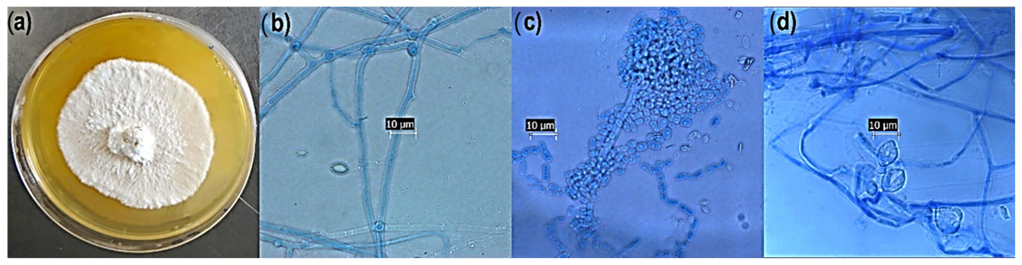
3.1. Morphological Identification

The macromorphology of isolate SH52 showed a white velvety mycelium, with a hard consistency and raised surface when grown on SDA medium (Figure 1a), consistent with the report of [15]. Exudates were noticeable after 14 days of growth. Microcultures were observed under an optical microscope (100×) showing a monomitic hyphal system, with hyaline septate hyphae (Figure 1b). Arthrospores, as a result of hyphal fragmentation, and ellipsoid basidiospores were also present (Figure 1c). Thick crystals (10 µm) were observed (Figure 1d) and were consistent with other reports about the Phlebiopsis genera [16]. There may not be enough observable morphological characters present to identify the isolate at a species level; however, this does not diminish the significance of the findings.

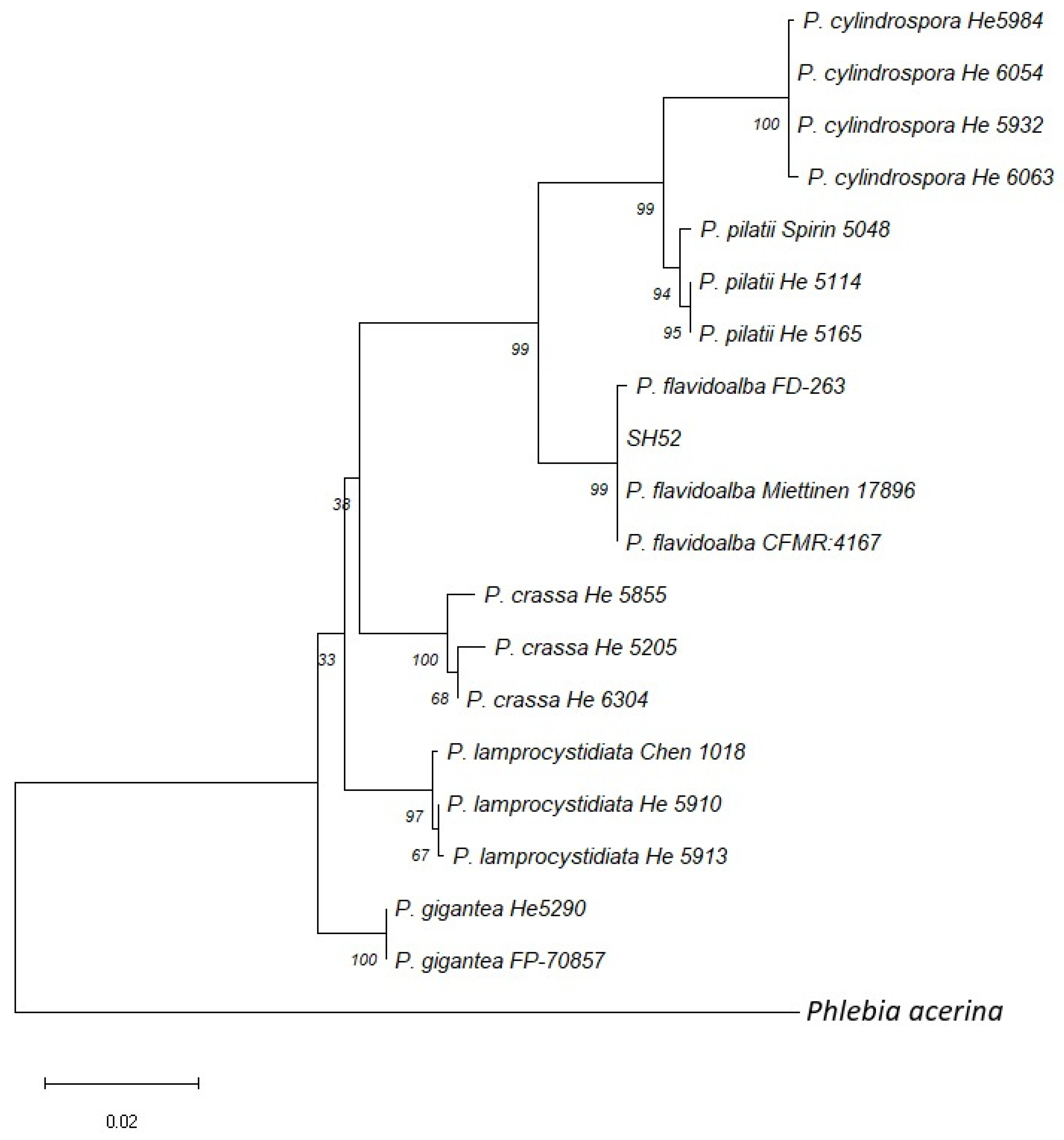
3.2. Molecular Identification and Phylogenetic Analysis

BLAST analysis (NCBI) of the ITS, tef 1, and 28S regions showed 90.6 to 100% identity with Phlebiopsis spp. A multilocus sequencing typing (MLST) analysis with concatenation of the ITS and 28S regions was performed to establish the phylogeny of the isolate SH52. The tef 1 region was excluded from the MLST analysis due to its underrepresentation in databases. A concatenated ITS-28S dataset from 20 fungal specimens from six different Phlebiopsis species and the outgroup was constructed. The concatenated alignment consisted of 1468 pb with 144 parsimony informative sites (106 and 38 from the ITS and 28S regions, respectively). The phylogenetic analysis established strong phylogenetic support (99%) of the isolate SH52 with the Phlebiopsis flavidoalba clade (Figure 2). The morphological and molecular results confirmed the identity of isolate SH52 as Phlebiopsis flavidoalba.

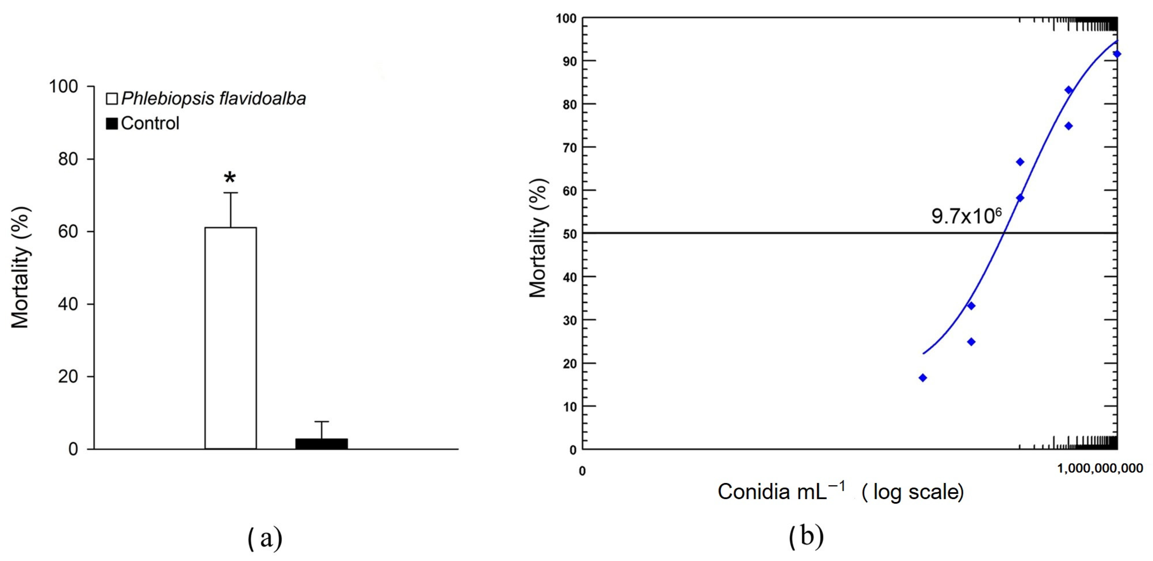
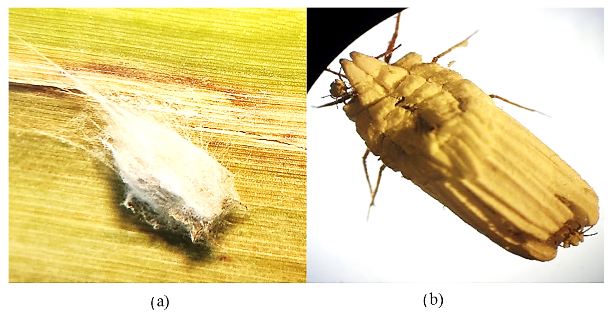
3.3. Pathogenicity and Virulence of Phlebiopsis flavidoalba SH52 Toward Praelongorthezia acapulcoa

The pathogenicity of P. flavidoalba SH52 was evaluated at a concentration of 1 × 10⁷ conidia mL−1. Insect mortality was observed three days after exposure to the fungus, reaching 61.1% mortality at day six. However, the experiment was continued until day 12, and the mean lethal time was calculated, resulting in 139.54 h, equivalent to 5.8 days, which is consistent with the mean lethal dose since 50% of the insects died before day 6.
The control group showed 2.7% mortality, and no mycelia were observed in those cadavers. The Student’s t-test indicated that there was a significant difference between the two values (p < 0.001). A probit analysis was performed to calculate the LC50, testing five concentrations, which yielded a mean lethal concentration of 9.7 × 10⁶ conidia mL−1, as shown in Figure 3.
Mycosis of insect cadavers was confirmed under a stereoscopic microscope (Figure 4a), and the mycelia generally developed eight days after the application of the fungal treatment.

4. Discussion

Praelongorthezia acapulcoa is a widely distributed pest in Morelos State, and in 2018, it was reported in five municipalities [20]. However, the pest was detected in seven municipalities in the present study. Based on field observations, the long-term control of P. acapulcoa using aqueous chemical pesticides may be limited, in part, due to the waxy coating produced by the insect [20]. Fungi as biological control agents represent a promising alternative to aqueous chemical pesticides, due to the hydrophobic properties of their propagule surfaces [21], which may facilitate attachment to the insect cuticle. Field explorations led to the identification of P. acapulcoa cadavers with visible signs of mycosis, from which P. flavidoalba SH52 was isolated and morphologically and molecularly identified.
The purpose of evaluating pre-reproductive females is because they still have the ovisac without the presence of the operculum through which stage 1 nymphs emerge when the fungus suspension is applied, and as soon as they emerge they remain in a gregarious state during the first molt and are more likely to be infected by the conidia applied to the pre-reproductive female. In stage 2, they are dispersed throughout the plant and are also expected to act as vectors of infection for the next molt, which is another advantage.
The basidiomycete P. flavidoalba is a wood-decaying fungus [22], and to the best of our knowledge, there are no reports describing its entomopathogenic capacity. Most commercial entomopathogenic fungi belong to the phylum Ascomycota (Beauveria spp., Metarhizium spp., and Isaria spp.). Nonetheless, some members of the Basidiomycota group are also known as entomopathogens [23]; for example, Septobasidium spp. are well known for infecting scale insects [24]. Ethyl acetate extracts from Pleurotus ostreatus and crude extracts from liquid cultures of the basidiomycete Pycnoporus cinnabarinus showed insecticidal activity against the rose aphid Macrosiphum rosae and larvae of Diatreaea magnifactella [25,26]. Basidiomycetes have bioactive compounds such as phenols, alkaloids, flavonoids, and lytic enzymes. The pathogenicity of Phlebiopsis flavidoalba SH52 was evaluated according to Koch’s postulates, showing that it was responsible for the death of P. acapulcoa. The LC50 of P. flavidoalba SH52 was similar to that of other reports using common entomopathogenic fungi against other pests [10] and two orders of magnitude lower than the LC50 of Phlebiopsis gigantea against the lepidopteran Hyblaea puera [9]. There are no reports on the biological control of P. acapulcoa or, as previously stated, the use of P. flavidoalba as a BCA; thus, comparisons with other BCAs should be made with caution. In terms of virulence, the LC50 of P. flavidoalba SH52 toward P. acapulcoa was comparable to that of Septobasidium sp. against Phyllophaga sp., according to [26], and other commercial entomopathogenic fungi [24], according to Selvaraj and Kaushik 2014 and Zhang et al. 2018 [27,28].
A mortality rate of 61% for Phlebiops under laboratory conditions would likely be considered low; however, so far, there is no universal ranking of whether a mortality rate is high, low, or medium. It is known that for a control, the mortality should not be higher than 20%; hence, it could be considered that any value higher than the maximum allowed for a control already indicates some potential as a biological control agent.
In conclusion, field explorations permitted the isolation of the basidiomycete P. flavidoalba SH52, which is capable of controlling P. acapulcoa, a pest of sugarcane and rice in the State of Morelos. Additional studies related to conidial production and quality, persistence under field conditions, and host range are necessary to determine its potential as a new BCA. This constitutes the first report of P. flavidoalba as a fungal entomopathogen and its use as a potential biological control agent of P. acapulcoa.

Author Contributions

Conceptualization, V.M.H.-V. and S.H.-H.; methodology, F.M.-P. and S.H.-H.; software, V.M.H.-V., F.M.-P. and S.H.-H.; validation, V.M.H.-V., F.M.-P. and G.P.-C.; formal analysis, V.M.H.-V. and F.M.-P.; investigation, V.M.H.-V.; resources, V.M.H.-V., F.M.-P. and G.P.-C.; data curation, F.M.-P. and S.H.-H.; writing—original draft preparation, S.H.-H.; writing—review and editing, F.M.-P., V.M.H.-V. and S.H.-H.; visualization, F.M.-P. and S.H.-H.; supervision, V.M.H.-V.; project administration, V.M.H.-V.; funding acquisition, F.M.-P. and V.M.H.-V. All authors have read and agreed to the published version of the manuscript.

Funding

This research received no external funding.

Institutional Review Board Statement

Not applicable.

Informed Consent Statement

Not applicable.

Data Availability Statement

The original contributions presented in this study are included in the article. Further inquiries can be directed to the corresponding authors.

Acknowledgments

The authors thank Marco Antonio Díaz Manjarrez (CESVMOR) for his valuable support during the field explorations and the Centro Nacional de Referencia de Control Biológico for the molecular identification of P. flavidoalba SH52.

Conflicts of Interest

The authors declare no conflicts of interest.

References

  1. Murphy, R. Sugarcane: Production Systems, Uses and Economic Importance; Nova Publishers: Hauppauge, NY, USA, 2017; ISBN 9781536108989.2017. [Google Scholar]
  2. Carapia Ruiz, V.E. Pralengorthezia Acapulcoa (MORRISON) (HEMIPTERA: ORTHEZIIDAE) UNA NUEVA PLAGA PARA LA CAÑA DE AZUCAR. Investig. Agropecu. 2009, 6, 163–172. [Google Scholar]
  3. Colombia Ministerio de Agricultura y Desarrollo Rural; Corpoica; Universidad de Antioquia. Cítricos Cultivo, Poscosecha e Industrialización; Corporación Universitaria Lasallista: Caldas, Colombia, 2012; ISBN 9789588406176.
  4. Chomnunti, P.; Hongsanan, S.; Aguirre-Hudson, B.; Tian, Q.; Peršoh, D.; Dhami, M.K.; Alias, A.S.; Xu, J.; Liu, X.; Stadler, M.; et al. The Sooty Moulds. Fungal Divers. 2014, 66, 1–36. [Google Scholar] [CrossRef]
  5. Konczné Benedicty, Z.; Kozár, F. Revision of Newsteadia (Homoptera: Coccoidea) of the Nearctic and Neotropic regions, with descriptions of new species. Acta Phytopathol. Entomol. Hung. 2001, 36, 123–142. [Google Scholar] [CrossRef]
  6. Afifah, L.; Aena, A.C.; Saputro, N.W.; Kurniati, A.; Maryana, R.; Lestari, A.; Abadi, S.; Enri, U. Maize Media Enhance the Conidia Production of Entomopathogenic Fungi Lecanicillium Lecanii Also Its Effective to Control the Weevil Cylas Formicarius (Fabricius) (Coleoptera: Brentidae). Agrivita 2022, 44, 513–525. [Google Scholar] [CrossRef]
  7. Serna-Domínguez, M.G.; Andrade-Michel, G.Y.; Rosas-Valdez, R.; Castro-Félix, P.; Arredondo-Bernal, H.C.; Gallou, A. High Genetic Diversity of the Entomopathogenic Fungus Beauveria bassiana in Colima, Mexico. J. Invertebr. Pathol. 2019, 163, 67–74. [Google Scholar] [CrossRef]
  8. Kidanu, S.; Hagos, L. Entomopathogenic Fungi as a Biological Pest Management Option: A Review. Int. J. Res. Stud. Agric. Sci. 2020, 6, 1–10. [Google Scholar] [CrossRef]
  9. Biji, C.P.; Sudheendrakumar, V.V.; Shabna, S. Fungal Endophytes, Phlebiopsis gigantea (Fr.) Jillich and Phanerochaete sordida (P. Karst.) J. Erikss and Ryvarden: New Aspirants in Biopesticide Scenario. J. Biol. Control 2011, 25, 114–117. [Google Scholar] [CrossRef]
  10. Gökçe, A.; Er, M.K. First Description of the Disease by Conidiobolus Osmodes on Tipula Paludosa Larvae with the Report of a Natural Epizootic. J. Invertebr. Pathol. 2003, 84, 83–89. [Google Scholar] [CrossRef]
  11. Zemek, R.; Konopická, J.; Jozová, E.; Skoková Habuštová, O. Virulence of Beauveria bassiana Strains Isolated from Cadavers of Colorado Potato Beetle, Leptinotarsa decemlineata. Insects 2021, 12, 1077. [Google Scholar] [CrossRef]
  12. Jaronski, S.T. Ecological Factors in the Inundative Use of Fungal Entomopathogens. BioControl 2010, 55, 159–185. [Google Scholar] [CrossRef]
  13. Milijaš Jotić, M.; Panevska, A.; Iacovache, I.; Kostanjšek, R.; Mravinec, M.; Skočaj, M.; Zuber, B.; Pavšič, A.; Razinger, J.; Modic, Š.; et al. Dissecting Out the Molecular Mechanism of Insecticidal Activity of Ostreolysin A6/Pleurotolysin B Complexes on Western Corn Rootworm. Toxins 2021, 13, 455. [Google Scholar] [CrossRef] [PubMed]
  14. Raja, H.A.; Miller, A.N.; Pearce, C.J.; Oberlies, N.H. Fungal Identification Using Molecular Tools: A Primer for the Natural Products Research Community. J. Nat. Prod. 2017, 80, 756–770. [Google Scholar] [CrossRef] [PubMed]
  15. Zhao, Y.N.; He, S.H.; Nakasone, K.K.; Wasantha Kumara, K.L.; Chen, C.C.; Liu, S.L.; Ma, H.X.; Huang, M.R. Global Phylogeny and Taxonomy of the Wood-Decaying Fungal Genus Phlebiopsis (Polyporales, Basidiomycota). Front. Microbiol. 2021, 12, 622460. [Google Scholar] [CrossRef]
  16. Chen, C.C.; Chen, C.Y.; Wu, S.H. Species Diversity, Taxonomy and Multi-Gene Phylogeny of Phlebioid Clade (Phanerochaetaceae, Irpicaceae, Meruliaceae) of Polyporales. Fungal Divers. 2021, 111, 337–442. [Google Scholar] [CrossRef]
  17. Domínguez-Galdámez, O.M.; Oliva-Llaven, M.Á.; Aguilar-Tipacamú, G.; Mendoza-Nazar, P.; Ruiz-Sesma, B.; Bautista-Trujillo, G.U.; Antonio, F. Evaluation of Beauveria sp. Strains, Conidial Concentration and Immersion Times on Mortality Rate of Bovine Tick (Boophilus sp). J. Appl. Biol. Biotechnol. 2016, 4, 64–68. [Google Scholar] [CrossRef]
  18. Demirci, S.N.Ş.; Altuntaş, H. Entomopathogenic Potential of Purpureocillium lilacinum against the Model Insect Galleria mellonella (Lepidoptera: Pyralidae). Environ. Exp. Biol. 2019, 17, 71–74. [Google Scholar] [CrossRef]
  19. Lei, C.; Sun, X. Comparing Lethal Dose Ratios Using Probit Regression with Arbitrary Slopes. BMC Pharmacol. Toxicol. 2018, 19, 61. [Google Scholar] [CrossRef]
  20. Carapia-Ruiz, V.E.; Romero-Hernández, J.P.; Hernández-Velázquez, V.M. Primer Reporte de la Escama Bandera Pralengorthezia acapulcoa1 en Arroz, en Morelos, México. Southwest. Entomol. 2019, 44, 349–351. [Google Scholar] [CrossRef]
  21. Wang, J.; Lovett, B.; St Leger, R.J. The Secretome and Chemistry of Metarhizium; a Genus of Entomopathogenic Fungi. Fungal Ecol. 2018, 38, 7–11. [Google Scholar] [CrossRef]
  22. Sánchez-Corzo, L.D.; Álvarez-Gutiérrez, P.E.; Meza-Gordillo, R.; Villalobos-Maldonado, J.J.; Enciso-Pinto, S.; Enciso-Sáenz, S. Lignocellulolytic Enzyme Production from Wood Rot Fungi Collected in Chiapas, Mexico, and Their Growth on Lignocellulosic Material. J. Fungi 2021, 7, 450. [Google Scholar] [CrossRef]
  23. Araújo, J.P.M.; Hughes, D.P. Diversity of Entomopathogens Fungi: Which Groups Conquered the Insect Body? Adv. Genet. 2016, 94, 1–39. [Google Scholar] [CrossRef] [PubMed]
  24. Liu, H.; Bauer, L.S. Susceptibility of Agrilus planipennis (Coleoptera: Buprestidae) to Beauveria bassiana and Metarhizium anisopliae. J. Econ. Entomol. 2006, 99, 1096–1103. [Google Scholar] [CrossRef] [PubMed]
  25. Díaz-Godínez, G.; Téllez-Téllez, M.; Rodríguez, A.; Obregón-Barbosa, V.; De Lourdes Acosta-Urdapilleta, M.; Villegas, E. Enzymatic, Antioxidant, Antimicrobial, and Insecticidal Activities of Pleurotus pulmonarius and Pycnoporus cinnabarinus Grown Separately in an Airlift Reactor. Bioresources 2016, 11, 4186–4200. [Google Scholar] [CrossRef]
  26. Asma Noshad, A.N.; Mudassar Iqbal, M.I.; Zafar Iqbal, Z.I.; Hamida Bibi, H.B.; Saifullah, S.; Salma Bibi, S.B.; Shah, H.U. Aphidicidal potential of ethyl acetate extract from Pleurotus ostreatus. Sarhad J. Agric. 2015, 31, 101–110. [Google Scholar] [CrossRef]
  27. Selvaraj, K.; Kaushik, H.D. Greenhouse Evaluation of Beauveria bassiana (Balsamo) Vuillemin against Aphis craccivora (Das) on Fenugreek. J. Appl. Nat. Sci. 2014, 6, 852–856. [Google Scholar] [CrossRef]
  28. Zhang, X.C.; Li, X.X.; Gong, Y.W.; Li, Y.R.; Zhang, K.L.; Huang, Y.H.; Zhang, F. Isolation, Identification, and Virulence of a New Metarhizium anisopliae Strain on the German Cockroach. J. Econ. Entomol. 2018, 111, 2611–2616. [Google Scholar] [CrossRef]
Figure 1. Morphology of isolate SH52 at five days of growth: (a) growth on SDA medium; (b) septate hyphae; (c) fragmentation of hyphae into arthrospores. Crystals were present after 12 days of growth (d). All micrographs were observed at 100×.
Microbiolres 15 00162 g001
Figure 2. Phylogenetic tree based on concatenation of the ITS and 28S regions. The tree was constructed by the maximum likelihood method (1000 bootstrap replications). Isolate SH52 grouped with the Phlebiopsis flavidoalba clade. The numbers at branch points represent bootstrap support values. Phlebia acerina MY51 was used as the outgroup.
Microbiolres 15 00162 g002
Figure 3. Pathogenicity (a) and virulence (b) of Phlebiopsis flavidoalba toward Praelongorthezia acapulcoa at day 12. The LC50 (9.7 × 10⁶ conidia mL−1) was estimated via probit analysis. The asterisk above the bar represents a significant difference according to Student’s t test (p < 0.001).
Microbiolres 15 00162 g003
Figure 4. Females of Praelongorthezia acapulcoa: (a) mycosic insect (eight days post-exposure to Phlebiopsis flavidoalba); (b) healthy specimen.
Microbiolres 15 00162 g004
Table 1. Primers for the amplification of the regions used for molecular identification.
RegionPrimer NameNucleotide Sequence (5′ → 3′)Reference
ITSITS5GGAAGTAAAAGTCGTAACAAGG[15]
ITS4TCCTCCGCTTATTGATATGC
Tef 1983FGCYCCYGGHCAYCGTGAYTTYAT[16]
2218RATGACACCRACRGCRACRGTYTG
28SLRORGTACCCGCTGAACTTAAGC[15]
LR5CCTGAGGGAAACTTCG
Table 2. GenBank accession numbers of the fungal sequences used for the phylogenetic analysis.
SpeciesITS28S
Phlebiopsis flavidoalba GC 1807-47MZ637050MZ637254
P. flavidoalba FD-263KP135402KP135271
P. flavidoalba Miettinen 17896KP135397KX752607
P. flavidoalba CFMR: 4167KX065957KX065991
Phlebiopsis gigantea He 5290MT386381MT447416
P. gigantea FP-70857KP135390KP135272
Phlebiopsis cylindrospora He 5932MT386404MT447445
P. cylindrospora He 5984MT386404MT447445
P. cylindrospora He 6054MT561716MT598030
P. cylindrospora He 6063MT561717MT598031
Phlebiopsis crassa He 5205MT452523MT447448
P. crassa He 5205MT452523MT447448
P. crassa He 5855MT452525MT447450
P. crassa He 6304MT561714MT598029
Phlebiopsis lamprocystidiata He 5910MT386383MT447419
P. lamprocystidiata He 5913MT386384MT447420
P. lamprocystidiata Chen 1018MT561709GQ470647
Phlebiopsis pilatii He 5114MT386385MT447421
P. pilatii He 5165MT386386MT447422
P. pilatii Spirin 5048KX752590KX752590
Phlebia acerina MY51MK592753PP478794
Disclaimer/Publisher’s Note: The statements, opinions and data contained in all publications are solely those of the individual author(s) and contributor(s) and not of MDPI and/or the editor(s). MDPI and/or the editor(s) disclaim responsibility for any injury to people or property resulting from any ideas, methods, instructions or products referred to in the content.

