Article

Cicer arietinum Extract Suppresses Lung Sepsis Induced by Cecal Ligation and Puncture in Rats

1
Department of Clinical Laboratory Sciences, Faculty of Applied Medical Sciences, University of Bisha, 255, Alnakhil, Bisha 67714, Saudi Arabia
2
Department of Medical Laboratory Sciences, College of Applied Medical Sciences, University of Bisha, Bisha 67714, Saudi Arabia
3
Lecture of Physiology, Basic Medical Science Department, Faculty of Dentistry, AlRyada University for Science and Technology, Cairo 32897, Egypt
*
Author to whom correspondence should be addressed.
Microbiol. Res. 2024, 15(3), 1939-1956; https://doi.org/10.3390/microbiolres15030130
Submission received: 12 August 2024 / Revised: 16 September 2024 / Accepted: 18 September 2024 / Published: 23 September 2024

Abstract

:
Sepsis is characterized by multiple organ dysfunction, which is now accepted to be due to oxidative damage. The lung is the first organ exposed to this damage, and its injury is one of the leading causes of death. Therefore, many pharmacological strategies are employed to attenuate sepsis. This study aimed to evaluate the in silico and in vitro antibacterial activity of Cicer arietinum extract (CAE) against Staphylococcus aureus, Escherichia coli, and Pseudomonas aeruginosa and the in vivo modulatory effect of CAE against sepsis induced by cecal ligation and puncture (CLP) in rats. This study identified seven bioactive components in Cicer arietinum extract, revealing promising interactions between these components and Staphylococcus aureus-PBP2a and Pseudomonas aeruginosa-PBP3 proteins, highlighting their potential as novel antibacterial agents. After ensuring the bactericidal ability of CAE against Staphylococcus aureus and Pseudomonas aeruginosa, an in vivo study was performed. Twenty-four rats were divided into sham-operated rats, CLP-septic rats, CLP rats treated with CAE (500 mg/kg b.wt), and CLP rats treated with hydrocortisone (25 mg/ kg b.wt). CAE was administered orally for 3 days post-operation, and animals were euthanized on the fourth day. Another twenty-four rats were used to study survival for 5 days. This study revealed that CAE, like hydrocortisone, can rescue CLP rats from death by suppressing lung procalcitonin (PCT) and MDA and enhancing SOD, CAT, and GSH levels significantly, as compared with the CLP group. The histopathological results were parallel with the biochemical results since the CLP rats treated with CAE had lower histological/inflammatory scores in the lung like hydrocortisone. The beneficial role of CAE may result from its antibacterial and antioxidant activities, and CAE can be considered as a lung antiseptic extract. This study provides a novel treatment for sepsis-induced ALI. However, the beneficial impact of CAE needs extensive study to obtain evidence.

1. Introduction

Sepsis is defined as the presence of pathogenic bacteria in the bloodstream that trigger systemic inflammatory response syndrome [1]. Despite recent significant studies on the management of the pathogenesis of sepsis, it remains a challenging clinical problem that is responsible for mortality [2]. The ultimate cause of death in patients with severe sepsis is multiple organ failure [3]. Recent investigations reveal that the lung is, frequently, the first affected organ in the sequential development of multiple organ failure induced by sepsis [4,5]. Sepsis of either pulmonary or non-pulmonary origin is the clinical precursor to acute lung injury (ALI) [6]. ALI is one of the respiratory complications that is characterized by the accumulation of a large number of neutrophils in the lungs and eventually leads to morbidity and mortality [4,7]. One hallmark of both sepsis and ALI is a breakdown of the alveolar epithelial barrier that leads to an increase in epithelial permeability and eventually ends with pulmonary edema [8,9]. Unfortunately, one of the most common causes of the rising mortality rate of sepsis is the late prognosis of its occurrence. Therefore, it is important to assess early diagnostic markers for sepsis. Procalcitonin (PCT) is a known biochemical indicator that exhibits greater specificity for the rapid diagnosis and prognosis of sepsis [10]. Thereby, PCT ascertains the sepsis occurrence in animal models and, hence, its analysis can be used to evaluate new antiseptic therapies. One of the causative mechanistic factors contributing to the ALI induced by sepsis is the generation of reactive oxygen species (ROS), due to the formation of free radicals [7]. This occurs due to an imbalance between prooxidant and antioxidant systems [2]. Under normal physiological conditions, a homeostatic balance exists between the formation of oxygen species and their removal by endogenous antioxidant compounds [11]. If the homeostatic balance is impaired, the overproduction of oxygen free radicals occurs followed by organ dysfunction [12]. Thus, estimates of ROS and PCT are considered urgent indices to develop new pharmacological approaches to this life-threatening disease.
Despite the many advanced attempts at sepsis treatment, no satisfactory therapy has been developed for ALI induced by sepsis. Unfortunately, most of the conventional antibiotics that are used for sepsis treatment are resistible by many microbial pathogens due to their overuse [13]. Thus, a new trend in sepsis treatment strategies attracts investigators working in this field. They postulated that since oxidative damage is central to the pathology of sepsis, antioxidants may act as a potential therapy. However, there is little evidence about the beneficial role of using antioxidant supplementation in patients with sepsis and organ dysfunction [14]. Therefore, the present study selects Cicer arietinum (chickpea) seed methanolic extract to evaluate its antibacterial and antiseptic activities in vitro and in vivo. The chickpea is a member of the Fabaceae family, and it is the third most important grain legume in the world based on total grain production, according to the Food and Agriculture Organization (FAO) yearbook production (1992). This study selects C. arietinum extract (CAE) due to the following facts that enable it to be a potent antiseptic supplement: (1) its antioxidant capacity [15] and (2) its in vitro antibacterial activity [16,17]. Thus, firstly, the current work assesses the in vitro antibacterial activity of CAE against the most common septic bacteria (Staphylococcus aureus, Escherichia coli, and Pseudomonas aeruginosa). Secondly, the lung antiseptic potency of the CAE is studied in vivo using a sepsis animal model. To the best of our knowledge, the effect of CAE on sepsis has not been investigated yet. To study the in vivo efficacy of the CAE for the treatment of sepsis, cecal ligation and puncture (CLP) are employed in male albino rats and compared with hydrocortisone (typically administered to septic patients). The present investigation uses CLP, as this sepsis model mimics the pathophysiology of human sepsis [18].

2. Materials and Methods

2.1. In Silico Study

The Penicillin-Binding Protein was chosen as a scarified protein to study the ability of our molecules to bind with the same binding sites of penicillin. The Penicillin-Binding Protein is a good standard model to test the antibacterial activity of CEA. Also, penicillin is the most effective antibacterial drug for the Staphylococcus aureus-PBP2a and Pseudomonas aeruginosa-PBP3 proteins. Therefore, the reason for choosing the Penicillin-Binding Protein is the huge activity of penicillin as an antibacterial drug on Staphylococcus aureus, Escherichia coli, and Pseudomonas aeruginosa. Also, the septic activity of these bacteria and the rapid mutations causing these bacteria to be antibiotic-resistant made it a good model to measure the effect of CEA extract molecules on its main binding sites.
As Jayaprakash and Das [19] mentioned, the GC-MS analysis of Cicer arietinum methanolic extract has 7 bioactive components, as reported in Table 1. By aiding ChemBio office and DrugBank database, the structures of these bioactive components have been retrieved. Also, by using the Protein Data Bank (PDB), the structure and PDB of Staphylococcus aureus-PBP2a (1 MWT) [20] protein and Pseudomonas aeruginosa-PBP3 (3OC2) [21] protein have been retrieved. By using UCSF Chimera Tool (Version 1.18 (build 42505) with a resolution of 3.00 Å, the PDB files were cleaned, missing side chains and loops were built, and the hydrogen bonds were optimized. Water molecules were also eliminated to clear the binding pockets that might confound and distort the pose search. The structure’s energy was verified and optimized via OPLS 2005 force field. This force field was used to add the hydrogen bonds and to determine the bond length. The grid maps were obtained from the ligand-binding sites of the PDB sum. Penicillin G structure has been retrieved from NCBI with PubChem CID (5904) (National Center for Biotechnology Information). Energy minimization and auto-dock vina for Penicillin G molecule against the two selected proteins individually were carried out. Auto Dock Vina Docking was repeated in 10 models, and the lowest binding affinity and zero RMSD were been chosen. The interactions between ligands and receptors were plotted using the Discovery Studio visualizer (v20.1.0.19295). Docking trials were conducted in triplicate (Table 2).

2.2. In Vitro Study

2.2.1. Preparation of CAE

One gram of Cicer arietinum seed powder was soaked in 4 mL of methanol and heated in a water bath at 60 °C for 1 h while being shaken. The obtained extract was centrifuged at 10,000 rpm for 20 min at 5 °C. Then, the obtained supernatant was filtered, concentrated, and lyophilized [22].

2.2.2. Antibacterial Activity of CAE

The potency of the CAE to inhibit the growth of bacteria was evaluated by Kirby–Bauer disc diffusion method [23]. Briefly, sterile Hi-sensitivity Mueller–Hinton agar plates were prepared, poured into Petri dishes, and allowed to solidify. The Petri plates were inoculated with 24 h old cultures of the three selected bacterial strains, which are Staphylococcus aureus ATCC 6538 (Gram-positive), Escherichia coli ATCC 8739, and Pseudomonas aeruginosa ATCC 9027 (Gram-negative). Bacterial strains were obtained from the American Type Culture Collection (ATCC), and the work was made in the Microanalytical center- Faculty of Science-Cairo University. The discs (0.8 cm) were saturated with 100 µL CAE (20 mg/mL), allowed to dry, and introduced into the upper layer of the seeded agar plate. The discs were then pressed lightly on the surface of the plate. The plate was incubated at 37 °C for 72 h and then observed for zone of inhibition. To assess the accurate antibacterial effect of CAE, a positive control (Gentamicin) and a negative control (methanol) were used. Finally, the zone of inhibition (area where there is no growth around the discs) was measured using a millimeter ruler.

2.3. In Vivo Study

2.3.1. Animals

Forty-eight rats were used in this experiment. They were divided into 24 for the main study and 24 for survival study. The Institutional Animal Care and Use Committee (IACUC) (CUFS/F/PHY/52/15) of the Faculty of Science, Cairo University, Egypt, reviewed and approved this experimental protocol, and the care and handling of animals were conducted according to the criteria outlined in the Guide for the Care and Use of Laboratory Animals (8th edition). This study used male albino rats (Rattus norvegicus) weighing 150–170 g purchased from the National Research Center (Egypt). The animals were housed in standard wire cages with sawdust bedding (five animals/cage) in a room maintained at 22 ± 2 °C with an alternating 12 h light/dark cycle. Prior to the initiation of the experiment, the animals were acclimatized for 1 week in the animal house. The animals were fed with standard laboratory animal food pellets and water ad libitum.

2.3.2. CLP Septic Model

In sterile conditions and sodium pentobarbital (30 mg/kg b.wt, i.p) anesthesia, rats underwent CLP operation, as described by Liu et al. [24]. Briefly, after shaving the abdominal fur, an abdominal midline incision was made; then, cecum was exteriorized and ligated just distal to the ileocecal valve to avoid intestinal obstruction. Then, it was punctured twice with an 18-gauge needle, and both holes were gently compressed to extrude small droplets of fecal matter. After that, the cecum was repositioned back into the animal abdominal cavity and the abdominal incision was closed in two layers using a 4-0 silk thread. In parallel, sham animals were subjected to all steps of the procedure, but the cecum was manipulated without being ligated or punctured. Following surgery, the rats were resuscitated with physiologic saline solution (3 mL/kg b.wt) to prevent dehydration. Postoperatively, the rats were housed in their cages and allowed free access to food and water. Then, the following treatments were performed.

2.3.3. Animal Grouping and Treatment Regimen

Twenty-four rats (six rats/each group) were classified into four groups; they were treated immediately after the CLP operation (Table 3). Group I served as a sham group, in which rats received distilled water orally for 3 days. Group II served as CLP-septic group, where rats received distilled water orally once/day for 3 days. Group III is the CLP-treated group, in which rats were administered CAE (500 mg/kg b.wt/day). Group IV is the CLP treated with hydrocortisone (25 mg/ kg b.wt/day), 2 h after CLP orally via gastric gavage for 3 days. The dosage of CAE used in this study was in accordance with the safety evaluation study of Fahmy et al. [25]. Rats were euthanized 24 h later to the last dose, and both lungs were excised and prepared for marker determination.

2.3.4. Sample Preparations

Markers determined in lung homogenate were considered as useful clinical markers in identifying the mechanistic action of the disease in the septic animals. Thereby, the current study detects the PCT and ROS, as they contribute to the sepsis incident mechanism in the lung homogenate.

2.3.5. Preparation of Lung Homogenate

At the 4th day, both lungs were rapidly excised from each euthanized rat and rinsed with ice-cold saline. The right ones were homogenized in homogenization buffer (0.1 M Tris-HCl buffer, pH 7.4). Homogenates were centrifuged at 6000 rpm for 15 min in cold centrifuge (Centurion K280R, Aberdeen, UK). The obtained supernatants were used to determine the following:
PCT level, quantified with an ELISA kit according to the manufacturer’s instructions (Weka Med Supplies Corp., Industrieweg, Belgium).
The oxidative stress (OxS) markers, including lipid peroxides (MDA), glutathione (GSH), superoxide dismutase (SOD), and catalase (CAT) levels, were used to evaluate the antioxidant status in the lung. These markers were determined according to the instruction of the prepared Biodiagnostic kits (Dokki, Giza, Egypt).

2.3.6. Histological Examination

The left lung for each was submerged in 10% buffered formalin for immediate fixation, embedded in paraffin wax, sectioned into 4 µm, and stained with hematoxylin and eosin (H&E). Morphological examinations of the lung tissues were performed in a blind manner by a histopathologist. Two histological sections per group were demonstrated in this study to illustrate the lung tissue structure in many fields in each group. The tissue morphological alterations were analyzed by light microscopy, and the histological scoring system was utilized to grade the severity degree of lung injury, as described by Yamanel et al. [26]. In brief, a scoring system was employed for four histologic features: edema, hemorrhage, leukocyte infiltration, and alveolar septal thickness. Each feature was quantitatively graded on a scale of 0–4 as follows: 0 (absent and appears normal); 1 (light); 2 (moderate); 3 (strong); 4 (intense). Then, the sum of all grades for each feature was calculated as the total score ranging between 0 and 16 (most severe).

2.3.7. Survival Study

Twenty-four rats were divided into sham group, CLP group, CLP + CAE group, and CLP + hydrocortisone (n = 6/group) to study their lifespan after CLP. The treatment protocol is like the abovementioned treatment regimen. This study aimed to assess whether the CAE can rescue rats from sepsis-induced death. Observation was made continuously and began immediately after the CLP operation, while the endpoint was set at 5 days after the CAE and hydrocortisone treatments. The survival rate was expressed as a percentage. The rescued rats were observed to ensure that they are healthy by external heart and respiratory rates and behavioral changes.

2.4. Statistical Analyses

The data of this study were expressed as mean ± SEM of six animals. The difference between groups was analyzed by one-way ANOVA followed by Duncan’s post hoc test. This study used Kruskal–Wallis; then, dual comparisons among groups were performed using the Student–Newman–Keuls method for statistical evaluation of histological scores. Survival data were analyzed by Kaplan–Meier and the log-rank statistics. Values of p < 0.05 were considered as statistically significant. Data analyses were performed using SPSS version 15.0 software.

3. Results

3.1. Docking Results

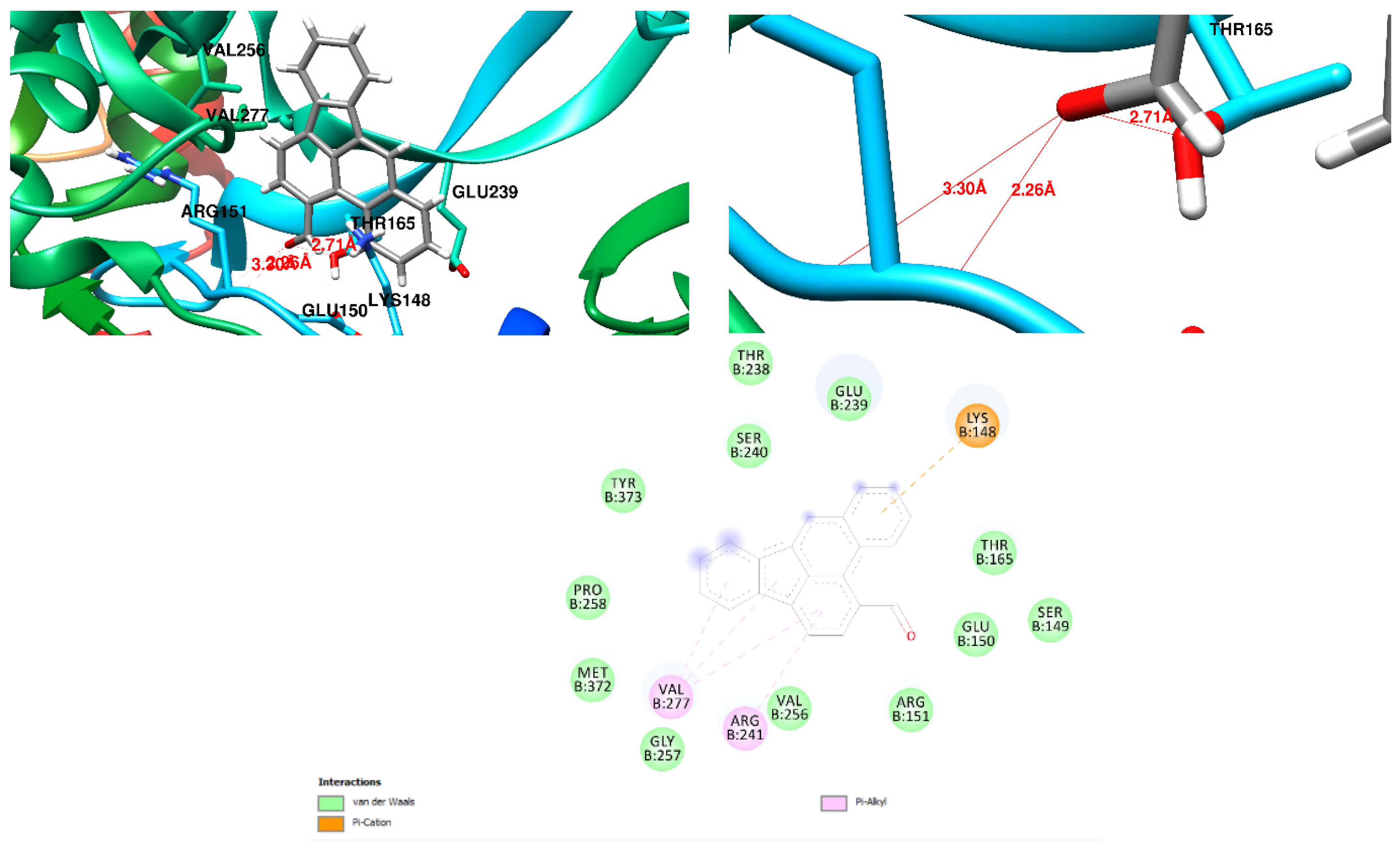
By using Autodock Vina tools, docking of the bioactive components and Penicillin G were performed on Pseudomonas aaeruginosa-PBP3 and Staphylococcus aureus-PBP2a proteins. The (cis)-2-nonadecene showed a docking score −4.3 kcal/mol with Staphylococcus aureus-PBP2a s (Figure 1) and a docking score −5.1 kcal/mol with Pseudomonas aeruginosa-PBP3 (Figure 2). According to the docking scores and molecule stability during the reaction, 1-formylbenzo[b]fluoranthene molecules showed the highest binding score, highest number of hydrogen bonding sites and highest stable molecules during the reaction. By identifying the binding sites of 1-formylbenzo[b]fluoranthene on the Staphylococcus aureus-PBP2a, it was found that the ligand has three hydrogens bound to Lys-148, Glu-150 and Arg-151 residues, respectively (Figure 3), with a docking score −8.6 Kcal/mol, as shown in Table 2. Also, by identifying the binding sites of 1-formylbenzo[b]fluoranthene on the Pseudomonas aeruginosa-PBP3, it was found that the ligand has no hydrogen bound with the receptor with a docking score −9.0 kcal/mol (Figure 4), as shown in Table 2. By identifying the binding sites of 7-Hydroxy-1-methoxy-6-methylanthraquinone on the Staphylococcus aureus-PBP2a, it was found that the ligand has no hydrogen bonds with a docking score −6.4 kcal/mol, as shown in Table 2. Further, 1-Tetradecene (CAS) showed a docking score with 4.3 kcal/mol Staphylococcus aureus-PBP2a (Figure 5) and a docking score −4.7 kcal/mol with Pseudomonas aeruginosa-PBP3 (Figure 6). 3-Dodecene showed a docking score −4.3 kcal/mol with Staphylococcus aureus-PBP2a (Figure 7). Cyclohexadecane (CAS) showed a docking score −4.7 kcal/mol with Pseudomonas aeruginosa-PBP3 (Figure 8). By identifying the binding sites of the 6-(Aminomethyl)-2-naphthol on the Staphylococcus aureus-PBP2a, it was found that the ligand has one hydrogen bound to the Ser 403 residue (Figure 9), with a docking score −6.8 kcal/mol, as shown in Table 2. And, by identifying the binding sites of the 6-(Aminomethyl)-2-naphthol on the Pseudomonas aeruginosa-PBP3, it was found that the ligand has one hydrogen bound to the Ala 57 residue (Figure 10), with a docking score −6.2 kcal/mol, as shown in Table 2. By identifying the binding sites of 7-Hydroxy-1-methoxy-6-methylanthraquinone on the Staphylococcus aureus-PBP2a, it has docking scores with −6.4 kcal/mol (Figure 11). Regarding Pseudomonas aeruginosa-PBP3, it was found that the ligand has one hydrogen bound to the Lys-197 residue (Figure 12), with a docking score −7.2 kcal/mol, as shown in Table 2. Cyclohexadecane (CAS) showed a docking score with Staphylococcus aureus-PBP2a of −6.6 kcal/mol (Figure 13), while Cyclohexadecane (CAS) showed a docking score with Pseudomonas aeruginosa-PBP3 of −7.2 kcal/mol (Figure 14). Also, Penicillin G showed two hydrogen bonds in the Arg-151 residue (Figure 15) and a binding score −6.8 Kcal/mol with Staphylococcus aureus-PBP2a, as shown in Table 2, and one hydrogen bound to the Asn-351 residue (Figure 16), with a binding score −7.1 Kcal/mol with Pseudomonas aeruginosa-PBP3, as shown in Table 2.

3.2. Antibacterial Activity of CAE

The results of the antibacterial test are presented in Table 4 and in Supplementary S1. The methanolic CAE has an inhibitory activity against Staphylococcus aureus and Pseudomonas aeruginosa but has no inhibition against Escherichia coli.

3.3. CAE Inhibits Sepsis by Decreasing Lung PCT Level

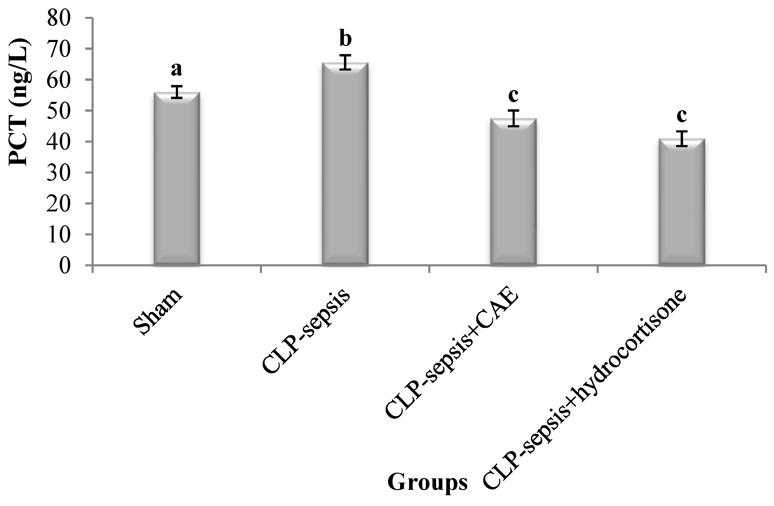
This study revealed the occurrence of sepsis in the CLP model, as a significant (p < 0.05) elevation in the lung PCT level was recorded in the CLP-septic group in comparison with the sham control group. Conversely, treatment of CLP-septic rats with the CAE or hydrocortisone significantly (p < 0.05) alleviated the elevated PCT level induced by CLP, as compared to the untreated CLP-septic rats (Figure 17).

3.4. CAE Inhibits Sepsis through Antioxidant System Enhancement

CLP rats showed a significant increase in the MDA level in lung tissue (p < 0.05), as compared to sham control rats (Table 5). The administration of CAE (500 mg/kg b.wt) or hydrocortisone to septic rats evoked a significant (p < 0.05) reduction in the lung MDA level, as compared to septic untreated rats. Sepsis induction caused a significant (p < 0.05) reduction in the lung GSH level, as compared with the sham control group. The treatment of septic rats with CAE or hydrocortisone reversed the lung GSH level significantly (p < 0.05), as compared with the sham group. Again, the lung SOD activity decreased significantly (p < 0.05) after CLP operation, as compared to the control SOD activity. However, the lung SOD level was significantly (p < 0.05) elevated after the septic rats were treated with CAE or hydrocortisone in comparison to those untreated rats. The level of lung CAT was not affected by CLP operation when compared with the sham level. Also, the administration of CAE or hydrocortisone caused a non-significant change in the lung CAT level, as compared with CLP-septic rats.

3.5. Histological Evaluation of Lung Tissue

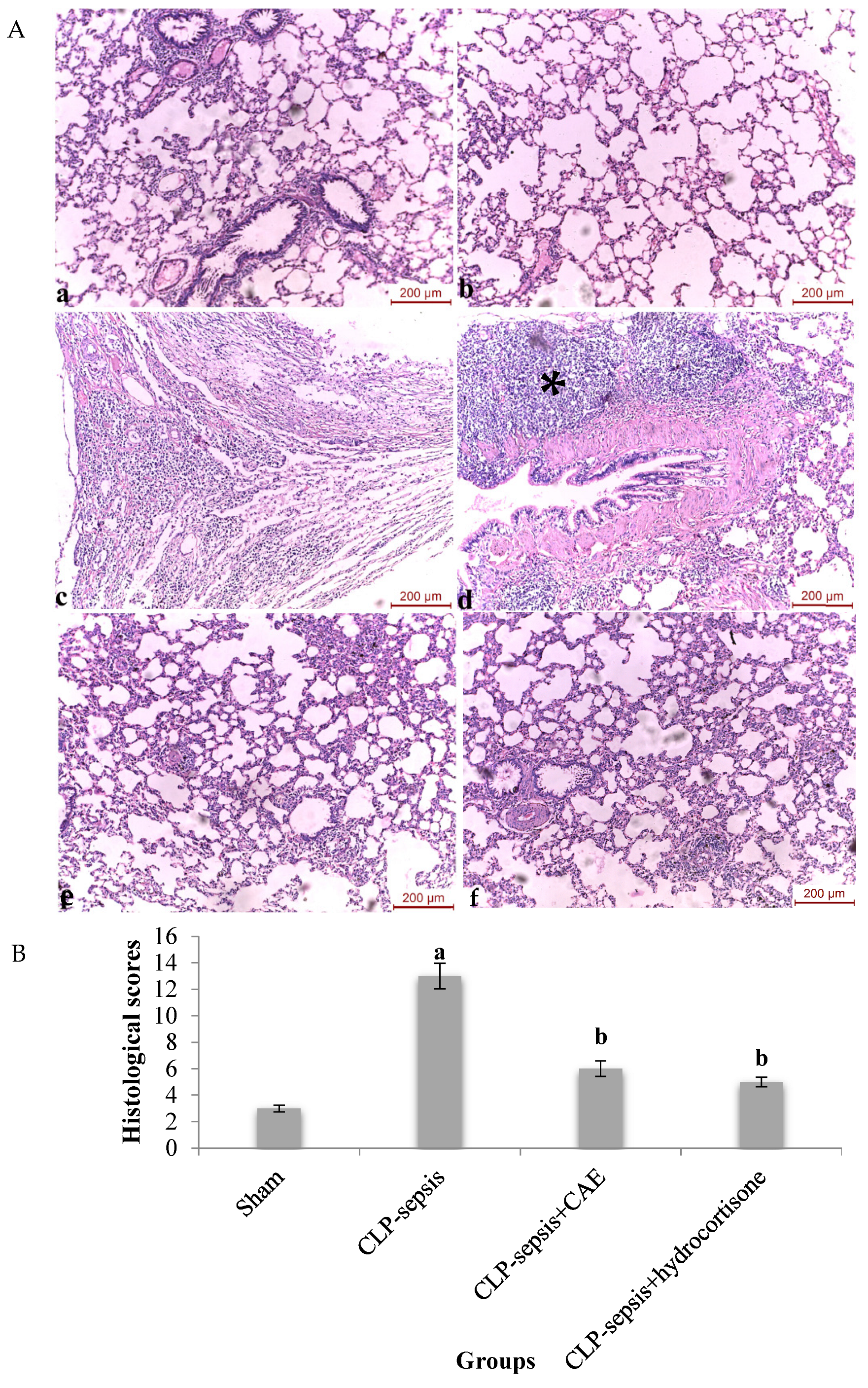
Generally, the lungs in the sham group showed a normal histological structure (Figure 18A(a,b)). Conversely, there were marked morphological alterations in lung tissues of the CLP-septic group, characterized by thickening of the alveolar wall and infiltration as well as the aggregation of neutrophils (Figure 18A(c,d)). However, these inflammatory alterations were markedly minimized in the CAE, hydrocortisone-treated groups (Figure 18A(e,f)). Consistent with these findings, the histological scores of lung tissue were significantly (p < 0.05) higher after the CLP procedure (Figure 18B). However, CLP combined with CAE administration or hydrocortisone resulted in a noticeable (p < 0.05) reduction in injury induced by CLP compared with the untreated CLP group.

3.6. Treatment with CAE Attenuates CLP-Induced Mortality

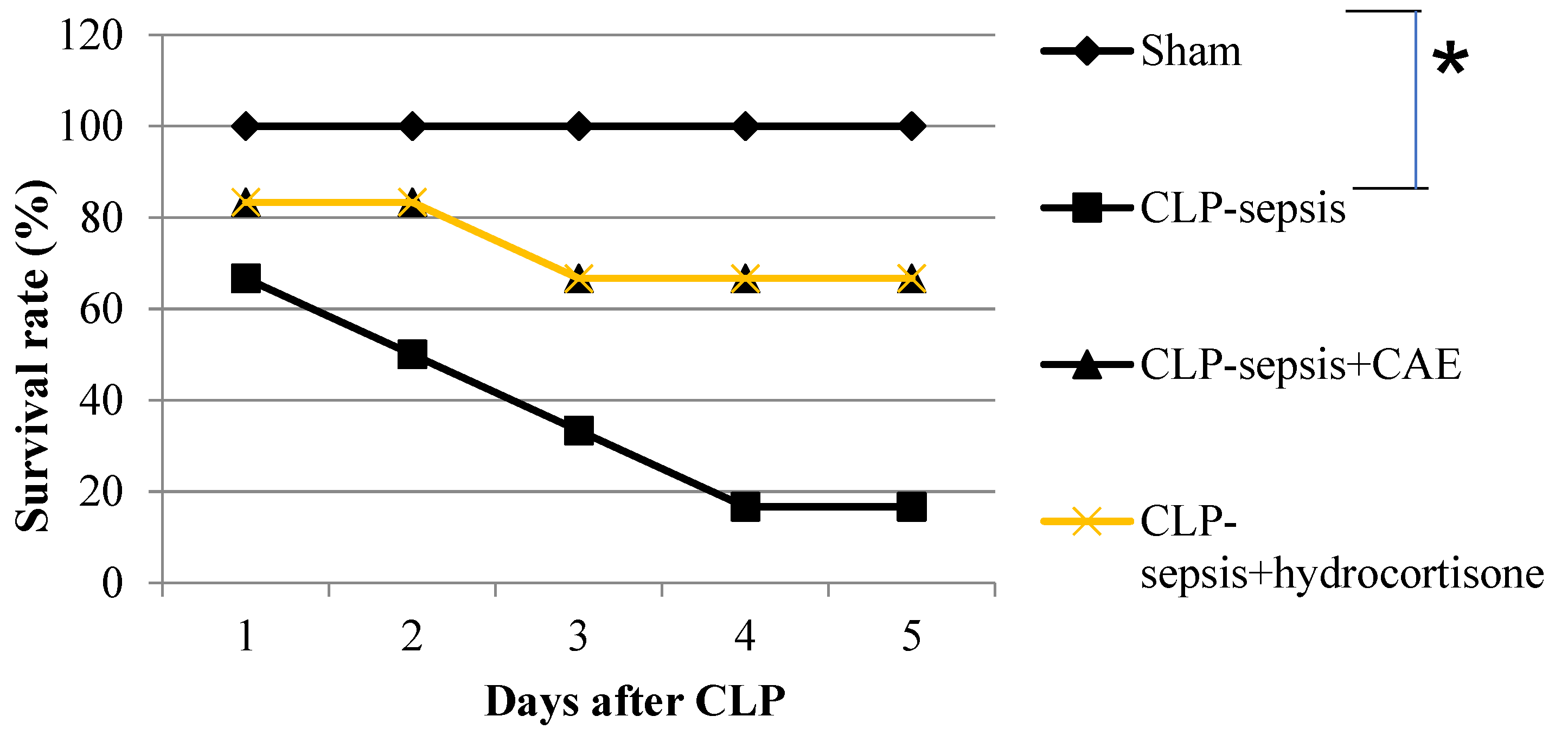
The present study revealed that the survival rate in the CLP group was 50% on day 2, and this percentage was decreased gradually until reaching 16.7% on day 5. In other words, an 83.3% mortality rate in the CLP-septic rats was recorded within 5 days. This is evidenced by a significant decline (p < 0.05) in the survival rate of the CLP-septic rats, as compared to sham rats, where 1/6 (16.7%) rats survived in the CLP-septic group. This study ascertained the survival improvement in the CAE, as it can rescue the septic rats from death. This was clarified by the survival rate percentage increment to 83.3% on day 2 and 66.7% on day 5. This means that 4/6 (66.7%) septic rats survived after CAE treatment, as compared with CLP-septic rats. The survival rate of CAE is similar to hydrocortisone (Figure 19).

4. Discussion

A study conducted by Jayaprakash and Das [19] focused on the GC-MS analysis of Cicer arietinum extract, identifying seven bioactive components in the methanolic extract. The structures of these bioactive components were retrieved by their chemical formula using ChemBio office and the DrugBank database (Table 1).
The docking study conducted in the present study aimed to investigate the interactions between bioactive components, specifically the differences in the interactions between Penicillin G (National Center for Biotechnology Information) and 1-formylbenzo[b]fluoranthene, with two important bacterial proteins: Pseudomonas aeruginosa-PBP3 [21] and Staphylococcus aureus-PBP2a [20]. Additionally, the structures of Staphylococcus aureus-PBP2a (1MWT) [20] and Pseudomonas aeruginosa-PBP3 (3OC2) [21] proteins were retrieved from the Protein Data Bank (PDB). The results revealed intriguing findings regarding the binding affinities, hydrogen bonding patterns, and stability of the molecules during the docking process.
Penicillin G exhibited notable interactions with both proteins, demonstrating two hydrogen bonds with Staphylococcus aureus-PBP2a and one hydrogen bond with Pseudomonas aeruginosa-PBP3. Despite its lower binding score with Pseudomonas aeruginosa-PBP3 compared to Staphylococcus aureus-PBP2a, the hydrogen bonding pattern suggests specific binding orientations and potential modes of action against these bacterial proteins.
Interestingly, 1-formylbenzo[b]fluoranthene emerged as a promising candidate, showing the best binding score, highest number of hydrogen bonds, and stability during the reaction compared to Penicillin G. The detailed analysis of its binding sites on Staphylococcus aureus-PBP2a revealed two hydrogen bonds, including interactions with key amino acids, like Lys-148, Glu-150, and Arg-151. Similarly, its binding to Pseudomonas aeruginosa-PBP3 showed no hydrogen bonds, yet it achieved a high docking score and interacted with critical amino acids, such as Arg-54, Ile-56, Pro-59, Glu-121, Phe-122, and Lys-197.
On the other hand, 7-Hydroxy-1-methoxy-6-methylanthraquinone showed no hydrogen bonds with PBP2a and only one with PBP3, indicating a higher interaction score compared to Penicillin G with PBP3. These results underscore the importance of considering both docking scores and hydrogen bonding patterns in evaluating the potential of bioactive compounds as antibacterial agents targeting PBPs.
These findings suggest that 1-formylbenzo[b]fluoranthene and 7-Hydroxy-1-methoxy-6-methylanthraquinone could be potent inhibitors against both Staphylococcus aureus-PBP2a and Pseudomonas aeruginosa-PBP3, potentially offering a new avenue for the development of antibacterial agents.
In sepsis, the lung is the first organ that loses its function in comparison with multiple organ dysfunction [4]. The present study adopted the CLP-induced sepsis model as it is considered the gold-standard model, since it is the best model resembling the clinical pathophysiology of human sepsis and represents an indirect insult similar to the pathogenesis of acute lung injury (ALI) [4]. Thus, the current study aimed to investigate if the Cicer arietinum extract (CAE) can alleviate the severity of CLP-induced sepsis in rats. In this context, the present study initially investigated the in vitro antibacterial potency of the CAE against the most common septic bacterial strains and found that the CAE (20 mg/mL) has promising inhibitory activity against Staphylococcus aureus and Pseudomonas aeruginosa bacterial strains but did not inhibit Escherichia coli bacterial growth. This finding agrees with the findings of Dalal et al. [16] and Verdrengh et al. [27]. The present work attributed the bactericidal activity of CAE to its ability to lyse the bacterial cell wall of S. aureus and P. aeruginosa, and this ability may be due to its phytoconstituents, as ascertained in a previous study [25]. Fahmy et al. [25] revealed that CAE contains carbohydrates, saponins, phytosterols, terpenoids, flavonoids, alkaloids, and tannins, in addition to its isoflavone (daidzein, genistein, formononetin, and biochanin A) contents. Zaki et al. [17] revealed the antibacterial effects of isoflavones of Cicer arietinum, particularly genistein. Genistein can change the bacterial nature, as it can (1) alter the bacterial cell morphology to filamentous cells; (2) inhibit the synthesis of DNA and RNA within 15 min after addition to a bacterial culture; (3) inhibit protein synthesis [28]. And, so, we expected that the genistein of the CAE made these obvious changes to the S. aureus and P. aeruginosa. On the other hand, it was reported that E. coli was resistant to most flavonoids. Additionally, the nucleic acids and proteins were unaffected in E. coli cultures [28]. Therefore, E. coli may be resisting the CAE for these reasons.
Regarding the in vivo study, the CLP surgical operation caused sepsis in rats. This is evidenced by a significant increase in the lung procalcitonin (PCT) that is a diagnostic sepsis marker. Interestingly, the CAE can restore the lung PCT level to its control value, as a significant decrease in the lung PCT level was observed in the CLP+CAE group. This means the potency of CAE to treat sepsis abnormalities may be through its antibacterial activity and its isoflavone content, particularly genistein, where the genistein blocks the invasion of pathogenic bacteria in mammalian epithelial cells [29]. On the other side, sepsis is associated with OxS, which is considered one of the causative factors in the development of sepsis-induced multiorgan failure [2]. Liu et al. [24] added that OxS is responsible for the severity of ALI. Thereby, diminished antioxidative defenses such as superoxide dismutase, catalase and glutathione eventually aid in sepsis occurrence and severity. The present investigation revealed that CLP surgery promotes a significant increase in the MDA level and significant depletion in GSH, CAT as well as the SOD levels in the lung. The non-significant finding of CAT may be due to the effect of other H2O2 decomposers, such as glutathione peroxidase (GPx). These findings agree with the findings of Bayir et al. [7] and Özkanlar et al. [30]. Generally, the lung suffers from a high risk of oxidative injury due to its exposure to the natural oxidizing nature of the atmosphere [31]. In the healthy lung, airway lining fluids and extracellular spaces are maintained in a reduced state as the levels of antioxidants and oxidants in the lung are balanced to preserve normal physiological functions. Crapo et al. [31] and Kinnula and Crapo [32] clarified that the decreases in antioxidants, as in the current findings, lead to several disturbances in lung tissue, such as (1) disrupting the oxidant/antioxidant equilibrium, (2) increasing the permeability of endothelial and alveolar epithelial cells, and (3) damaging the liquid transferring ability of alveolar epithelial cells. Liu et al. [24] summarized the relation between OxS and sepsis as the production of ROS as the main anti-microbicidal mechanism by which the upregulation of ROS induces tissue damage in sepsis and ended by ALI. And, hence, OxS is considered one of the first events that ultimately lead to lung damage. However, CAE suppresses OxS generated by CLP surgery; this is manifested by decreasing the increased MDA level of the septic rats significantly and replenishing the GSH, CAT, and SOD contents of the lung. The reversible effect of the CAE against lung oxidative damage may be due to the antioxidant efficacy of the CAE that was revealed in a previous study [15,33]. They proved that CAE could neutralize the free radicals, interact with the oxidative chain reaction, and enhance the antioxidant status by increasing the antioxidant system. The current results match the concept of Ercan and Ozdemir [2], who mentioned that an enhancement in the antioxidant status in septic animal models/patients may be protective. Additionally, CAE may lower the lung microvascular permeability and minimize lung damage, as Gadek et al. [34] disclosed that the oral intake of an antioxidant reduced the microvascular permeability of the lung. For these reasons, the present work can describe the CAE as an antiseptic extract.
To confirm the lung antiseptic efficacy of the CAE, an ongoing study observed the effect of the CAE on the mortality induced by CLP surgery and examined whether the current extract ameliorates the lung tissue abnormalities. The present findings proved that the survival rate of the CLP rats declined sharply at 5 days post-CLP (16.7%). A previous study revealed that when sepsis is accompanied by respiratory disorders such as ALI, the death in intensive care units reaches a mortality rate over 40% [35]. Ritter et al. [36] disclosed that MDA and SOD levels are markers of early mortality in septic rats, and this may explain why the MDA and SOD levels changed in the septic rats in the current study. However, the CLP rats post-treated with CAE survived with a rate of 66.7%, similar to hydrocortisone. This indicates that the administration of CAE improved the survival of CLP-septic rats. The survival improvement of the CAE may be due to (1) its potency to scavenge OxS induced by CLP, (2) its antiseptic capacity that comes from genistein, one of the CAE components, that is able to inhibit the invasion of pathogenic bacteria in vivo; this was proved by the decreased PCT level in post-treated rats. These findings are similar with hydrocortisone administered to septic patients [37].
Regarding the histopathological examination in the lungs of the CLP rats with or without CAE treatment, the present work found a marked difference in terms of histological scores between the sepsis group and the CAE-treated group. The lung sections stained with hematoxylin and eosin showed that CLP-induced septic rats had thickened alveolar septa with increased inflammatory and interstitial cell infiltration. These descriptions are in harmony with the findings of Liu et al. [24] and Özkanlar et al. [30] and confirmed the ALI incidence in the CLP rats. Abraham [38] mentioned that inflammatory cell infiltration into the lung tissue is considered a critical characteristic in the pathogenesis of ALI. Liu et al. [24] emphasized that OxS, which was achieved in this study, is an indicator of inflammatory processes. Guo and Ward [39] interpreted that OxS of lung tissue induced by sepsis initiated by products of activated lung macrophages and infiltrated neutrophils that propagate to lung tissue and ends with tissue damage and organ dysfunction. Thus, this may argue for the appearance of inflammatory infiltration in the lung tissue of septic rats. CAE post-treatment challenged this damage by minimizing the inflammatory cell infiltration and alveolar thickness, similar to hydrocortisone, and this indicates that CAE may reduce pulmonary vascular permeability. This result together with other findings verify that CAE appears to have a modulatory effect on ALI induced by CLP. This was ascertained by the attenuation of inflammatory cell infiltration of lung tissue, reduction in PCT and OxS, antibacterial activity, and rescuing rats from death.

5. Conclusions

This study provides a novel lung antiseptic extract, CAE. The current study revealed that CAE has the capacity to improve the lung injury induced by CLP. We expect that CAE exerts its antiseptic effect through inhibiting the PCT, OxS production, and inflammatory cell infiltration in lung tissue. Thus, CAE may be beneficial in clinical applications of lung septic cases. But, clinical trials are urgent to fully clarify the potential role of CAE in sepsis-induced ALI.

Supplementary Materials

The following supporting information can be downloaded at: https://www.mdpi.com/article/10.3390/microbiolres15030130/s1, Supplementary S1: In vitro antibacterial activity of CAE.

Author Contributions

Conceptualization and investigation, A.A.A. and S.I.; methodology, software, and data curation, S.I. and K.I.G.; validation, M.H.A.-A. and K.I.G.; resources, M.H.A.-A. and K.I.G.; writing—original draft preparation, S.I. and K.I.G.; writing—review and editing, M.H.A.-A. and K.I.G.; visualization and supervision, A.A.A., M.H.A.-A. and K.I.G.; project administration and funding acquisition, A.A.A., M.H.A.-A. and K.I.G. All authors have read and agreed to the published version of the manuscript.

Funding

This research received no external funding.

Institutional Review Board Statement

The animal study protocol was approved by the Institutional Animal Care and Use Committee (IACUC) (CUFS/F/PHY/52/15) of the Faculty of Science, Cairo University, Egypt.

Informed Consent Statement

Not applicable.

Data Availability Statement

Data are contained within the article.

Acknowledgments

Authors are thankful to the Deanship of Graduate Studies and Scientific Research at University of Bisha for supporting this work through the Fast-Track Research Support Program.

Conflicts of Interest

The authors report no conflicts of interest. The authors alone are responsible for the content and writing of the paper.

References

  1. Dai, W.; Li, Y.; Liu, X.; Wang, N.; Luo, P.; Kong, L. Protective effects of Nigella sativa L. seeds aqueous extract-based silver nanoparticles on sepsis-induced damages in rats. Inorg. Chem. Commun. 2024, 166, 112594. [Google Scholar] [CrossRef]
  2. Ercan, M.; Ozdemir, S. The contribution to studies of the effect of β-glucan on plasma viscosity in a rat sepsis model. Med. Sci. Discov. 2015, 2, 148–153. [Google Scholar] [CrossRef]
  3. Vincent, J.L.; Nelson, D.R.; Williams, M.D. Is worsening multiple organ failure the cause of death in patients with severe sepsis? Crit. Care Med. 2011, 39, 1050–1055. [Google Scholar] [CrossRef] [PubMed]
  4. Xu, L.; Bao, H.-G.; Si, Y.-N.; Han, L.; Zhang, R.; Cai, M.-M.; Shen, Y. Effects of adiponectin on acute lung injury in cecal ligation and puncture-induced sepsis rats. J. Surg. Res. 2013, 183, 752–759. [Google Scholar] [CrossRef]
  5. Ware, L.B.; Matthay, L.B. The acute respiratory distress syndrome. N. Engl. J. Med. 2000, 342, 1334–1349. [Google Scholar] [CrossRef]
  6. Fein, A.M.; Calalang-Colucci, M.G. Acute lung injury and acute respiratory distress syndrome in sepsis and septic shock. Crit. Care Clin. 2000, 16, 289–317. [Google Scholar] [CrossRef]
  7. Bayir, Y.; Albayrak, A.; Can, I.; Karagoz, Y.; Cakir, A.; Suleyman, H.; Uyanik, H.; Yayla, N.; Polat, B.; Karakus, E.; et al. Nigella sativa as a potential therapy for the treatment of lung injury caused by cecal ligation and puncture-induced sepsis model in rats. Cell Mol. Biol. 2012, 58, 1680–1687. [Google Scholar]
  8. Wheeler, A.P.; Bernard, G.R. Acute lung injury and the acute respiratory distress syndrome: A clinical review. Lancet 2007, 369, 1553–1564. [Google Scholar] [CrossRef]
  9. Xia, W.; Pan, Z.; Zhang, H.; Zhou, Q.; Liu, Y. ERRα protects against sepsis-induced acute lung injury in rats. Mol. Med. 2023, 29, 76. [Google Scholar] [CrossRef]
  10. Raoofi, R.; Salmani, Z.; Moradi, F.; Sotoodeh, A.; Sobhanian, S. Procalcitonin as a marker for early diagnosis of sepsis. Am. J. Infect. Dis. 2014, 10, 15–20. [Google Scholar] [CrossRef]
  11. Tsai, K.; Hsu, T.; Kong, C.; Lin, K.; Lu, F. Is the endogenous peroxyl-radical scavenging capacity of plasma protective in systemic inflammatory disorders in humans? Free. Radic. Biol. Med. 2000, 28, 926–933. [Google Scholar] [CrossRef]
  12. Macdonald, J.; Galley, H.F.; Webster, N.R. Oxidative stress and gene expression in sepsis. Br. J. Anaesth. 2003, 90, 221–232. [Google Scholar] [CrossRef] [PubMed]
  13. Pradipta, I.S.; Sodik, D.C.; Lestari, K.; Parwati, I.; Halimah, E.; Diantini, A.; Abdulah, R. Antibiotic resistance in sepsis patients: Evaluation and recommendation of antibiotic use. N. Am. J. Med. Sci. 2013, 5, 344–352. [Google Scholar] [CrossRef]
  14. Nagar, H.; Piao, S.; Kim, C.S. Role of Mitochondrial Oxidative Stress in Sepsis. Acute Crit. Care 2018, 33, 65–72. [Google Scholar] [CrossRef] [PubMed]
  15. Sayed, A.A.; Abbas, O.A.; Saad, M.A.; Marie, M.S. Cicer arietinum extract ameliorate γ-irradiation disorders via modulation of oxidative/antioxidative pathway. J. Photochem. Photobiol. B 2018, 183, 46–56. [Google Scholar] [CrossRef] [PubMed]
  16. Dalal, K.; Ahlawat, S.; Munjal, H.; Patra, A. Antibacterial activity of roots of Cicer arietinum Linn. J. Chem. Pharm. Res. 2010, 2, 43–46. [Google Scholar]
  17. Zaki, M.; Zaid, A.; Magd, M.; Abou Zid, S. Antibacterial effects of isoflavones isolated from Cicer arietinum. Int. J. Nat. Prod. Res. 2013, 2, 1–5. [Google Scholar]
  18. Brooks, H.F.; Osabutey, C.K.; Moss, R.F.; Andrews, P.L.; Davies, D.C. Caecal ligation and puncture in the rat mimics the pathophysiological changes in human sepsis and causes multi-organ dysfunction. Metab. Brain Dis. 2007, 22, 353–373. [Google Scholar] [CrossRef]
  19. Jayaprakash, B.; Das, A. Extraction and Characterization of ChickPEA (Cicer arietinum) Extract with Immunostimulant Activity in BALB/C MICE. Asian Pac. J. Cancer Prev. 2018, 19, 803–810. [Google Scholar]
  20. Lim, D.C.; Strynadka, N.C.J. Structure of penicillin G acyl-Penicillin binding protein 2a from methicillin resistant Staphylococcus aureus strain 27r at 2.45 A resolution. Nat. Struct. Biol. 2002, 9, 870–876. [Google Scholar]
  21. Sainsbury, S.; Bird, L.; Stuart, D.I.; Owens, R.J.; Ren, J. Oxford Protein Production Facility (OPPF). Crystal structure of penicillin-binding protein 3 from Pseudomonas aeruginosa. J. Mol. Biol. 2011, 405, 173–184. [Google Scholar] [CrossRef] [PubMed]
  22. Marzouk, M.; Soliman, A.M.; Fahmy, S.R.; Sayed, A.A. Ameliorative effects of Cicer arietinum extract and Coelatura aegyptiaca shell powder on estrogen sensitive organs in ovariectomized rats. World Appl. Sci. J. 2014, 31, 863–872. [Google Scholar]
  23. Bauer, A.W.; Kirby, W.M.; Sherris, J.C.; Turck, M. Antibiotic susceptibility testing by a standardized single disk method. Am. J. Clin. Pathol. 1966, 45, 493–496. [Google Scholar] [CrossRef] [PubMed]
  24. Liu, A.; Wang, W.; Fang, H.; Yang, Y.; Jiang, X.; Liu, S.; Hu, J.; Hu, Q.; Dahmen, U.; Dirsch, O. Baicalein protects against polymicrobial sepsis-induced liver injury via inhibition of inflammation and apoptosis in mice. Eur. J. Pharmacol. 2015, 748, 45–53. [Google Scholar] [CrossRef]
  25. Shen, L.; Mo, H.; Cai, L.; Kong, T.; Zheng, W.; Ye, J.; Qi, J.; Xiao, Z. Losartan prevents sepsis induced acute lung injury and decreases activation of nuclear factor kappa B and mitogen-activated protein kinases. Shock. 2009, 31, 500–506. [Google Scholar] [CrossRef]
  26. Yamanel, L.; Kaldirim, U.; Oztas, Y.; Coskun, O.; Poyrazoglu, Y.; Durusu, M.; Cayci, T.; Ozturk, A.; Demirbas, S.; Yasar, M.; et al. Ozone therapy and hyperbaric oxygen treatment in lung injury in septic rats. Int. J. Med. Sci. 2011, 8, 48–55. [Google Scholar] [CrossRef]
  27. Verdrengh, M.; Collins, L.V.; Bergin, P.; Tarkowski, A. Phytoestrogen genistein as an anti-staphylococcal agent. Microbes Infect. 2004, 6, 86–92. [Google Scholar] [CrossRef]
  28. Ulanowska, K.; Tkaczyk, A.; Konopa, G.; Wegrzyn, G. Differential antibacterial activity of genistein arising from global inhibition of DNA.; RNA and protein synthesis in some bacterial strains. Arch. Microbiol. 2006, 184, 271–278. [Google Scholar] [CrossRef]
  29. Hong, H.; Landauer, M.R.; Foriska, M.A.; Ledney, G.D. Antibacterial activity of the soy isoflavone genistein. J. Basic. Microbiol. 2006, 46, 329–335. [Google Scholar] [CrossRef]
  30. Özkanlar, S.; Koç, F.; Karakuş, E.; Güvenalp, Z.; Oruç, E.; Özbek, H. The protective effects of Peganum harmala extract on lung and kidney in sepsis induced by cecal ligation and perforation in rats. Kafkas Univ. Vet. Fak. Derg. 2015, 21, 367–375. [Google Scholar]
  31. Crapo, J.D. Oxidative stress as an initiator of cytokine release and cell damage. Eur. Respir. J. Suppl. 2003, 44, 4s–6s. [Google Scholar] [CrossRef] [PubMed]
  32. Kinnula, V.L.; Crapo, J.D. Superoxide dismutases in the lung and human lung diseases. Am. J. Respir. Crit. Care Med. 2003, 167, 1600–1619. [Google Scholar] [CrossRef] [PubMed]
  33. Fahmy, S.R.; Soliman, A.M.; Sayed, A.A.; Marzouk, M. Possible antiosteoporotic mechanism of Cicer arietinum extract in ovariectomized rats. Int. J. Clin. Exp. Pathol. 2015, 8, 3477–3490. [Google Scholar] [PubMed]
  34. Gadek, J.E.; DeMichele, S.J.; Karlstad, M.D.; Pacht, E.R.; Donahoe, M.; Albertson, T.E.; Chi, V.H.; Ann, K.W.; Jeffrey, L.N.; Mojtaba, N. Effect of enteral feeding with eicosapentaenoic acid; gamma-linolenic acid; and antioxidants in patients with acute respiratory distress syndrome. Enteral Nutrition in ARDS Study Group. Crit. Care Med. 1999, 27, 1409–1420. [Google Scholar] [CrossRef]
  35. Rubenfeld, G.D.; Caldwell, E.; Peabody, E.; Weaver, J.; Martin, D.P.; Neff, M.; Stern, E.J.; Hudson, L.D. Incidence and outcomes of acute lung injury. N. Engl. J. Med. 2005, 353, 1685–1693. [Google Scholar] [CrossRef]
  36. Ritter, C.; Andrades, M.; Frota, M.L.C.; Weaver, J.; Martin, D.P.; Neff, M.; Klamt, F.; Pinheiro, C.T.S.; Menna-Barreto, S.S.; Moreira, J.C.F.; et al. Oxidative parameters and mortality in sepsis induced by cecal ligation and perforation. Intens. Care Med. 2003, 29, 1782–1789. [Google Scholar] [CrossRef]
  37. Keivanpour, H.; Zamzam, R.; Mojtahedzadeh, M.; Delnavazi, M.R.; Sharifan, A.; Sabzevari, O. 6-Gingerol anti-inflammatory and antioxidant properties protect against heart and liver dysfunction in rats with sepsis. Pharmacol. Res.-Mod. Chin. Med. 2024, 12, 100470. [Google Scholar] [CrossRef]
  38. Abraham, E. Neutrophils and acute lung injury. Crit. Care Med. 2003, 31, S195. [Google Scholar] [CrossRef]
  39. Guo, R.; Ward, P.A. Role of oxidants in lung injury during sepsis. Antioxid. Redox Signal. 2007, 9, 1991–2002. [Google Scholar] [CrossRef]
Figure 1. Docking of (cis)-2-nonadecene on Staphylococcus aureus-PBP2a.
Microbiolres 15 00130 g001
Figure 2. Docking (cis)-2-nonadecene on Pseudomonas aeruginosa-PBP3.
Microbiolres 15 00130 g002
Figure 3. Docking of 1-formylbenzo[b]fluoranthene on Staphylococcus Aureus-PBP2a, showing 3 hydrogen bonds as ligand oxygen to THR 165 oxygen, 2.71 Å, ARG 151 hydrogen to ligand oxygen, 2.26 Å and ligand oxygen to THR 151 oxygen, 3.30 Å.
Microbiolres 15 00130 g003
Figure 4. Docking of 1-formylbenzo[b]fluoranthene on Pseudomonas aeruginosa-PBP3.
Microbiolres 15 00130 g004
Figure 5. Docking of 1-Tetradecene (CAS) on Staphylococcus aureus-PBP2a.
Microbiolres 15 00130 g005
Figure 6. Docking of 1-Tetradecene (CAS) on Pseudomonas aeruginosa-PBP3.
Microbiolres 15 00130 g006
Figure 7. Docking of 3-Dodecene, (Z)- on Staphylococcus aureus-PBP2a.
Microbiolres 15 00130 g007
Figure 8. Docking of 3-Dodecene, (Z)- on Pseudomonas aeruginosa-PBP3.
Microbiolres 15 00130 g008
Figure 9. Docking of 6-(Aminomethyl)-2-naphthol on Staphylococcus aureus-PBP2a, showing 1 Hydrogen bonds, ligand oxygen to Ser 403 Oxygen, 2.24 Å.
Microbiolres 15 00130 g009
Figure 10. Docking of 6-(Aminomethyl)-2-naphthol on Pseudomonas aeruginosa-PBP3, showing 1 Hydrogen bond, ligand hydrogen to Ala 57 Oxygen, 2.29 Å.
Microbiolres 15 00130 g010
Figure 11. Docking of 7-Hydroxy-1-methoxy-6-methylanthraquinone on Staphylococcus aureus-PBP2a.
Microbiolres 15 00130 g011
Figure 12. Docking of 7-Hydroxy-1-methoxy-6-methylanthraquinone on Pseudomonas aeruginosa-PBP3, showing 1 Hydrogen Bound, Lys 197 Hydrogen to Ligand Oxygen, 2.17 Å.
Microbiolres 15 00130 g012
Figure 13. Docking of Cyclohexadecane (CAS) on Staphylococcus aureus-PBP2a.
Microbiolres 15 00130 g013
Figure 14. Docking of Cyclohexadecane (CAS) on Pseudomonas aeruginosa-PBP3.
Microbiolres 15 00130 g014
Figure 15. Docking of Penicillin G on Staphylococcus aureus-PBP2a, showing 2 Hydrogen Bounds, ARG 151 hydrogen to ligand oxygen, 2.36 Å and ARG 151 hydrogen to ligand oxygen, 2.41 Å.
Microbiolres 15 00130 g015
Figure 16. Docking of Penicillin G on Pseudomonas aeruginosa- PBP3, showing 1 Hydrogen Bound, showing ASN 351 hydrogen to ligand oxygen, 2.39 Å.
Microbiolres 15 00130 g016
Figure 17. Effect of CAE on lung PCT level of septic rats. Values are mean ± SEM (n = 6). Values with different letters are significantly different (p < 0.05). Data were analyzed by ANOVA post hoc with Duncan test.
Microbiolres 15 00130 g017
Figure 18. (A) Representative photomicrographs of lung histology (H&E) showing the effect of CAE treatment on sepsis-induced histopathological changes. Photomicrograph of a pulmonary section from a sham-operated rats showing normal alveolar structure (A) (a,b). Photomicrograph of a lung section of septic rats showing leukocyte infiltration (*) and alveolar septal thickening (black arrow) (c,d). Photomicrograph of a lung section of septic rats treated with CAE and hydrocortisone seems as normal structure (e,f; respectively). (B) Histological scores of lung injury. Data were compared by Kruskal–Wallis and the Student–Newman–Keuls method. a p < 0.05 versus sham group, b p < 0.05 versus CLP-septic group.
Microbiolres 15 00130 g018
Figure 19. Alterations in the survival rate within 5 days after CLP. The survival rate was estimated by the Kaplan–Meier method and compared by log-rank test. * p < 0.05 versus sham. Note the same readings for CAE and hydrocortisone, so the lines above them.
Microbiolres 15 00130 g019
Table 1. Bioactive components identified in methanolic extract of C. arietinum by GC-MS analysis.
S.NoRTName of CompoundM.FormulaM.Weight
129.187-Hydroxy-1-methylanthraquinoneC16H12O4268
232.296-(Aminomethyl)-2-naphtholC11H11NO173
313.78Cyclohexadecane CASC16H32224
417.22(cis)-2-nonadeceneC19H38266
520.991-formylbenzo[b]fluorantheneC21H12O280
610.601-Tetradecene (CAS)C14H28196
724.54(cis)-2-nonadeceneC19H38266
87.183-Dodecene,(Z)-C12H24168
Table 2. Docking Scores and hydrogen bonding of the bioactive components identified in methanolic extract of C. arietinum against Staphylococcus aureus-PBP2a and Pseudomonas aeruginosa-PBP3.
Free Binding Energy of Temperature (T) = 298.15 KStaphylococcus aureus-PBP2a (1 MWT) Docking ScorePseudomonas aeruginosa-PBP3 (3OC2) Docking Score
Penicillin G−6.9 kcal/mol
(2 Hydrogen Bonds)
−7.1 kcal/mol (1 Hydrogen Bond)
ASN 351 Hydrogen to Ligand oxygen, 2.39 Å
  • ARG 151 hydrogen to ligand oxygen, 2.36 Å
  • ARG 151 hydrogen to ligand oxygen, 2.41 Å
(cis)-2-nonadecene−4.3 kcal/mol
(0 Hydrogen Bonds)
−5.1 kcal/mol
(0 Hydrogen Bond)
1-formylbenzo[b]fluoranthene−8.6 kcal/mol
(3 Hydrogen Bonds)
−9.0 kcal/mol
(0 Hydrogen Bond)
  • Ligand Oxygen to THR165 oxygen, 2.71 Å
  • ARG 151 hydrogen toligand oxygen, 2.26 Å
  • Ligand Oxygen to THR151 oxygen, 3.30 Å
1-Tetradecene (CAS)−4.3 kcal/mol
(0 Hydrogen Bonds)
−4.7 kcal/mol
(0 Hydrogen Bond)
3-Dodecene, (Z)-−4.3 kcal/mol
(0 Hydrogen Bonds)
−4.7 kcal/mol
(0 Hydrogen Bond)
6-(Aminomethyl)-2-naphthol−6.8 kcal/mol
(1 Hydrogen Bonds)
Ligand Oxygen to Ser 403 Oxygen, 2.24 Å
−6.2 kcal/mol
(1 Hydrogen Bond)
Ligand hydrogen to Ala 57 Oxygen, 2.29Å
7-Hydroxy-1-methoxy-6-methylanthraquinone−6.4 kcal/mol
(0 Hydrogen Bonds)
−7.2 kcal/mol
(1 Hydrogen Bond)
Lys 197 Hydrogen to Ligand Oxygen, 2.17Å
Cyclohexadecane (CAS)−6.6 kcal/mol
(0 Hydrogen Bonds)
−7.2 kcal/mol
(0 Hydrogen Bond)
Table 3. Experimental design and treatment regimen.
GroupsDistilled WaterCAE
(500 mg/kg b.wt)
HydrocortisoneAdministration Time
(Days)
Euthanization Time
(Days)
I.
Sham control rats
+34th
II.
CLP-septic rats
+34th
III.
CLP-septic rats + CAE
+34th
IV.
CLP-septic rats + hydrocortisone
+34th
Table 4. Antibacterial activity of the CAE against different Gram-positive and Gram-negative bacteria.
Test OrganismZone of Inhibition (mm)
CAEPositive ControlNegative Control
S. aureus1219-
P. aeruginosa1215-
E. coli-17-
Table 5. Effect of CAE on the levels of lung oxidative stress/antioxidant enzymes disorder of septic rats.
Parameter MDA
(nmol/g Tissue)
GSH
(mg/g Tissue)
SOD
(U/g Tissue)
CAT
(U/g Tissue)
Groups
Sham2.127 ± 0.304 a0.399 ± 0.075 a404.965 ± 42.518 a0.925 ± 0.158 a
CLP-sepsis5.470 ± 1.390 b0.163 ± 0.026 b160.813 ± 12.335 b0.599 ± 0.090 a
CLP-sepsis + CAE1.329 ± 0.265 a1.038 ± 0.082 c402.040 ± 43.825 a0.748 ± 0.086 a
CLP-sepsis + hydrocortisone0.998 ± 0.008 a1.001 ± 0.067 c385.972 ± 34.863 a0.698 ± 0.064 a
Values are mean ± SEM (n = 6). Values with different superscript letters are significantly different (p < 0.05). Data were analyzed by ANOVA post hoc with Duncan test.
Disclaimer/Publisher’s Note: The statements, opinions and data contained in all publications are solely those of the individual author(s) and contributor(s) and not of MDPI and/or the editor(s). MDPI and/or the editor(s) disclaim responsibility for any injury to people or property resulting from any ideas, methods, instructions or products referred to in the content.