Communication

Antagonistic Interactions in Onychomycosis: Antifungal Activity of Extracts from Pure and Mixed Cultures of Candida parapsilosis and Trichophyton spp.

by
Thiago Henrique Lemes
1,*,
Julyanna Andrade Silva Nascentes
1,
Luis Octávio Regasini
1,
João Paulo Zen Siqueira
2,
Glaucia Rigotto
3,
Ludmilla Tonani
3,
Marcia Regina von Zeska Kress
3 and
Margarete Teresa Gottardo de Almeida
2,*
1
Departament of Microbiology Institute of Biosciences, Humanities and Exact Sciences, São Paulo State University (UNESP), São José do Rio Preto 15054-000, Brazil
2
Department of Dermatological, Infectious, and Parasitic Diseases, São José do Rio Preto School of Medicine (FAMERP), São José do Rio Preto 15090-000, Brazil
3
Department of Clinical, Toxicological, and Bromatological Analyses, Ribeirao Preto School of Pharmaceutical Sciences (USP), University of Sao Paulo, Ribeirão Preto 14040-903, Brazil
*
Authors to whom correspondence should be addressed.
Microbiol. Res. 2024, 15(2), 880-888; https://doi.org/10.3390/microbiolres15020057
Submission received: 25 February 2024 / Revised: 15 May 2024 / Accepted: 20 May 2024 / Published: 22 May 2024

Abstract

:
Onychomycoses are nail infections that require prolonged therapy and have high recurrence rates. Dermatophytes are the main etiological agents of these infections, followed by yeasts and non-dermatophyte filamentous fungi. The limited antifungal arsenal used to treat onychomycosis and the change in the susceptibility profile of these agents contribute to the chronicity and recalcitrant profile of infections. The present study aimed to determine the antifungal activity of extracts obtained from pure and mixed cultures of Candida parapsilosis, Trichophyton mentagrophytes, and Trichophyton rubrum. Additionally, in vivo toxicity tests with Galleria mellonella and time-kill assays were carried out. The susceptibility profiles of dermatophytes were determined using a microdilution technique with minimum inhibitory concentrations (MICs) between 250 and 8000 µg/mL. The time-kill assay, compared to growth control, resulted in the death of dermatophytes within 48 h. No toxicity of the extracts was detected in experiments with Galleria mellonella larvae under the test conditions. The extracts of pure and mixed cultures of Candida parapsilosis and dermatophytes present antifungal activity against T. mentagrophytes and T. rubrum. Isolating and identifying compounds in the extracts may allow the development of new therapeutic approaches to control fungal infections.

1. Introduction

Onychomycoses are infections with worldwide incidence, clinically characterized by discoloration, hyperkeratosis, and onycholysis of the nail plate [1]. Dermatophytes, which account for 70% of cases, are fungi that can degrade keratinized tissues such as skin, hair, and nails. Among the prevalent species, Trichophyton rubrum (TR) and members of the T. mentagrophytes (TM) complex are the most isolated [2]. Recently, studies indicate an increase in nail infections caused by yeasts of the genus Candida, including the species C. albicans and C. parapsilosis (CP), and non-dermatophyte filamentous fungi such as Scopulariopsis brevicaulis, Acremonium spp., Aspergillus spp., Fusarium spp., and Neoscytalidium [3,4,5], occurring before or concomitantly with dermatophytes [6]. In infections of mixed etiology, interactions between species promote the production of various metabolites and the expression of virulence factors, such as hydrolytic enzymes, that are capable of regulating morphogenesis, growth, and biofilm formation. These mechanisms are essential for the survival of microorganisms in hostile environments, helping them to tolerate the action of antifungal drugs and evade the host’s immune response [7,8]. Considering the increase in cases of onychomycosis caused by different species of fungi, the knowledge of the competitive interactions between microorganisms constitutes a challenge, providing a new scenario for novel therapeutic approaches in the treatment of these infections [9,10]. The present study evaluated the antifungal activity of extracts obtained from pure and mixed cultures of CP, TM, and TR against strains of Trichophyton spp.

2. Materials and Methods

2.1. Microorganisms

Clinical isolates of CP (1), TM (6), and TR (6) from the collection of the Microbiology Laboratory of the School of Medicine in São José do Rio Preto (FAMERP), Brazil, from the American Type Culture Collection (T. mentagrophytes 11481—TMATCC) and from the Central Bureau of Fungal Cultures (Centraalbureau voor Schimmelcultures—T. rubrum 118892—TRCBS) were used. All isolates were registered in the National System for the Management of Genetic Heritage and Associated Traditional Knowledge (SisGen) under protocol number AF41CDD.

2.2. Extracts

To obtain the CP culture extract, colonies grown on Sabouraud Dextrose Agar were incubated at 35 °C for 24 h. An inoculum suspension (5 mL) in sterile saline (0.85%), with turbidity corresponding to MacFarland Scale 10, was inoculated into 500 mL of Sabouraud Dextrose Broth (SDB) and incubated at 35 °C for 72 h. For pure culture extracts of TM and TR, dermatophyte colonies with seven days of growth on Potato Dextrose Agar were covered with sterile saline. Subsequently, the mixture of conidia and hyphae in the suspension (5 mL) was transferred to 500 mL of SDB and incubated at 35 °C for 120 h. In preparing mixed culture extracts with CP and TM (CPTM) and with CP and TR (CPTR), 5 mL of inoculum of CP and previously prepared samples of Trichophyton spp. were transferred to 500 mL of SDB and incubated at 35 °C for 120 h. The culture medium containing the inoculum was then filtered through a 0.2 μm Millipore membrane and subjected to liquid-liquid extraction with ethyl acetate (250 mL culture medium/50 mL ethyl acetate). The extraction procedure was repeated three times, allowing total extraction of the metabolites in the culture filtrate. The acetate phase was subjected to drying using a rotary evaporator. The mass of the resulting compound was solubilized in a 10% solution of dimethyl sulfoxide (DMSO) with sterile water to carry out subsequent tests.

2.3. Susceptibility Profile of Dermatophyte Strains against Extracts and Antifungal Drugs

The susceptibility profiles of TR strains in respect to extracts and drugs were determined by the microdilution technique following the protocol described in document M38-3rd of the Clinical Laboratory Standard Institute (CLSI) with modifications [11]. The dermatophytes were cultivated on Potato Dextrose Agar at 35 °C for seven days. After this period, the colony surface, covered with 5 mL of sterile saline (0.85%), was scraped with a sterile loop. The inoculum was adjusted with a spectrophotometer (530 nm), followed by dilution at a ratio of 1:50 in RPMI 1640 medium (Sigma-Aldrich®—Rosen Park Media Institute, Saint Louis, MO, USA) with a final concentration of 0.4–5 × 10⁴ cells/mL. Subsequently, 0.1 mL of the suspension was inoculated in 96-well polystyrene plates containing RPMI 1640 medium at different extract concentrations (8000–1.9 µg/mL) and drugs were tested: itraconazole 4–0.16 µg/mL, fluconazole and terbinafine 64–0.001 µg/mL. Sterility control wells (containing 0.2 mL of RPMI) and growth controls (containing inoculum and RPMI only) were prepared. Plates were incubated at 35 °C for 120 h. The MICs of fluconazole and itraconazole were determined as the lowest concentration capable of inhibiting 80% of fungal growth (MIC80); concentrations capable of inhibiting 100% (MIC100) were considered for the extracts and for terbinafine. For the minimum fungicidal concentration (MFC), an aliquot of each well of the plates prepared with extracts and inoculum was transferred to Petri dishes containing Sabouraud Dextrose Agar. The MFC was defined as the lowest concentration of the extract to inhibit visible growth on a solid medium. All tests were performed in triplicate.

2.4. Time-Kill Assay

The time-kill assay was conducted according to the method described by Klepser et al. with Trichophyton spp. The MFC values obtained were used for this assay. The dermatophyte inoculum was prepared as described previously. Suspensions of 0.1 mL of conidia were added to 4.9 mL of SDB to, obtain a final concentration corresponding to 0.4–5 × 10⁴ colony-forming units (CFU/mL). The inoculum after preparation was diluted at a ratio of 1:1 with the extract (0.2 mL of the extract with 0.2 mL of the inoculum). Tubes containing only inoculum were included to evaluate growth control. At predetermined times (8 h, 24 h, 48 h), an aliquot of 30 µL was taken from each sample and transferred to Sabouraud Dextrose Agar (DIFCO®, Detroit, MI, USA) plates using a Drigalski loop and incubated at 35 °C for 120 h. Subsequently, the number of colony-forming units (CFU) was counted. The percentage of inhibition was calculated by comparing with the growth control using the colony counting method [12].

2.5. Toxicity Test with Galleria mellonella

Toxicity tests of the extracts were performed using G. mellonella larvae following the experimental model of toxicity according to the protocol described by Ignasiak and Maxwell. Five G. mellonella larvae (275 ± 25 mg) in the sixth instar of development were separated in Petri dishes (90 × 15 mm2) for each condition. Artificial inoculation was achieved by injecting 5 μL of extracts at concentrations of 2000–8000 µg/mL using a Hamilton micro syringe for gas chromatography (model 7000.5 KH: 10 μL). The experimental controls were untouched larvae (naïve), larvae inoculated with 10% ethanol (positive toxicity control), and 10% DMSO (negative toxicity control). Inoculated larvae were deprived of food and direct light, incubated at 37 °C, and scored for viability at 24 h intervals for 5 days. Differences in resulting survival plots were evaluated using the Origin software (pro 9.1) with the Mantel–Cox test (Log-rank method) [13].

3. Results

3.1. Susceptibility Profile of Dermatophyte Strains against Extracts and Antifungal Drugs

The results for the susceptibility profile of dermatophyte strains against antifungal drugs are shown in Table 1. MIC values for fluconazole ranged from 2 to 32 µg/mL, with the highest values (32 µg/mL) being found for the TM strains (TM5094, TM6085, and TM6007) and the lowest (2 µg/mL) for the TR strains (RCBS, TR7604, and TR7984). Itraconazole values ranged from 0.5 to 2 µg/mL, with the highest (2 µg/mL) for the TM5094 strain and the lowest (0.25 µg/mL) for TRCBS. For terbinafine, the highest MIC value (64 µg/mL) found was for the clinical strain TR7259; the other strains had values of 0.03 µg/mL.
The MIC values found for the extracts ranged from 250 to 8000 µg/mL (Table 2). A range of lower values was observed for the CP extracts against TR and CPTM against the TM and TR strains (250 and 500 µg/mL). In contrast, a higher MIC range was observed for CPTR and TR extracts (1000–8000 µg/mL). MFC values followed this trend, with smaller intervals for CPTM (250 and 1000 µg/mL) and CP (500 and 1000 µg/mL); CPTR and TR extracts showed higher MFC values (2000–8000 µg/mL).
Considering MIC and MFC geometric mean values, the CP extract showed the best activity against TR and TM at 885 and 1284 µg/mL, respectively. The TM and CPTM extracts showed the second-best activity, with MIC geometric means of 1557 and 1577 µg/mL, respectively. The lowest antifungal activity was observed for CPTR and TR with values of 3035 and 2883 µg/mL for MICs and 4157 µg/mL for MFCs (Table 3).

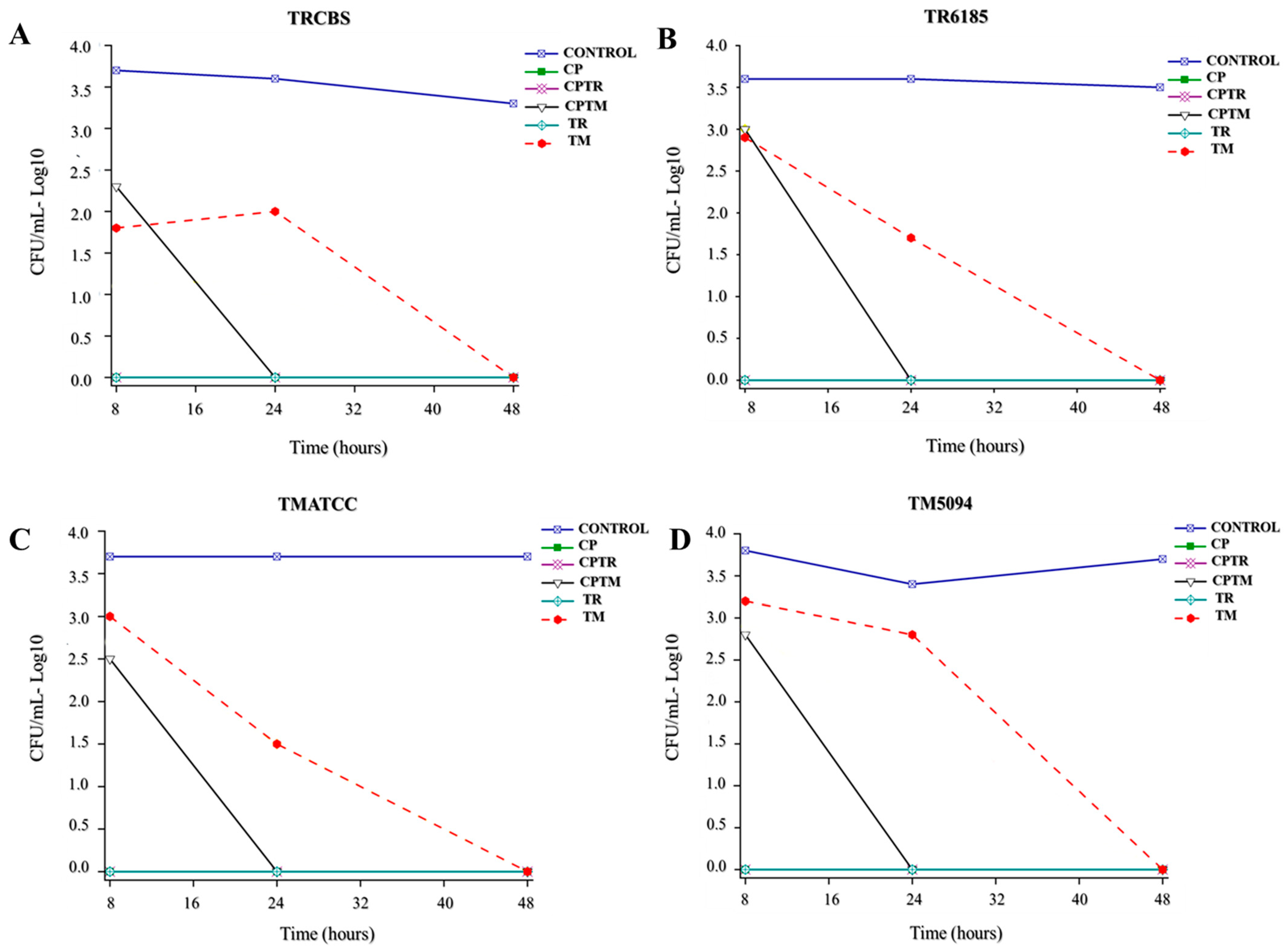
3.2. Time-Kill Assay

The time-kill assay was carried out for 48 h, exposing the TRCBS, TR6185, TMATCC, and TM5094 strains to the extracts (Figure 1).
The maximum reduction in CFU/mL for the dermatophytes was observed at 48 h, with an inhibition rate of 99.9% compared to the growth control. CP, TR, and CPTR extracts showed the fastest reduction time for all dermatophyte strains (8 h), followed by CPTM (24 h—Table 4).

3.3. Toxicity Test with Galleria mellonella

The toxicity tests using G. mellonella show the non-toxicity of the extracts at concentrations of 2000–8000 µg/mL with the survival rate of 80–100% of the larvae injected with the compounds. Toxicity tests using G. mellonella showed that the CP, TR1, and CPTM extracts tested were not toxic after five days, with a 100% survival rate of the injected larvae. Similarly, TM and CPTR extracts were not considered toxic, with an 80% survival rate (Figure 2).

4. Discussion

Clinical laboratory case reports of onychomycosis caused by Candida, filamentous non-dermatophytes and Trichophyton spp. have been common in recent years. Performing an analysis by direct examination often identifies fungi with two or more different structures (e.g., hyphae and yeast); however, fungal growth is often observed only for a single species [14,15].
The biological interference between the dermatophytes TM and TR with Candida when cultivated in a solid medium was demonstrated by Lemes et al. The anti-Trichophyton activity of extracts produced from pure cultures of Candida spp. corroborates the microbial growth interference [16]. The current study shows that mixed culture extracts also have antifungal activity against dermatophytes demonstrating the presence of antifungal compounds in these cultures. The individual biological characteristics of each strain justify the differences in MICs since the strains originate from different sources of infection. The unstable environment causes adaptation and response with biological changes as a defense or tolerance mechanism.
In the nails, mixed interactions between microorganisms promote the production of essential metabolites for survival, with a variety of virulence factors such as proteases, lipases, and biofilm formation, in addition to resistance to host immune responses or the action of drugs. In this context, Mohammadi et al., analyzing the virulence profile of Candida species isolated from nails, observed significant correlations between the MICs of fluconazole and itraconazole and biofilm production [17]. One study coordinated by Oliver et al. on chemical profiles of metabolites showed a variety of molecules produced by species of Candida spp. isolated from different sources of infection. In this study, 66 different metabolites were identified. These metabolic pathways are mainly related to energy production and virulence mechanisms [18].
A metabolomics analysis of TR by Ciesielska et al. identified compounds involved in amino acid metabolism, carbohydrate metabolism related to glycolysis, the tricarboxylic acid (TCA) cycle, and nucleotide and energy metabolisms. Due to the limited availability of nutrients, an increased production of substances and specific protective mechanisms such as metabolites that act as energy carriers (GTP), adenosine triphosphate (ATP), and uridine-5′-triphosphate (UTP) were detected in the control medium [19]. In the present study, it was not possible to identify the metabolites produced in pure and mixed cultures. Therefore, investigations are necessary to identify these compounds and better understand the interactions. Although the compounds in the extracts have not been identified, the antifungal activity described here opens possibilities for new therapeutic approaches to manage onychomycosis.
Considering the confluence of the species mentioned above, such as dermatophytes and Candida spp., in the present study, the data support the assertion that the synthesis of biologically derived compounds may be altered under growth conditions, whether isolated or mixed, given that the original environments harbor antagonistic or synergistic interferents. This phenomenon elucidates the variation in MIC values of the extracts when originating from mixed cultures.
The kinetics of the time-kill assay provides information on the microbicidal action dynamics of the tested compounds, an essential tool in the antimicrobial analysis of potential antifungal compounds [20]. The ability of extracts to inhibit fungal growth shows fungicidal characteristics against TM and TR strains, with maximum inhibition within 48 h.
The use of G. mellonella for toxicity tests has gained attention from the scientific community in recent years. Compared to traditional mammalian models, G. mellonella have certain advantages, such as being easy to obtain for large-scale experiments, simple and easy to handle without the need for special equipment, and being exempt from approval by research ethics committees [21]. The similarity with the innate immune response of mammals such as having cellular and humoral defenses should also be highlighted. Additionally, it is possible to establish the dosage of potential drug candidates in mammals [22].

5. Conclusions

The extracts of pure and mixed cultures of CP and dermatophytes present antifungal activity against TM and TR, which may influence the laboratory diagnosis of mixed infections. These results can explain cases of mixed fungal infections where only one of the etiologic agents grows in culture. Furthermore, isolating and identifying the compounds in extracts may allow new therapeutic approaches to control fungal infections.

Author Contributions

T.H.L.: study design/development and methodology/collection of data and data analysis/interpretation, writing all/sections of the manuscript. M.T.G.d.A.: study design/development and methodology/collection of data and data analysis/interpretation, manuscript revision. J.A.S.N.: methodology and obtention of extracts. L.O.R.: methodology and obtention of extracts. J.P.Z.S.: manuscript revision. G.R., L.T., and M.R.v.Z.K.: Toxicity test with Galleria mellonella assay. All authors have read and agreed to the published version of the manuscript.

Funding

The authors declare that they received financial support from the National Council for the Improvement of Higher Education (CAPES–01) and the National Council for Scientific and Technological Development (CNPq).

Data Availability Statement

The datasets generated during and/or analyzed during the current study are available from the corresponding author on reasonable request.

Acknowledgments

The authors would like to thank the Multi-user Laboratory (LMU) at São Jose do Rio Preto (FAMERP).

Conflicts of Interest

The authors declare no conflicts of interest that might be perceived to influence the results and/or discussion reported in this paper.

References

  1. Leung, A.K.C.; Lam, J.M.; Leong, K.F.; Hon, K.L.; Barankin, B.; Leung, A.A.M.; Wong, A.H.C. Onychomycosis: An Updated Review. Recent Pat. Inflamm. Allergy Drug Discov. 2020, 14, 32–45. [Google Scholar] [CrossRef] [PubMed]
  2. Gupta, A.K.; Taborda, V.B.A.; Taborda, P.R.O.; Shemer, A.; Summerbell, R.C.; Nakrieko, K.A. High prevalence of mixed infections in global onychomycosis. PLoS ONE 2020, 15, e0239648. [Google Scholar] [CrossRef] [PubMed]
  3. Andrés, T.S.; Alexandro, B. Candida Onychomycosis: An Old Problem in Modern Times. Curr. Fungal Infect. Rep. 2020, 14, 209–216. [Google Scholar] [CrossRef]
  4. Rather, S.; Keen, A.; Shah, F.Y.; Yaseen, A.; Farooq, S.; Bakhshi, A. Candidal Onychomycosis: Clinicoepidemiological Profile, Prevailing Strains, and Antifungal Susceptibility Pattern—A Study from a Tertiary Care Hospital. Indian J. Dermatol. 2021, 66, 132–137. [Google Scholar] [CrossRef] [PubMed]
  5. Gupta, A.K.; Stec, N.; Summerbell, R.C.; Shear, N.H.; Piguet, V.; Tosti, A.; Piraccini, B.M. Onychomycosis: A review. J. Eur. Acad. Dermatol. Venereol. 2020, 34, 1972–1990. [Google Scholar] [CrossRef] [PubMed]
  6. Martínez-Herrera, E.O.; Arroyo-Camarena, S.; Tejada-García, D.L.; Porras-López, C.F.; Arenas, R. Onychomycosis due to opportunistic molds. An. Bras. Dermatol. 2015, 90, 334–337. [Google Scholar] [CrossRef] [PubMed]
  7. Gaurav, V.; Bhattacharya, S.N.; Sharma, N.; Datt, S.; Kumar, P.; Rai, G.; Singh, P.K.; Taneja, B.; Das, S. Terbinafine resistance in dermatophytes: Time to revisit alternate antifungal therapy. J. Med. Mycol. 2021, 31, 101087. [Google Scholar] [CrossRef] [PubMed]
  8. Gupta, A.K.; Stec, N. Recent advances in therapies for onychomycosis and its management. F1000Research 2019, 8, 968. [Google Scholar] [CrossRef] [PubMed]
  9. Lemes, T.H.; Torrezan, G.S.; Polaquini, C.R.; Octavio, L.; Paziani, H.; von Zeska Kress, M.R.; Siqueira, J.P.Z.; Teresa, M.; Almeida, G. In vitro antifungal activity of Candida culture extracts against Trichophyton rubrum and Trichophyton mentagrophytes. Braz. J. Case Rep. 2021, 1, 135–152. [Google Scholar] [CrossRef]
  10. Tian, X.; Ding, H.; Ke, W.; Wang, L. Quorum Sensing in Fungal Species. Annu. Rev. Microbiol. 2021, 75, 449–469. [Google Scholar] [CrossRef]
  11. M38Ed3 Filamentous Fungi Antifungal Susceptibility Test [Internet]. Clinical & Laboratory Standards Institute. Available online: https://clsi.org/standards/products/microbiology/documents/m38/ (accessed on 21 July 2022).
  12. Klepser, M.E.; Ernst, E.J.; Lewis, R.E.; Ernst, M.E.; Pfaller, M.A. Influence of test conditions on antifungal time-kill curve results: Proposal for standardized methods. Antimicrob. Agents Chemother. 1998, 42, 1207–1212. [Google Scholar] [CrossRef] [PubMed]
  13. Ignasiak, K.; Maxwell, A. Galleria mellonella (greater wax moth) larvae as a model for antibiotic susceptibility testing and acute toxicity trials. BMC Res. Notes 2017, 10, 428. [Google Scholar] [CrossRef] [PubMed]
  14. Gómez-Sáenz, A.; Arenas, R. Mixed Onychomycosis. A Case Report Caused by Trichophyton rubrum, Fusarium sp. and Candida albicans. Dermatol. Cosmet. Medica Quir. 2020, 18, 48–50. [Google Scholar]
  15. Youssef, A.B.; Kallel, A.; Azaiz, Z.; Jemel, S.; Bada, N.; Chouchen, A.; Belhadj-Salah, N.; Fakhfakh, N.; Belhadj, S.; Kallel, K. Onychomycosis: Which fungal species are involved? Experience of the Laboratory of Parasitology-Mycology of the Rabta Hospital of Tunis. J. Mycol. Med. 2018, 28, 651–654. [Google Scholar] [CrossRef] [PubMed]
  16. Lemes, T.H.; Maschio-Lima, T.; Brizzotti-Mazuchi, N.S.; Siqueira, J.P.Z.; de Almeida, M.T.G. O estudo da interferência biológica entre dermatófitos e leveduras em onicomicoses. Int. J. Health Sci.-PDVS 2021, 1, 75–86. [Google Scholar]
  17. Mohammadi, F.; Ghasemi, Z.; Familsatarian, B.; Salehi, E.; Sharifynia, S.; Barikani, A.; Hosseini, M.A. Relationship between antifungal susceptibility profile and virulence factors in Candida albicans isolated from nail specimens. Rev. Soc. Bras. Med. Trop. 2020, 53, e20190214. [Google Scholar] [CrossRef] [PubMed]
  18. Oliver, J.C.; Laghi, L.; Parolin, C.; Foschi, C.; Marangoni, A.; Liberatore, A.; Dias, A.L.T.; Cricca, M.; Vitali, B. Metabolic profiling of Candida clinical isolates of different species and infection sources. Sci. Rep. 2020, 10, 16716. [Google Scholar] [CrossRef]
  19. Ciesielska, A.; Kawa, A.; Kanarek, K.; Soboń, A.; Szewczyk, R. Metabolomic analysis of Trichophyton rubrum and Microsporum canis during keratin degradation. Sci. Rep. 2021, 11, 3959. [Google Scholar] [CrossRef]
  20. Bax, H.I.; Bakker-Woudenberg, I.A.J.M.; de Vogel, C.P.; van der Meijden, A.; Verbon, A.; de Steenwinkel, J.E.M. The role of the time-kill kinetics assay as part of a preclinical modeling framework for assessing the activity of anti-tuberculosis drugs. Tuberculosis 2017, 105, 80–85. [Google Scholar] [CrossRef]
  21. Junqueira, J.C.; Mylonakis, E.; Borghi, E. Galleria mellonella experimental model: Advances and future directions. Pathog. Dis. 2021, 79, ftab021. [Google Scholar] [CrossRef]
  22. Piatek, M.; Sheehan, G.; Kavanagh, K. Galleria mellonella: The Versatile Host for Drug Discovery, In Vivo Toxicity Testing and Characterising Host-Pathogen Interactions. Antibiotics 2021, 10, 1545. [Google Scholar] [CrossRef] [PubMed]
Figure 1. Graphical representation of the dermatophyte death kinetics in contact with the following extracts: (A) TRCBS; (B) TR6185; (C) TMATCC; (D) TM5094. CP: C. parapsilosis; CPTR: C. parapsilosis and T. rubrum; CPTM: C. parapsilosis and T. mentagrophytes; TR: T. rubrum; TM: T. mentagrophytes.
Microbiolres 15 00057 g001
Figure 2. Survival (%) of Galleria mellonella larvae exposed to extracts (2000–8000 µg/mL) with monitoring at 24 h intervals for five days. CP: C. parapsilosis; CPTR: C. parapsilosis and T. rubrum; CPTM: C. parapsilosis and T. mentagrophytes; TR: T. rubrum; TM: T. mentagrophytes.
Microbiolres 15 00057 g002
Table 1. Minimum inhibitory concentration (MIC) values of the drugs fluconazole, itraconazole, and terbinafine against dermatophyte strains.
MIC (µg/mL)
FluconazoleItraconazoleTerbinafine
TMATCC40.50.03
TM50943220.03
TM54191610.03
TM60073210.03
TM60853210.03
TM73891610.03
TRCBS20.250.03
TR618540.50.03
TR619540.50.03
TR628440.50.03
TR7259320.564.0
TR760420.50.03
TR798420.50.03
Table 2. Minimum inhibitory concentration (MIC) and minimum fungicidal concentration (MFC) values for fluconazole, itraconazole, and terbinafine against dermatophyte strains.
MIC (µg/mL)
TMATCCTM5094TM5419TM6007TM6085TM7389
CP1000–20001000–20001000–20001000–20001000–20001000–2000
CPTM250–8000250–8000500–8000250–8000250–8000250–8000
CPTR2000–80004000–80004000–80004000–80004000–80004000–8000
TM1000–80002000–80001000–80001000–80001000–80001000–8000
TR2000–80002000–80004000–80004000–80004000–80004000–8000
MIC (µg/mL)
TRCBSTR6185TR6195TR6284TR7604TR7984
CP500–2000500–2000500–2000250–500500–2000250–2000
CPTM250–8000250–8000250–8000250–1000500–8000250–8000
CPTR2000–40002000–80002000–40001000–20002000–40002000–4000
TM1000–40001000–80002000–8000250–20001000–8000250–4000
TR1000–40001000–80002000–800010002000–80001000–8000
MFC (µg/mL)
TMATCCTM5094TM5419TM6007TM6085TM7389
CP1000–20001000–20001000–20001000–20001000–20001000–2000
CPTM500–8000500–8000500–8000500–80001000–8000500–8000
CPTR4000–80004000–80004000–80004000–80004000–80004000–8000
TM1000–80002000–80002000–80001000–80001000–80001000–8000
TR2000–80004000–80004000–80004000–80004000–80004000–8000
MFC (µg/mL)
TRCBSTR6185TR6195TR6284TR7604TR7984
CP1000–20001000–20001000–20001000–2000500–1000500–2000
CPTM250–8000250–8000500–8000500–4000500–8000500–8000
CPTR2000–40002000–80002000–8000200040002000–4000
TM1000–40001000–80002000–80001000–40002000–40001000–4000
TR1000–80002000–80002000–80001000–40004000–80002000–4000
CP: C. parapsilosis; CPTR: C. parapsilosis and T. rubrum; CPTM: C. parapsilosis and T. mentagrophytes; TR: T. rubrum; TM: T. mentagrophytes and T. mentagrophytes.
Table 3. The minimum inhibitory concentration (MIC) and minimum fungicidal concentration (MFC) geometric mean values for the extracts against the dermatophyte strains.
Geometric Mean (µg/mL)
TRTMTRTM
MICMFCMICMFCMICMFC
CP6891212113713618851284
CPTM141417141759261915772119
CPTR224534294104504030354157
TM134322451805329915572722
TR213334293899504028834157
CP: C. parapsilosis; CPTR: C. parapsilosis and T. rubrum; CPTM: C. parapsilosis and T. mentagrophytes; TR: T. rubrum; TM: T. mentagrophytes; TRTM: T. rubrum and T. mentagrophytes.
Table 4. The colony-forming unit (CFU/mL) of dermatophyte strains exposed to extracts for different times.
TRCBSTR6185
8 h24 h48 h 8 h24 h48 h
CONTROL508338121955CONTROL395742583450
CP000CP000
CPTR000CPTR000
CPTM18300CPTM111700
TR000TR000
TM671001TM817541
TMATCCTM5094
8 h24 h48 h 8 h24 h48 h
CONTROL517354655217CONTROL597127454753
CP000CP000
CPTR000CPTR000
CPTM28300CPTM66700
TR000TR000
TM1000330TM15336330
CP: C. parapsilosis; CPTR: C. parapsilosis and T. rubrum; CPTM: C. parapsilosis and T. mentagrophytes; TR: T. rubrum; TM: T. mentagrophytes.
Disclaimer/Publisher’s Note: The statements, opinions and data contained in all publications are solely those of the individual author(s) and contributor(s) and not of MDPI and/or the editor(s). MDPI and/or the editor(s) disclaim responsibility for any injury to people or property resulting from any ideas, methods, instructions or products referred to in the content.

