Article

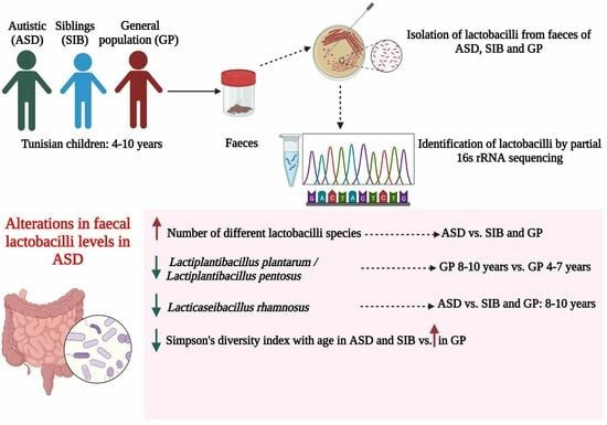
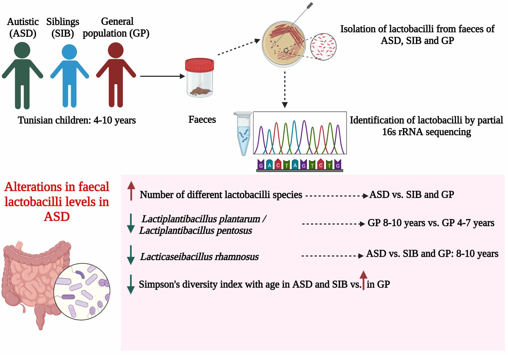
Lactobacilli Profile in Faecal Samples of Tunisian Children Diagnosed with Autism Spectrum Disorder

by
Mariem Chamtouri
1,2,
Abderrahmen Merghni
3,
Naoufel Gaddour
4,
Maha Mastouri
2,
Silvia Arboleya
1,5,* and
Clara G. de los Reyes-Gavilán
1,5,*
1
Department of Microbiology and Biochemistry of Dairy Products, Instituto de Productos Lácteos de Asturias (IPLA-CSIC), 33300 Villaviciosa, Spain
2
Laboratory of Transmissible Diseases and Biologically Active Substances LR99ES27, Faculty of Pharmacy, University of Monastir, Monastir 5000, Tunisia
3
Laboratory of Antimicrobial Resistance LR99ES09, Faculty of Medicine of Tunis, University of Tunis El Manar, Tunis 1068, Tunisia
4
Unit of Child Psychiatry, Monastir University Hospital, Monastir 5000, Tunisia
5
Diet, Microbiota, and Health Group, Instituto de Investigación Sanitaria del Principado de Asturias (ISPA), 33011 Oviedo, Spain
*
Authors to whom correspondence should be addressed.
Microbiol. Res. 2023, 14(3), 1225-1237; https://doi.org/10.3390/microbiolres14030082
Submission received: 17 July 2023 / Revised: 15 August 2023 / Accepted: 21 August 2023 / Published: 24 August 2023

Abstract

:
Alterations in faecal lactobacilli in autistic children have been reported, but little is known related to age and disorder severity. We used a culture-based method and partial 16S rRNA gene sequencing to isolate and identify lactobacilli strains from faeces of Tunisian autistic children (ASD group) and compared them with strains isolated from siblings (SIB) and children from the general population (GP). The ASD group displayed an increased number of different species compared to SIB and GP. Differences in species abundance with age accounted for a significant decrease in the abundance of Lactiplantibacillus plantarum/Lactiplantibacillus pentosus isolates in the GP at the age of 8–10 years compared to the age of 4–7 years, and to a significantly lower abundance of Lacticaseibacillus rhamnosus in the ASD group with respect to SIB and the GP at the age of 8–10 years. Simpson’s and Shannon–Wiener indices showed a more pronounced species diversity increase with age in the GP group compared to the ASD and SIB groups. Minor differences were found in lactobacilli prevalence and in species diversity between children with severe and mild-to-moderate ASD. Overall, we found substantial differences in the profile of faecal lactobacilli species in the ASD and GP groups at the age of 8–10 years.

Graphical Abstract

1. Introduction

Autism spectrum disorder (ASD) is an early-onset neurodevelopmental pathology characterised by impaired social communication and interaction and repetitive and stereotyped behaviours or interests [1]. While the aetiology of ASD remains largely unknown, a strong body of evidence revealed that alterations in the composition of the gut microbiota are associated with the development of autism [2].
Several research studies have analysed the intestinal microbiota of autistic children using culture-dependent methods as well as high-throughput DNA sequencing techniques [3,4,5]. An imbalance in the relative abundance of Bacteroidota (formerly named as Bacteroidetes) and Bacillota (formerly named as Firmicutes) phyla has been found, with a decrease in the Bacteroidota/Bacillota ratio in faecal samples of children with ASD [6]. A higher relative abundance of Collinsella, Corynebacterium and Dorea genera and of the lactobacilli group have been reported in faeces from ASD children compared to those from controls [7]. Other authors found reduced levels of Bifidobacterium, Prevotella, and Akkermansia municiphila in the gut microbiota of autistic children [8,9,10]. To date, different and sometimes contradictory results have been reported in studies conducted on the gut microbiota composition in ASD, which can be partly attributed to the heterogeneity of some factors that can influence the microbiota such as dietary and social habits, environment, and ethnicity [11].
Lactobacilli are Gram-positive, non-sporulating, catalase-negative and fermentative group of lactic acid bacteria comprising different closely related genera [12]. These microorganisms belong to the phylum Bacillota and some members of this group are found in the human and animal gut microbiota. Lactobacilli can be clustered into two major metabolic groups based on the hexose catabolic pathway: (i) homofermentative, which metabolise hexoses via the Embden–Meyerhoff pathway to pyruvate as the main metabolic intermediate and lactic acid as the main end product, and (ii) heterofermentative, displaying a metabolism of hexoses via the phosphoketolase pathway to be converted into pyruvate and acetyl-phosphate as key intermediates, and lactic acid, acetic acid and/or ethanol as end products [12,13].
Most of the studies focusing on ASD microbiota have found alterations in the lactobacilli population when comparing the ASD group with the general population (GP). Some of them have reported high levels of lactobacilli in the faeces of ASD children [3,6,7,11], whereas De Angelis et al. found reduced levels of lactobacilli in ASD with respect to children from the control group [4]. These findings, although apparently contradictory, point towards a possible association of lactobacilli with the development of autism.
Ligilactobacillus ruminis was the most frequently isolated and predominant species found in the gut microbiota of Indian ASD children [11], whereas no identification at the species level within the lactobacilli group was carried out in other studies with different human populations within the lactobacilli group [3,4,6,7]. Indeed, the identity of lactobacilli isolated from the faeces of children with ASD is still poorly known. To our knowledge, the prevalence, species diversity, and characteristics of lactobacilli isolated from the gut microbiota of Tunisian individuals with ASD have not been previously explored. Our aim in the present work was to unveil differences in faecal lactobacilli composition among Tunisian children with ASD, their siblings and a Tunisian children’s control group (ASD, SIB and GP groups, respectively), along with the putative effects of age and the severity of the disorder. To those ends, we isolated and identified the predominant lactobacilli strains from faecal samples and we compared the profiles obtained among the three groups of children. To our knowledge, this is the first report analysing differences in the profile of faecal lactobacilli in ASD in Tunisia and one of the few works in the literature considering the influences of age and disorder severity on the faecal microbiota composition in autism.

2. Materials and Methods

2.1. Subjects and Faecal Sample Collection

Faecal samples were collected from 74 Tunisian children aged between 4 and 10 years. The children were divided into three groups: 28 ASD (22 boys and 6 girls; with a mean age of 7.93 ± 2.05 years), 18 age-matched SIB (brothers and sisters of autistic children, 5 boys and 13 girls; with a mean age of 7.56 ± 1.89 years), and 28 age- and sex-matched unrelated GP children (22 boys and 6 girls; with a mean age of 7.29 ± 2.09 years). Autistic children were recruited at the Child and Adolescent Psychiatry Unit, Department of Psychiatry, Fattouma Bourguiba University Hospital, Monastir, Tunisia. The children from the general population were recruited from kindergartens and/or schools from the same social group. An ASD diagnosis was carried out by qualified personnel according to the Diagnostic and Statistical Manual for Mental Disorders (DSM-5) [14], the Autism Diagnostic Inventory-Revised (ADI-R) [15], and the Autism Diagnostic Observation Schedule-2 (ADOS-2) [16]. The severity of ASD was assessed using the Childhood Autism Rating Scale [17] (CARS). The exclusion criteria for all the participants were: having neurological disorders not strictly related to autism, type 1 diabetes, genetic syndromes, celiac disease, food intolerance, or inflammatory bowel disease. Subjects in this study were not treated with antibiotics or antifungals and had not taken probiotics and/or prebiotics for at least one month prior to sampling. Demographic and clinical data of the participants were described in detail in a previous study [18]. The study was approved by the Ethics Committee of the Faculty of Medicine of Monastir (reference IORG 0009738 Nº18/ OMB 0990-0279) and by the Bioethics Committee of CSIC (reference 172/2020), and informed written consent was obtained from the parents or legal guardians of each child before their inclusion in the study.

2.2. Isolation of Lactobacilli Strains and Culture Conditions

One gram of each fresh faecal sample was resuspended in 10 mL sterile distilled water and homogenised by vortexing for 5 min. Then, 10 µL of this homogenate was spread-plated on MRS agar (Biokar, Beaubois, France) and incubated under anaerobic conditions at 37 ° C for 48–72 h. Colonies of various morphologies and sizes were picked at random from plates and re-isolated again on MRS agar plates to obtain pure cultures. Pure isolated colonies were then inoculated in MRS broth, stained with the Gram method and tested for their ability to produce the enzyme catalase. Isolates suspected to be Gram-positive bacilli/cocobacilli and catalase-negative were stored in 30% glycerol (v/v) at −20 °C for further identification [19].

2.3. Identification of Lactobacilli Strains Using Partial 16S rRNA Gene Sequencing

Frozen pure isolates were thawed and cultured anaerobically on MRS broth for 24 h, centrifuged and the cell pellet was used for DNA extraction and amplification reactions. The identification of strains was carried out by partial sequence analysis of the 16S rRNA gene. Briefly, the bacterial DNA was extracted from the cell pellets of pure microbial cultures by using the Gen EluteTM Bacterial Genomic DNA Kit (Sigma-Aldrich, St. Louis, MO, USA), following the manufacturer’s instructions. The 16S rRNA was partially amplified using the universal primers plb 16 (5′-AGAGTTTGATCCTGGCTCAG-3′) and mlb 16 (5′-GGCTGCTGGCACGTAGTTAG-3′) [20]. The PCR reaction mixtures (25 μL) contained 1 μL of genomic DNA, 12.5 μL of 2X DreamTaq Green PCR Master Mix (Thermo Fisher Scientific, Waltham, MA, USA), 1 μL of each forward and reverse primer (10 μM), and 9.5 μL of molecular biology-grade water (Sigma-Aldrich). The PCR conditions were as follows: one initial denaturing step at 95 °C for 5 min, 30 cycles at 94 °C for 45 s (denaturation), 55 °C for 60 s (annealing), 72 °C for 35 s (elongation), and a final extension step at 72 °C for 10 min. DNA amplification was performed in a SimpliAmp Thermal Cycler (Applied Biosystems, Foster City, CA, USA), verified by electrophoresis on 1% agarose gel (Condalab, Torrejon de Ardoz, Spain) stained with Midori Green Advanced DNA stain (NIPPON Genetics Europe, Germany) and visualised under UV light by using a G box machine (SynGene, Cambridge, UK). Furthermore, the PCR products were purified using the GenElute PCR Clean-Up Kit (Sigma-Aldrich) and sequenced at Macrogen Inc. (Madrid, Spain) in an ABI3730XL automated sequencer (Applied Biosystems). The sequences were compared to those in the NCBI database using the BLASTn tool (https://blast.ncbi.nlm.nih.gov/Blast.cgi, accessed on 27 February 2023).

2.4. Prevalence and Species Diversity

The prevalence of lactobacilli isolates in stools of the different groups of children was estimated as the percentage of positive stool samples (with at least one lactobacilli colony isolated and identified) with respect to the total stool samples analysed in the group considered. Species lactobacilli abundance was calculated as the percentage of each species among the total lactobacilli isolates identified in each group of children.
The species diversity in each group of children considered in this study was calculated taking into account the number of species and the relative abundance of each species by using Simpson’s (D) and the Shannon–Wiener (H) diversity indices, which emphasise evenness and richness of species, respectively.
Simpson’s diversity index (D) was calculated by applying the following formula:
D =   1 n ( n 1 ) N ( N 1 )
The Shannon–Wiener diversity index (H) was calculated applying the formula:
H =   ( n N ) ln ( n N )
where
D: Simpson’s diversity index;
H: Shannon–Wiener diversity index;
n: number of colonies from each individual species;
N: total number of colonies from all species;
ln: natural logarithm;
Σ: sum of the calculations for all species.
The values of D range between 0 and 1 [21,22], and the values of H usually range between 0.5 and 5 [23]; the higher the value, the greater the diversity.

2.5. Statistical Analysis

Statistical analyses were carried out using the SPSS statistical pack version 26.0 (IBM SPSS, Inc., Chicago, IL, USA). Comparison of the prevalence of lactobacilli, species abundance and proportion of homofermentative and heterofermentative species among the ASD, SIB, and GP groups, as well as among the same groups stratified by age (4–7 years and 8–10 years) and by the severity of the disorder in the ASD group (mild-to-moderate with a CARS score of 30–36 and severe with a score of 37–60), was performed using the chi-square test (χ2) or Fisher’s exact test. A comparison of specific lactobacilli species abundance between children from the two age ranges considered in this study (4–7 years and 8–10 years) within the same group (ASD, SIB and GP) was also performed using the same statistical tools. Differences were considered significant at p-values ≤ 0.05.

3. Results

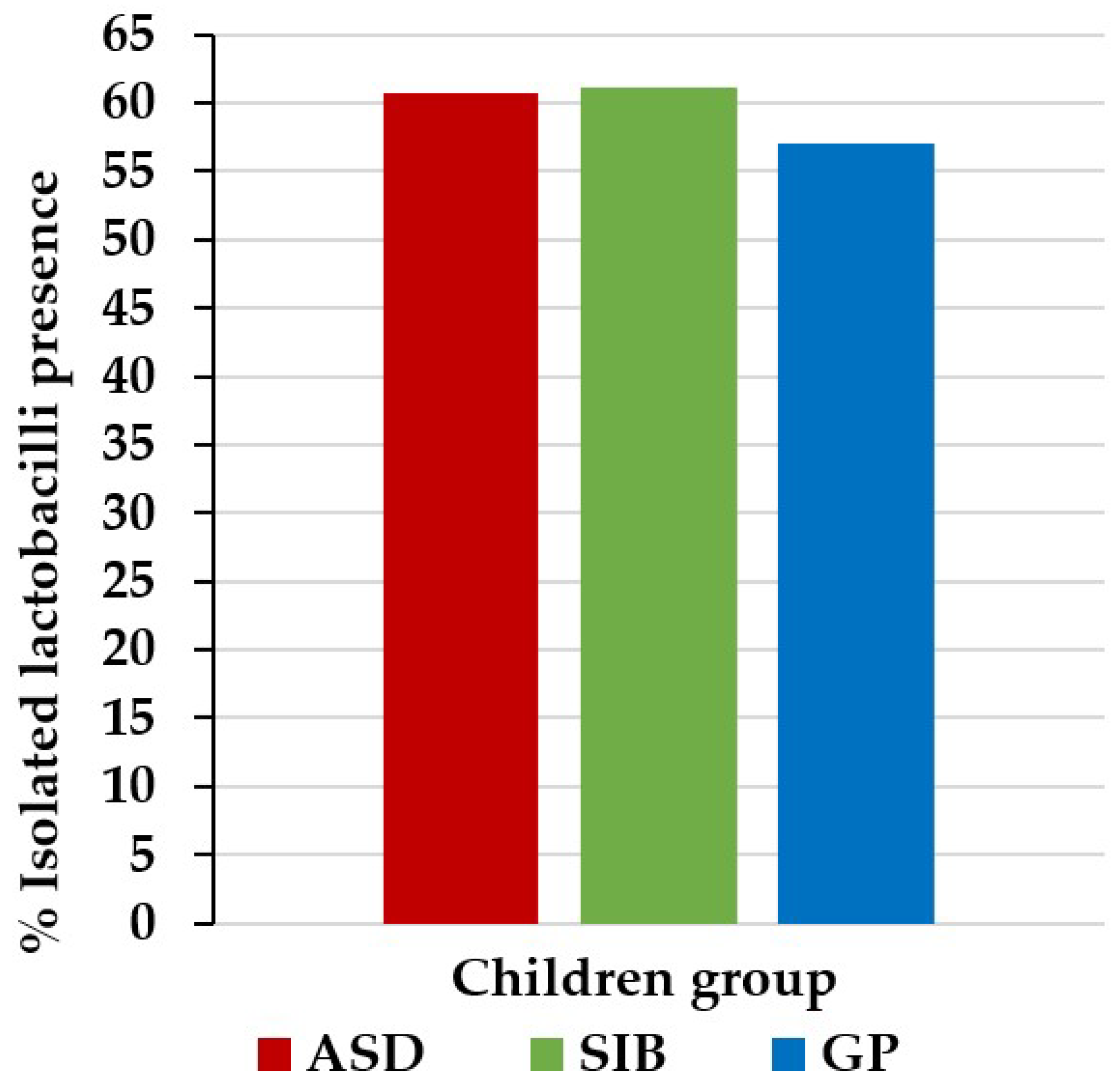
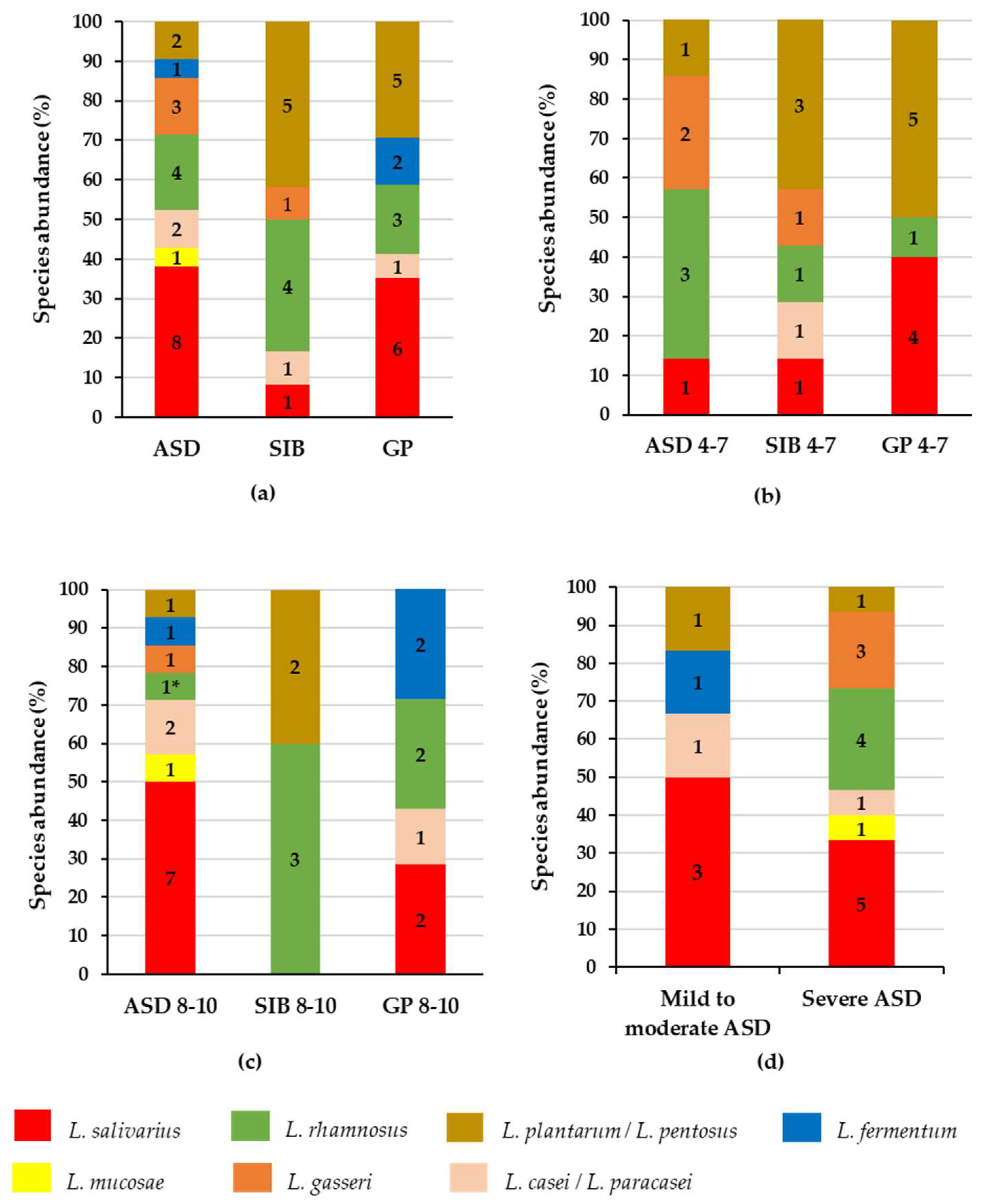
3.1. Prevalence, Species Distribution and Characteristics of Lactobacilli in Stools of ASD, SIB and GP Children

A total of 50 Gram-positive and catalase-negative isolated colonies were identified as members of the lactobacilli group using partial 16S rRNA gene sequencing (Table S1). Out of these isolates, 21 were from faecal samples from ASD children (in four of these samples, two strains were obtained), 12 isolates were from SIB of the autistic children (one of these samples provided two strains) and 17 were from samples from the GP group (one of the samples had two isolated strains). In this way, we isolated lactobacilli colonies from faecal samples of 17 ASD, 11 SIB and 16 GP children (Figure 1). No statistically significant difference was found in the prevalence of lactobacilli among the faecal samples from the three groups of children (p > 0.05).
Partial sequencing of the 16S rRNA gene identified two strains as Lacticaseibacillus casei/Lacticaseibacillus paracasei and the other two as Lactiplantibacillus plantarum/Lactiplantibacillus pentosus due to the high genome similarities between the species L. casei and L. paracasei and between L. plantarum and L. pentosus. Therefore, in the rest of the paper, isolates identified as L. casei/L. paracasei or L. paracasei and as L. plantarum/L. pentosus or L. plantarum will be called L. casei/L. paracasei or L. plantarum/L. pentosus, respectively. Among the lactobacilli isolated from stools of the ASD group, Ligilactobacillus salivarius (n = 8; 38.1%) was the most abundant species, followed by Lacticaseibacillus rhamnosus (n = 4; 19%), Lactobacillus gasseri (n = 3; 14.3%), L. plantarum/L. pentosus (n = 2; 9.5%), L. casei/L. paracasei (n = 2; 9.5%), Limosilactobacillus fermentum (n = 1; 4.8%) and Limosilactobacillus mucosae (n = 1; 4.8%). In the SIB group, the following species were identified in decreasing order of abundance: L. plantarum/L. pentosus (n = 5; 41.7%), L. rhamnosus (n = 4; 33.3%), L. salivarius (n = 1; 8.3%), L. casei/L. paracasei (n = 1; 8.3%) and L. gasseri (n = 1; 8.3%). The isolates from the GP group were identified as L. salivarius (n = 6, 35.3%), L. plantarum/L. pentosus (n = 5, 29.4%), L. rhamnosus (n = 3; 17.6%), L. fermentum (n = 2; 11.8%) and L. casei/L. paracasei (n = 1; 5.9%) (Figure 2a). No significant difference in species abundance was found among the three groups of children (p > 0.05). Nonetheless L. plantarum/L. pentosus were less abundant in the ASD group compared to the SIB and GP groups, L. gasseri was only found in faeces from the ASD and SIB groups and L. mucosae was only present in stools from the ASD group.
L. salivarius, L. rhamnosus, L. plantarum, L. pentosus, L. paracasei, L. casei, and L. gasseri are considered homofermentative species, whereas L. fermentum and L. mucosae are heterofermentative [12]. In the faecal samples from ASD children, 19 isolated colonies (90.5%) were identified as belonging to homofermentative species and only 2 isolates as heterofermentative (9.5%). In the SIB group, all isolates (100%) were identified as homofermentative species. In samples from GP children, 15 colonies (88.2%) corresponded to homofermentative microorganisms, whereas 2 colonies (11.8%) were from microorganisms belonging to heterofermentative species. When comparing proportions of homofermentative and heterofermentative species among the ASD, SIB and GP groups, no statistically significant difference was found (p > 0.05) among them (Table 1).
Simpson’s (D) and Shannon–Wiener (H) diversity indices were determined for all species identified within each group of children, as well as for homofermentative microorganisms, providing a unique numerical value of each index for each group. The indices were not used to calculate the diversity of the heterofermentative group due to the low number of lactobacilli species considered as heterofermentative among our identified isolates. When considering all lactobacilli species, the D index was the same in the ASD, SIB and GP groups (D = 0.8), whereas the ASD and SIB groups displayed a slightly higher homofermentative species diversity (D = 0.8) than GP (D = 0.7) (Table 2). However, the H index was higher in the ASD group than in the SIB and GP groups when considering all species (H = 1.7 in ASD vs. 1.4 and 1.5 in SIB and GP groups, respectively) as well as homofermentative species (H = 1.5 in ASD vs. 1.4 and 1.2 in SIB and GP groups, respectively) (Table 2).

3.2. Prevalence, Species Abundance and Diversity of Lactobacilli by Age

The prevalence, species abundance, and diversity of lactobacilli in the children’s groups were also analysed separately in children aged between 4 and 7 years (n = 35, 22 boys and 13 girls; with a mean age of 5.74 ± 1.48 years) and in children aged between 8 and 10 years (n = 39, 27 boys and 12 girls; with a mean age of 9.05 ± 1.68 years). We established this sample stratification since at the age of 4 to 7 years most autistic children in Tunisia stay at home, whereas at the age of 8 to 10 years most children are assisted or are institutionalised in specialised centres.
In the age of 4–7 years, lactobacilli were isolated from faecal samples of 5 ASD children (50%), 6 SIB (54.5%) and 10 GP individuals (71.4%) (Table S2). The isolates from the ASD group were identified as L. rhamnosus (n = 3; 42.9%), L. gasseri (n = 2; 28.6%), L. plantarum/L. pentosus (n = 1; 14.3%), and L. salivarius (n = 1; 14.3%). Isolates from the SIB group belonged to the species L. plantarum/L. pentosus (n = 3; 42.9%), L. salivarius (n = 1; 14.3%), L. casei/L. paracasei (n = 1; 14.3%), L. gasseri (n = 1; 14.3%) and L. rhamnosus (n = 1; 14.3%). Isolates from samples from the GP group were identified as L. plantarum/L. pentosus (n = 5, 50%), L. salivarius (n = 4; 40%) and L. rhamnosus (n = 1; 10%) (Figure 2b). Although L. salivarius and L. plantarum/L. pentosus were more abundant in GP than in the ASD group and L. rhamnosus was more abundant in ASD children, no significant differences in species abundance among groups of children were found at the ages of 4 to 7 years (p > 0.05). In this age range, all lactobacilli isolates were homofermentative species (Table 1), with a lower diversity (D and H indices) in the GP than in the ASD and SIB groups (Table 2).
In the age range of 8–10 years, lactobacilli were isolated from stools from 12 ASD (66.7%), 5 SIB (71.4%) and 6 GP children (42.9%) (Table S2). The ASD isolates were identified as L. salivarius (n = 7; 50%), L. paracasei/L. casei (n = 2; 14.3%), L. fermentum (n = 1; 7.1%), L. gasseri (n = 1; 7.1%), L. mucosae (n = 1; 7.1%), L. rhamnosus (n = 1; 7.1%) and L. plantarum/L. pentosus (n = 1; 7.1%). In the SIB group, the isolates were identified as L. rhamnosus (n = 3; 60%) and L. plantarum/L. pentosus (n = 2; 40%). In faeces from GP children, the isolates were identified as L. salivarius (n = 2; 28.6%), L. rhamnosus (n = 2; 28.6%), L. fermentum (n = 2; 28.6%) and L. casei/L. paracasei (n = 1; 14.3%) (Figure 2c). Remarkably, L. rhamnosus was significantly less abundant in ASD than in SIB and GP children at this range of age (p = 0.05). In the ASD group, 12 isolates (85.7%) were identified as homofermentative species and 2 (14.3%) as heterofermentative. In the SIB group, all isolates (n = 5; 100%) were homofermentative. In the GP group, five of the isolates (71.4%) were homofermentative and two (28.6%) were heterofermentative. However, when comparing the proportions of homofermentative and heterofermentative species at 8–10 years of age, no significant differences were found among the ASD, SIB and GP groups (Table 1). In the age range of 8–10 years, Simpson’s diversity index (D) for all species as well as for homofermentative isolates was slightly higher in the GP than in the ASD group, whereas the Shannon–Wiener index (H) remained higher in ASD than in the GP and SIB groups (Table 2).
Finally, we analysed the variations in abundance of specific lactobacilli species for each group of children (ASD, SIB and GP) between the two ranges of age considered (Table 3). Despite some minor differences observed, the only significant change found in the transition from 4–7 years to 8–10 years was a decrease in the proportion of L. plantarum/L. pentosus (p = 0.04) in the group of GP children that did not occur in the ASD and SIB groups. As a consequence, the ratio of L. rhamnosus to L. salivarius species decreased at the age of 8–10 years in the ASD group (3 at 4–7 years vs. 0.1 at 8–10 years), whereas it increased in the GP group (0.25 at 4–7 years vs. 1 at 8–10 years). Notably, in the two ranges of age considered, we observed that Simpson’s diversity index decreased while aged 8–10 in the ASD and SIB groups, while it increased in the GP group with respect to the 4–7-year age category. An increase in the Shannon–Wiener index was found in both ASD and GP groups from 4–7 years to 8–10 years, this increase being more pronounced in the GP group and coincident with a clear increase in the diversity of homofermentative species in this GP group.
In short, some differences in the distribution of lactobacilli species were found between faeces from ASD and GP children, with L. plantarum/L. pentosus and L. rhamnosus accounting for the more prominent differences observed at the ages of 8–10 years but not in younger children.

3.3. Prevalence, Species Abundance and Diversity of Lactobacilli by Severity of the Disorder

We stratified the group of ASD children into two subgroups according to the severity of the disorder following the CARS score, mild-to-moderate (n = 11) and severe (n = 17), and analysed the differences in the prevalence, abundance and diversity of lactobacilli species between these two subsets.
Lactobacilli were isolated from faecal samples from 6 children with mild-to-moderate ASD (54.5%) and 11 children with severe ASD (64.7%). Out of the 21 isolates from the ASD group, 6 of them were from individuals with mild-to-moderate ASD and 15 from children with severe ASD (Table S2). In stools of children with mild-to-moderate ASD, the isolates were identified as L. salivarius (n = 3; 50%), L. plantarum/L. pentosus (n = 1; 16.7%), L. fermentum (n = 1; 16.7%) and L. casei/L. paracasei (n = 1; 16.7%). In children with severe ASD, the isolates were identified as L. salivarius (n = 5; 33.3%), L. rhamnosus (n = 4; 26.7%), L. gasseri (n = 3; 20%), L. plantarum/L. pentosus (n = 1; 6.7%), L. mucosae (n = 1; 6.7%) and L. casei/L. paracasei (n = 1; 6.7%) (Figure 2d). Out of the six isolates from children with mild-to-moderate ASD, five (83.3%) of them were homofermentative and one (16.7%) was heterofermentative. In children with severe ASD, 14 isolates (93.3%) were homofermentative and 1 (6.7%) was heterofermentative (Table 1).
Simpson’s diversity index (D) for all species was the same in the two ASD subgroups, whereas the Shannon–Wiener index increased from children with mild-to-moderate to children with severe symptoms of ASD. The children with a severe degree of the disorder displayed a higher homofermentative species diversity than children with mild-to-moderate ASD on both diversity indices (Table 2).
No significant differences were found in the prevalence, species abundance and proportions of homofermentative and heterofermentative species of lactobacilli isolates from the faecal samples from the two ASD subgroups. Remarkably, some lactobacilli species such as L. mucosae, L. rhamnosus and L. gasseri were only isolated from faeces of children with a severe degree of the disorder.

4. Discussion

ASD is one of the most common neurodevelopmental disorders and its prevalence has increased rapidly in recent years. Several studies have demonstrated an alteration in the gut microbiota composition of autistic children as compared to typically developing individuals [2]. It has been suggested that lactobacilli, which naturally inhabit the gastrointestinal tract of humans and animals, can display altered levels in the faeces of autistic children [3,4,7]. Nonetheless, the data on the link between this group of microorganisms and autism are inconsistent to date and little is known about the prevalence, species identity and characteristics of lactobacilli in autistic children. Thus, in this study, we used a culture-based method to isolate lactobacilli strains from the faeces of Tunisian autistic children and identified them using partial 16S rRNA gene sequencing. We aimed to determine the possible variations in the faecal lactobacilli population in Tunisian autistic children through comparisons with a group of SIB and individuals from the GP, with special focus on the effects of age and ASD severity.

4.1. Prevalence and Species Abundance of Lactobacilli in Stools of ASD, SIB and GP Groups

Although we obtained a slightly higher prevalence of lactobacilli isolates in the faeces of children with ASD and their SIB compared to the GP group, these differences did not reach statistical significance.
Among the lactobacilli species identified in our work, L. salivarius was the most frequently isolated from the faeces of the ASD group, followed by considerably lower proportions of other species, including L. rhamnosus. L. salivarius was also the most abundant species in the GP group, whereas L. plantarum/L. pentosus were the most abundant species isolated from faecal samples of the SIB group. It has been reported that L. salivarius can induce in vitro the secretion of proinflammatory cytokines such as TNF-α by dendritic cells [24]. Elevated levels of certain proinflammatory cytokines such as IL-1, IL-6, and IL-12p40 have been found in the plasma of autistic children and have been strongly associated with the impaired communication and aberrant behaviours observed in autism [25]. In this way, we could tentatively speculate that there is a relationship between elevated levels of L. salivarius in the gut and a more proinflammatory status in autistic children.
In contrast to the study of Pullikan et al. [11], we did not find the species L. ruminis, with recognised immunomodulatory properties, in the faeces of our autistic children. However, the differential presence of some other species between the groups of children deserves some comment. L. gasseri was isolated from faeces from the ASD and SIB groups but not from the faeces from the GP group. The strain L. gasseri NK109, isolated from a human faecal sample, has been demonstrated to reduce colitis and neuroinflammation in mice via modulation of gut microbiota, leading to the attenuation of cognitive deficits and depression as well as to the improvement of neuropsychiatric disorders [26]. L. mucosae was only detected by us in the faeces from ASD children, and not from the other two groups of children. Recently, it has been reported that L. mucosae NK41, isolated from healthy human gut microbiota, can alleviate neuropsychiatric disorders by regulating altered gut microbiota in a mouse model [27]. Thus, the species L. gasseri and L. mucosae, mainly isolated in the present work from faecal samples from ASD children and generally considered as beneficial for health, may be playing a protective and beneficial role in some way for autistic children. Although we cannot know if the balance between the different lactobacilli species may influence the symptoms of the disorder and intestinal health, our results suggest differences in the profile of lactobacilli species between autistic children and children from the general population that would need a more in-depth study.

4.2. Prevalence, Species Abundance and Diversity of Lactobacilli by Age and Disorder Severity

One of the most important factors affecting the prevalence and diversity of lactobacilli in the gut microbiota is age [28]. Hence, here we analysed the prevalence, species diversity and abundance of lactobacilli strains by stratifying our sample into two age ranges (4–7 years and 8–10 years). In this way, we found some variations in the abundance of lactobacilli species according to age. Two remarkable changes occurred in species abundance as a function of age. First, at the ages of 8–10 years, L. rhamnosus was significantly less abundant in ASD children than in the SIB and GP groups. Second, a significant decrease in the abundance of L. plantarum/L. pentosus was found in the faeces from GP children at the ages of 8–10 years compared to 4–7-year-old children, a shift that did not occur in the faecal samples from SIB and ASD children. L. rhamnosus is known for its immunomodulatory and anti-inflammatory effects, and many strains have been widely used as probiotics [29,30,31]. In contrast, the proinflammatory or modulatory effect of L. salivarius has been found to be strain-dependent [32]. We tentatively speculate that the balance between L. rhamnosus and L. salivarius in the faeces from ASD children at more advanced ages may be related to the inflammatory status of their immune system, although this aspect deserves to be investigated in more depth. In contrast to the more pronounced differences in the lactobacilli species profiles found in older children, we recently reported in the same population that there were more pronounced differences in the profile of faecal amino acids in younger children (4 to 7 years) than in those with more advanced ages (8 to 10 years) [18].
The species diversity in the three groups of children was also influenced by age. Simpson’s and Shannon–Wiener diversity indices were lower in the GP group than in the ASD and SIB groups at the ages of 4–7 years while Simpson’s index was higher in the GP group than in the ASD and SIB groups at the ages of 8–10 years. The evolution of species diversity with age accounts for the increase in both indices at the ages of 8–10 years in the GP group, in contrast with the less pronounced or non-occurring increase in ASD and SIB groups. The infant’s gut microbiota initially exhibits a low diversity that increases with age from infancy to adulthood. In general, a healthy gut microbiota in infants and young children is characterised by a low species diversity versus a high species diversity in adults and older healthy individuals [33]. Here, in our study, we found that GP children showed an increase in lactobacilli species diversity and homofermentative species with age, which could be related to a healthier gut microbiota in the GP group.
No significant differences were found in terms of autism severity, despite a slightly higher abundance of lactobacilli species in children with severe symptoms compared to children with mild-to-moderate autism, the presence of some species only in individuals with severe symptoms of the disorder, and higher Shannon–Wiener diversity in the group with severe ASD symptoms.
This is one of the few studies currently available about faecal lactobacilli isolates in ASD and the sole considering the influence of age on the faecal lactobacilli species composition in autistic children. The results obtained highlighted clear differences in lactobacilli species composition between autistic and non-autistic children as a function of age.

5. Conclusions

The results obtained in the present work point to the few but clear differences in the profiles of faecal lactobacilli species from ASD and GP Tunisian children as related to age. Although the low number of lactobacilli isolates precludes formulating a sound hypothesis, these preliminary results lay eyes on one of the most controversial microbial group from the gut microbiota in ASD, lactobacilli. Moreover, this work emphasises the need for further studies with a larger human sample size and the use of culturomic approaches to reveal the importance of specific microbial groups in different neurodevelopmental disorders.

Supplementary Materials

The following supporting information can be downloaded at: https://www.mdpi.com/article/10.3390/microbiolres14030082/s1, Table S1: Molecular identification of lactobacilli strains isolated from stools of autistic children (ASD), their siblings (SIB) and children from the general population (GP); Table S2: Number and prevalence of lactobacilli isolates in stools from autistic children (ASD), siblings (SIB) and children from the general population (GP) stratified by age (4–7 years and 8–10 years) and according to the severity of disorder (mild-to-moderate and severe). P-values are indicated for statistical comparisons among the three groups of children and between the two ranges of severity of disorder for the presence or absence of lactobacilli isolates in stool samples.

Author Contributions

Conceptualisation, A.M., S.A. and C.G.d.l.R.-G.; methodology, M.C. and S.A.; software, M.C.; validation, M.C., S.A. and C.G.d.l.R.-G.; formal analysis, M.C.; investigation, M.C., S.A. and C.G.d.l.R.-G.; resources, N.G.; data curation, M.C.; writing—original draft preparation, M.C., S.A. and C.G.d.l.R.-G.; writing—review and editing, M.C., A.M., N.G., M.M., S.A. and C.G.d.l.R.-G.; visualisation, M.C.; supervision, project administration and funding acquisition, M.M. and C.G.d.l.R.-G. All authors have read and agreed to the published version of the manuscript.

Funding

This work was supported by the project COOPA2022, financed by CSIC through the call icoop+ 2018, and by the project AYUD/2021/50981 of the Principality of Asturias, to support the activity of their research groups. Mariem Chamtouri received a 3-month fellowship from the University of Monastir to carry out the current study in collaboration with IPLA-CSIC Asturias, Spain. Silvia Arboleya was the recipient of a postdoctoral researcher contract (ITM-Junior-22) awarded by the Biosanitary Research Foundation in Asturias (FINBA, Spain).

Institutional Review Board Statement

This study was conducted in accordance with the Declaration of Helsinki and approved by the Ethical Committee of the Faculty of Medicine of Monastir, Tunisia (code IORG 0009738 Nº18/ OMB 0990-0279, date of approval: 3 May 2019) and by the Ethics Committee from the Spanish Research Council (CSIC) (code 172/2020, date of approval: 17 September 2021).

Informed Consent Statement

Written informed consent was obtained from all parents of children included in the study.

Data Availability Statement

All data generated or analysed during this study are included in this article and its supplementary information files.

Acknowledgments

The authors thank all children and their families for participating in this study.

Conflicts of Interest

The authors declare no conflict of interest.

References

  1. Genovese, A.; Butler, M.G. Clinical assessment, genetics, and treatment approaches in autism spectrum disorder (ASD). Int. J. Mol. Sci. 2020, 21, 4726. [Google Scholar] [CrossRef] [PubMed]
  2. Cao, X.; Liu, K.; Liu, J.; Liu, Y.W.; Xu, L.; Wang, H.; Zhu, Y.; Wang, P.; Li, Z.; Wen, J.; et al. Dysbiotic Gut Microbiota and Dysregulation of Cytokine Profile in Children and Teens with Autism Spectrum Disorder. Front. Neurosci. 2021, 15, 635925. [Google Scholar] [CrossRef] [PubMed]
  3. Adams, J.B.; Johansen, L.J.; Powell, L.D.; Quig, D.; Rubin, R.A. Gastrointestinal flora and gastrointestinal status in children with autism—Comparisons to typical children and correlation with autism severity. BMC Gastroenterol. 2011, 11, 22. [Google Scholar] [CrossRef] [PubMed]
  4. De Angelis, M.; Piccolo, M.; Vannini, L.; Siragusa, S.; De Giacomo, A.; Serrazzanetti, D.I.; Cristofori, F.; Guerzoni, M.E.; Gobbetti, M.; Francavilla, R. Fecal Microbiota and Metabolome of Children with Autism and Pervasive Developmental Disorder Not Otherwise Specified. PLoS ONE 2013, 8, e76993. [Google Scholar] [CrossRef]
  5. Liu, S.; Li, E.; Sun, Z.; Fu, D.; Duan, G.; Jiang, M.; Yu, Y.; Mei, L.; Yang, P.; Tang, Y.; et al. Altered gut microbiota and short chain fatty acids in Chinese children with autism spectrum disorder. Sci. Rep. 2019, 9, 287. [Google Scholar] [CrossRef]
  6. Tomova, A.; Husarova, V.; Lakatosova, S.; Bakos, J.; Vlkova, B.; Babinska, K.; Ostatnikova, D. Gastrointestinal microbiota in children with autism in Slovakia. Physiol. Behav. 2015, 138, 179–187. [Google Scholar] [CrossRef]
  7. Strati, F.; Cavalieri, D.; Albanese, D.; De Felice, C.; Donati, C.; Hayek, J.; Jousson, O.; Leoncini, S.; Renzi, D.; Calabrò, A.; et al. New evidences on the altered gut microbiota in autism spectrum disorders. Microbiome 2017, 5, 24. [Google Scholar] [CrossRef]
  8. Finegold, S.M.; Dowd, S.E.; Gontcharova, V.; Liu, C.; Henley, K.E.; Wolcott, R.D.; Youn, E.; Summanen, P.H.; Granpeesheh, D.; Dixon, D.; et al. Pyrosequencing study of fecal microflora of autistic and control children. Anaerobe 2010, 16, 444–453. [Google Scholar] [CrossRef]
  9. Wang, L.; Christophersen, C.T.; Sorich, M.J.; Gerber, J.P.; Angley, M.T.; Conlon, M.A. Low relative abundances of the mucolytic bacterium Akkermansia muciniphila and Bifidobacterium spp. in feces of children with autism. Appl. Environ. Microbiol. 2011, 77, 6718–6721. [Google Scholar] [CrossRef]
  10. Kang, D.W.; Park, J.G.; Ilhan, Z.E.; Wallstrom, G.; LaBaer, J.; Adams, J.B.; Krajmalnik-Brown, R. Reduced Incidence of Prevotella and Other Fermenters in Intestinal Microflora of Autistic Children. PLoS ONE 2013, 8, e68322. [Google Scholar] [CrossRef]
  11. Pulikkan, J.; Maji, A.; Dhakan, D.B.; Saxena, R.; Mohan, B.; Anto, M.M.; Agarwal, N.; Grace, T.; Sharma, V.K. Gut Microbial Dysbiosis in Indian Children with Autism Spectrum Disorders. Microb. Ecol. 2018, 76, 1102–1114. [Google Scholar] [CrossRef] [PubMed]
  12. Zheng, J.; Wittouck, S.; Salvetti, E.; Franz, C.M.A.P.; Harris, H.M.B.; Mattarelli, P.; O’toole, P.W.; Pot, B.; Vandamme, P.; Walter, J.; et al. A taxonomic note on the genus Lactobacillus: Description of 23 novel genera, emended description of the genus Lactobacillus beijerinck 1901, and union of Lactobacillaceae and Leuconostocaceae. Int. J. Syst. Evol. Microbiol. 2020, 70, 2782–2858. [Google Scholar] [CrossRef] [PubMed]
  13. Gänzle, M.G. Lactic metabolism revisited: Metabolism of lactic acid bacteria in food fermentations and food spoilage. Curr. Opin. Food Sci. 2015, 2, 106–117. [Google Scholar] [CrossRef]
  14. American Psychiatric Association. Diagnostic and Statistical Manual of Mental Disorders, 5th ed.; DSM-5; American Psychiatric Association: Washington, DC, USA, 2013. [Google Scholar] [CrossRef]
  15. Lord, C.; Rutter, M.; Le Couteur, A. Autism Diagnostic Interview-Revised: A Revised Version of a Diagnostic Interview for Caregivers of Individuals with Possible Pervasive Developmental Disorders. J. Autism Dev. Disord. 1994, 24, 659–685. [Google Scholar] [CrossRef]
  16. Lord, C.; Rutter, M.; Goode, S.; Heemsbergen, J.; Jordan, H.; Mawhood, L.; Schopler, E. Autism diagnostic observation schedule: A standardized observation of communicative and social behavior. J. Autism Dev. Disord. 1989, 19, 185–212. [Google Scholar] [CrossRef]
  17. Schopler, E.; Reichler, R.J.; DeVellis, R.F.; Daly, K. Toward objective classification of childhood autism: Childhood Autism Rating Scale (CARS). J. Autism Dev. Disord. 1980, 10, 91–103. [Google Scholar] [CrossRef]
  18. Chamtouri, M.; Merghni, A.; Salazar, N.; Redruello, B.; Gaddour, N.; Mastouri, M.; Arboleya, S.; de Los Reyes-Gavilán, C.G. An Overview on Fecal Profiles of Amino Acids and Related Amino-Derived Compounds in Children with Autism Spectrum Disorder in Tunisia. Molecules 2023, 28, 3269. [Google Scholar] [CrossRef]
  19. Derriche, I.; Nogacka, A.M.; Salazar, N.; Ruas-Madiedo, P.; Gueimonde, M.; Bensalah, F.; de Los Reyes-Gavilán, C.G. Effect of inulin-type fructans and galactooligosaccharides on cultures of Lactobacillus strains isolated in Algeria from camel’s milk and human colostrum. Food Sci. Technol. Int. 2021, 27, 223–233. [Google Scholar] [CrossRef]
  20. Kullen, M.J.; Sanozky-Dawes, R.B.; Crowell, D.C.; Klaenhammer, T.R. Use of the DNA sequence of variable regions of the 16S rRNA gene for rapid and accurate identification of bacteria in the Lactobacillus acidophilus complex. J. Appl. Microbiol. 2000, 89, 511–516. [Google Scholar] [CrossRef]
  21. Simpson, E. Measurement of Diversity. Nature 1949, 163, 688. [Google Scholar] [CrossRef]
  22. Kim, B.R.; Shin, J.; Guevarra, R.B.; Lee, J.H.; Kim, D.W.; Seol, K.H.; Lee, J.H.; Kim, H.B.; Isaacson, R. Deciphering diversity indices for a better understanding of microbial communities. J. Microbiol. Biotechnol. 2017, 27, 2089–2093. [Google Scholar] [CrossRef]
  23. Ortiz-Burgos, S. Shannon-Weaver Diversity Index. In Encyclopedia of Estuaries, 1st ed.; Kennish, M.J., Ed.; Springer: Dordrecht, The Netherlands, 2016; pp. 572–573. [Google Scholar] [CrossRef]
  24. O’Hara, A.M.; O’Regan, P.; Fanning, Á.; O’Mahony, C.; MacSharry, J.; Lyons, A.; Bienenstock, J.; O’Mahony, L.; Shanahan, F. Functional modulation of human intestinal epithelial cell responses by Bifidobacterium infantis and Lactobacillus salivarius. Immunology 2006, 118, 202–215. [Google Scholar] [CrossRef] [PubMed]
  25. Ashwood, P.; Krakowiak, P.; Hertz-Picciotto, I.; Hansen, R.; Pessah, I.; Van de Water, J. Elevated plasma cytokines in autism spectrum disorders provide evidence of immune dysfunction and are associated with impaired behavioral outcome. Brain Behav. Immun. 2011, 25, 40–45. [Google Scholar] [CrossRef] [PubMed]
  26. Yun, S.W.; Kim, J.K.; Lee, K.E.; Oh, Y.J.; Choi, H.J.; Han, M.J.; Kim, D.H. A probiotic Lactobacillus gasseri alleviates Escherichia coli-induced cognitive impairment and depression in mice by regulating il-1β expression and gut microbiota. Nutrients 2020, 12, 3441. [Google Scholar] [CrossRef] [PubMed]
  27. Kim, J.K.; Lee, K.E.; Lee, S.A.; Jang, H.M.; Kim, D.H. Interplay Between Human Gut Bacteria Escherichia coli and Lactobacillus mucosae in the Occurrence of Neuropsychiatric Disorders in Mice. Front. Immunol. 2020, 1, 273. [Google Scholar] [CrossRef] [PubMed]
  28. Ghosh, T.S.; Arnoux, J.; O’Toole, P.W. Metagenomic analysis reveals distinct patterns of gut Lactobacillus prevalence, abundance, and geographical variation in health and disease. Gut Microbes 2020, 12, 1822729. [Google Scholar] [CrossRef] [PubMed]
  29. Mathipa-Mdakane, M.G.; Thantsha, M.S. Lacticaseibacillus rhamnosus: A Suitable Candidate for the Construction of Novel Bioengineered Probiotic Strains for Targeted Pathogen Control. Foods 2022, 11, 785. [Google Scholar] [CrossRef]
  30. Johansson, M.A.; Björkander, S.; Mata Forsberg, M.; Qazi, K.R.; Salvany Celades, M.; Bittmann, J.; Eberl, M.; Sverremark-Ekström, E. Probiotic lactobacilli modulate Staphylococcus aureus-induced activation of conventional and unconventional T cells and NK cells. Front. Immunol. 2016, 7, 273. [Google Scholar] [CrossRef]
  31. Chen, J.F.; Zhuang, Y.; Jin, S.B.; Zhang, S.L.; Yang, W.W. Probiotic Lactobacillus rhamnosus GG (LGG) restores intestinal dysbacteriosis to alleviate upregulated inflammatory cytokines triggered by femoral diaphyseal fracture in adolescent rodent model. Eur. Rev. Med. Pharmacol. Sci. 2021, 25, 376–389. [Google Scholar] [CrossRef]
  32. Drago, L.; Nicola, L.; Iemoli, E.; Banfi, G.; De Vecchi, E. Strain-dependent release of cytokines modulated by Lactobacillus salivarius human isolates in an in vitro model. BMC Res. Notes 2010, 3, 44. [Google Scholar] [CrossRef]
  33. Yao, Y.; Cai, X.; Ye, Y.; Wang, F.; Chen, F.; Zheng, C. The Role of Microbiota in Infant Health: From Early Life to Adulthood. Front. Immunol. 2021, 12, 708472. [Google Scholar] [CrossRef] [PubMed]
Figure 1. Prevalence of lactobacilli strains isolated from stools in autistic children (ASD), siblings (SIB) and children from the general population (GP) groups.
Microbiolres 14 00082 g001
Figure 2. Species abundance of lactobacilli (%) isolated from faecal samples of autistic children (ASD), their siblings (SIB), and children from the general population (GP) considered as a whole (a), stratified by age—children aged between 4 and 7 years (b) and children aged between 8 and 10 years (c)—and in subgroups of ASD according to the severity of the disorder: children with mild-to-moderate ASD and children with severe ASD (d). The numbers inside the stacked bars indicate the absolute number of isolates from each species. Significant differences (p ≤ 0.05) in species abundance among groups are marked with an asterisk, indicating the group with the lowest abundance.
Microbiolres 14 00082 g002
Table 1. Proportions (%) of homofermentative and heterofermentative lactobacilli species in faecal samples from autistic children (ASD), their siblings (SIB), and children from the general population (GP) as a whole, stratified by age (4–7 years and 8–10 years) and by severity of the disorder within the group of ASD according to the CARS score (mild-to-moderate ASD and severe ASD).
Lactobacilli Species by Fermentation Type
Homofermentative n (%)Heterofermentative n (%)
Whole three groupsASD19 (90.5)2 (9.5)
SIB12 (100)0 (0)
GP15 (88.2)2 (11.8)
p-value0.487
4–7 yearsASD 4–77 (100)0 (0)
SIB 4–77 (100)0 (0)
GP 4–710 (100)0 (0)
p-value-
8–10 yearsASD 8–1012 (85.7)2 (14.3)
SIB 8–105 (100)0 (0)
GP 8–105 (71.4)2 (28.6)
p-value0.395
ASD severityMild-to-moderate ASD5 (83.3)1 (16.7)
Severe ASD14 (93.3)1 (6.7)
p-value0.5
Table 2. Simpson’s diversity index and Shannon–Wiener diversity index of lactobacilli species isolated from faecal samples from autistic children (ASD), their siblings (SIB), and children from the general population (GP) as a whole and stratified by age (4–7 years and 8–10 years) and from subgroups of autistic children according to the severity of the disorder (mild-to-moderate ASD and severe ASD).
Simpson’s Diversity Index Shannon–Wiener Diversity Index
All Species Homofermentative Species All SpeciesHomofermentative Species
Whole three groupsASD0.80.81.71.5
SIB0.80.81.41.4
GP0.80.71.51.2
4–7 yearsASD 4–70.80.81.31.3
SIB 4–70.90.91.51.5
GP 4–70.60.60.90.9
8–10 yearsASD 8–100.80.71.61.2
SIB 8–100.70.70.70.7
GP 8–100.90.81.41.1
ASD severityMild-to-moderate ASD0.80.71.21
Severe ASD0.80.81.61.4
Table 3. Abundance of specific lactobacilli species isolated from each group of children: autistic children (ASD), siblings (SIB) and children from the general population (GP) in the two age ranges (4–7 years and 8–10 years).
Abundance Number of Lactobacilli
Isolates from ASD (n = 21)
Number of Lactobacilli
Isolates from SIB (n = 12)
Number of Lactobacilli
Isolates from GP (n = 17)
4–7 y (n = 7)8–10 y (n = 14) 4–7 y (n = 7)8–10 y (n = 5)4–7 y (n = 10)8–10 y (n = 7)
Ligilactobacillus salivariusNumber of isolates, n (%)1 (14.3)7 (50)1 (14.3)0 (0)4 (40)2 (28.6)
p-value 0.1711
Lacticaseibacillus rhamnosusNumber of isolates, n (%)3 (42.9)1 (7.1)1 (14.3)3 (60)1 (10)2 (28.6)
p-value0.090.220.54
Lactobacillus gasseriNumber of isolates, n (%)2 (28.6)1 (7.1)1 (14.3)0 (0)--
p-value0.251
Lactiplantibacillus plantarum/Lactiplantibacillus pentosusNumber of isolates, n (%)1 (14.3)1 (7.1)3 (42.9)2 (40)5 (50)0 (0)
p-value110.04 *
Lacticaseibacillus paracasei/Lacticaseibacillus caseiNumber of isolates, n (%)0 (0)2 (14.3)1 (14.3)0 (0)0 (0)1 (14.3)
p-value0.5310.41
Limosilactobacillus mucosaeNumber of isolates, n (%)0 (0)1 (7.1)----
p-value1
Limosilactobacillus fermentumNumber of isolates, n (%)0 (0)1 (7.1)--0 (0)2 (28.6)
p-value1 0.15
* Significant difference (p ≤ 0.05).
Disclaimer/Publisher’s Note: The statements, opinions and data contained in all publications are solely those of the individual author(s) and contributor(s) and not of MDPI and/or the editor(s). MDPI and/or the editor(s) disclaim responsibility for any injury to people or property resulting from any ideas, methods, instructions or products referred to in the content.

