Article

Antibacterial Effect and Mode of Action of Secondary Metabolites from Fungal Endophyte Associated with Aloe ferox Mill

by
Tsolanku Sidney Maliehe
*,
Melusi Mbambo
,
Mduduzi Innocent Nqotheni
,
Ngema Siyanda Senzo
and
Jabulani Siyabonga Emmanuel Shandu
Department of Biochemistry and Microbiology, Faculty of Science, Agriculture and Engineering, University of Zululand, Private Bag X1001, Kwadlangezwa 3886, South Africa
*
Author to whom correspondence should be addressed.
Microbiol. Res. 2022, 13(1), 90-101; https://doi.org/10.3390/microbiolres13010007
Submission received: 24 November 2021 / Revised: 20 January 2022 / Accepted: 21 January 2022 / Published: 3 February 2022

Abstract

:
The constant increase in drug resistance, occurrence of incurable diseases and high medical costs, have necessitated bio-prospecting of fungi as alternative sources of therapeutic compounds. This study aimed at assessing the antibacterial effect and mode of action of secondary metabolites from fungal endophyte associated with Aloe ferox Mill. Endophytic fungus was isolated from the gel of A. ferox and identified by internal transcribed spacer (ITS) rRNA gene sequence analysis. The targets of antibacterial activity were assessed based on minimum inhibitory concentration (MIC) and the effect of the extract on respiratory chain dehydrogenase (RCD) and membrane integrity. Fourier transform-infrared spectrophotometer (FTIR) was employed to ascertain functional groups. The fungus with the most promising antibiotic-production was identified as Aspergillus welwitschiae MK450668.1. Its extract exhibited antibacterial activity with the MIC values of 0.5 and 1 mg/mL against Staphylococcus aureus (ATCC 25925) and Escherichia coli (ATCC 25922). It demonstrated the inhibitory effect on the RCD activity and destruction of membrane integrity on the test bacteria. FTIR spectrum revealed hydroxyl, amine and alkene groups. A. welwitschiae MK450668.1 serves as a potential source of effective compounds to combat the challenge of drug resistance.

1. Introduction

In recent years, an alarming rise in infections has been noticed owing to multidrug resistance (MDR). MDR is a phenomenon whereby microorganisms develop resistance to more than one class of antimicrobials [1]. MDR poses a threat to human health and the economy, as it is responsible for over 7 million deaths worldwide and a total economic loss of more than USD 20 billion per year [2,3]. If there are no efforts made to intervene, the number of deaths is estimated to rise to 10 million and the costs to rise to $100 trillion by 2050 [4]. Thus, the constant increase of MDR, the occurrence of incurable diseases and high medical costs have led to a shift of attention to alternative sources of pharmacologically important compounds such as endophytic fungi [5].
Endophytic fungi are microorganisms that reside inside plant tissues without inducing any apparent disease symptoms. More than one million different endophytic fungal strains have been reported to inhabit about 300,000 plant species [6]. Most fungal endophytes that have been reported belong to the Basidiomycota or Ascomycota [7,8]. Fungal endophytes function as stimulants of plant growth by synthesising phytohormones, enhancing nutrient accessibility by mobilising soil insoluble nutrients, and by protecting host plants against abiotic and biotic stress [9,10]. In addition, fungal endophytes are an important source of various biologically active secondary metabolites [11]. They are a prolific source of metabolites belonging to alkaloids, terpenes, diterpenes, coumarins, lactones phenylpropanoids, flavonoids, polyketides, tannins, peptides, lignans, and phenolics [12]. Many of these metabolites have a broad spectrum of antimicrobial action and other pharmacological properties [13]. However, although literature reports pharmacological activities such as antimicrobial activity, few publications state their mechanism of action. Moreover, fungal metabolites are a comparatively less exploited group [14].
Aloe ferox is a polymorphollic, single-stemmed aloe plant, endemic to the Western Cape and KwaZulu-Natal provinces in South Africa [15]. It is a very important species of the genus Aloe, especially from a medical point of view. A. ferox Mill is recognised in the pharmaceutical and cosmetics industries due to its components that are characterised by their disinfecting, cleansing, laxative, moisturising, anticancer and anti-inflammatory capabilities [16]. Its constituents, such as moisture, sugars, amino acids, elements, and organic acids, are good substrates for microbial growth [17]. However, despite several studies of the medicinal effects of A. ferox, there is still limited scientific literature and knowledge regarding its fungal endophytes.
Therefore, this study aimed at assessing the antibacterial effect and mode of action of secondary metabolites from fungal endophytes associated with A. ferox Mill. The endophytic fungi were isolated and identified using conventional and molecular techniques. The antimicrobial activity and mechanism of its extract were evaluated by micro-dilution and p-iodonitrotetrazolium violet (INT) assays. Moreover, the fungal extract was characterised by evaluating the functional groups and different classes of metabolites using Fourier transform-infrared spectrophotometer (FTIR) and standard methods, respectively.

2. Materials and Methods

2.1. Collection of Plant Samples

A. ferox Mill was selected as a source of endophytic fungi because of its traditional pharmacological relevancies in treating several ailments. The aloe was collected from the University of Zululand, Dlangezwa campus in KwaZulu Natal, South Africa (latitude 28.7532° S, longitude 31.8935° E, altitude 117 m) in April 2021. The climate of this area is a humid subtropical climate with an annual average temperature of 21.9 °C and precipitation of 113 mm.

2.2. Plant Treatment

Fresh leaves were explanted from the aloe, placed in a clean plastic bag, and taken to the Department of Botany for allocation of the voucher specimen number. The voucher specimen number MM01 was assigned to the plant and deposited in the University of Zululand Herbarium [ZULU]. Thereafter, the aloe was taken to the Microbiology laboratory for processing. The leaves were washed with tap water to remove dirt and soil particles and then air-dried. The leaves were fragmented into 15 segments of 4 cm, surface sterilised by dipping into 70% ethanol for 2 min, followed by 2% sodium hypochlorite treatment for 4 min and 70% ethanol treatment for 2 min. The fragments were rinsed five times in autoclaved distilled water and dried under a laminar airflow chamber. Thereafter, the gel was extracted aseptically using a sterile knife into a sterile beaker and ground by a sterile mortar [18].

2.3. Fungal Isolation

The ground tissue was serially diluted in sterile saline solution (0.85% NaCl). About 100 μL of each dilute (10−1 and 10−2) and the undiluted sample was pipetted onto potato dextrose agar (PDA, Merck, Darmstadt, Germany) plates. Plates were incubated at 30 °C and inspected for hyphal growth for 10 days. The last-wash water during the plant treatment procedure was also sprayed onto PDA plates and served as a negative control to evaluate the success of sterilisation. The hyphal tips from emerging fungal growth were isolated and sub-cultured on PDA plates under the same culture conditions as described previously to obtain pure cultures. The endophytic fungal isolates were selected based on their morphological features, such as structure, colour, and growth pattern of the colony [19].

2.4. Screening for Production of Antimicrobial Compounds

2.4.1. Primary Screening

The different fungal isolates were screened for antibiotic production using the cross-streak method against S. aureus (ATCC 25925) and E. coli (ATCC 25922). Briefly, the isolates were streaked in the middle of the production agar medium (yeast extract 3 g; peptone 3 g; casein 3 g; starch 8 g; K2HPO4 0.5 g; MgSO4.7H2O 0.5 g; NaCl 2 g; agar, 15 g in one litre of sterilised distilled water) as a straight line. The agar plates were incubated at 30 °C for 72 h. Thereafter, the test bacterial strains at exponential phase were adjusted to 1 × 10⁶ colony-forming units per millilitre (CFU/mL) and streaked perpendicularly to the isolates. Agar plates that were streaked with the test bacteria were used as a control. The plates were then incubated at 37 °C for 24–48 h, and thereafter, observed for the formation of the inhibition zones [20].

2.4.2. Secondary Screening

Fungal isolates that inhibited the test bacteria during primary screening were selected for secondary screening based on their ability to inhibit S. aureus (ATCC 25925) and E. coli (ATCC 25922). Isolates were inoculated into a conical flask of 100 mL containing production broth medium (yeast extract 3 g; peptone 3 g; casein 3 g; starch 8 g; K2HPO4 0.5 g; MgSO4.7H2O 0.5 g; NaCl 2 g; in one Litre sterilised distilled water) and incubated for 7 days at 30 °C in a shaking incubator at 160 rpm. The culture broths were centrifuged for 30 min at 5000 rpm. Thereafter, antimicrobial activity was evaluated by the agar well diffusion method. Briefly, the two test bacterial inoculums, at the logarithm growth phase, were adjusted to 1 × 10⁶ CFU/mL. Thereafter, the bacterial lawns were prepared on Muller Hinton agar plates, followed by the boring of the wells (6 mm diameter). The cell-free supernatant (100 µL) was pipetted into the wells and incubated at 37 °C for 24 h. The agar plates with un-inoculated supernatant served as controls and zones of inhibition were recorded in millilitres [21].

2.5. Secondary Metabolite Production Phase

With the aim of identifying the production phase of the secondary metabolites of the most promising strain—FUN01—in the fermentation broth, the conical flask (100 mL) containing 50 mL of the medium (yeast extract 3 g; peptone 3 g; casein 3 g; starch 8 g; K2HPO4 0.5 g; MgSO4.7H2O 0.5 g; NaCl 2 g; in one litre of sterilised distilled water) was inoculated with the fungus and incubated at 30 °C at the shaking speed of 160 rpm. Thereafter, the broth (2 mL) was collected daily for 9 days and centrifuged (5000 rpm, 30 min) to remove the cells. The cell-free supernatant (100 µL) was used to evaluate the antibacterial activity of the excreted secondary metabolites using the agar well diffusion method as described previously.

2.6. Molecular Identification of the Fungus

The DNA of the fungal isolate with the most promising antibiotic production was extracted using Genomic DNA Extraction Kits (Sangon Biotech, Shanghai, China). The internal transcribed spacer (ITS) region of rRNA (ITS1 and ITS4) of the fungal isolate was amplified using the universal primers: ITS1: 5′-TCCGTAGGTGAACCTGCGG-3′ and ITS4: 5′-TCCTCCGCTTATTGATATGC-3′. The 30 μL reaction mixture contained 15 μL OneTaq™ Quick Load 2x Master Mix (NEB, Catalogue No. M0486), 2 μL for each of forward and reverse primers (5 μM), 2 μL gDNA (10 ng/μL), and 9 μL sterilised water. Polymerase chain reaction (PCR) was programmed as 95 °C at 1 min, 30 circles of 94 °C at 35 s, 50 °C at 45 s and 72 °C at 35 s and a final extension for 72 °C at 10 min. The PCR products were envisaged using gel electrophoresis on 1% agarose gel, followed by gel extraction using Zymoclean TM Gel DNA Recovery kit (Zymo Research, California, USA, Catalogue No. D4001). The obtained DNA fragments were sequenced in the forward and reverse directions on Nimagen, BrilliantdyeTM Terminator Cycle Sequencing Kit V3.1, BRD3-100/1000 and purified using Zymo Research, ZR-96, DNA Sequencing Clean-up KitTM, Catalogue No. D4050. The purified fragments were examined by ABI PRISM™ 3500xl Genetic Analyser. The purified sequence products were analysed using CLC Bio Main Workbench v7.6, followed by a BLAST search using the US National Center for Biotechnology Information (NCBI) database to determine the closest fungal strain [22].

2.7. Extraction of the Secondary Metabolites

The identified isolate was inoculated into the production medium and cultivated for 7 days at 30 °C in a shaking incubator at 160 rpm. The broth culture was centrifuged for 30 min at 5000 rpm. Thereafter, a three-solvent system comprising of chloroform, ethanol and ethyl acetate was used for the extraction of the secondary metabolites. A ratio of 1:1 for each solvent to the cell-free supernatant was utilised, separately. The extractable secondary metabolites were obtained by evaporating the solvents under a laminar airflow cabinet. The acquired extracts were pooled together and dissolved in 10% dimethyl sulfoxide (DMSO) [23].

2.8. Antibacterial Activity of the Extract

2.8.1. Minimum Inhibitory Concentration (MIC) of the Extract

The antibacterial activity of the fungal extract was assessed in terms of MIC using a rapid Mueller Hinton broth micro-dilution method with INT solution (0.2 mg/mL) as an indicator [24]. Before the evaluation of MIC, fresh S. aureus (ATCC 25925) and E. coli (ATCC 25922) were adjusted to 1 × 10⁶ CFU/mL. DMSO (10%) was used as negative control, while ciprofloxacin served as a positive control.

2.8.2. Minimum Bactericidal Concentration (MBC) of the Extract

The MBC was assessed by withdrawing 20 µL of bacterial suspensions from the wells that demonstrated no growth during the MIC evaluation. The suspensions were pipetted into 50 µL of nutrient broth (NB) in a sterile 96 micro-well plate. The plate was incubated at 37 °C for 24 h. Thereafter, 40 µL of INT was transferred into the plate and re-incubated for 30 min at 37 °C. The lowest concentration that killed the bacteria was regarded as the MBC [25].

2.9. Mode of Action

2.9.1. Determination of the Respiratory Chain Dehydrogenase Activity

The effect of the extract on the respiratory chain dehydrogenase (RCD) activity of S. aureus (ATCC 25925) and E. coli (ATCC 25922) was investigated by the INT method. Bacteria were cultured on NB, incubated overnight at 37 °C and adjusted to 106 CFU/mL. Thereafter, 1 mL of the bacterial suspensions was added into 2 mL of 0.1 mol/L glucose solution, 2 mL of 0.05 mol/L Tris-HCl buffer (pH = 8.6) and 2 mL of 1 mg/mL INT solution. After agitation, the extract (MIC) was pipetted and incubated at 37 °C for 6 h. Two drops of concentrated sulphuric acid were added into the test tubes to stop the reaction. Thereafter, 5 mL of n-butyl ethanol was used for the extraction of the products. The upper organic phase was centrifuged at 5000 rpm for 15 min. The optical density at 490 nm was measured using n-butyl ethanol as a blank. The cells that were boiled for 30 min to inactivate their enzymes served as the negative control, while the positive control was the cells whose enzymes were maintained active by not boiling [26].

2.9.2. Effect of the Extract on the Release of 260 nm Absorbing Materials

S. aureus (ATCC 25925) and E. coli (ATCC 25922) were inoculated in NB and incubated at 37 °C for 24 h. Thereafter, the bacterial cells were adjusted to 106 CFU/mL and 5 mL was re-incubated for 4 h at 37 °C, 160 rpm with the MIC of the extract. The control was incubated without the extract. The mixtures were then centrifuged (5000 rpm, 10 min) and the concentration of the cell constituents in 1 mL of the cell-free supernatant was measured at 260 nm using a spectrophotometer (Spectro-quant, Merck Pharo 100) [27].

2.10. Characterisation of the Bacterial Extract

2.10.1. Chemical Composition

The well-established methods were utilised during the screening of different classes of compounds within the extract. The classes of chemical constituents that were screened were terpenoids, flavonoids, cardiac glycosides, coumarins and saponins [28].

2.10.2. Assessment of Functional Groups

Fourier transform-infrared spectrophotometer (FT-IR) (PerkinElmer UATR TWO, 2000; Germany) was used to ascertain the different functional groups within the fungal extract. The extract was grinded with KBr at 25 °C and assessed at a wavenumber ranging from 4000 to 400 cm−1 [29].

2.11. Statistical Analysis

The experiments were performed in triplicate and data was expressed as mean ± standard deviation. The statistical analyses were done by one-way analysis of variance and was considered to be significantly different at p < 0.05.

3. Results and Discussion

Fungal strains are known for the production of diverse secondary metabolites with a wide range of pharmaceutical applications. Tannin, terpenoid, ferulic acid, cinnamic, phenols and flavonoid are some compounds that are characterised by profound antimicrobial action and have been isolated from different endophytic fungi [30]. Among fungi, Aspergillus species are ubiquitous and are recognised for the biosynthesis of a large number of enzymes and drug-lead compounds and to degrade a wide range of organic biomass significant for bioenergy conversion [31]. Endophytic Aspergillus flavus, Aspergillus terreus-F7, Aspergillus terreus EF6 and Aspergillus fumigatus, among Aspergillus species, have been found to possess antioxidant, anti-fungal and antibacterial activities [32,33,34,35]. In the course of our efforts to combat MDR, endophytic fungi associated with A. ferox Mill were isolated. During the sterilisation procedure, there was no mycelium growth on the control plates, indicating the success of the sterilisation procedure [36]. However, there was mycelium colonisation on some plates that were inoculated with the gel extract. A total of 15 fungi were isolated on PDA. In the preliminary screening, about seven isolates displayed growth inhibition against at least one test bacteria (Table 1). After secondary screening for antibiotic production, out of seven isolates that showed growth inhibition against the bacterial strains during primary screening, FUN01 was the most promising antibiotic producer. FUN01 had a blackish colony with a white to yellowish edge. Due to financial constraints, only isolate FUN01 was identified using the ITS rRNA sequencing technique (Table 1). In the GenBank database, the FUN01showed 99% similarities to Aspergillus welwitschiae with the accession number MH545928.1 (Table 1). A. welwitschiae is a filamentous fungus that belongs to the Nigri group of the genus Aspergillus. It is a saprotroph known to decompose plant material [37]. It has been reported to produce secondary metabolites with antibacterial [38] and anti-virulence activities [39] and is used as biocontrol [40]. It is also utilised to synthesise protein drug uricase [41] and enzyme lipase [42].

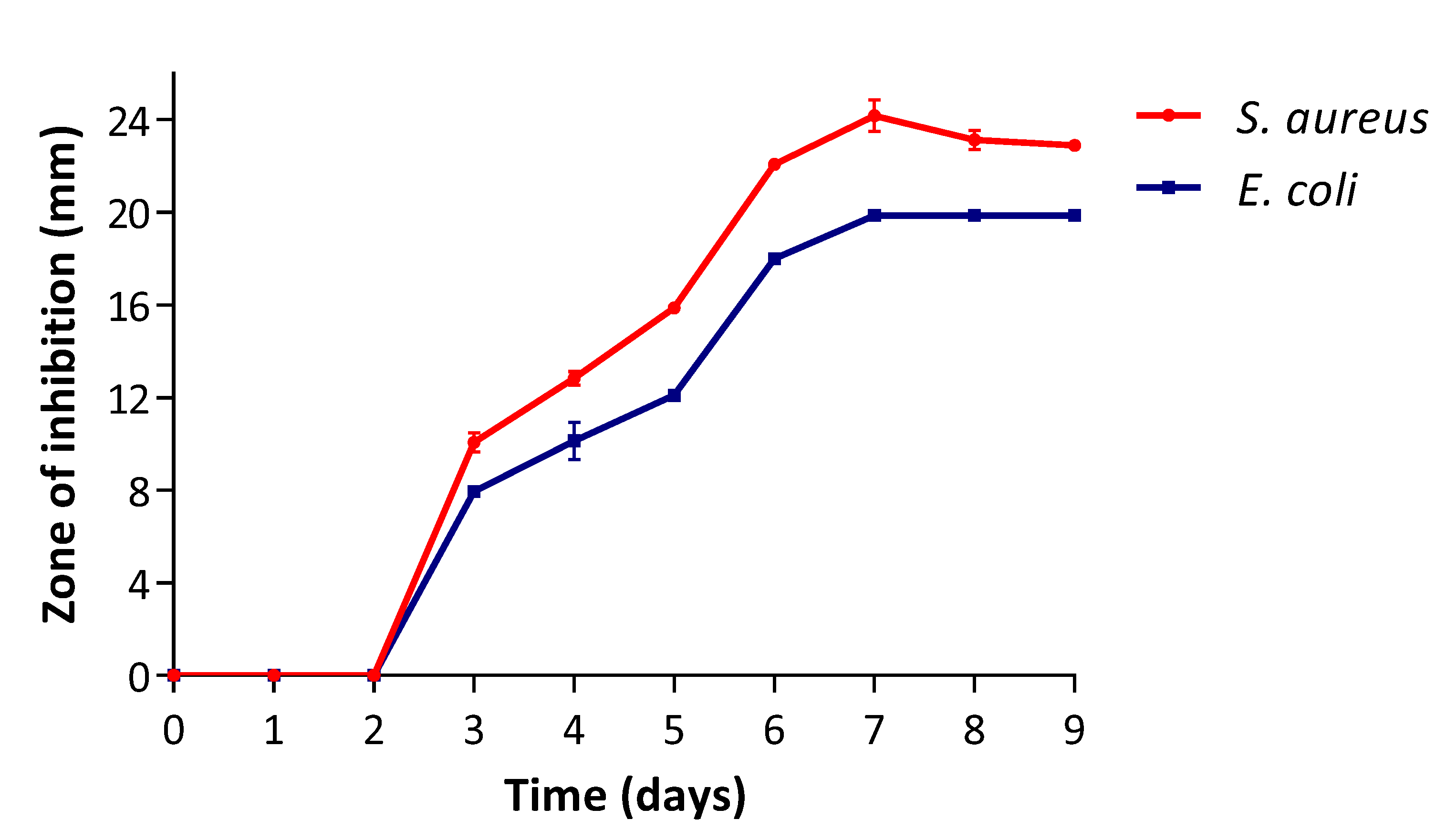
3.1. Secondary Metabolite Production Stage

The optimum phase for production of the antibacterial secondary metabolites by the most active isolate—FUN01—was assessed as influenced by the elapsing time, and the results are presented in Figure 1. There were no zones of inhibition observed within 2 days of fermentation against S. aureus (ATCC 25925) and E. coli (ATCC 25922). This might have been due to the fact that the fungus was at the lag and exponential phases, during which the cells were metabolically active, synthesising primary metabolites responsible for its growth and not producing secondary metabolites. The zones of inhibition were observed after 3 days with the maximum zones shown at day 7 against both bacterial strains. However, the zones were more profound on S. aureus (ATCC 25925) than on E. coli (ATCC 25922), implying S. aureus (ATCC 25925) to be more sensitive to the produced secondary metabolites. After 7 days, the zones were constant on E. coli (ATCC 25922) and there was insignificant decrease at day 8 and 9 on S. aureus (ATCC 25925). The decrease might be due to autolysis that might have occurred as the nutrients depleted [43]. In addition, at day 7, the inhibitive intensity of the secondary metabolites in the broth implied that the fungus produced active antibacterial metabolites optimally at its stationary phase. The results are correlated by those of Khalil et al. [44], whereby Aspergillus strain CMB-M81F initiated the production of secondary metabolites with the transition to its stationary phase.

3.2. Antibacterial Activity of the Fungal Extract
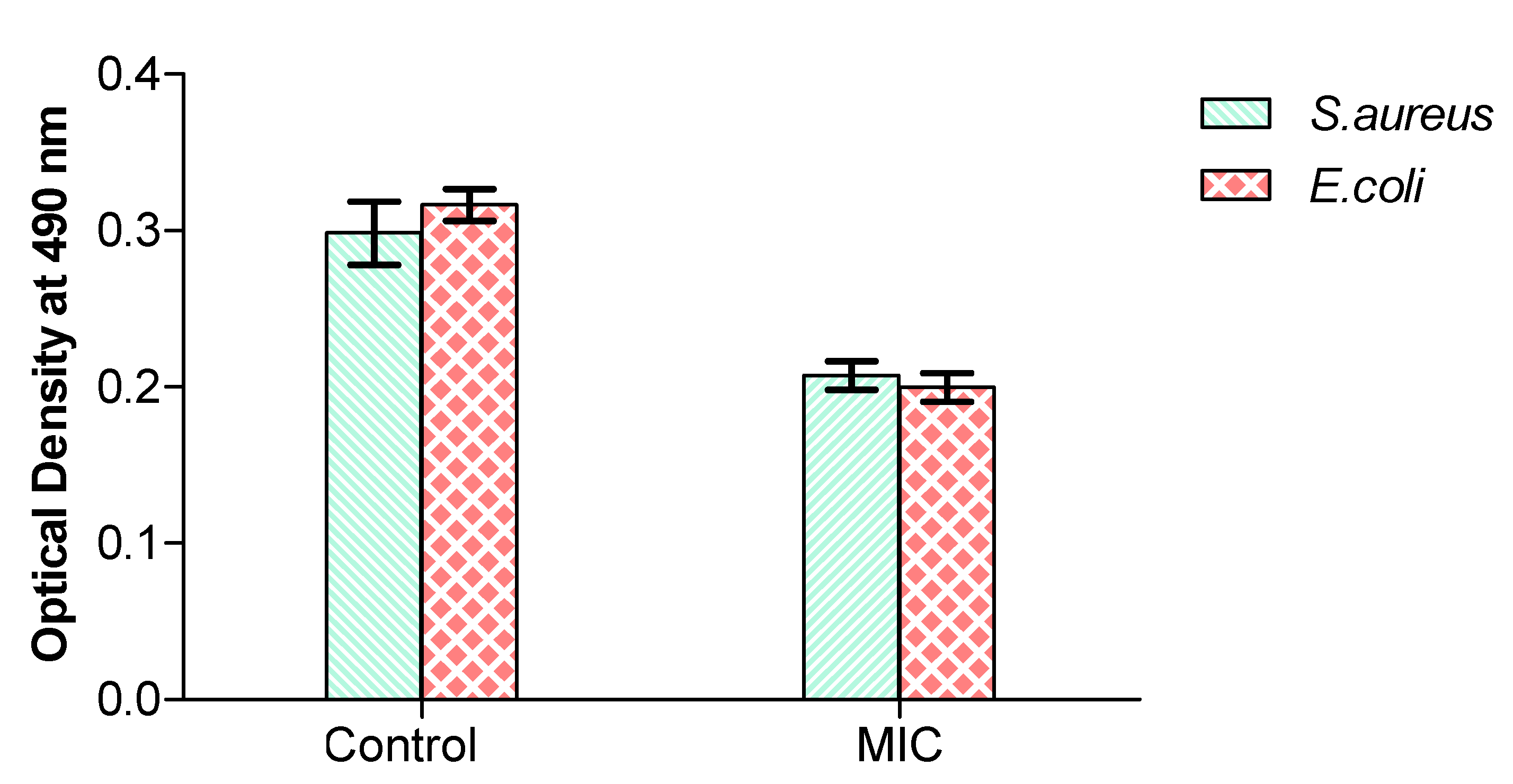
There is a dire need to find novel antimicrobial candidates to combat MDR among pathogens. Therefore, the exploration of fungal endophytes as a source of novel bioactive metabolites is significant for the discovery and development of potent antimicrobial agents [45]. Thus, the antibacterial activity of the extract from the isolated fungal endophyte was evaluated and the results are shown in Table 2. The extract revealed antibacterial activity against S. aureus (ATCC 25925) and E. coli (ATCC 25922) with MIC values of 1 mg/mL, respectively. The activity of the extract was noteworthy (MIC values ≤ 1 mg/mL) on both bacteria [46]. It was also worth noting that the extract was equally effective against both the Gram-positive strain and the Gram-negative one, implying that the activity was not affected by the difference in their cell membranes. Elisha et al. [47] observed similar results, whereby the Gram-negative strains were more susceptible to the extract than the Gram-positive bacteria.
The MBC was assessed against S. aureus (ATCC 25925) and E. coli (ATCC 25922). The extract showed an MBC value greater than the tested concentration (2 mg/mL) against both bacteria. The results implied that the extract had only a bacteriostatic effect [48]. In a previous study, secondary metabolites from A. welwitschiae were also reported to demonstrate significant antimicrobial activity [37]. The profound antibacterial activity observed in this study suggests that A. welwitschiae MH545928.1 might be playing a vital role in the protection of A. ferox Mill against pathogens. Moreover, A. welwitschiae MH545928.1 has the potential to serve as a source of antimicrobial agents of medicinal and biotechnological importance.

3.3. RCD Activity

Respiration is the main energy-producing process for microbial growth. Thus, the destruction of the respiratory system is considered the target for antimicrobial substances [26]. The effect of the extract on the RCD of the test bacteria was evaluated and the results revealed that the extract inhibited its activity (Figure 2). When the extract with MIC was added, the absorbance value decreased to 0.214 and 0.205 for S. aureus and E. coli, respectively, in comparison to the controls, which were 0.298 and 0.326 for S. aureus and E. coli, respectively. The inactivation of the bacteria’s respiratory chain meant that the oxidative phosphorylation was disturbed and that the bacteria’s energy production pathway was tempered, consequently resulting in the inhibition of the test bacteria [49]. Therefore, it can be concluded that the extract inhibited the test bacterial by interfering with the activity of the respiratory chain dehydrogenase. The results are in agreement with the study by Gomaa [50], whereby antimicrobial agents were able to exert their effect by destructing the activity of bacterial respiratory chain dehydrogenase.

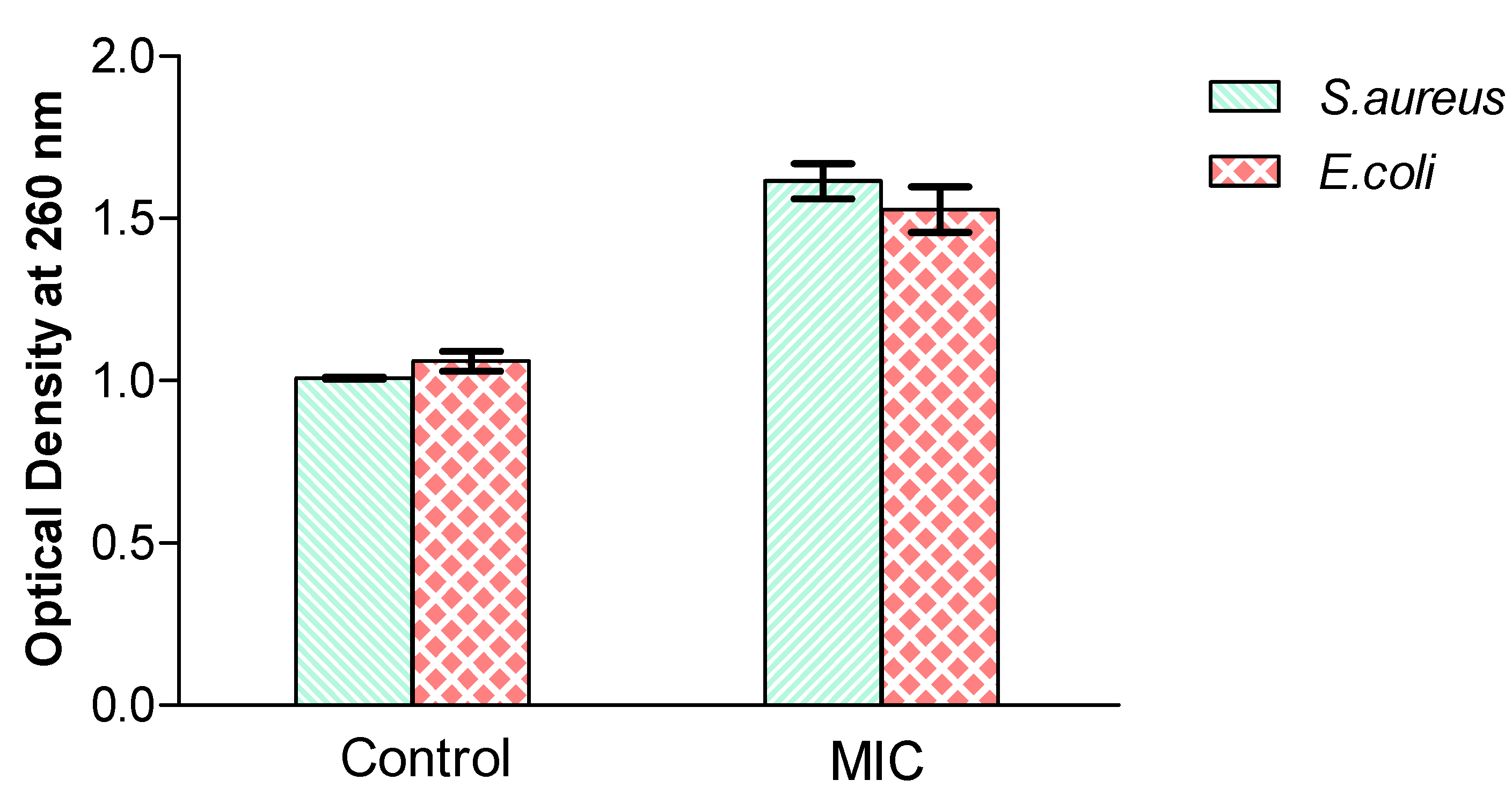
3.4. Effect of the Extract on the Release of 260 nm Absorbing Materials

The cell membrane’s integrity is one of the major factors important for the microbial growth and metabolism process. The cytoplasmic contents are regarded as important units of microbial structural substances and their leakage can lead to microbial death [49]. Thus, the effect of the extract (MIC) on the integrity of the cell membrane was investigated and the results are shown in Figure 3. The OD260 values of S. aureus and E. coli treated with the MIC of the extract were 1.55 and 1.47, respectively. It is worth noting that the OD260 values of S. aureus and E. coli treated with the MIC were significantly (p < 0.05) higher than that of the controls, which were 0.85 and 1 for S. aureus and E. coli, respectively.
The literature states that the availability of cell leakage markers (260 nm absorbing materials) in a culture broth serves as an indicator of the malfunction of the integrity of the cell membranes, implying the destruction of the cell membranes and the discharge of cell constituents, which might lead to bacterial inhibition [51]. The 260 nm absorbing materials, such as nucleic acids and proteins, are obligatory for bacteria growth and their leaching may lead to cell death. Thus, the fungal extract in this study showed to have induced its bacteriostatic effect on the test bacteria by damaging their cell membranes. These results are consistent with the findings by Tang et al. [52], which indicated that the extract inhibited E. coli and S. aureus by destroying their cell membranes.

3.5. Chemical Constituents of the Extract

The fungal endophytes usually have a strong relationship with the host plants due to the long-term association with the hosts and, thus, tend to produce secondary metabolites that mimic those of the host plants [53]. To date, over 200 secondary metabolites have been reported to be from endophytic fungi. Terpenoids, coumarins and cardiac glycosides were detected present, whereas flavonoids and saponins tested negative (Table 3). Terpenoids are secondary metabolites secreted as a crucial defence mechanism against abiotic and biotic stress conditions. They are reported to exert antimicrobial activity mainly by interfering with the respiratory pathway [54]. Cardiac glycosides are subgroups of steroids reformed from terpenoids and are often associated with antimicrobial effects [55,56]. Coumarins are heterocyclic compounds produced from benzene and pyrone rings containing oxygen. Coumarins derivatives are well known for their diverse biological activities, which include antimicrobial action [57]. Therefore, the detected secondary metabolites (coumarins, terpenoids and cardiac glycosides) might have resulted in the observed bacteriostatic effect in this study. Thus, A. welwitschiae MK450668.1 has the potential to serve as a natural source of these compounds for medicinal purposes.

3.6. Functional Groups of the Extract

The functional groups of the extract were identified using FTIR and the results are illustrated in Figure 4. The extract showed strong and broad absorption at 3293.3 cm1, as a result of O-H stretching and its compound class’s alcohol. The O-H group increases the polarity of molecules and improves their solubility in polar solvents [58]. Zhang et al. [59] pointed out that the antibacterial activities of some compounds are often due to the O-H group. There was also a strong absorption peak at 1640 cm−1, due to C=C stretching or N-H bending vibrations and its compound class’s alkene and amine groups. Alkene and amine groups are reported to possess antibacterial potency [60]. The extract further displayed a medium absorption peak at 1413 cm−1, as a result of O-H bending indicative of alcohol. Lastly, it revealed a strong absorption at 991 cm−1, due to C=C bending, corresponding to an alkene. Generally, it can be concluded that the presence of the observed characteristic functional groups could be responsible for the antibacterial activity of the fungal extract [61].

4. Conclusions

In this study, out of seven isolated endophytic fungi, isolate FUN01 showed the most promising ability for antibiotic production and was identified as A. welwitschiae MH545928.1 based on ITS sequence analysis. Its extract revealed a noteworthy antibacterial activity against S. aureus (ATCC 25925) and E. coli (ATCC 25922). The extract exerted a bacteriostatic effect against the bacterial strains by inhibiting the respiratory chain dehydrogenase (RCD) activity and damaging the membrane integrity. The profound antibacterial activity was attributed to the identified functional groups and synergistic effect of the different classes of compounds within the extract. The obtained information shows the potentiality of the endophytic A. welwitschiae MH545928.1 as a source for therapeutic bioactive compounds. For further research, the identification of the individual constituents within the extract and the assessment of their in vivo activities needs to be done.

Author Contributions

Conceptualisation, J.S.E.S. and T.S.M.; formal analysis, T.S.M. and M.M.; investigation, M.I.N. and M.M.; supervision, J.S.E.S. and T.S.M.; writing—original draft, T.S.M.; writing—review and editing, J.S.E.S. and N.S.S. All authors have read and agreed to the published version of the manuscript.

Funding

This research received no external funding.

Institutional Review Board Statement

Not applicable.

Informed Consent Statement

Not applicable.

Data Availability Statement

Not applicable.

Acknowledgments

The authors would like to acknowledge the staff and postgraduate students at the University of Zululand, Department of Biochemistry and Microbiology for their support.

Conflicts of Interest

The authors declare no conflict of interest.

References

  1. Orsi, R.H.; den Bakker, H.C.; Wiedmann, M. Listeria monocytogenes lineages: Genomics, evolution, ecology, and phenotypic characteristics. Int. J. Med. Microbiol. 2011, 301, 79–96. [Google Scholar] [CrossRef] [PubMed]
  2. Mujaddidi, N.; Nisa, S.; Al Ayoubi, S.; Bibi, Y.; Khan, S.; Sabir, M.; Zia, M.; Ahmad, S.; Qayyum, A. Pharmacological properties of biogenically synthesized silver nanoparticles using endophyte Bacillus cereus extract of Berberis lyceum against oxidative stress and pathogenic multidrug-resistant bacteria. Saudi J. Biol. Sci. 2021, 28, 6432–6440. [Google Scholar] [CrossRef] [PubMed]
  3. World Health Organization. Global Action Plan for the Prevention and Control of Noncommunicable Diseases 2013–2020; World Health Organization: Geneva Switzerland, 2013. [Google Scholar]
  4. Kebede, Y.; Hayder, A.; Girma, K.; Abamecha, F.; Alemayehu, G.; Abebe, L.; Sudhakar, M.; Birhanu, Z. Primary school students’ poetic malaria messages from Jimma zone, Oromia, Ethiopia: A qualitative content analysis. BMC Public Health 2021, 21, 1688. [Google Scholar] [CrossRef] [PubMed]
  5. Mitra, I.; Huang, B.; Mousavi, N.; Ma, N.; Lamkin, M.; Yanicky, R.; Shleizer-Burko, S.; Lohmueller, K.E.; Gymrek, M. Patterns of de novo tandem repeat mutations and their role in autism. Nature 2021, 589, 246–250. [Google Scholar] [CrossRef]
  6. Pál, S.; Tóth, R.; Nosanchuk, J.; Vágvölgyi, C.; Németh, T.; Gácser, A. A Candida parapsilosis overexpression collection reveals genes required for pathogenesis. J. Fungi 2021, 7, 97. [Google Scholar] [CrossRef]
  7. Jain, R.; Bhardwaj, P.; Pandey, S.S.; Kumar, S. Arnebia euchroma, a plant species of cold desert in the Himalayas, harbors beneficial cultivable endophytes in roots and leaves. Front. Microbiol. 2021, 12, 696667. [Google Scholar] [CrossRef]
  8. Khalmuratova, R.; Shin, H.-W. Crosstalk between mucosal inflammation and bone metabolism in chronic rhinosinusitis. Clin. Exp. Otorhinolaryngol. 2021, 14, 43. [Google Scholar] [CrossRef]
  9. Martinez-Klimova, E.; Rodríguez-Peña, K.; Sánchez, S. Endophytes as sources of antibiotics. Biochem. Pharmacol. 2017, 134, 1–17. [Google Scholar] [CrossRef]
  10. Khare, E.; Mishra, J.; Arora, N.K. Multifaceted interactions between endophytes and plant: Developments and prospects. Front. Microbiol. 2018, 9, 2732. [Google Scholar] [CrossRef]
  11. Moghaddam, N.; Dabard, C.; Dufour, M.; Po, H.; Xu, X.; Pons, T.; Lhuillier, E.; Ithurria, S. Surface modification of CdE (E: S, Se, and Te) nanoplatelets to reach thicker nanoplatelets and homostructures with confinement-induced intraparticle type I energy level alignment. J. Am. Chem. Soc. 2021, 143, 1863–1872. [Google Scholar] [CrossRef]
  12. Mulla, K.P.; Dandu, C.; Thirumalanadhuni, V.; Shaik, S.; Shaik, I.A.; Nimmanapalli, Y. Phytochemical, elemental, physico-chemical, HPTLC and anticancer investigations of Ceropegia spiralis Wight. Tuber extracts. Pharma Innov. J. 2021, 10, 95–105. [Google Scholar] [CrossRef]
  13. Song, E.; Zhang, C.; Israelow, B.; Lu-Culligan, A.; Prado, A.V.; Skriabine, S.; Lu, P.; Weizman, O.-E.; Liu, F.; Dai, Y. Neuroinvasion of SARS-CoV-2 in human and mouse brain. J. Exp. Med. 2021, 218, e20202135. [Google Scholar] [CrossRef] [PubMed]
  14. Wu, C.; Chen, X.; Cai, Y.; Zhou, X.; Xu, S.; Huang, H.; Zhang, L.; Zhou, X.; Du, C.; Zhang, Y. Risk factors associated with acute respiratory distress syndrome and death in patients with coronavirus disease 2019 pneumonia in Wuhan, China. JAMA Intern. Med. 2020, 180, 934–943. [Google Scholar] [CrossRef] [PubMed] [Green Version]
  15. O’Brien, C.; Van Wyk, B.-E.; Van Heerden, F. Physical and chemical characteristics of Aloe ferox leaf gel. South Afr. J. Bot. 2011, 77, 988–995. [Google Scholar] [CrossRef] [Green Version]
  16. Šírová, K.; Vaculík, M. Toxic effects of cadmium on growth of Aloe ferox Mill. South Afr. J. Bot. 2021. [Google Scholar] [CrossRef]
  17. Galli, C.L.; Cinelli, S.; Ciliutti, P.; Melzi, G.; Marinovich, M. Aloe-emodin, a hydroxyanthracene derivative, is not genotoxic in an in vivo comet test. Regul. Toxicol. Pharmacol. 2021, 124, 104967. [Google Scholar] [CrossRef]
  18. Nxumalo, C.I.; Ngidi, L.S.; Shandu, J.S.E.; Maliehe, T.S. Isolation of endophytic bacteria from the leaves of Anredera cordifolia CIX1 for metabolites and their biological activities. BMC Complementary Med. Ther. 2020, 20, 300. [Google Scholar] [CrossRef]
  19. Lu, Y.; Chen, C.; Chen, H.; Zhang, J.; Chen, W. Isolation and identification of endophytic fungi from Actinidia macrosperma and investigation of their bioactivities. Evid. Based Complementary Altern. Med. 2012, 2012, 382742. [Google Scholar] [CrossRef] [Green Version]
  20. Dudeja, S.; Chhokar, V.; Badgujjar, H.; Chauhan, R.; Soni, S.; Beniwal, V.; Kumar, A. Isolation and screening of antibiotic producing fungi from solid-state waste. Polymorphism 2020, 4, 59–71. [Google Scholar]
  21. Darshit, R.; Pandya, D.D. Screening and characteristic study of antimicrobial actinomycetes from near-by soil of medicinal plants. Int. J. Pharm. Pharma. Sci. 2018, 10, 66. [Google Scholar] [CrossRef]
  22. Pryce, T.; Palladino, S.; Kay, I.; Coombs, G. Rapid identification of fungi by sequencing the ITS1 and ITS2 regions using an automated capillary electrophoresis system. Med Mycol. 2003, 41, 369–381. [Google Scholar] [CrossRef] [PubMed]
  23. Sharma, D.; Pramanik, A.; Agrawal, P.K. Evaluation of bioactive secondary metabolites from endophytic fungus Pestalotiopsis neglecta BAB-5510 isolated from leaves of Cupressus torulosa D. Don. 3 Biotech 2016, 6, 210. [Google Scholar] [CrossRef] [Green Version]
  24. Eloff, J.N. A sensitive and quick microplate method to determine the minimal inhibitory concentration of plant extracts for bacteria. Planta medica 1998, 64, 711–713. [Google Scholar] [CrossRef] [PubMed] [Green Version]
  25. Nkala, B.A. The Cytotoxic Effects, Anti-Iflammatory, Antioxidant, Antibacterial, and Antidiabetic Properties of Eight Selected South African Plants for Medicinal Purposes. Ph.D. Thesis, University of KwaZulu Natal, Durban, South Africa, 2020. [Google Scholar]
  26. Guo, F.; Chen, Q.; Liang, Q.; Zhang, M.; Chen, W.; Chen, H.; Yun, Y.; Zhong, Q.; Chen, W. Antimicrobial activity and proposed action mechanism of linalool against pseudomonas fluorescens. Front. Microbiol. 2021, 12, 49. [Google Scholar] [CrossRef] [PubMed]
  27. Huang, W.; Wang, J.-Q.; Song, H.-Y.; Zhang, Q.; Liu, G.-F. Chemical analysis and in vitro antimicrobial effects and mechanism of action of Trachyspermum copticum essential oil against Escherichia coli. Asian Pac. J. Trop. Med. 2017, 10, 663–669. [Google Scholar] [CrossRef] [PubMed]
  28. Ali, S.; Khan, M.R.; Sajid, M.; Zahra, Z. Phytochemical investigation and antimicrobial appraisal of Parrotiopsis jacquemontiana (Decne) Rehder. BMC Complementary Altern. Med. 2018, 18, 1–15. [Google Scholar] [CrossRef] [PubMed] [Green Version]
  29. Ngema, S.; Basson, A.; Maliehe, T. Synthesis, characterization and application of polyacrylamide grafted bioflocculant. Phys. Chem. Earth Parts A/B/C 2020, 115, 102821. [Google Scholar] [CrossRef]
  30. Yadav, G.; Meena, M. Bioprospecting of endophytes in medicinal plants of Thar Desert: An attractive resource for biopharmaceuticals. Biotechnol. Rep. 2021, 30, e00629. [Google Scholar] [CrossRef]
  31. Vesth, T.C.; Nybo, J.L.; Theobald, S.; Frisvad, J.C.; Larsen, T.O.; Nielsen, K.F.; Hoof, J.B.; Brandl, J.; Salamov, A.; Riley, R. Investigation of inter-and intraspecies variation through genome sequencing of Aspergillus section Nigri. Nat. Genet. 2018, 50, 1688–1695. [Google Scholar] [CrossRef] [Green Version]
  32. Ravindran, C.; Naveenan, T.; Varatharajan, G.R.; Rajasabapathy, R.; Meena, R.M. Antioxidants in mangrove plants and endophytic fungal associations. Bot. Mar. 2012, 55, 269–279. [Google Scholar] [CrossRef]
  33. Da Silva, I.P.; Brissow, E.; Kellner Filho, L.C.; Senabio, J.; de Siqueira, K.A.; Vandresen Filho, S.; Damasceno, J.L.; Mendes, S.A.; Tavares, D.C.; Magalhães, L.G. Bioactive compounds of Aspergillus terreus—F7, an endophytic fungus from Hyptis suaveolens (L.) Poit. World J. Microbiol. Biotechnol. 2017, 33, 1–10. [Google Scholar] [CrossRef] [PubMed]
  34. Sornakili, A.; Thankappan, S.; Sridharan, A.; Nithya, P.; Uthandi, S. Antagonistic fungal endophytes and their metabolite-mediated interactions against phytopathogens in rice. Physiol. Mol. Plant Pathol. 2020, 112, 101525. [Google Scholar] [CrossRef]
  35. Liu, J.; Song, Y.; Zhang, Z.; Wang, L.; Guo, Z.; Zou, W.; Tan, R. Aspergillus fumigatus CY018, an endophytic fungus in Cynodon dactylon as a versatile producer of new and bioactive metabolites. J. Biotechnol. 2004, 114, 279–287. [Google Scholar] [CrossRef]
  36. Almaary, K.S.; Alharbi, N.S.; Kadaikunnan, S.; Khaled, J.M.; Rajivgandhi, G.; Ramachandran, G.; Kanisha, C.C.; Murugan, M.; Alanzi, K.F.; Manoharan, N. Anti-bacterial effect of marine sea grasses mediated endophytic actinomycetes against K. pneumoniae. J. King Saud Univ. Sci. 2021, 33, 101528. [Google Scholar] [CrossRef]
  37. Quintanilha-Peixoto, G.; Torres, R.O.; Reis, I.M.A.; Oliveira, T.A.S.d.; Bortolini, D.E.; Duarte, E.A.A.; Ariston de Carvalho Azevedo, V.; Brenig, B.; Aguiar, E.R.G.R.; Soares, A.C.F. Calm before the storm: A glimpse into the secondary metabolism of Aspergillus welwitschiae, the etiologic agent of the sisal bole rot. Toxins 2019, 11, 631. [Google Scholar] [CrossRef] [Green Version]
  38. Han, J.; Yang, N.; Wei, S.; Jia, J.; Lin, R.; Li, J.; Bi, H.; Song, F.; Xu, X. Dimeric hexylitaconic acids from the marine-derived fungus Aspergillus welwitschiae CUGBMF180262. Nat. Prod. Res. 2020, 36, 1–8. [Google Scholar] [CrossRef]
  39. Loges, L.A.; Silva, D.B.; Paulino, G.V.; Landell, M.F.; Macedo, A.J. Polyketides from marine-derived Aspergillus welwitschiae inhibit Staphylococcus aureus virulence factors and potentiate vancomycin antibacterial activity in vivo. Microb. Pathog. 2020, 143, 104066. [Google Scholar] [CrossRef]
  40. Ying, L.; Zhong, D.; Peng, D.-L.; LIU, S.-M.; Kong, L.-A.; Huan, P.; Xiang, C.; LI, Z.-C.; Huang, W.-K. Evaluation of the biocontrol potential of Aspergillus welwitschiae against the root-knot nematode Meloidogyne graminicola in rice (Oryza sativa L.). J. Integr. Agric. 2019, 18, 2561–2570. [Google Scholar]
  41. El-Naggar, N.E.-A.; El-Shweihy, N.M. Bioprocess development for L-asparaginase production by Streptomyces rochei, purification and in-vitro efficacy against various human carcinoma cell lines. Sci. Rep. 2020, 10, 1–21. [Google Scholar] [CrossRef]
  42. Vanzela, D.O.A.; Massi, F.P.; de Oliveira, A.L.M.; Fungaro, M.H.P.; Sartori, D. Isolation and identification of aspergillus section Nigri, and genotype associated with ochratoxin A and fumonisin B 2 production in garlic marketed in Brazil. Curr. Microbiol. 2020, 77, 1–9. [Google Scholar] [CrossRef]
  43. Almeida, C.; Bills, G.; González-Menéndez, V.; Martin, J.; Tormo, J.R.; Genilloud, O. Time-dependent production of the bioactive peptides endolides A and B and the polyketide mariline A from the sponge-derived fungus Stachylidium bicolor 293K04. Fermentation 2017, 3, 45. [Google Scholar] [CrossRef] [Green Version]
  44. Khalil, Z.G.; Kalansuriya, P.; Capon, R.J. Lipopolysaccharide (lps) stimulation of fungal secondary metabolism. Mycology 2014, 5, 168–178. [Google Scholar] [CrossRef] [PubMed] [Green Version]
  45. Abdalla, M.A.; Aro, A.O.; Gado, D.; Passari, A.K.; Mishra, V.K.; Singh, B.P.; McGaw, L.J. Isolation of endophytic fungi from South African plants, and screening for their antimicrobial and extracellular enzymatic activities and presence of type I polyketide synthases. South Afr. J. Bot. 2020, 134, 336–342. [Google Scholar] [CrossRef]
  46. Asong, J.A.; Amoo, S.O.; McGaw, L.J.; Nkadimeng, S.M.; Aremu, A.O.; Otang-Mbeng, W. Antimicrobial activity, antioxidant potential, cytotoxicity and phytochemical profiling of four plants locally used against skin diseases. Plants 2019, 8, 350. [Google Scholar] [CrossRef] [Green Version]
  47. Elisha, I.L.; Botha, F.S.; McGaw, L.J.; Eloff, J.N. The antibacterial activity of extracts of nine plant species with good activity against Escherichia coli against five other bacteria and cytotoxicity of extracts. BMC Complementary Altern. Med. 2017, 17, 1–10. [Google Scholar] [CrossRef] [Green Version]
  48. Dejoies, L.; Le Neindre, K.; Reissier, S.; Felden, B.; Cattoir, V. Distinct expression profiles of regulatory RNAs in the response to biocides in Staphylococcus aureus and Enterococcus faecium. Sci. Rep. 2021, 11, 6892. [Google Scholar] [CrossRef]
  49. Liu, C.H.; Zhang, E.; Wong, G.T.F.; Hyun, S. Factors associated with depression, anxiety, and PTSD symptomatology during the COVID-19 pandemic: Clinical implications for US young adult mental health. Psychiatry Res. 2020, 290, 113172. [Google Scholar] [CrossRef]
  50. Gomaa, E.Z. Silver nanoparticles as an antimicrobial agent: A case study on Staphylococcus aureus and Escherichia coli as models for Gram-positive and Gram-negative bacteria. J. Gen. Appl. Microbiol. 2017, 63, 36–43. [Google Scholar] [CrossRef] [Green Version]
  51. Dong, S.; Yang, X.; Zhao, L.; Zhang, F.; Hou, Z.; Xue, P. Antibacterial activity and mechanism of action saponins from Chenopodium quinoa Willd. husks against foodborne pathogenic bacteria. Ind. Crop. Prod. 2020, 149, 112350. [Google Scholar] [CrossRef]
  52. Tang, H.; Chen, W.; Dou, Z.-M.; Chen, R.; Hu, Y.; Chen, W.; Chen, H. Antimicrobial effect of black pepper petroleum ether extract for the morphology of Listeria monocytoge nes and Salmonella typhimurium. J. Food Sci. Technol. 2017, 54, 2067–2076. [Google Scholar] [CrossRef]
  53. Jayaram, A.; Deer, E.; Amaral, L.M.; Campbell, N.; Vaka, V.R.; Cunningham, M.; Ibrahim, T.; Cornelius, D.C.; LaMarca, B.B. The role of tumor necrosis factor in triggering activation of natural killer cell, multi-organ mitochondrial dysfunction and hypertension during pregnancy. Pregnancy Hypertens. 2021, 24, 65–72. [Google Scholar] [CrossRef] [PubMed]
  54. Alibi, S.; Ramos-Vivas, J.; Selma, W.B.; Mansour, H.B.; Boukadida, J.; Navas, J. Virulence of clinically relevant multidrug resistant Corynebacterium striatum strains and their ability to adhere to human epithelial cells and inert surfaces. Microb. Pathog. 2021, 155, 104887. [Google Scholar] [CrossRef] [PubMed]
  55. Pandey, D.; Pandey, V.C. Sacred plants from ancient to modern era: Traditional worshipping towards plants conservation. Trop. Plant Res. 2016, 3, 136–141. [Google Scholar]
  56. Mahale, S.; Goswami-Giri, A. Composition and characterization of refined oil compared with its crude oil from waste obtained from Mangifera indica. Asian J. Research Chem. 2011, 4, 1415–1419. [Google Scholar]
  57. Al-Majedy, Y.K.; Kadhum, A.A.H.; Al-Amiery, A.A.; Mohamad, A.B. Coumarins: The antimicrobial agents. Syst. Rev. Pharm. 2017, 8, 62. [Google Scholar] [CrossRef]
  58. Abd Hamid, H.; Mutazah, R.; Yusoff, M.M.; Abd Karim, N.A.; Razis, A.F.A. Comparative analysis of antioxidant and antiproliferative activities of Rhodomyrtus tomentosa extracts prepared with various solvents. Food Chem. Toxicol. 2017, 108, 451–457. [Google Scholar] [CrossRef] [Green Version]
  59. Zhang, L.-L.; Zhang, L.-F.; Xu, J.-G. Chemical composition, antibacterial activity and action mechanism of different extracts from hawthorn (Crataegus pinnatifida Bge.). Sci. Rep. 2020, 10, 1–13. [Google Scholar]
  60. Burman, S.; Bhattacharya, K.; Mukherjee, D.; Chandra, G. Antibacterial efficacy of leaf extracts of Combretum album Pers. against some pathogenic bacteria. BMC Complementary Altern. Med. 2018, 18, 213. [Google Scholar] [CrossRef] [Green Version]
  61. Dhivya, S.; Kalaichelvi, K. Phytochemical studies and gas chromatography-mass spectrometry analysis of sarcostemma brevistigma, wight & arn. Asian J. Pharm. Clin. Res. 2017, 10, 462–466. [Google Scholar]
Figure 1. The zones of inhibition produced by the secondary metabolites as influenced by the elapsing time.
Microbiolres 13 00007 g001
Figure 2. The effect of the extract on respiratory chain dehydrogenase.
Microbiolres 13 00007 g002
Figure 3. The effect of the extract on the release of 260 nm absorbing material from bacterial strains.
Microbiolres 13 00007 g003
Figure 4. Identification of the functional groups of the extract.
Microbiolres 13 00007 g004
Table 1. Zones of inhibition obtained during the screening of the production of the production of antimicrobial agents by the isolates and their identities.
Primary ScreeningSecondary Screening
IsolatesS. aureusE. coliS. aureusE. coliFungal NameGenBank
Accession
Number
FUN01+++++++++++Aspergillus welwitschiaeMH545928.1
FUN02+++++++Un-identifiedUn-identified
FUN03++++++Un-identifiedUn-identified
FUN04++++Un-identifiedUn-identified
FUN05++++Un-identifiedUn-identified
FUN06+++Un-identifiedUn-identified
FUN07+Un-identifiedUn-identified
FUN08 Un-identifiedUn-identified
FUN09 Un-identifiedUn-identified
FUN010 Un-identifiedUn-identified
FUN011 Un-identifiedUn-identified
FUN012 Un-identifiedUn-identified
FUN013 Un-identifiedUn-identified
FUN014 Un-identifiedUn-identified
FUN015 Un-identifiedUn-identified
Inhibition zone diameter index: + (≤9 mm) weak activity, ++ (10–20 mm) moderate activity, +++ (≥21 mm) strong activity and − denotes no activity.
Table 2. MIC and MBC values of the fungal extract.
BacteriaExtractCiprofloxacin
MIC
(mg/mL)
MBC
(mg/mL)
MIC
(µg/mL)
MBC
(µg/mL)
S. aureus (ATCC 25925)1>20.0150.031
E. coli (ATCC 25922)1>20.0150.031
Table 3. The qualitative chemical components of the fungal extract.
CompoundsPresence/Absence of Compounds
Terpenoids+
Flavonoids
Cardiac glycosides+
Coumarins+
Saponins
Key: + signifies presence and − shows absence.
Publisher’s Note: MDPI stays neutral with regard to jurisdictional claims in published maps and institutional affiliations.

