Article

Manganese Sulfate Nanocomposites Fabricated by Hot-Melt Extrusion for Chemodynamic Therapy of Colorectal Cancer

1
College of Pharmacy, Kangwon National University, Chuncheon 24341, Republic of Korea
2
Department of Animal Resources Science, College of Animal Life Sciences, Kangwon National University, Chuncheon 24341, Republic of Korea
3
School of Animal Life Convergence Science, Hankyong National University, Anseong 17579, Republic of Korea
4
Institute of Applied Humanimal Science, Hankyong National University, Anseong 17579, Republic of Korea
5
Darby Genetics Inc., Anseong 17529, Republic of Korea
6
Department of Bio-Health Technology, College of Biomedical Science, Kangwon National University, Chuncheon 24341, Republic of Korea
7
Department of Chemistry and Biochemistry, Food and Dairy Innovation Center, Boise State University, Boise, ID 83725, USA
8
Daehwa Pharmaceutical Co., Ltd., Seoul 06699, Republic of Korea
*
Author to whom correspondence should be addressed.
These authors equally contributed to this work.
Pharmaceutics 2023, 15(7), 1831; https://doi.org/10.3390/pharmaceutics15071831
Submission received: 3 June 2023 / Revised: 22 June 2023 / Accepted: 25 June 2023 / Published: 27 June 2023
(This article belongs to the Section Nanomedicine and Nanotechnology)

Abstract

:
The development of metal salts-based nanocomposites is highly desired for the Fenton or Fenton-like reaction-based chemodynamic therapy of cancer. Manganese sulfate (MnSO4)-dispersed nanoparticles (NPs) were fabricated with a hot-melt extrusion (HME) system for the chemodynamic therapy of colorectal cancer in this study. MnSO4 was homogeneously distributed in polyethylene glycol (PEG) 6000 (as a hydrophilic polymer) with the aid of surfactants (Span 80 and Tween 80) by HME processing. Nano-size distribution was achieved after dispersing the pulverized extrudate of MnSO4-based composite in the aqueous media. The distribution of MnSO4 in HME extrudate and the interactions between MnSO4 and pharmaceutical additives were elucidated by Fourier-transform infrared, X-ray diffractometry, X-ray photoelectron spectroscopy, and scanning electron microscopy analyses. Hydroxyl radical generation efficiency by the Fenton-like chemistry capability of Mn2+ ion was also confirmed by catalytic assays. By using the intrinsic H2O2 in cancer cells, MnSO4 NPs provided an elevated cellular reactive oxygen species level, apoptosis induction capability, and antiproliferation efficiency. The designed HME-processed MnSO4 formulation can be efficiently used for the chemodynamic therapy of colorectal cancer.

1. Introduction

Hot-melt extrusion (HME) has been widely engaged for preparing drug formulations due to its continuous process, cost-effectiveness, large scale-up production, no downstream process, high throughput characteristic, solvent-free property, and unit operation [1,2,3]. Several machine and operation factors, such as screw type (e.g., single, twin, and multi screws), screw design, screw speed, feed rate, barrel temperature, and die shape, may govern the physicochemical properties of extrudates [1]. Additionally, physical and chemical features of active pharmaceutical ingredients, carriers, and plasticizers should be considered for the optimization of HME process [1]. In the commercial market, several products have been supplied as shaped (rod or ring) systems and amorphous or crystalline dispersion for ophthalmic inserts, implants, devices, and oral formulations [2]. HME-processed products may have the following outcomes: solubility/bioavailability improvement, taste masking, and controlled/targeted drug release [2]. Various types of pharmaceutical dosage forms, such as films, granules, multicomponent systems (e.g., salts, co-amorphous systems, and co-crystal systems), nanoparticles, pellet, self-microemulsifying drug delivery systems, semi-solid products (e.g., creams, gels, and ointments), and solid implants, can be prepared by the HME technique [3]. These days, an HME system has been coupled with other downstream equipment (e.g., high-pressure homogenizer, pelletizer, and 3D printer) for producing advanced formulations [3].
In cases of organic small chemicals, HME has been selected for improving aqueous solubility, dissolution, and absorption, controlling drug release, and masking bitter tastes [4,5,6,7]. However, there were few reports regarding the fabrication of delivery systems for inorganic substances by HME technique [8,9,10]. In our previous studies [8,9,10], hydrophilic polymer and surfactants have been introduced to make colloidal systems of mineral salts (e.g., ZnSO4, FeSO4, and CuSO4) after dispersing in aqueous media. Herein, polyethylene glycol (PEG) 6000 was used as a hydrophilic polymer and Span 80 and Tween 80 were added as non-ionic surfactants for the homogeneous dispersion of MnSO4. PEG 6000 can act as a polymer matrix for the distribution of MnSO4 and both Span 80 and Tween 80 can reduce the surface tension during the particle formation process in the aqueous media. The combination of PEG 6000, Span 80, and Tween 80 has been successfully applied to make colloidal systems of ZnSO4, FeSO4, and CuSO4 [8,9,10]. Although the same feeding ratio of PEG 6000, Span 80, and Tween 80 has been used for HME processing, those metal salts (e.g., ZnSO4, FeSO4, and CuSO4) in our previous studies have different physicochemical properties (i.e., solubility and dispersibility) compared with MnSO4. HME processing and formulation design were successfully applied to a MnSO4 molecule in this study. Those HME-processed mineral salts-based particles exhibited nano-sized structures following dispersion in aqueous media. Nano-sized particulate systems may generally possess several advantages such as specific delivery to the target region, the improvement of drug safety and efficacy, and controlled drug cargo release, as reported [11]. This study may have critical implications in the fabrication of MnSO4-based nanoparticles (NPs) with polymer and surfactants by HME processing.
Moreover, HME-processed MnSO4 NPs were applied to the chemodynamic therapy of colorectal cancer in this investigation. Cancer cells may have higher H2O2 levels compared to normal cells; therefore, a H2O2-responsive system may possess cancer cell targeting capability [12]. Mn2+ ion, usually provided by MnO2, can provide a Fenton-like reaction; therefore, it can generate hydroxyl radical from cellular H2O2 [13,14,15]. In the current study, MnSO4 has been used as the source of Mn2+ ion, and the HME process was introduced to make nano-size structures for enhanced entry to cancer cells. The generation of toxic hydroxyl radicals may inhibit the proliferation of cancer cells through the elevation of the cellular reactive oxygen species (ROS) level and the induction of other cell death mechanisms (e.g., apoptosis). It is expected that the developed MnSO4 NPs may be efficient chemodynamic therapeutic agents for colorectal cancer.

2. Materials and Methods

2.1. Materials

MnSO4 (MnSO4·H2O) was acquired from TMC Co., Ltd. (Anyang, Republic of Korea). PEG 6000 was provided by Samchun Pure Chemical Co., Ltd. (Pyeongtaek, Republic of Korea). Span 80 and Tween 80 were purchased from Daejung Chemical & Metals Co., Ltd. (Siheung, Republic of Korea). 2′,7′-Dichlorofluorescin diacetate (DCFH-DA), 3,3′,5,5′-tetramethylbenzidine dihydrochloride hydrate (TMB DH), and methylene blue (MB) were purchased from Sigma–Aldrich (Saint Louis, MO, USA). Dulbecco’s modified Eagle’s medium (DMEM), penicillin-streptomycin, and fetal bovine serum (FBS) were obtained from Gibco Life Technologies, Inc. (Grand Island, NY, USA).

2.2. Production and Particle Characterization of MnSO4 Extrudate-Based Nanocomposites

2.2.1. Production of MnSO4 Formulations by HME

MnSO4, PEG 6000, Span 80, and Tween 80 were blended at a 20:64:12:4 ratio (w/w/w/w) prior to putting those materials into the extruder [8,9,10,16]. Those blended materials were translocated to the hopper part of HME (45 g/min rate). A hot-melt extruder installed with twin-screw (STS-25HS, Hankook E.M. Ltd., Pyeongtaek, Republic of Korea) and a round-shaped die (1 mm diameter) was utilized for the fabrication of MnSO4-based extrudates. Temperatures in the barrel and die parts were set as 45 °C and 40 °C, respectively. The screw speed was maintained at 150 rpm in this HME step. After moving through the conveying and kneading parts in the barrel, specimens were extruded from the die part. Extruded materials were hardened and pulverized by the grinder (HBL-3500S, Samyang Electronics Co., Gunpo, Republic of Korea).

2.2.2. Particle Characterization of MnSO4 NPs

The mean diameter of the MnSO4 powder was determined by laser diffraction particle size analysis. MnSO4 was dispersed in ethanol and the size-dependent volume density profile was obtained by a laser diffraction particle size analyzer (Mastersizer 3000, Malvern Instruments Ltd., Malvern, UK) [17].
The particle properties of MnSO4 NPs in distilled water (DW) were investigated. Hydrodynamic diameter, polydispersity index, and zeta potential values of MnSO4 NPs dispersed in DW (at 10 mg/mL) were measured by dynamic light scattering and laser Doppler methods (ELS-Z1000; Otsuka Electronics, Tokyo, Japan) [8,9,10].
The morphology of MnSO4 NPs (in DW) was observed by transmission electron microscopy (TEM). MnSO4 NPs (10 mg/mL in DW) were put onto the copper grid with film, the sample was stained with uranyl acetate (1%), and the liquid content was dried in an air stream for 10 min. The particle shape was observed by TEM (CM120; Philips, Amsterdam, The Netherlands).
The content of Mn in MnSO4 NPs was determined by inductively coupled plasma-optical emission spectrometry (ICP-OES; Optima 7300 DV, PerkinElmer, Inc., Waltham, MA, USA). Before ICP-OES analysis, the specimen was dissolved in nitric acid.
Incubation time-dependent mean diameter and size distribution features of MnSO4 NPs were determined by a dynamic light scattering method (ELS-Z1000, Otsuka Electronics). MnSO4 NPs (at 10 mg/mL) were dispersed in phosphate buffered saline (PBS, pH 7.4) or FBS solution (50%, v/v) and they were incubated for 24 h. After incubating for 0.5, 1, 3, 6, and 24 h, hydrodynamic diameter and polydispersity index values were measured.

2.3. Solid-State Studies

2.3.1. Fourier-Transform Infrared (FT-IR) Spectroscopy Analysis

Alteration in the chemical functions of MnSO4, freeze-dried mixture (MnSO4, PEG 6000, Span 80, and Tween 80), and MnSO4 NPs was studied by FT-IR analysis. Transmittance (%) values of MnSO4, the freeze-dried mixture, and MnSO4 NPs were scanned using a Frontier FT-IR spectrometer (PerkinElmer Inc., Buckinghamshire, UK). FT-IR spectra were obtained in the attenuated total reflectance mode. Transmittance (%) values of MnSO4, the freeze-dried mixture, and MnSO4 NPs were monitored in a 400–4000 cm−1 wavenumber range.

2.3.2. X-ray Diffractometry (XRD) Assay

The crystal properties of MnSO4, the freeze-dried mixture, and MnSO4 NPs were explored by a Philips X′Pert PRO MPD diffractometer (PANalytical Corp., Almero, The Netherlands). The intensity values according to angle (2θ) at the 5–80° range were measured. The generator voltage and tube current were set as 40 kV and 30 mA, respectively. The scan time per step and scan step size were 8.67 s and 0.013°, respectively.

2.3.3. X-ray Photoelectron Spectroscopy (XPS) Study

The elemental composition of MnSO4 and MnSO4 NPs was determined by an XPS (K-Alpha+, Thermo Fisher Scientific, East Grinstead, UK) system. The atomic contents (%) in MnSO4 (Mn 2p, O 1s, Ca 2p, C 1s, and S 2p) and MnSO4 NPs (Mn 2p, O 1s, C 1s, and S 2p) were measured. Al Kα X-ray (as a source gun) was used in this test. Peak patterns of C 1s, O 1s, Mn 2p, and S 2p were analyzed in a narrow binding energy range.

2.3.4. Field Emission Scanning Electron Microscopy (FE-SEM)/Energy Dispersive Spectrometry (EDS) Study

FE-SEM (JSM-7900F, JEOL, Tokyo, Japan) combined with EDS was applied to identify the distribution of elements on MnSO4 and MnSO4 NPs. Atomic contents of C, O, S, and Mn in MnSO4 and MnSO4 NPs were quantitatively determined.

2.4. Catalytic Assays

2.4.1. 3,3′,5,5′- Tetramethylbenzidine (TMB) Assay

The hydroxyl radical generation activity was examined by a TMB-based assay [18]. Each specimen (0.2 mL) of MnSO4 and MnSO4 NPs (at 1000 μg/mL MnSO4 concentration) was put into the microtube and 0.03% H2O2 solution (0.2 mL) was added. Then, 10 mM TMB DH (0.4 mL) was added to each sample and they were incubated at 37 °C for reaction. The reactant (0.2 mL) was acquired at 0, 10, 20, and 30 min and the corresponding absorbance was detected at 650 nm by a microplate reader (SpectraMax i3, Molecular Devices, Sunnyvale, CA, USA).

2.4.2. MB Assay

The hydroxyl radical scavenging effect was studied by an MB-based assay [19]. Each specimen (0.5 mL) of MnSO4 and MnSO4 NPs (at 1000 μg/mL MnSO4 concentration) was put into a dialysis tube (molecular weight cut-off: 12–14 kDa). An H2O2 solution (0.03%, 2.5 mL) was added to that sample and they were incubated at 37 °C for 30 min. Subsequently, an MB solution (20 μg/mL) was added to that mixture and the reactant (0.2 mL) was collected at 0, 15, 30, 60, and 120 min. The absorbance at 650 nm was measured by a microplate reader.

2.5. In Vitro Anticancer Activity Tests

2.5.1. Antiproliferation Assay

The antiproliferation potential of MnSO4 and MnSO4 NPs was assessed in CT-26 (colon carcinoma) cells with a colorimetric assay. CT-26 cells were obtained from the Korean Cell Line Bank (Seoul, Republic of Korea). Those cells were cultured in DMEM containing FBS (10%, v/v) and penicillin-streptomycin (1%, v/v) at 37 °C. CT-26 cells (at 5.0 × 10³ cells per well) were seeded in 96-well plate and they were incubated at 37 °C for 24 h. MnSO4 and MnSO4 NPs (at 10, 25, 50, 100, and 200 μg/mL MnSO4 concentrations) were applied to the cells and they were incubated for 72 h. After eliminating each sample, CellTiter 96® AQueous One Solution Cell Proliferation Assay Reagent (Promega Corp., Fitchburg, WI, USA) was applied to the cells and incubated at 37 °C. The absorbance at 490 nm was detected by a microplate reader.

2.5.2. Cellular ROS Assay

CT-26 cells were added to a 6-well plate at 1.0 × 10⁵ cells/well density. The next day, each specimen of MnSO4 and MnSO4 NPs (at 200 μg/mL MnSO4 concentration) was treated to the cells and they were incubated for 24 h. The cells were washed with cold PBS two times and stained by DCFH-DA (10 μM) for 20 min at 37 °C. Then, the cells were resuspended with FBS solution (2%, v/v). The fluorescence intensity of cellular ROS was measured by flow cytometry (BD Bioscience, San Diego, CA, USA).

2.5.3. Apoptosis Assay

CT-26 cells were seeded in a 6-well plate at a density of 1.0 × 10⁵ cells per well. Each specimen of MnSO4 and MnSO4 NPs (at 200 μg/mL MnSO4 concentration) was treated to the cells and incubated for 24 h. The cells were washed with cold PBS two times and resuspended in the binding buffer at a concentration of 1.0 × 10⁶ cells/mL. The cells were stained with FITC Annexin V and propidium iodide (PI) in the binding buffer and analyzed by flow cytometry.

2.6. Data Analysis

Statistical analyses of data were performed with a two-tailed t-test and analysis of variance. Each experiment was repeated at least three times. Data are provided as the mean ± standard deviation (SD).

3. Results and Discussion

3.1. Fabrication and Particle Property Tests of MnSO4 NPs

MnSO4 NPs were fabricated by the HME process for the chemodynamic therapy of cancer in this study (Figure 1). MnSO4 was homogeneously dispersed in hydrophilic polymer (PEG 6000) and non-ionic surfactants (Span 80 and Tween 80) by a twin-screw-based HME system. As observed in our previous reports [8,9], the combination of PEG 6000, Span 80, and Tween 80 was successfully engaged to prepare nano-sized particles of FeSO4 or CuSO4 following their dispersion in the aqueous environment. MnSO4/PEG 6000/Span 80/Tween 80 extrudate was acquired by the HME process and was pulverized for the convenient preparation of nano-size vesicles after dispersing in the aqueous media. Nano-sized particles of MnSO4 may be easily introduced to cancer cells [20] and they may attribute to the conversion of intrinsic H2O2 to hydroxyl radical by a Fenton-like reaction of Mn2+ ion.
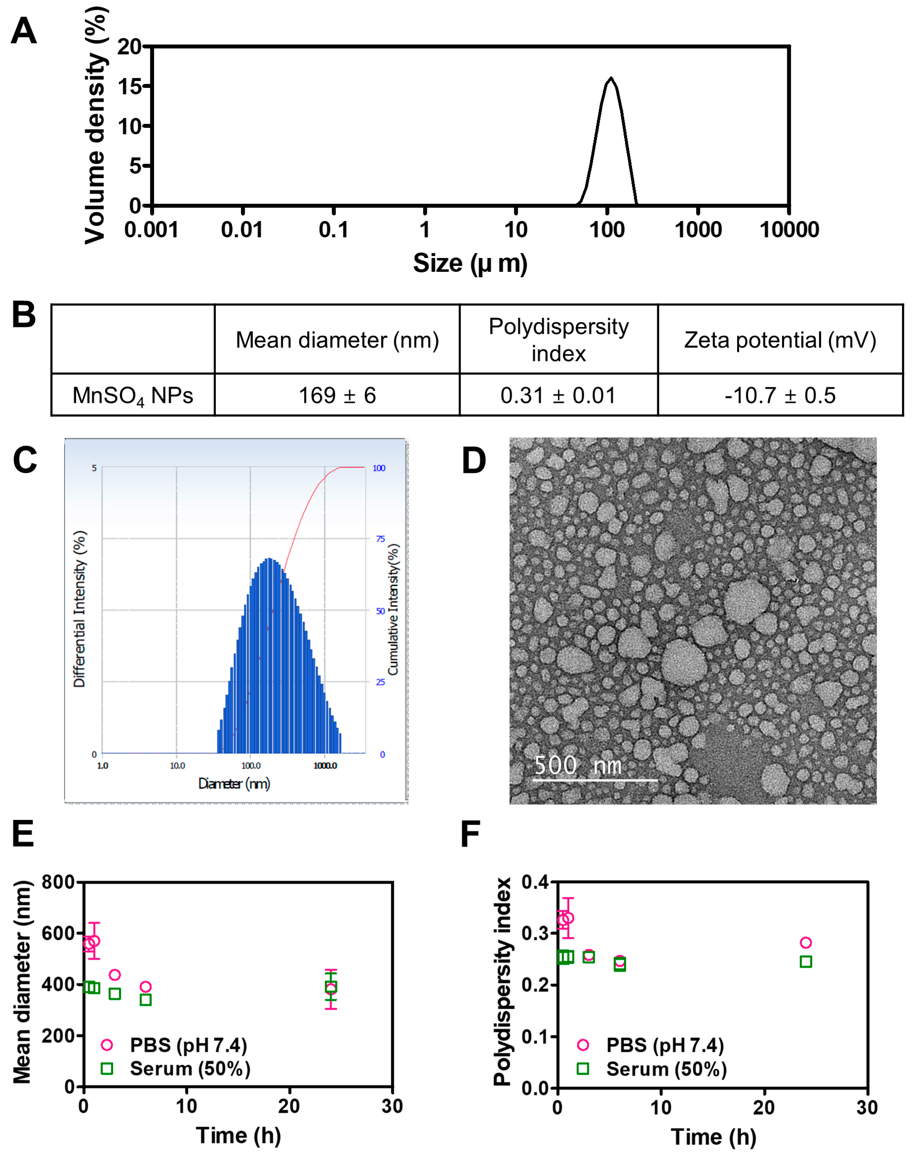
The particle features of MnSO4 NPs produced by the HME technique were explored as shown in Figure 2. The diameter of MnSO4 dispersed in ethanol was determined by a particle size analyzer (Figure 2A). The Dv (10), Dv (50), and Dv (90) values of MnSO4 were 78, 116, and 169 μm, respectively. The micron size of the MnSO4 powder was confirmed by particle size analysis.
The particle properties of HME-processed MnSO4 composites (MnSO4 NPs) were explored by size, distribution, zeta potential, encapsulation efficiency, and morphology analyses (Figure 2B–F). The hydrodynamic diameter of MnSO4 NPs in DW was 169 nm, and that group exhibited unimodal size distribution (Figure 2B,C). MnSO4 seems to be successfully dispersed in the hydrophilic polymer (PEG 6000) and surfactants (Span 80 and Tween 80), reducing the surface tension of particles. By the aid of twin-screw-enabled HME processing, MnSO4 seems to be homogeneously dispersed in the polymer matrix and it produced nano-sized particles in the aqueous media. Therefore, a significant size reduction effect from micron (MnSO4 powder) to nano (MnSO4 NPs) size was accomplished with the HME process with the hydrophilic polymer and surfactants. The zeta potential value of MnSO4 NPs was −10.7 mV and spherical in shape, with the corresponding diameter, was also shown in the TEM image (Figure 2B,D). The encapsulation efficiency of MnSO4 in MnSO4 NPs was around 100%, indicating the successful encapsulation of MnSO4 in polymer/surfactant-based particles.
The particle stability in the biological matrices was evaluated by particle size analysis in aqueous buffer and serum media (Figure 2E,F). In both PBS (pH 7.4) and serum solution (50% FBS), the initial hydrodynamic diameters of MnSO4 NPs were higher than that in DW (Figure 2B,E). This may be due to the interactions between MnSO4 NPs and buffer salts in PBS or proteins in serum media. Nevertheless, the hydrodynamic diameters of MnSO4 NPs in PBS (pH 7.4) and serum media (50% FBS) after 24 h incubation were 381 and 392 nm, respectively. The polydispersity index values of MnSO4 NPs in PBS (pH 7.4) and serum media (50% FBS) after 24 h incubation were 0.28 and 0.25, respectively. In particular, the existence of the PEG layer in MnSO4 NPs may inhibit the opsonization (which can lead to early elimination by mononuclear phagocyte system) and attribute to the maintenance of individual particles without the formation of aggregation [21,22]. Those results mean the maintenance of the nano-size even after incubation in the aqueous buffer or serum conditions, simulating the biological fluids. This may imply the safe application of developed MnSO4 NPs for cancer treatment.

3.2. Solid-State Features of MnSO4 NPs

The solid-state features of fabricated MnSO4 NPs by HME process were explored by FT-IR, XRD, XPS, and SEM-EDS tests (Figure 3, Figure 4, Figure 5 and Figure 6 and Figures S1–S4, Tables S1 and S2). The existence of MnSO4 and its interactions with pharmaceutical additives were elucidated by FT-IR analysis (Figure 3 and Table S1). MnSO4 was characterized by several shifts, such as v1SO42− (1017 cm−1), v3SO42− (1090 cm−1), and the O–H stretching band (3140 cm−1) (Figure 3A) [23]. Those peaks were also observed at 960 cm−1, 1094 cm−1, and 3433 cm−1, respectively, in the freeze-dried mixture group (Figure 3B). MnSO4 in MnSO4 NPs was also featured by v1SO42− (960 cm−1), v3SO42− (1096 cm−1), and the O–H stretching band (3411 cm−1) (Figure 3C). The FT-IR data of the freeze-dried mixture and MnSO4 NPs groups were almost similar, indicating similar interactions between MnSO4 and pharmaceutical excipients during freeze-drying and the HME process (Table S1). The unaltered doublet Mn–O peaks of MnSO4 (529/508 cm−1) in MnSO4 NPs (529/509 cm−1) envisaged the incorporation of MnSO4 in MnSO4 NPs [24,25,26]. In particular, peaks at 1740 cm−1 and 2884/1467 cm−1 were attributed to the C=O stretching of ester functionality of Span 80 and Tween 80 and the C–H stretching/deformation bands of –CH2– of Span 80, Tween 80, and PEG 6000, respectively [27]. The appearance of alkane- and ester-specific peaks in the FT-IR spectra of MnSO4 NPs indicated the successful formation of MnSO4-based nanovesicles by the HME process.
Figure 3. FT-IR spectra of (A) MnSO4, (B) freeze-dried mixture, and (C) MnSO4 NPs. Wavenumber-dependent transmittance value (%T) is plotted.
Pharmaceutics 15 01831 g003
The crystalline feature of MnSO4 included in NPs was explored by XRD assay (Figure 4). Representative peaks were shown at 18.04°, 25.38°, 28.37°, and 34.81° in the spectrum of MnSO4 [28]. Characteristic shifts were observed at 19.23°, 23.34°, and 25.42° in the profile of the MnSO4 NPs group. Those peaks in MnSO4 NPs group were very similar to those in freeze-dried mixture group with attenuated intensity. The HME process can likely be attributed to less crystallization of the ingredients compared to freeze-drying. The existence of PEG 6000, Span 80, and Tween 80 and their interactions with MnSO4 were observed in the XRD profile of the MnSO4 NPs group.
Figure 4. XRD patterns of (A) MnSO4, (B) freeze-dried mixture, and (C) MnSO4 NPs.
Pharmaceutics 15 01831 g004
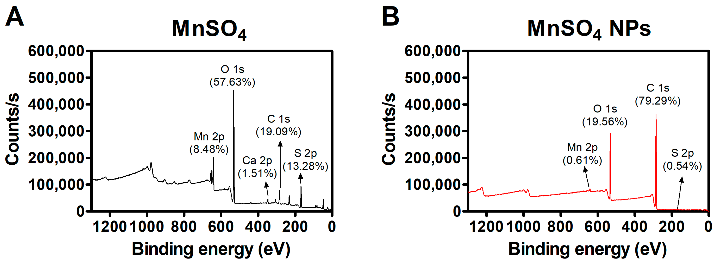
The atomic distribution in the outer surface of prepared specimens was investigated with an XPS assay (Figure 5 and Figure S1–S4, Table S2). The contents of Mn 2p, S 2p, and O 1s in the MnSO4 group were 8.48%, 13.28%, and 57.63%, respectively. However, in the MnSO4 NPs group, the values of Mn 2p, S 2p, and O 1s were changed to 0.61%, 0.54%, and 19.56%, respectively. HME processing with pharmaceutical excipients seems to alter the atomic distribution in fabricated MnSO4 NPs. The MnSO4 molecule might move to the inner part of the NP sample according to the data of XPS analysis. A dramatic change in C 1s and O 1s values between MnSO4 and MnSO4 NPs groups may be due to the existence of PEG 6000, Span 80, and Tween 80 in the tested region.
Figure 5. XPS data of (A) MnSO4 and (B) MnSO4 NPs.
Pharmaceutics 15 01831 g005
The incorporation of PEG 6000, Span 80, and Tween 80 could also be inferred from the deconvoluted C 1s and O 1s spectrum of MnSO4 NPs. The deconvoluted C 1s peaks were observed at 284.53, 286.05, and 288.70 eV, attributed to –CH2–/–CH=CH–, >CH–OH/–CH2–OH, and –(C=O)–O–, respectively (Figure S1 and Table S2) [29]. Moreover, two distinct peaks were detected at 532.35 and 533.22 eV in the deconvoluted O 1s spectrum of MnSO4 NPs assigned to –(C=O)–O– and >CH–OH/–CH2OH (Figure S2B) [30].
The peaks at 654.55 and 642.01 eV attributed to Mn 2p1/2 and Mn 2p3/2 in Mn 2p spectrum of MnSO4 [31] shifted to 654.29 and 641.59 eV, respectively, in the Mn 2p spectrum of MnSO4 NPs (Figure S3). Such a relatively lesser negative shift (i.e., –0.26 and –0.42 eV, respectively) of Mn 2p peaks implied the prevalence of weak ionic interaction between Mn(II) of MnSO4 and –(C=O)–O–/>CH–OH/–CH2–OH of PEG 6000, Span 80, and Tween 80 [32]. It was further confirmed by the shift of O 1s and S 2p peaks of SO42− from 531.68 and 168.29/169.33 eV ) of MnSO4 to 531.57 and 164.65/168.75 eV (Figures S2B and S4B) in MnSO4 NPs, respectively.
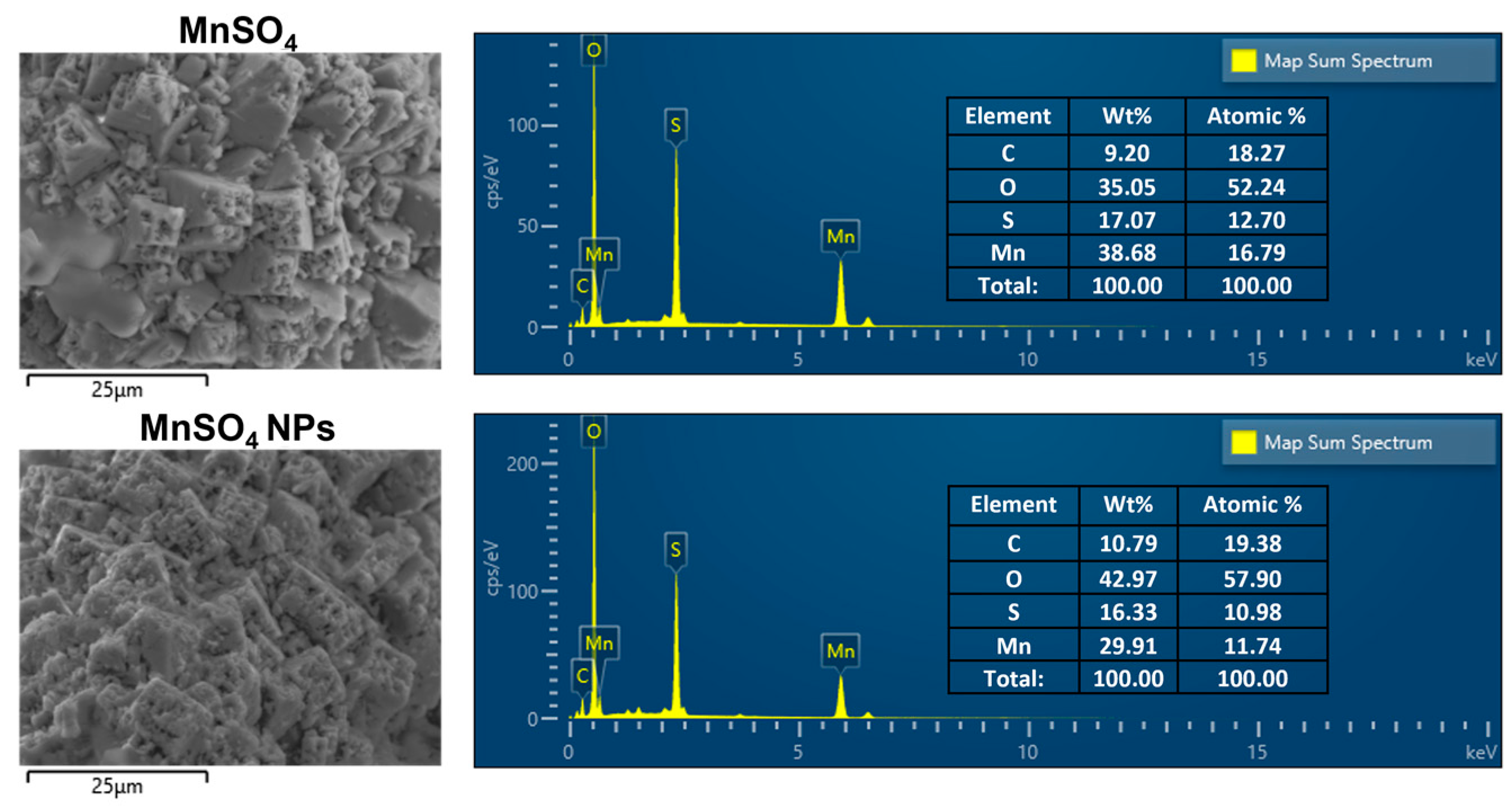
Atomic localization was also investigated with SEM equipped with an EDS technique (Figure 6). The atomic percentage values of Mn and S in the MnSO4 group were 16.79% and 12.70%, respectively. They were clearly changed to 11.74% (Mn) and 10.98% (S) in the MnSO4 NPs group. The alteration of tested atoms in the MnSO4 NPs group compared to the MnSO4 group indicates the formation of nano-sized particles composed of MnSO4, PEG 6000, Span 80, and Tween 80 by the HME process. All of these findings revealed in the solid-state studies suggest the successful fabrication of MnSO4-based nanovesicles following dispersion in the aqueous media.
Figure 6. SEM images and EDS analysis data of MnSO4 and MnSO4 NPs. Weight portion (Wt%) and atomic contents (Atomic %) are included in the inset.
Pharmaceutics 15 01831 g006

3.3. Catalytic Features

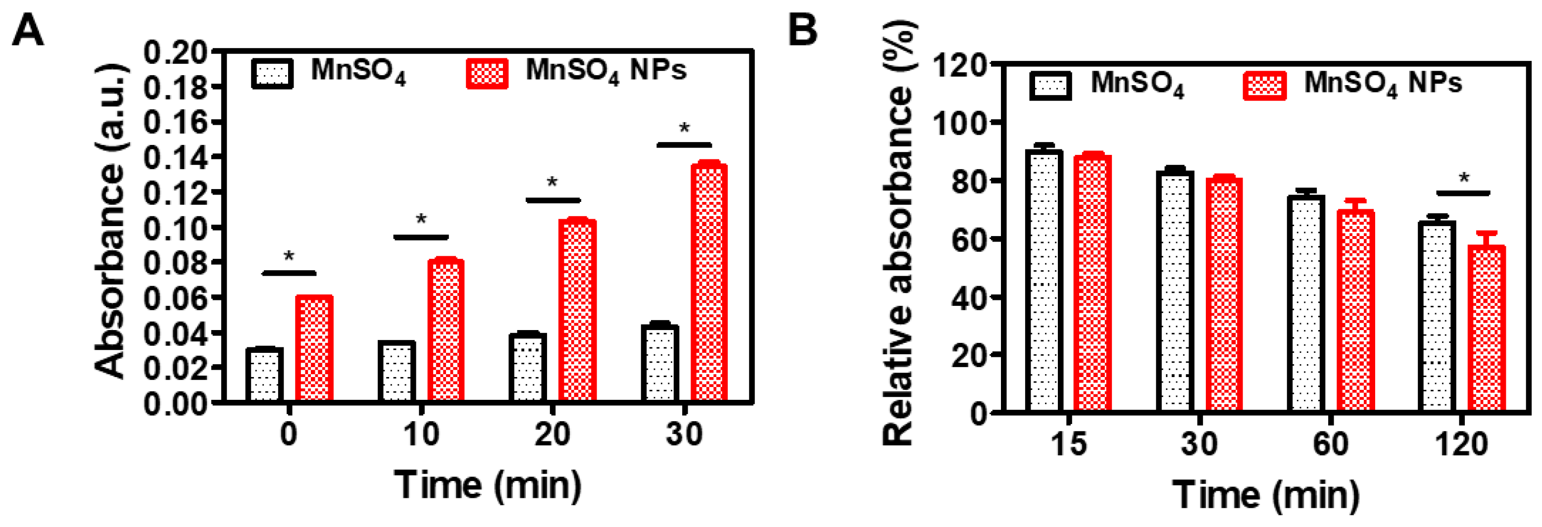
The catalytic activity of developed MnSO4 NPs was investigated with TMB and MB assays (Figure 7). Mn2+ ion (from MnSO4) can degrade cellular H2O2 to OH radical via a Fenton-like reaction [33]. The H2O2 level in the tumor cell is clearly higher than that in normal cells; therefore, this H2O2-responsive catalytic vehicle may possess tumor-selective therapeutic effects. OH radical may oxidize TMB (colorless) to its blue product; thus, its absorbance at the 650 nm wavelength can be used for the assessment of OH radical generation [34]. In this study (Figure 7A), there was an obvious difference between MnSO4 and MnSO4 NPs groups (p < 0.05). Of note, an increasing pattern of the absorbance value was observed in the MnSO4 NPs group rather than the MnSO4 group. Compared to micron-sized MnSO4 powder, MnSO4 NPs may possess large surface area in the aqueous media. This may result in elevated Fenton-like reaction-based conversion of H2O2 to OH radical.
The OH radical generation efficiency of designed MnSO4 NPs was also assessed by MB-based assay (Figure 7B). It is known that OH radical may attack the C–S+=C group as the first step of MB degradation and the degradation of the aromatic ring will make the MB solution transparent [35]. The MB degradation rate can be used for the evaluation of OH radical production [36]. As shown in Figure 7B, both MnSO4 and MnSO4 NPs groups had decreasing profiles of MB for 120 min incubation. Interestingly, there was a significant difference between MnSO4 and MnSO4 NPs groups at 120 min (p < 0.05). An approximately 43% reduction in the absorbance value of MB in the MnSO4 NPs group may imply the continuous production of OH radical during the tested reaction process. All of these data from catalytic assays suggest the successful generation of hydroxyl radicals aiming at the chemodynamic therapy of cancer.

3.4. In Vitro Anticancer Activities

The anticancer potential of developed MnSO4 NPs was explored in CT-26 cells (Figure 8). The antiproliferation activity of designed MnSO4 formulations was evaluated by an MTS-based assay (Figure 8A). Both MnSO4 and MnSO4 NPs groups had concentration-dependent cytotoxicity profiles. The IC50 values (presented as MnSO4 concentration in both groups) of MnSO4 and MnSO4 NPs were 100.5 and 86.0 μg/mL, respectively. In particular, the MnSO4 NPs group exhibited higher cytotoxicity than MnSO4 group at 200 μg/mL MnSO4 concentration (p < 0.05). Compared to the micron size of MnSO4, MnSO4 NPs had colloidal size range in the aqueous media (Figure 2). A smaller particle size may have higher surface area; therefore, MnSO4 NPs will produce a higher OH radical amount in cancer cells rather than MnSO4 based on the Fenton-like reaction. Particle size reduction with HME processing and the aid of hydrophilic polymer and surfactants may support the higher cytotoxicity of MnSO4 NPs compared to MnSO4. This finding suggests the efficient antiproliferation potential of colorectal cancer cells following MnSO4 NPs treatment.
Cellular ROS level was investigated after applying MnSO4 and MnSO4 NPs to CT-26 cells (Figure 8B). At current tested conditions, the cellular ROS level of the MnSO4 NPs group was 42% higher than the MnSO4 group (p < 0.05). As revealed in the results of catalytic assays (Figure 7), Mn2+ ion seems to provide Fenton-like chemistry-related OH radical generation from intrinsic H2O2, which is highly accumulated in cancer cells [13,14,15]. The smaller particle size of MnSO4 NPs, compared to MnSO4, may be the reason for the enhanced ROS level in cancer cells, principally due to the improved OH radical conversion.
The apoptosis induction capability of developed NPs was also assessed by annexin V-FITC and PI staining assay (Figure 8C). The sum of percentage in the upper right (UR) and lower right (LR) panels implies the population of apoptosis after applying each treatment. In observed data (Figure 8C), both MnSO4 and MnSO4 NPs induced a higher apoptosis rate than the control (no treatment) group, meaning the apoptosis induction capacity of the Mn2+ ion. Notably, the population percentage of (UR + LR) panel in the MnSO4 NPs group was significantly higher than those of the control and MnSO4 groups (p < 0.05). An elevated cellular ROS level may lead to improved apoptosis induction efficiency, ultimately explaining the higher antiproliferation potential. All of these experimental data in CT-26 cells may indicate the application of MnSO4 NPs to its chemodynamic therapy.

4. Conclusions

Micron-sized MnSO4 powder was dispersed in the hydrophilic polymer (PEG 6000) and non-ionic surfactants (Span 80 and Tween 80) to make nano-sized particles after dispersing in aqueous media with the aid of HME processing. A twin-screw-installed HME system with temperature control successfully produced a homogeneous extrudate composed of MnSO4 and pharmaceutical excipients. Following pulverization, those HME-processed MnSO4 particles exhibited a nano-size diameter, spherical shape, and negative zeta potential. The proper distribution of MnSO4 in the hydrophilic polymer with surfactants was demonstrated by several solid-state studies. The hydroxyl radical generation activity of MnSO4 NPs was elucidated by catalytic assays. In colorectal cancer (CT-26) cells, Mn2+ ion-related Fenton-like chemistry converted intrinsic H2O2 to OH radical and elevated the cellular ROS level, apoptosis induction efficiency, and antiproliferation potential. Developed MnSO4 NPs may be one of the promising candidates for the chemodynamic therapy of colorectal cancer.

Supplementary Materials

The following supporting information can be downloaded at: https://www.mdpi.com/article/10.3390/pharmaceutics15071831/s1, Figure S1: XPS spectrum of C 1s in MnSO4 NPs; Figure S2: XPS spectra of O 1s in (A) MnSO4 and (B) MnSO4 NPs; Figure S3: XPS spectra of Mn 2p in (A) MnSO4 and (B) MnSO4 NPs; Figure S4: XPS spectra of S 2p in (A) MnSO4 and (B) MnSO4 NPs; Table S1: FT-IR spectroscopy data of MnSO4, freeze-dried mixture, and MnSO4 NPs; Table S2: XPS analytical data MnSO4 and MnSO4 NPs.

Author Contributions

Conceptualization, S.C., B.-J.C., W.-S.K. and H.-J.C.; methodology, D.I.J., S.K., J.S.K., M.K. (Minju Kim), K.Y.K., M.O.K.A., W.-S.K. and H.-J.C.; validation, D.I.J. and S.K.; formal analysis, D.I.J., S.K., M.K. (Mrinmoy Karmakar) and S.C.; investigation, D.I.J., S.K., J.S.K., S.Y.L., M.K. (Minju Kim), K.Y.K. and M.O.K.A.; data curation, D.I.J., S.K., J.S.K., M.K. (Mrinmoy Karmakar) and H.-J.C.; writing—original draft preparation, D.I.J., S.K., S.C. and H.-J.C.; writing—review and editing, D.I.J., S.K., and H.-J.C.; visualization, D.I.J., S.K. and H.-J.C.; supervision, B.-J.C., W.-S.K. and H.-J.C.; project administration, B.-J.C., W.-S.K. and H.-J.C.; funding acquisition, B.-J.C., W.-S.K. and H.-J.C. All authors have read and agreed to the published version of the manuscript.

Funding

This work was supported by the Korea Institute of Planning and Evaluation for Technology in Food, Agriculture, Forestry and Fisheries (IPET) through the Agri-Bio Industry Technology Development Program, funded by the Ministry of Agriculture, Food and Rural Affairs (MAFRA) (No. 116073-03-2-CG000) and the National Research Foundation of Korea (NRF) grant funded by the Korea government (Ministry of Science and ICT and Ministry of Education) (2018R1A6A1A03025582 and 2020R1C1C1003945).

Institutional Review Board Statement

Not applicable.

Informed Consent Statement

Not applicable.

Data Availability Statement

The data presented in this study are available on request from the corresponding author.

Acknowledgments

FT-IR, XRD, and XPS analyses were conducted in the Central Laboratory of Kangwon National University.

Conflicts of Interest

The authors declare no conflict of interest.

References

  1. Patil, H.; Tiwari, R.V.; Repka, M.A. 2016. Hot-melt extrusion: From theory to application in pharmaceutical formulation. AAPS PharmSciTech 2016, 17, 20–42. [Google Scholar] [CrossRef] [PubMed] [Green Version]
  2. Simões, M.F.; Pinto, R.M.A.; Simões, S. Hot-melt extrusion in the pharmaceutical industry: Toward filing a new drug application. Drug Discov. Today 2019, 24, 1749–1768. [Google Scholar] [CrossRef] [PubMed]
  3. Tambe, S.; Jain, D.; Agarwal, Y.; Amin, P. Hot-melt extrusion: Highlighting recent advances in pharmaceutical applications. J. Drug Deliv. Sci. Technol. 2021, 63, 102452. [Google Scholar] [CrossRef]
  4. Almotairy, A.; Alyahya, M.; Althobaiti, A.; Almutairi, M.; Bandari, S.; Ashour, E.A.; Repka, M.A. Disulfiram 3D printed film produced via hot-melt extrusion techniques as a potential anticervical cancer candidate. Int. J. Pharm. 2023, 635, 122709. [Google Scholar] [CrossRef] [PubMed]
  5. Bagde, A.; Kouagou, E.; Singh, M. Formulation of topical flurbiprofen solid lipid nanoparticle gel formulation using hot melt extrusion technique. AAPS PharmSciTech 2022, 23, 257. [Google Scholar] [CrossRef]
  6. Patel, H.; Palekar, S.; Patel, A.; Patel, K. Ibrutinib amorphous solid dispersions with enhanced dissolution at colonic pH for the localized treatment of colorectal cancer. Int. J. Pharm. 2023, 641, 123056. [Google Scholar] [CrossRef]
  7. Xu, Y.; Yan, G.; Wen, X.; Wu, L.; Deng, R.; Liang, Q.; Zhang, L.; Chen, H.; Feng, X.; He, J. Preparation, evaluation, and pharmacokinetics in beagle dogs of a taste-masked flunixin meglumine orally disintegrating tablet prepared using hot-melt extrusion technology and D-optimal mixture design. Eur. J. Pharm. Sci. 2022, 168, 106019. [Google Scholar] [CrossRef]
  8. Koo, J.S.; Lee, S.Y.; Nam, S.; Azad, M.O.K.; Kim, M.; Kim, K.; Chae, B.J.; Kang, W.S.; Cho, H.J. Preparation of cupric sulfate-based self-emulsifiable nanocomposites and their application to the photothermal therapy of colon adenocarcinoma. Biochem. Biophys. Res. Commun. 2018, 503, 2471–2477. [Google Scholar] [CrossRef]
  9. Koo, J.S.; Lee, S.Y.; Azad, M.O.K.; Kim, M.; Hwang, S.J.; Nam, S.; Kim, S.; Chae, B.J.; Kang, W.S.; Cho, H.J. Development of iron(II) sulfate nanoparticles produced by hot-melt extrusion and their therapeutic potentials for colon cancer. Int. J. Pharm. 2019, 558, 388–395. [Google Scholar] [CrossRef]
  10. Lee, S.Y.; Nam, S.; Choi, Y.; Kim, M.; Koo, J.S.; Chae, B.J.; Kang, W.S.; Cho, H.J. Fabrication and characterizations of hot-melt extruded nanocomposites based on zinc sulfate monohydrate and Soluplus. Appl. Sci. 2017, 7, 902. [Google Scholar] [CrossRef] [Green Version]
  11. Thapa, R.K.; Kim, J.O. Nanomedicine-based commercial formulations: Current developments and future prospects. J. Pharm. Investig. 2023, 53, 19–33. [Google Scholar] [CrossRef]
  12. López-Lázaro, M. Dual role of hydrogen peroxide in cancer: Possible relevance to cancer chemoprevention and therapy. Cancer Lett. 2007, 252, 1–8. [Google Scholar] [CrossRef]
  13. Lin, L.S.; Song, J.; Song, L.; Ke, K.; Liu, Y.; Zhou, Z.; Shen, Z.; Li, J.; Yang, Z.; Tang, W.; et al. Simultaneous Fenton-like ion delivery and glutathione depletion by MnO2-based nanoagent enhances chemodynamic therapy. Angew. Chem. Int. Ed. Engl. 2018, 57, 4902–4906. [Google Scholar] [CrossRef] [PubMed]
  14. Pan, Y.; Zhu, Y.; Xu, C.; Pan, C.; Shi, Y.; Zou, J.; Li, Y.; Hu, X.; Zhou, B.; Zhao, C.; et al. Biomimetic yolk-shell nanocatalysts for activatable dual-modal-image-guided triple-augmented chemodynamic therapy of cancer. ACS Nano 2022, 16, 19038–19052. [Google Scholar] [CrossRef] [PubMed]
  15. Qian, J.; Su, L.; He, J.; Ruan, R.; Wang, J.; Wang, Z.; Xiao, P.; Liu, C.; Cao, Y.; Li, W.; et al. Dual-modal imaging and synergistic spinal tumor therapy enabled by hierarchical-structured nanofibers with cascade release and postoperative anti-adhesion. ACS Nano 2022, 16, 16880–16897. [Google Scholar] [CrossRef]
  16. Kim, M.J.; Hosseindoust, A.; Kim, K.Y.; Moturi, J.; Lee, J.H.; Kim, T.G.; Mun, J.Y.; Chae, B.J. Improving the bioavailability of manganese and meat quality of broilers by using hot-melt extrusion nano method. Brit. Poult. Sci. 2022, 63, 211–217. [Google Scholar] [CrossRef] [PubMed]
  17. Choi, J.W.; Lee, S.Y.; Cho, E.J.; Jeong, D.I.; Kim, D.D.; Kim, H.C.; Cho, H.J. Gas generating microspheres for immediate release of Hsp90 inhibitor aiming at postembolization hypoxia in transarterial chemoembolization therapy of hepatocellular carcinoma. Int. J. Pharm. 2021, 607, 120988. [Google Scholar] [CrossRef] [PubMed]
  18. Jeong, D.I.; Kim, S.; Lee, S.Y.; Kim, H.J.; Lee, J.; Lee, K.; Cho, H.J. Iron sulfate-reinforced hydrogel reactors with glucose deprivation, serial reactive oxygen species generation, ferroptosis induction, and photothermal ablation for cancer therapy. Chem. Eng. J. 2022, 438, 135584. [Google Scholar] [CrossRef]
  19. Lee, S.Y.; Park, J.; Jeong, D.I.; Hwang, C.; Lee, J.; Lee, K.; Kim, H.J.; Cho, H.J. Ferrocene and glucose oxidase-installed multifunctional hydrogel reactors for local cancer therapy. J. Control. Release 2022, 349, 617–633. [Google Scholar] [CrossRef] [PubMed]
  20. Kiio, T.M.; Park, S. Physical properties of nanoparticles do matter. J. Pharm. Investig. 2021, 51, 35–51. [Google Scholar] [CrossRef]
  21. Haque, S.T.; Karim, M.E.; Othman, I.; Chowdhury, E.H. Mitigating off-target distribution and enhancing cytotoxicity in breast cancer cells with alpha-ketoglutaric acid-modified Fe/Mg-CA nanoparticles. J. Pharm. Investig. 2022, 52, 367–386. [Google Scholar] [CrossRef]
  22. Zalba, S.; Ten Hagen, T.L.M.; Burgui, C.; Garrido, M.J. Stealth nanoparticles in oncology: Facing the PEG dilemma. J. Control. Release 2022, 351, 22–36. [Google Scholar] [CrossRef]
  23. Guo, X.; Xiao, H.S.; Wang, F.; Zhang, Y.H. Micro-Raman and FTIR spectroscopic observation on the phase transitions of MnSO4 droplets and ionic interactions between Mn2+ and SO42-. J. Phys. Chem. A 2010, 114, 6480–6486. [Google Scholar] [CrossRef] [PubMed]
  24. Chen, H.; Dong, X.; Shi, J.; Zhao, J.; Hua, Z.; Gao, J.; Ruan, M.; Yan, D. Templated synthesis of hierarchically porous manganese oxide with a crystalline nanorod framework and its high electrochemical performance. J. Mater. Chem. 2007, 17, 855–860. [Google Scholar] [CrossRef]
  25. Wang, H.; Lu, Z.; Qian, D.; Li, Y.; Zhang, W. Single-crystal α-MnO2 nanorods: Synthesis and electrochemical properties. Nanotechnology 2007, 18, 115616. [Google Scholar] [CrossRef]
  26. Stella, C.; Soundararajan, N.; Ramachandran, K. Structural, optical, dielectric and magnetic properties of Mn1−xCoxO2 nanowires. Superlattices Microstruc. 2014, 71, 203–210. [Google Scholar] [CrossRef]
  27. Nosal, H.; Moser, K.; Warzała, M.; Holzer, A.; Stańczyk, D.; Sabura, E. Selected fatty acids esters as potential PHB-V bioplasticizers: Effect on mechanical properties of the polymer. J. Polym. Environ. 2021, 29, 38–53. [Google Scholar] [CrossRef]
  28. Nayl, A.A.; Ismail, I.M.; Aly, H.F. Recovery of pure MnSO4∙H2O by reductive leaching of manganese from pyrolusite ore by sulfuric acid and hydrogen peroxide. Int. J. Miner. Process. 2011, 100, 116–123. [Google Scholar] [CrossRef]
  29. Bhatt, S.; Pulpytel, J.; Mirshahi, M.; Arefi-Khonsari, F. Catalyst-free plasma-assisted copolymerization of poly(ε-caprolactone)-poly(ethylene glycol) for biomedical applications. ACS Macro Lett. 2012, 1, 764–767. [Google Scholar] [CrossRef]
  30. Mondal, H.; Karmakar, M.; Ghosh, N.N.; Maiti, D.K.; Chattopadhyay, P.K.; Singha, N.R. One-pot synthesis of sodium alginate-grafted-terpolymer hydrogel for As(III) and V(V) removal: In situ anchored comonomer and DFT studies on structures. J. Environ. Manag. 2021, 294, 112932. [Google Scholar] [CrossRef]
  31. Xu, W.; Li, H.; Zheng, Y.; Lei, W.; Wang, Z.; Cheng, Y.; Qi, R.; Peng, H.; Lin, H.; Yue, F.; et al. Atomic insights into Ti Doping on the stability enhancement of truncated octahedron LiMn2O4 nanoparticles. Nanomaterials 2021, 11, 508. [Google Scholar] [CrossRef] [PubMed]
  32. Mondal, H.; Karmakar, M.; Dutta, A.; Mahapatra, M.; Deb, M.; Mitra, M.; Roy, J.S.D.; Roy, C.; Chattopadhyay, P.K.; Singha, N.R. Tetrapolymer network hydrogels via gum ghatti-grafted and N−H/C−H-activated allocation of monomers for composition-dependent superadsorption of metal ions. ACS Omega 2018, 3, 10692–10708. [Google Scholar] [CrossRef] [PubMed]
  33. Cao, W.; Jin, M.; Yang, K.; Chen, B.; Xiong, M.; Li, X.; Cao, G. Fenton/Fenton-like metal-based nanomaterials combine with oxidase for synergistic tumor therapy. J. Nanobiotechnol. 2021, 19, 325. [Google Scholar] [CrossRef] [PubMed]
  34. Song, H.; Li, X.; He, Y.; Peng, Y.; Pan, J.; Niu, X.; Zhao, H.; Lan, M. Colorimetric evaluation of the hydroxyl radical scavenging ability of antioxidants using carbon-confined CoOx as a highly active peroxidase mimic. Mikrochim. Acta 2019, 186, 354. [Google Scholar] [CrossRef]
  35. Khan, I.; Saeed, K.; Zekker, I.; Zhang, B.; Hendi, A.H.; Ahmad, A.; Ahmad, S.; Zada, N.; Ahmad, H.; Shah, L.A.; et al. Review on methylene blue: Its properties, uses, toxicity and photodegradation. Water 2022, 14, 242. [Google Scholar] [CrossRef]
  36. Satoh, A.Y.; Trosko, J.E.; Masten, S.J. Methylene blue dye test for rapid qualitative detection of hydroxyl radicals formed in a Fenton’s reaction aqueous solution. Environ. Sci. Technol. 2007, 41, 2881–2887. [Google Scholar] [CrossRef]
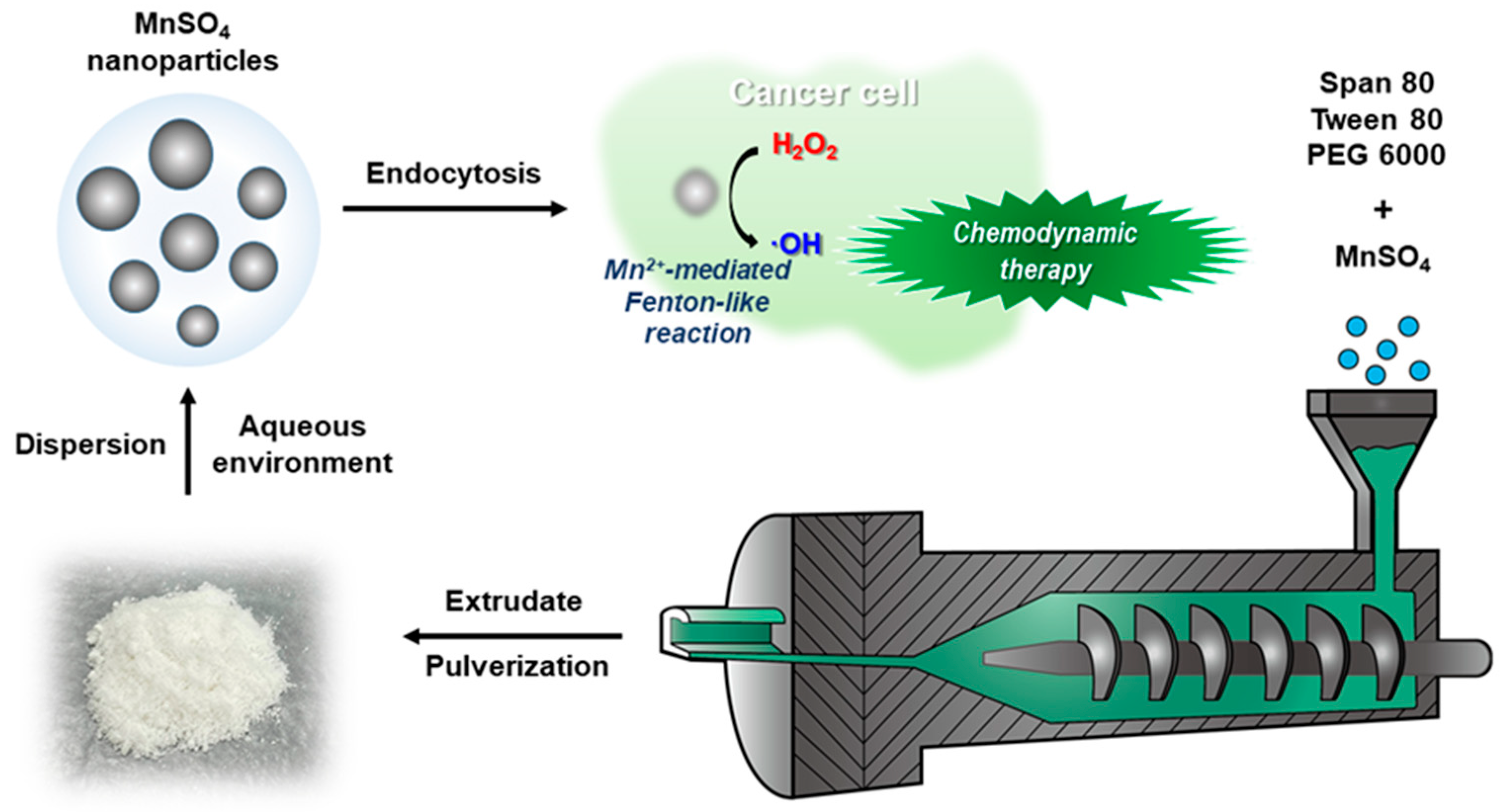
Figure 1. Schematic illustration of MnSO4 NPs production by HME process.
Pharmaceutics 15 01831 g001
Figure 2. Particle characteristics of HME-processed MnSO4 NPs. (A) Particle size distribution of MnSO4. (B) Mean diameter, polydispersity index, and zeta potential values of MnSO4 NPs. Mean diameter and polydispersity index values were calculated by measuring 50 cycles per each sample. Average and deviation data were acquired with three batches of sample. Each point represents mean ± SD (n = 3). (C) Particle size distribution of MnSO4 NPs dispersed in DW. (D) TEM image of MnSO4 NPs dispersed in DW. Scale bar length: 500 nm. (E) Incubation time-dependent mean diameter profiles in PBS (pH 7.4) and serum with DW (50%, v/v). Each point represents mean ± SD (n = 3). (F) Incubation time-dependent polydispersity index profiles in PBS (pH 7.4) and serum with DW (50%, v/v). Each point represents mean ± SD (n = 3).
Pharmaceutics 15 01831 g002
Figure 7. Catalytic assays of MnSO4 and MnSO4 NPs. (A) TMB assay data of MnSO4 and MnSO4 NPs. Each point represents mean ± SD (n = 3). * p < 0.05, between two groups. (B) MB assay data of MnSO4 and MnSO4 NPs. Each point represents mean ± SD (n = 3). * p < 0.05, between two groups.
Pharmaceutics 15 01831 g007
Figure 8. In vitro anticancer activity tests of MnSO4 and MnSO4 NPs in CT-26 cells. (A) Antiproliferation efficacy data of MnSO4 and MnSO4 NPs. Each point represents mean ± SD (n = 4). * p < 0.05, between two groups. (B) Cellular ROS assay data of MnSO4 and MnSO4 NPs. Each point represents mean ± SD (n = 3). * p < 0.05, between two groups. (C) Apoptosis induction capability of MnSO4 and MnSO4 NPs. UL: upper left; UR: upper right; LL: lower left; LR: lower right. Each point represents mean ± SD (n = 3). * p < 0.05, among indicated groups.
Pharmaceutics 15 01831 g008
Disclaimer/Publisher’s Note: The statements, opinions and data contained in all publications are solely those of the individual author(s) and contributor(s) and not of MDPI and/or the editor(s). MDPI and/or the editor(s) disclaim responsibility for any injury to people or property resulting from any ideas, methods, instructions or products referred to in the content.