Article

Membranolytic Activity Profile of Nonyl 3,4-Dihydroxybenzoate: A New Anti-Biofilm Compound for the Treatment of Dermatophytosis

by
Caroline B. Costa-Orlandi
1,
Níura M. Bila
1,2,
Jean Lucas C. Bonatti
1,
Carolina O. Vaso
1,
Mariana B. Santos
3,
Carlos R. Polaquini
3,
Mariana M. Santoni Biasioli
4,
Rondinelli D. Herculano
5,
Luis O. Regasini
3,
Ana Marisa Fusco-Almeida
1 and
Maria José S. Mendes-Giannini
1,*
1
Department of Clinical Analysis, School of Pharmaceutical Sciences, São Paulo State University (U.N.E.S.P.), Araraquara 14800-903, SP, Brazil
2
Department of Para-Clinic, School of Veterinary, Eduardo Modlane University (UEM), Maputo 257, Mozambique
3
Department of Chemistry and Environmental Sciences, Institute of Biosciences, Humanities and Exact Sciences, São Paulo State University (U.N.E.S.P.), Sao Jose do Rio Preto 15054-000, SP, Brazil
4
Department of Biological Sciences, School of Pharmaceutical Sciences, São Paulo State University (U.N.E.S.P.), Araraquara 14800-903, SP, Brazil
5
Department of Bioprocesses and Biotechnology, School of Pharmaceutical Sciences, São Paulo State University (U.N.E.S.P.), Araraquara 14800-903, SP, Brazil
*
Author to whom correspondence should be addressed.
Pharmaceutics 2023, 15(5), 1402; https://doi.org/10.3390/pharmaceutics15051402
Submission received: 25 November 2022 / Revised: 17 February 2023 / Accepted: 21 February 2023 / Published: 4 May 2023
(This article belongs to the Special Issue Recent Advances in Antifungal Drugs)

Abstract

:
The ability of dermatophytes to live in communities and resist antifungal drugs may explain treatment recurrence, especially in onychomycosis. Therefore, new molecules with reduced toxicity that target dermatophyte biofilms should be investigated. This study evaluated nonyl 3,4-dihydroxybenzoate (nonyl) susceptibility and mechanism of action on planktonic cells and biofilms of T. rubrum and T. mentagrophytes. Metabolic activities, ergosterol, and reactive oxygen species (ROS) were quantified, and the expression of genes encoding ergosterol was determined by real-time PCR. The effects on the biofilm structure were visualized using confocal electron microscopy, scanning electron microscopy (SEM), and transmission electron microscopy (TEM). T. rubrum and T. mentagrophytes biofilms were susceptible to nonyl and resistant to fluconazole, griseofulvin (all strains), and terbinafine (two strains). The SEM results revealed that nonyl groups seriously damaged the biofilms, whereas synthetic drugs caused little or no damage and, in some cases, stimulated the development of resistance structures. Confocal microscopy showed a drastic reduction in biofilm thickness, and transmission electron microscopy results indicated that the compound promoted the derangement and formation of pores in the plasma membrane. Biochemical and molecular assays indicated that fungal membrane ergosterol is a nonyl target. These findings show that nonyl 3,4-dihydroxybenzoate is a promising antifungal compound.

1. Introduction

Dermatophytosis is among the most prevalent mycoses worldwide, affecting a large proportion of the population regardless of age, immunological status, sex, or race [1,2,3,4]. The need for prolonged treatment, recurrence of infections, frequent reports of resistance, and the possibility of biofilm formation, especially in onychomycoses, are the main limitations for the treatment of this disease [5,6,7,8,9,10].
Azoles and allylamines are classically used to treat dermatophytosis and can be administered orally and/or topically, alone or in combination, depending on the severity of the infection and patient prognosis [7,11,12,13]. Terbinafine, fluconazole, and itraconazole are the most commonly used drugs for systemic treatment. Ketoconazole and griseofulvin are prescribed according to the clinical manifestations of dermatophytosis but are no longer recommended for the treatment of onychomycosis [11,14,15]. It is important to note that oral administration of fluconazole is not approved in some countries for the treatment of dermatophytic onychomycoses [15,16]. Despite their high efficacy, systemic antifungals should be used with caution because of the greater likelihood of adverse effects than topical administration, including interactions with other drugs, congestive heart failure, and hepatotoxicity [15].
Terbinafine, cyclopirox, and amorolfine are still widely used as topical treatments in some countries, while efinaconazole, tavaborol, and luliconazole have gained prominence owing to their safety and efficacy [7,17,18,19,20,21]. Topical therapy is generally nontoxic to patients because systemic absorption does not occur, minimizing the likelihood of interactions with other drugs and the emergence of severe adverse effects. Topical drug administration has disadvantages in the case of onychomycosis owing to low penetration into the nail plate caused by several physiological factors in addition to the composition of the pharmaceutical form. The cost of newer antifungals is high, making it difficult for many people to adhere to the treatment regimen for a long time [7,15,16].
Recurrence and reinfection, common in onychomycoses, may be related to biofilm formation and other factors [8,9,10,22]. Most infections caused by microorganisms are associated with the formation of sessile communities and three-dimensional structures covered by a complex polymeric extracellular matrix [23,24,25]. This phenotype promotes advantageous protection for microorganisms, which is provided by physical and genetic factors such as the barrier effect of biomass, microenvironment composition, concentration of metabolites and gases, and modification of gene regulation [25,26,27].
Fungal biofilms are difficult to treat and eradicate using currently available antifungals [28,29]. The dose of an antifungal agent required to eradicate certain fungal biofilms can exceed the recommended therapeutic concentration [28]. Therefore, novel compounds capable of eradicating fungal biofilms are being investigated.
Nonyl 3,4-dihydroxybenzoate (nonyl) is a synthetic derivative of protocatechuic acid. Protocatechuic acid and its derivatives have been studied and exhibit a broad spectrum of biological and antifungal activities [7,30,31,32,33,34,35]. Our group has described its fungicidal activity [30,35] and its effectiveness in reducing the metabolic activity and topography of dermatophyte biofilms [7]. However, further studies are needed to demonstrate the damage caused by this compound to the ultrastructure of biofilms compared to conventional antifungals, in addition to investigating its mechanism of action in fungal cells. Therefore, this study aimed to determine the susceptibility of mature biofilms and planktonic cells of T. rubrum and T. mentagrophytes to nonyl, compare these results to the activities of three conventional antifungal drugs, and study its mechanism of action in dermatophytes.

2. Materials and Methods

2.1. Dermatophytes Tested

Standard references (T. rubrum ATCC 28189 INCQS 40051 and T. mentagrophytes ATCC 11481 INCQS 40118) and clinical strains (T. rubrum 143 and T. mentagrophytes 66) were used. ATCC strains were obtained from the Clinical Mycology Laboratory of the School of Pharmaceutical Sciences at the Universidade Estadual Paulista, Araraquara, SP, Brazil. Clinical strains were kindly provided by Prof. Dr. Maria Aparecida Resende-Stoianoff from the Clinical Mycology Laboratory at the Federal University of Minas Gerais and isolated from patients with onychomycosis at the dermatology outpatient clinic of a tertiary care hospital. This study was published by Costa-Orlandi et al. [3] and approved by the Ethics Committees of the Federal University of Minas Gerais and Hospital Santa Casa de Misericordia de Belo Horizonte (Ethics 004.0.107.000-06/094-2009). The fungi were maintained in sterile distilled water, cultivated on potato dextrose agar (Difco, BD biosciences, Sparks, MD, USA) and incubated at 28 °C for seven days [36].

2.2. Synthesis of Nonyl 3,4-Dihydroxybenzoate

Nonyl protocatechuate was prepared as previously described by Faria et al. [37], Soares et al. [30], and Costa-Orlandi et al. [7]. Briefly, the synthesis was performed by standard esterification reactions using protocatechuic acid (Sigma-Aldrich, St. Louis, MO, USA) and nonyl alcohol, in N′,N′-dicyclohexylcarbodiimide (DCC) and p-dioxane. The residue was partitioned with ethyl acetate and filtered. The filtrate was washed with NaHCO3 solution and water and dried over MgSO4. The crude product was purified over a silica gel column (0.06–0.02 mm, A.C.R.O.S. Organics, Waltham, MA, USA) and eluted using CHCl3/CH3OH (98:2). The structure of the compound was confirmed using 1H and 13C Nuclear Magnetic Resonance spectroscopy. NMR spectra were recorded using a Bruker spectrometer (300 MHz, 7.1 T). The purity of the compound was determined by high-performance liquid chromatography with a photodiode array detector (HPLC-PAD) using the peak area. HPLC-PAD chromatograms were obtained on Agilent Technologies 1220 Infinity equipment, a photodiode array system (Agilent Technologies Model 1260 Infinity), and an Agilent Zorbaz Eclipse Plus C-18 column (250 mm × 4.6 mm, 5 µm) using methanol:water (98:2) as the mobile phase (1.0 mL/min). The NMR spectra and HPLC chromatograms are presented in the Supplementary Material (Figures S1–S3).

2.3. Preparation of Conventional Antifungals and Nonyl Protocatechuate (Nonyl 3,4-Dihydroxybenzoate)

Fluconazole, griseofulvin, and terbinafine were purchased from Sigma-Aldrich (St. Louis, MO, USA). Stock solutions of the antifungal drugs were prepared according to document M-38 A2 proposed by the Clinical and Laboratory Standards Institute (C.L.S.I.) [36]. Stock solutions of fluconazole were prepared in sterile distilled water at concentrations ranging from 0.625 mg/L to 5120 mg/L. For terbinafine and griseofulvin, stock solutions in DMSO were prepared at concentrations ranging from 0.1 to 3200 mg/L for the former and from 6.25 to 51,200 mg/L for the latter. Working solutions were prepared from the stock solutions in RPMI 1640 medium with glutamine, without sodium bicarbonate, and with phenol red as a pH indicator (Gibco Life Technologies, Grand Island, NY, USA), buffered with MOPS (Sigma-Aldrich, St. Louis, MO, USA). Working solutions of fluconazole and griseofulvin were prepared to obtain concentrations ranging from 0.0625 to 512 mg/L. For terbinafine, working solutions were prepared in the range of 0.001–32 mg/L. Nonyl was aseptically diluted in DMSO to obtain a DMSO concentration in the working solution of less than 1%. Working solutions were prepared at concentrations ranging from 0.015 to 500 mg/L.

2.4. Effects of Antifungal Drugs and Nonyl 3,4-Dihydroxybenzoate on T. rubrum and T. mentagrophytes, Planktonic Cells, and Mature Biofilms

One hundred microliters of the working solutions of the antifungals and the compound at twice the final desired concentration were added to 96-well microdilution plates to evaluate the effects of the antifungals and nonyl against planktonic cells. Inoculums were prepared in RPMI 1640 medium to reach a final concentration corresponding to 1 × 10⁶ cells/mL, and 100 µL of the fungal suspensions were added to each well along with their respective controls. The microplates were incubated at 37 °C for 72 h. According to our previously established protocol, biofilms were formed in 96-well plates with RPMI 1640 medium [10]. Biofilm susceptibility assays were conducted as described by Costa-Orlandi et al. [4,7]. Briefly, the culture medium was aspirated after maturation, and the wells were washed with 0.85% sterile saline. Then, 100 µL of each dilution of different concentrations of the antifungals and the compound were distributed in 96-well plates with their respective controls: the sterility control of the medium and the untreated biofilms. The microplates were incubated at 37 °C for 72 h. The XTT reduction assay was used to quantify the metabolic activities of planktonic cells and biofilms. The plates were read at 490 nm using a spectrophotometer (Microplate Reader iMarkTM; Bio-Rad, Hercules, CA, USA). Inhibition was defined as a reduction in metabolic activity of at least 50%.

2.5. Scanning Electron Microscopy (SEM) of Treated Biofilms

SEM was performed to visualize the effects of nonyl and antifungal drugs on fungal biofilm topography. Biofilms were formed in 24-well plates and treated with fluconazole (512 mg/L), griseofulvin (512 mg/L), terbinafine (32 mg/L), or nonyl (250 mg/L). The plates were incubated at 37 °C for 72 h. The samples were fixed and dried, and the bottoms of the plates were cut using a scalpel, according to Costa-Orlandi et al. [4,7,10], Bila et al. [20], Garcia et al. [38], and Vaso et al. [39]. The samples were fixed on carbon tape, mounted on aluminum cylinders with silver, and placed in a high-vacuum evaporator for gold plating. The topographies of the biofilms were analyzed using a scanning electron microscope (SEM; JEOL JSM-6610LV Peabody, MA, USA) at the School of Dentistry of the Sao Paulo State University (U.N.E.S.P.) Araraquara.

2.6. Confocal Microscopy of Treated Biofilms

Confocal microscopy was used to visualize the thickness of biofilms after antifungal treatment. Biofilms were grown in 24-well plates containing sterile round coverslips. After maturation, the culture medium was aspirated, and the biofilms were washed with sterile saline. Terbinafine (1 mL, 32 mg/L) and nonyl (250 mg/L) were then added. The plates were incubated for 72 h at 37 °C. The untreated biofilms were used as controls. A solution of C.A.A.F. (Concanavalin A—conjugated to Alexa Fluor 488—Molecular Probes, Eugene, OR, USA, 25 µM) and FUN 1 (Molecular Probes, USA, 10 µM) was prepared. This mixture was added to wells containing the treated and untreated biofilms, and the plates were incubated at 37 °C for 45 min while protected from light. Then the wells were washed, and the coverslips were removed, covered with 4 μL of Fluoromount-G (Sigma-Aldrich), and deposited on microscope slides for observation under a confocal microscope Leica TCS SP5 (Leica Microsystems, Wetzlar, Germany) from the School of Medicine of the University of Sao Paulo (U.S.P.) Ribeirão Preto. The images were processed using L.A.S. AF 1.8.2 build 1465 Leica Microsystems CMS Gmgh [4,10,40].

2.7. Determination of the Mechanism of Action of Nonyl

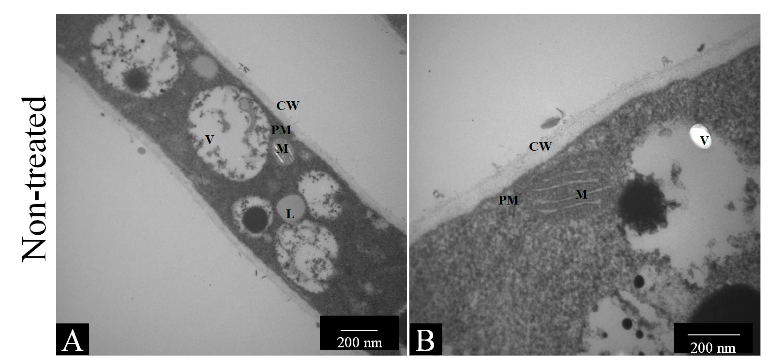
2.7.1. Transmission Electron Microscopy

To verify the damage caused by the compounds to the fungal ultrastructure, 1 × 10⁶ cells/mL of T. rubrum ATCC 28189 were treated with low concentrations of nonyl (1 and 0.5 mg/L), fluconazole (64 mg/L), and amphotericin (1 mg/L) as a control. After the incubation period, the contents of the microdilution plate wells were transferred to 15-mL conical tubes, washed three times with phosphate-buffered saline (PBS, pH 7.2) three times after centrifugation, washed again, and fixed with 2.5% glutaraldehyde solution in sodium cacodylate buffer (Electron Microscopy Sciences, Fisher Scientific, Waltham, MA, USA). After fixation, cells were washed and exposed to potassium dichromate and uranyl acetate. The fungal elements were washed, soaked in agar, and cut into small cubes (approximately 0.5 to 1 mm3), which were dehydrated using increasing concentrations of ethanol. The samples were then placed in Spurr resin, sectioned using an ultramicrotome, and observed under a Jeol JEM-100 CXII transmission electron microscope equipped with a Hamamatsu ORCA-HR digital camera at the School of Medicine of the University of Sao Paulo (U.S.P.) Ribeirao Preto [41].

2.7.2. Quantification of Fungal Membrane Sterols

For the quantification of fungal membrane sterols, five microliters of a 1 × 10⁶ conidia/mL suspension were placed in contact with sub-inhibitory concentrations of nonyl (7.8 mg/L), fluconazole (128 mg/L), and amphotericin B (1 mg/L). Untreated fungi were used as controls. The suspensions were incubated for 7 days at 37 °C. After this period, the tubes were centrifuged at 4 °C at 5000 rpm for 30 min; the supernatant was removed, and the dry weight was determined. Approximately 3 mL of a 25% KOH alcoholic solution was added to sterile glass tubes containing 1 g of dry weight and shaken for 1 min. The tubes were then placed in a water bath at 85 °C for 1 h and cooled to room temperature. After cooling, a mixture of 1 mL of sterile distilled water and 3 mL of n-heptane (Sigma-Aldrich) was added to the sterile glass beads. Vigorous vortexing was performed for 15 min. Supernatants were removed, placed in 1.5 mL microtubes, and stored at −20 °C overnight. The following day, the samples were read using an ultraviolet-visible spectrophotometer at a wavelength of 282 nm. A calibration curve was constructed using standard ergosterol (Sigma-Aldrich) [20,39,42].

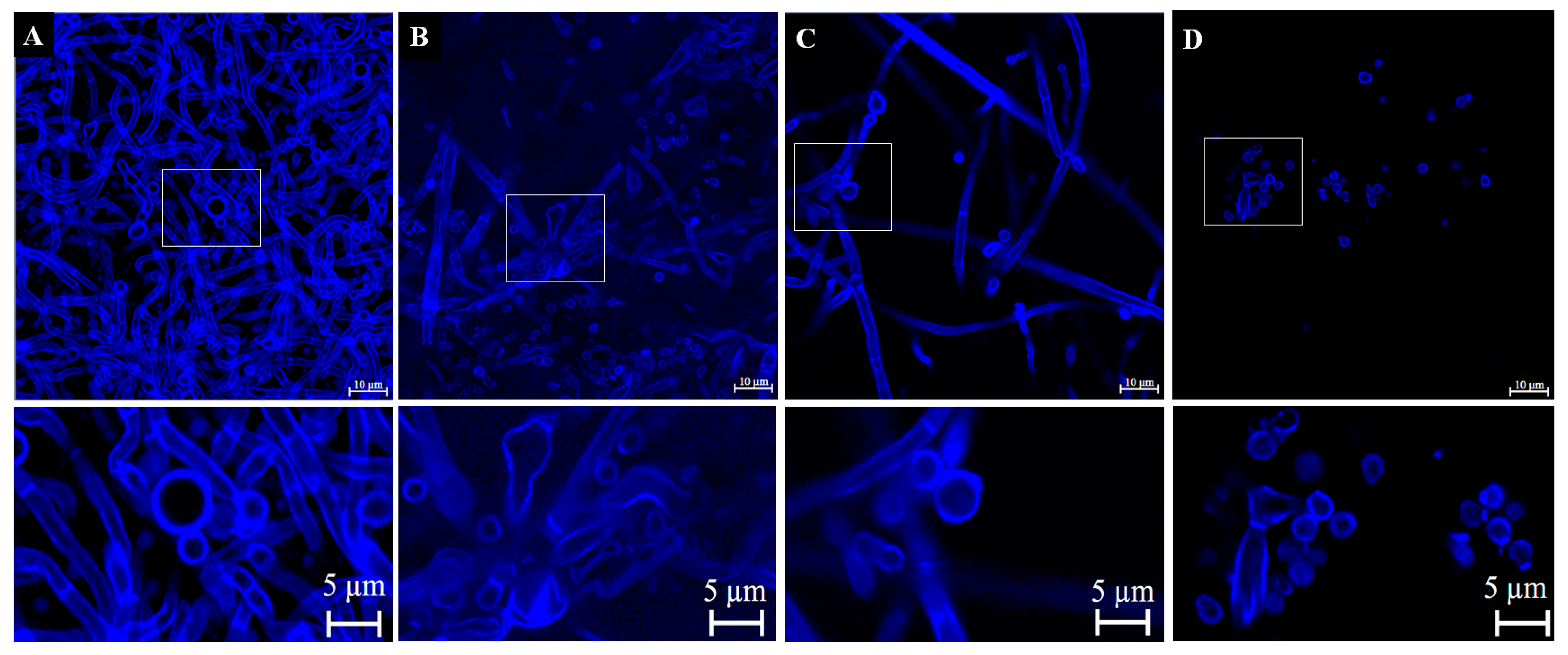
2.7.3. Confocal Laser Scanning Microscopy after Staining with Calcofluor White

For confocal microscopy, a 1 × 10⁶ cells/mL fungal suspension was treated with the same concentrations of nonyl, fluconazole, and amphotericin B in 24-well plates (Kasvi) containing sterile round coverslips. After 96 h of incubation, the plates were centrifuged, the culture medium was gently removed, and the wells were washed with PBS after successive centrifugation steps. A working solution of Calcofluor White (Sigma-Aldrich) was prepared at a concentration of 100 mg/L, and 30 µL of this solution was added to the wells. The plates were then reincubated at 37 °C for 45 min and protected from light. The wells were then washed with sterile PBS, and the plates were centrifuged. Coverslips were carefully removed and poured into 4 μL of Fluoromount-G (Sigma-Aldrich), previously deposited on microscope slides for observation under a confocal microscope, the ZEISS LSM 800 with Airyscan with image capture and processing software ZEN BLUE 2.3 System [20,43].

2.7.4. Quantification of Reactive Oxygen Species (ROS)

Intracellular production of ROS before and after treatment of T. rubrum ATCC 28189 with nonyl was evaluated using 50 µM of 2,7 dichlorodihydrofluorescein diacetate (H2DCFDA, Invitrogen by Thermo Fisher Scientific, Waltham, MA, USA). Amphotericin B and hydrogen peroxide (10 mM) were used as controls. The same concentrations of the compounds as in Section 2.7.1 and Section 2.7.2 were used for treatment. All samples were incubated with H2DCFDA for 30 min at room temperature and subsequently analyzed on a BD FACSCanto I flow cytometer using a 530/30 nm wavelength filter for fluorescence detection. The data were analyzed using FlowJo 10.1 software [44,45].

2.7.5. Apoptosis/Necrosis

The Annexin V-FITC apoptosis detection kit (A9210; Sigma-Aldrich) was used according to the manufacturer’s recommendations. To this, 1.5 mL of the fungal suspension at a final concentration of 1 × 10⁶ cells/mL was treated with nonyl (7.8 mg/L), amphothericin B (4 mg/L), or fluconazole (256 mg/L). The samples were analyzed using a BD FACSCanto I flow cytometer.

2.7.6. Analysis of Differential Gene Expression of T. rubrum ATCC 28189 Treated with Nonyl by Real-Time PCR

The relative expression of genes related to ergosterol synthesis after treatment of T. rubrum with nonyl protocatechuate was evaluated using real-time PCR (qPCR) and compared to untreated fungal gene expression. Total RNA was extracted using the Illustra RNAspin Isolation Kit (G.E. Life Sciences) according to the manufacturer’s guidelines. The concentration and purity were determined by reading in a Nanodrop™ 2000 spectrophotometer (Thermo Scientific™, Waltham, MA, USA) at absorbances of 260 and 280 nm, and the integrity was evaluated by capillary electrophoresis using the Agilent 2100 Bioanalyzer equipment (Agilent Technologies, Palo Alto, CA, USA). Specific primers are represented in Table 1 [46,47,48]. Total RNA was treated with the DNase I kit (Sigma-Aldrich), and complementary DNA (cDNA) was synthesized using a high-capacity cDNA reverse transcription kit (Applied Biosystems), according to the manufacturer’s guidelines. The qPCRs were performed on the 7500 Real-Time PCR Instrument (Applied Biosystems by Thermo Fisher Scientific) of the School of Pharmaceutical Sciences of U.N.E.S.P. Araraquara with the Power SYBR™ Green PCR Master Mix detection system (Applied Biosystems by Thermo Fisher Scientific).
Reactions were prepared in a total volume of 20 µL, using 52 ng of cDNA and 0.5 µM of each primer. The relative expression was calculated by the 2−∆∆Ct method, using the glyceraldehyde-3-phosphate dehydrogenase (GAPDH) and DNA-dependent RNA polymerase II (rpb2) genes as endogenous [46,49,50].

2.8. Statistical Analysis

All assays were performed in triplicate and in three independent experiments. Data were analyzed using an analysis of variance with Bonferroni’s post-hoc test. Data generated by qPCR were analyzed using the Student’s t-test. All data was generated using GraphPad Prism 5.0 software. Statistical significance was set at p less than 0.05.

3. Results

3.1. Susceptibilities of T. rubrum and T. mentagrophytes Planktonic and Biofilm Forms to Antifungal Drugs and Nonyl 3, 4-Dihydroxybenzoate

Table 2 shows a comparison of the metabolic activities of T. rubrum and T. mentagrophytes in the planktonic and biofilm forms after treatment with different drugs and nonyl. For all strains, planktonic cells were more susceptible to treatment than biofilm forms. The concentrations of fluconazole that reduced at least 50% of the metabolic activity (MIC50) of the planktonic cells of T. rubrum ATCC 28189, T. rubrum 143, T. mentagrophytes ATCC 11481, and T. mentagrophytes 66 were 128, 32, 2, and 16 mg/L, respectively. Griseofulvin reduced the metabolic activity by ≥50% at 256 mg/L for T. rubrum 143, 4 mg/L for T. rubrum ATCC 28189, and 1 mg/L for T. mentagrophytes ATCC 11481 and T. mentagrophytes 66. Terbinafine showed an inhibition at 0.015 mg/L for T. mentagrophytes ATCC 11481, T. rubrum ATCC 28189, and T. rubrum 143, and 0.06 mg/L for T. mentagrophytes at 0.125 mg/L 66. Nonyl reduced the metabolic activity to 7.8 mg/L for T. mentagrophytes ATCC 11481 and 15.6 mg/L for the other strains.
The mature biofilms of T. rubrum 143, T. rubrum ATCC 28189, T. mentagrophytes ATCC 11481, and T. mentagrophytes 66 exposed to fluconazole and griseofulvin exhibited metabolic activity values similar to those observed in the untreated controls, with SMIC50 > 512 mg/L (Table 2). Treatment with terbinafine significantly reduced the metabolic activity of T. rubrum 143 and T. mentagrophytes ATCC 11489 biofilms at concentrations of 0.5 and 16 mg/L, respectively. In contrast, for T. rubrum ATCC 28189 and T. mentagrophytes 66 biofilms, the SMIC50 values were greater than the highest concentration tested (>32 mg/L). Nonyl-treated biofilms obtained a significant reduction in metabolic activity at concentrations of 62.5 mg/L for T. rubrum species and 125 and 250 mg/L for T. mentagrophytes ATCC 11489 and T. mentagrophytes 66, respectively (Table 2). These results showed that the compound was more effective against biofilms than fluconazole and griseofulvin for all the strains tested.

3.2. Scanning Electron Microscopy of Treated Biofilms

SEM images showed that fluconazole and griseofulvin had minimal or no effect on T. rubrum biofilms (Figure 1L,M,Q,R). Terbinafine caused no apparent damage to T. rubrum ATCC 28189 hyphae (Figure 1N); however, visualization of T. rubrum 143 biofilms revealed the presence of destroyed hyphae and a reduction in the extracellular matrix (Figure 1S). Nonyl severely damaged T. rubrum ATCC 28189 (Figure 1O) and T. rubrum 143 (Figure 1T) biofilms, resulting in wilted hyphae, cell wall collapse, and a dramatic reduction in the extracellular matrix. However, few intact hyphae were observed in T. rubrum 143 (Figure 1J).
Concerning T. mentagrophytes biofilms, treatment with fluconazole (Figure 1B,G) and griseofulvin (Figure 1C,H) did not cause visible damage to the structures of the microbial communities. Moreover, griseofulvin stimulated the production of arthroconidia along the entire biofilm length. These structures, which are considered resistant, were observed especially in biofilms formed by T. mentagrophytes 66 (Figure 1C). Although terbinafine caused slight damage to T. mentagrophytes 66 biofilms (Figure 1D), severe damage was observed in the biofilms formed by the ATCC strain (Figure 1H). Nonyl caused significant damage to T. mentagrophytes 66 (Figure 1D) and T. mentagrophytes ATCC 11481 (Figure 1I) biofilms, causing leakage of cytoplasmic material and cell wall collapse. Interestingly, small round structures such as atrophied microconidia or perhaps extracellular vesicles (E.V.) were observed next to the damaged T. mentagrophytes hyphae treated with nonyl (white arrows in Figure 1D,I).

3.3. Confocal Microscopy of Treated Biofilms

Confocal microscopy of the biofilms treated with terbinafine and nonyl was performed to determine the thickness of the microbial communities after exposure to these compounds. The analysis showed that untreated T. rubrum ATCC 28189 and the clinical isolate 143 biofilms had an average thickness of 94.67 μm (Figure 2A) and 40.28 μm (Figure 2D), respectively. For the T. rubrum ATCC strain, treatment with terbinafine or nonyl significantly reduced the thickness of the biofilms on average to 33.23 µm (Figure 2B) and 39.28 µm (Figure 2C), respectively. There was no reduction in the thickness of biofilms of T. rubrum 143 treated with terbinafine (Figure 2E) or untreated (Figure 2F); however, in the latter, it was possible to observe a much less dense biofilm than in the control and terbinafine treatments. Untreated biofilms of T. mentagrophytes ATCC 11481 and the clinical isolate 66 showed an average thickness of 60.55 µm (Figure 2G) and 60.42 µm (Figure 2J). Treatment with terbinafine or nonyl significantly reduced the thickness of the biofilms formed by both T. mentagrophytes ATCC (Figure 2H,I) and the clinical isolate (Figure 2K,L).

3.4. Determination of the Mechanism of Action of Nonyl Protocatechuate

3.4.1. Transmission Electron Microscopy (TEM)

Microscopic results showed that nonyl and the drugs caused significant changes in the hyphae of T. rubrum ATCC 28189 (Figure 3). Figure 3A,B show the longitudinal sections of untreated T. rubrum hyphae with intact cell walls, plasma membranes, and organelles. Treatment with nonyl at a concentration of 0.5 mg/L caused an increase in vacuoles and plasma membrane derangement in some areas (Figure 3C,D). Treatment with 1 mg/L of nonyl caused significant damage, with vacuoles covering almost the entire cytoplasm (Figure 3E). There was a possible rupture of the plasma membrane and an apparent osmotic imbalance (Figure 3F). However, there was no change in cell morphology, indicating that there was no significant change in the cell wall. Fluconazole treatment resulted in plasma membrane derangement (Figure 3G,H), while amphotericin B treatment resulted in visible pore formation and leakage of cytoplasmic content (Figure 3I,J).

3.4.2. Quantification of Fungal Membrane Sterols

Sterol levels were reduced after treatment with nonyl (p < 0.01) and control antifungals (p < 0.001) (Figure 4).

3.4.3. Verification of Cell Wall Morphology and Damage by Calcofluor White Staining and Confocal Microscopy

The results in Figure 5 show that in the untreated control, the morphology was intact, with cell walls well-demarcated by calcofluor (Figure 5A). Conformational changes were observed in the cells treated with nonyl (Figure 5B), fluconazole (Figure 5C), and amphotericin B (Figure 5D), which showed irregular shapes. Despite the damage caused by the nonyl groups, there was no impairment of the cell wall structure. Calcofluor is a non-specific fluorophore that binds to cell wall chitin, mainly in the polysaccharides β 1, 3, and β 1,4 [51].

3.4.4. ROS Quantification

Intracellular quantification of ROS showed that nonyl treatment did not induce the production of reactive oxygen species when compared to the untreated control (p > 0.05) (Figure 6). The control treatment with hydrogen peroxide best induced the production of ROS compared to the other treatments (p < 0.001).

3.4.5. Determination of the Mechanisms of Cell Death by Apoptosis/Necrosis

Nonyl, fluconazole, and amphotericin B significantly induced necrotic cell death compared to that in the untreated control (p < 0.05). Among the treated groups, the most significant induction was caused by the protocatechuic acid derivative (63%) (p < 0.001) (Figure 7).

3.4.6. Analysis of Differential Gene Expression of T. rubrum ATCC MYA-4438 Treated with Nonyl Protocatechuate by Real-Time PCR

The relative expression results showed a negative modulation of Erg11 (p < 0.01) after treatment with nonyl (Figure 8), proving the membranolytic action of the protocatechuic acid derivative. Erg 11 encodes the enzyme lanosterol 14 α-demethylase, the main target of azole antifungals [47]. Gene modulation of Erg1 was insignificant, indicating that this compound does not act in the initial pathway of ergosterol biosynthesis (squalene epoxidase), similar to terbinafine.

4. Discussion

In this study, the susceptibilities of T. rubrum and T. mentagrophytes to the antifungal drugs fluconazole, griseofulvin, terbinafine, and a compound derivative of protocatechuic acid were tested under biofilm and planktonic conditions. Nonyl caused significant damage to the biofilms formed by all strains at a 62.5 mg/L concentration. Similar damage was observed in terbinafine-treated biofilms in T. mentagrophytes ATCC 11481 and T. rubrum 143. Furthermore, it is noteworthy that mature dermatophyte biofilms were resistant to all concentrations of fluconazole and griseofulvin. Although treatment with terbinafine damaged the topography of some biofilms and decreased their metabolic activity and thickness, many intact cells and a reasonable amount of extracellular matrix were still observed. Furthermore, it was necessary to apply terbinafine concentrations higher than 32 mg/L to eradicate biofilms from the other two strains, which is therapeutically problematic [52,53,54]. Variability in the strain-dependent resistance of dermatophyte biofilms is evident in the literature [4,7,20,38,55,56,57], which reinforces the need for increased testing to assist in the treatment of infections involving fungal communities.
Regarding griseofulvin in the SEM images, T. mentagrophytes biofilms treated with this drug produce structures such as arthroconidia throughout their length. Arthroconidia are resistant to certain antifungal drugs and adverse environmental conditions [58]. T. mentagrophytes spores remain viable after exposure to different concentrations of antifungal drugs such as griseofulvin [59].
Our SEM results also showed that treatment with nonyl in T. rubrum and T. mentagrophytes caused significant damage to the biofilms, resulting in the leakage of cytoplasmic contents and cell wall collapse. However, the antimicrobial effect of compounds derived from phenolic acids increases with an increasing number of alkyl side chains [30,60], which may explain the potency of nonyl 3,4-dihydroxybenzoate, which has nine carbons in the side alkyl chain. According to Kubo et al. [32], the hydrophilic portion of phenolic compounds can bind to the hydrophilic portion of the cell membrane, enabling it to enter and disrupt the lipid bilayer. Regarding T. mentagrophytes biofilms treated with the compound, in addition to the destruction of hyphae, we observed small round structures that remained intact after treatment, similar in size and morphology to extracellular vesicles (E.V.) that may be secreted after the stress caused by treatment of the biofilm. Previous studies have reported the presence of E.V. in the culture supernatants of C. albicans [61,62], C. neoformans [63], H. capsulatum [64], P. brasiliensis [65], Malassezia sympodialis [66], and T. interdigitale [67]. In fungal infections, secretory mechanisms play a key role in virulence, and the identification of molecules associated with these vesicles and secretions is crucial [62,68].
Microorganisms in biofilm form may resist antimicrobials at concentrations up to 5000 times higher than those of drugs required to eradicate planktonic or free forms of the same microorganism [24,25,29,69]. Thus, there is an urgent need to acquire new knowledge about the pathophysiology of fungal biofilms to aid the search for new antimicrobial molecules with anti-biofilm activity [24,25,70]. The current study confirmed the resistance of biofilms formed by the two main dermatophyte species to some of the most commonly used antifungal agents worldwide, especially in developing countries, where these mycoses are most prevalent. In addition, it highlights the potent activity of a synthetic compound originating from protocatechuic acid, which can be isolated from several plants and vegetables and is commercially available.
Regarding the mechanism of action, nonyl caused significant changes in the ultrastructure of the hyphae, an effect similar to that observed for antifungals of the azole and allylamine classes, which act on the biosynthesis of ergosterol, damaging the cell membrane [71]. TEM electron microscopy showed that the compound binds directly to non-sterol lipids in the fungal cell membrane, similar to the polyene class, leading to increased wall permeability and subsequent membrane lysis. AMB, the main drug in the polyene class, forms channels in the fungal cell membrane that allow ions and organic compounds from the cytoplasm to escape, thus destroying the membrane’s function [45,72,73].
Fungal membranes are composed of several lipids, the most abundant of which is ergosterol. The results showed that treating dermatophytes with nonyl, fluconazole, and amphotericin B reduced the ergosterol levels. Azoles inhibit the cytochrome P450-dependent enzyme system responsible for the conversion of lanosterol to ergosterol [74]. In contrast, AMB binds to ergosterol, creating pores that cause osmotic imbalance. Recent hypotheses have shown that this drug has a “sponge” effect, making it capable of extracting ergosterol from membranes. [75,76,77]. Vaso et al. [39] quantified ergosterol in the fungal cell membrane and found lower amounts of ergosterol in both azoles and polyenes.
Chitin is the leading cell wall polysaccharide, formed mainly by β-1,3 and β-1,4 glucans. Calcofluor white is a nonspecific fluorophore that binds to these components and emits fluorescence when excited [51,78]. Our results revealed that the nonyl group caused irregularities in the hyphal morphology and rupture of the fungal membrane, as observed by TEM, leading to an osmotic imbalance and irregular morphology. After treatment with fluconazole, there was no damage to the cell wall; after treatment with AMB, there was a reduction in the number of cells and inhibition of hyphal formation. Bila et al. [20] observed that for dermatophytes, fluconazole did not cause damage to cells, corroborating the results of the present study.
The nonyl treatment did not induce significant ROS production compared to the untreated control. When compared to the controls, treatment with hydrogen peroxide was the one that best induced ROS production, followed by AMB and fluconazole. Singulani et al. [79] obtained similar results for C. neoformans, and Bila et al. [20] obtained similar results for dermatophytes. The authors showed that AMB induced ROS production. This secondary mechanism of action leads to cell death via oxidative stress [45,80,81]. Vandenbosch et al. [82] showed that strains of Candida albicans treated with miconazole and ketoconazole induced ROS production.
Nonyl protocatechuate induces necrotic cell death in more than 60% of cells. These results prove that this compound has a cell membrane as its primary target and has an amphiphilic nature, which breaks the plasma membrane and alters its fluidity and integrity, mainly because of its role in ergosterol biosynthesis. In cell death assays, annexin V, which specifically binds to phosphatidylserine, is widely used to detect the affinity of phosphatidylserine for the outer bilayer of the plasma membrane at the beginning of the apoptotic process. During necrosis and late apoptosis, there is an influx of propidium iodide, which stains these cells [79,83]. Apoptosis is programmed cell death associated with cellular homeostasis. The characteristic changes in apoptotic cells include ROS accumulation, DNA breaks, caspase activation, and phosphatidylserine externalization [83]. Necrosis is death resulting from direct cell injury and is defined as the swelling and lysis of cells and organelles [84,85].
Finally, in gene expression analysis, negative modulation of Erg11 was confirmed after treatment with nonyl, demonstrating the membranolytic action of the protocatechuic acid derivative. Erg 11 encodes the enzyme lanosterol 14 α-demethylase, the main target of azole antifungals [47]. Gene modulation of Erg1 was insignificant, indicating that this compound does not act in the initial pathway of ergosterol biosynthesis (squalene epoxidase), similar to terbinafine.

5. Conclusions

This work demonstrated the effective anti-biofilm activity of the compound nonyl 3,4-dihydroxybenzoate against different strains of T. rubrum and T. mentagrophytes and showed that nonyl activity was better than the tested antifungal drugs. Various biochemical and molecular assays have demonstrated membranolytic activity in fungal cells. These findings must be considered because onychomycosis is a mycosis that is difficult, time-consuming, and expensive to treat, with high rates of relapse and treatment abandonment by a large part of the population. Furthermore, these results pave the way for new studies that enable this compound to evolve in the antifungal pipeline and become a drug candidate.

Supplementary Materials

The following supporting information can be downloaded at: https://www.mdpi.com/article/10.3390/pharmaceutics15051402/s1, Figure S1. 1H NMR spectrum of nonyl protocatechuate (300 MHz, CDCl3). Figure S2. 13C NMR spectrum of nonyl protocatechuate (75 MHz, DMSO-d6). Figure S3. HPLC-PAD chromatogram [MeOH:H2O (98:2); 254 nm].

Author Contributions

Conceptualization, C.B.C.-O. and M.J.S.M.-G.; methodology, C.B.C.-O., N.M.B., J.L.C.B., C.O.V., M.B.S., C.R.P., L.O.R., M.M.S.B. and R.D.H.; formal analysis, C.B.C.-O. and N.M.B.; investigation, C.B.C.-O. and N.M.B.; data curation, C.B.C.-O. and N.M.B.; writing—original draft preparation, C.B.C.-O., N.M.B. and C.O.V.; writing—review and editing, C.B.C.-O., N.M.B., C.O.V., L.O.R., R.D.H., M.M.S.B., A.M.F.-A. and M.J.S.M.-G.; supervision, M.J.S.M.-G. and A.M.F.-A.; project administration, M.J.S.M.-G.; funding acquisition, M.J.S.M.-G. and A.M.F.-A. All authors have read and agreed to the published version of the manuscript.

Funding

This research was funded by Fundação de Amparo à Pesquisa do Estado de São Paulo—F.A.P.E.S.P. [grants 2017/18388-6 (C.B.C.-O.), 2019/22188-8 (N.M.B.), 2020/15586-4 (C.O.V.), and 2018/02785-9 (M.J.S.M.-G.)]; Coordenação de Aperfeiçoamento de Pessoal de Nível Superior (CAPES) [finance code 001, 88887.500765/2020-00 (C.O.V.)], Conselho Nacional de Desenvolvimento Científico e Tecnológico (CNPq) 142049/2019-0 (N.M.B), 105072/2018-4 (J.L.C.B.) and [134559/2018-5 (C.O.V.)]; Instituto de Bolsa de Estudos (I.B.E.)—Moçambique [IBE150/2017 (N.M.B.)] and Pró-Reitoria de Pós-Graduação (P.R.O.P.G.), PDI-UNESP-00.

Institutional Review Board Statement

Not applicable.

Informed Consent Statement

Not applicable.

Data Availability Statement

Not applicable.

Conflicts of Interest

The authors declare no conflict of interest.

References

  1. Havlickova, B.; Czaika, V.A.; Friedrich, M. Epidemiological trends in skin mycoses worldwide. Mycoses 2008, 51 (Suppl. 4), 2–15. [Google Scholar] [CrossRef]
  2. Zhan, P.; Liu, W. The Changing Face of Dermatophytic Infections Worldwide. Mycopathologia 2017, 182, 77–86. [Google Scholar] [CrossRef]
  3. Costa-Orlandi, C.B.; Magalhães, G.M.; Oliveira, M.B.; Taylor, E.L.; Marques, C.R.; de Resende-Stoianoff, M.A. Prevalence of dermatomycosis in a Brazilian tertiary care hospital. Mycopathologia 2012, 174, 489–497. [Google Scholar] [CrossRef]
  4. Costa-Orlandi, C.B.; Martinez, L.R.; Bila, N.M.; Friedman, J.M.; Friedman, A.J.; Mendes-Giannini, M.J.S.; Nosanchuk, J.D. Nitric Oxide-Releasing Nanoparticles Are Similar to Efinaconazole in Their Capacity to Eradicate Trichophyton rubrum Biofilms. Front. Cell. Infect. Microbiol. 2021, 11, 684150. [Google Scholar] [CrossRef]
  5. Gupta, A.K.; Taborda, V.B.A.; Taborda, P.R.O.; Shemer, A.; Summerbell, R.C.; Nakrieko, K.A. High prevalence of mixed infections in global onychomycosis. PLoS ONE 2020, 15, e0239648. [Google Scholar] [CrossRef] [PubMed]
  6. Gupta, A.K.; Stec, N.; Summerbell, R.C.; Shear, N.H.; Piguet, V.; Tosti, A.; Piraccini, B.M. Onychomycosis: A review. J. Eur. Acad. Dermatol. Venereol. 2020, 34, 1972–1990. [Google Scholar] [CrossRef] [PubMed]
  7. Costa-Orlandi, C.B.; Serafim-Pinto, A.; da Silva, P.B.; Bila, N.M.; Bonatti, J.L.d.C.; Scorzoni, L.; Singulani, J.d.L.; Santos, C.T.d.; Nazaré, A.C.; Chorilli, M.; et al. Incorporation of Nonyl 3,4-Dihydroxybenzoate Into Nanostructured Lipid Systems: Effective Alternative for Maintaining Anti-Dermatophytic and Antibiofilm Activities and Reducing Toxicity at High Concentrations. Front. Microbiol. 2020, 11, 1154. [Google Scholar] [CrossRef]
  8. Gupta, A.K.; Wang, T.; Cooper, E.A. Dermatophytomas: Clinical Overview and Treatment. J. Fungi 2022, 8, 742. [Google Scholar] [CrossRef]
  9. Gupta, A.K.; Foley, K.A. Evidence for biofilms in onychomycosis. G. Ital. Dermatol. Venereol. 2019, 154, 50–55. [Google Scholar] [CrossRef]
  10. Costa-Orlandi, C.B.; Sardi, J.C.; Santos, C.T.; Fusco-Almeida, A.M.; Mendes-Giannini, M.J. In vitro characterization of Trichophyton rubrum and T. mentagrophytes biofilms. Biofouling 2014, 30, 719–727. [Google Scholar] [CrossRef]
  11. Gupta, A.K.; Cooper, E.A. Update in antifungal therapy of dermatophytosis. Mycopathologia 2008, 166, 353–367. [Google Scholar] [CrossRef]
  12. Martinez-Rossi, N.M.; Peres, N.T.; Rossi, A. Antifungal resistance mechanisms in dermatophytes. Mycopathologia 2008, 166, 369–383. [Google Scholar] [CrossRef] [PubMed]
  13. Gupta, A.K.; Foley, K.A.; Versteeg, S.G. New Antifungal Agents and New Formulations Against Dermatophytes. Mycopathologia 2017, 182, 127–141. [Google Scholar] [CrossRef] [PubMed]
  14. Gupta, A.K.; Paquet, M.; Simpson, F.C. Therapies for the treatment of onychomycosis. Clin. Dermatol. 2013, 31, 544–554. [Google Scholar] [CrossRef]
  15. Gupta, A.K.; Mays, R.R.; Versteeg, S.G.; Shear, N.H.; Piguet, V. Update on current approaches to diagnosis and treatment of onychomycosis. Expert Rev. Anti-Infect. Ther. 2018, 16, 929–938. [Google Scholar] [CrossRef]
  16. Falotico, J.M.; Lipner, S.R. Updated Perspectives on the Diagnosis and Management of Onychomycosis. Clin. Cosmet. Investig. Dermatol. 2022, 15, 1933–1957. [Google Scholar] [CrossRef] [PubMed]
  17. Gupta, A.K.; Stec, N. Recent advances in therapies for onychomycosis and its management. F1000Research 2019, 8, 968. [Google Scholar] [CrossRef]
  18. Feng, X.; Xie, J.; Zhuang, K.; Ran, Y. Efficacy and tolerability of luliconazole cream 1% for dermatophytoses: A meta-analysis. J. Dermatol. 2014, 41, 779–782. [Google Scholar] [CrossRef]
  19. Zane, L.T.; Plattner, J.; Chanda, S.; Coronado, D.; Merchant, T.; Alley, M.R.; Gupta, A.K. Tavaborole topical solution, 5% for the treatment of toenail onychomycosis. Drugs Today 2015, 51, 599–607. [Google Scholar] [CrossRef]
  20. Bila, N.M.; Costa-Orlandi, C.B.; Vaso, C.O.; Bonatti, J.L.C.; de Assis, L.R.; Regasini, L.O.; Fontana, C.R.; Fusco-Almeida, A.M.; Mendes-Giannini, M.J.S. 2-Hydroxychalcone as a Potent Compound and Photosensitizer Against Dermatophyte Biofilms. Front. Cell. Infect. Microbiol. 2021, 11, 679470. [Google Scholar] [CrossRef]
  21. Zeichner, J.A. New Topical Therapeutic Options in the Management of Superficial Fungal Infections. J. Drugs Dermatol. 2015, 14, s35–s41. [Google Scholar]
  22. Gupta, A.K.; Daigle, D.; Carviel, J.L. The role of biofilms in onychomycosis. J. Am. Acad. Dermatol. 2016, 74, 1241–1246. [Google Scholar] [CrossRef]
  23. Ali, F.A.; Bakir, S.H.; Haji, S.H.; Hussen, B.M. Evaluation of blaGES-5 and bla veb-1 genes with multidrug-resistant extend, pandrug resistance patterns (MDR, XDR, PDR), and biofilm formation in Pseudomonas aeruginosa isolates. Cell. Mol. Biol. 2021, 67, 52–60. [Google Scholar] [CrossRef]
  24. Suwal, N.; Subba, R.K.; Paudyal, P.; Khanal, D.P.; Panthi, M.; Nassan, M.A.; Alqarni, M.; Batiha, G.E.; Koirala, N. Antimicrobial and antibiofilm potential of Curcuma longa Linn. Rhizome extract against biofilm producing Staphylococcus aureus and Pseudomonas aeruginosa isolates. Cell. Mol. Biol. 2021, 67, 17–23. [Google Scholar] [CrossRef] [PubMed]
  25. Zhang, L.; Jin, M.; Sun, M. Inhibition characteristics of biofilm structure of Staphylococcus aureus. Cell. Mol. Biol. 2020, 66, 204–211. [Google Scholar] [CrossRef] [PubMed]
  26. Mahmood, K.I.; Najmuldeen, H.H.R.; Rachid, S.K. Physiological regulation for enhancing biosynthesis of biofilm-inhibiting secondary metabolites in Streptomyces cellulosae. Cell. Mol. Biol. 2022, 68, 33–46. [Google Scholar] [CrossRef]
  27. Costa-Orlandi, C.B.; Bila, N.M.; Vaso, C.O.; da Silva Pires, A.C.M.; de Matos Silva, S.; Medina Alarcón, K.P.; Marcos, C.M.; Fusco-Almeida, A.M.; Mendes-Giannini, M.J.S. Chapter 31—Polymicrobial biofilms: Impact on fungal pathogenesis. In Understanding Microbial Biofilms; Das, S., Kungwani, N.A., Eds.; Academic Press: Cambridge, MA, USA, 2023; pp. 521–567. [Google Scholar]
  28. Ramage, G.; Rajendran, R.; Sherry, L.; Williams, C. Fungal biofilm resistance. Int. J. Microbiol. 2012, 2012, 528521. [Google Scholar] [CrossRef] [PubMed]
  29. Costa-Orlandi, B.C.; Sardi, C.J.; Pitangui, S.N.; de Oliveira, C.H.; Scorzoni, L.; Galeane, C.M.; Medina-Alarcón, P.K.; Melo, C.W.; Marcelino, Y.M.; Braz, D.J.; et al. Fungal Biofilms and Polymicrobial Diseases. J. Fungi 2017, 3, 22. [Google Scholar] [CrossRef] [PubMed]
  30. Soares, L.; Gullo, F.; Sardi, J.; Pitangui, N.; Costa-Orlandi, C.; Sangalli-Leite, F.; Scorzoni, L.; Regasini, L.; Petônio, M.; Souza, F.; et al. Anti-Trichophyton Activity of Protocatechuates and Their Synergism with Fluconazole. Evid.-Based Complement. Altern. Med. 2014, 2014, 957860. [Google Scholar] [CrossRef]
  31. Nihei, K.; Nihei, A.; Kubo, I. Rational design of antimicrobial agents: Antifungal activity of alk(en)yl dihydroxybenzoates and dihydroxyphenyl alkanoates. Bioorg. Med. Chem. Lett. 2003, 13, 3993–3996. [Google Scholar] [CrossRef]
  32. Kubo, I.; Fujita, K.; Nihei, K. Molecular design of multifunctional antibacterial agents against methicillin resistant Staphylococcus aureus (MRSA). Bioorg. Med. Chem. 2003, 11, 4255–4262. [Google Scholar] [CrossRef] [PubMed]
  33. Liu, W.H.; Hsu, C.C.; Yin, M.C. In vitro anti-Helicobacter pylori activity of diallyl sulphides and protocatechuic acid. Phytother. Res. 2008, 22, 53–57. [Google Scholar] [CrossRef] [PubMed]
  34. Yin, M.C.; Chao, C.Y. Anti-Campylobacter, anti-aerobic, and anti-oxidative effects of roselle calyx extract and protocatechuic acid in ground beef. Int. J. Food Microbiol. 2008, 127, 73–77. [Google Scholar] [CrossRef] [PubMed]
  35. Medina-Alarcon, K.P.; Singulani, J.L.; Voltan, A.R.; Sardi, J.C.O.; Petronio, M.S.; Santos, M.B.; Polaquini, C.R.; Regasini, L.O.; Bolzani, V.S.; da Silva, D.H.S.; et al. Alkyl Protocatechuate-Loaded Nanostructured Lipid Systems as a Treatment Strategy for Paracoccidioides brasiliensis and Paracoccidioides lutzii In Vitro. Front. Microbiol. 2017, 8, 1048. [Google Scholar] [CrossRef] [PubMed]
  36. CLSI Document M38-A2; Reference Method for Broth Dilution Antifungal Susceptibility Testing of Filamentous Fungi; Aproved Standard—Second Edition. CLSI: Wayne, PA, USA, 2008.
  37. de Faria, C.M.; Nazaré, A.C.; Petrônio, M.S.; Paracatu, L.C.; Zeraik, M.L.; Regasini, L.O.; Silva, D.H.; da Fonseca, L.M.; Ximenes, V.F. Protocatechuic acid alkyl esters: Hydrophobicity as a determinant factor for inhibition of NADPH oxidase. Curr. Med. Chem. 2012, 19, 4885–4893. [Google Scholar] [CrossRef]
  38. Garcia, L.M.; Costa-Orlandi, C.B.; Bila, N.M.; Vaso, C.O.; Gonçalves, L.N.C.; Fusco-Almeida, A.M.; Mendes-Giannini, M.J.S. A Two-Way Road: Antagonistic Interaction Between Dual-Species Biofilms Formed by Candida albicans/Candida parapsilosis and Trichophyton rubrum. Front. Microbiol. 2020, 11, 1980. [Google Scholar] [CrossRef]
  39. Vaso, C.O.; Bila, N.M.; Pandolfi, F.; De Vita, D.; Bortolami, M.; Bonatti, J.L.C.; De Moraes Silva, R.A.; Gonçalves, L.N.C.; Tudino, V.; Costi, R.; et al. Evaluation of the Anti-Histoplasma capsulatum Activity of Indole and Nitrofuran Derivatives and Their Pharmacological Safety in Three-Dimensional Cell Cultures. Pharmaceutics 2022, 14, 1043. [Google Scholar] [CrossRef]
  40. Martinez, L.R.; Casadevall, A. Susceptibility of Cryptococcus neoformans biofilms to antifungal agents in vitro. Antimicrob. Agents Chemother. 2006, 50, 1021–1033. [Google Scholar] [CrossRef]
  41. Ghannoum, M.; Isham, N.; Henry, W.; Kroon, H.A.; Yurdakul, S. Evaluation of the morphological effects of TDT 067 (terbinafine in Transfersome) and conventional terbinafine on dermatophyte hyphae in vitro and in vivo. Antimicrob. Agents Chemother. 2012, 56, 2530–2534. [Google Scholar] [CrossRef]
  42. Arthington-Skaggs, B.A.; Jradi, H.; Desai, T.; Morrison, C.J. Quantitation of ergosterol content: Novel method for determination of fluconazole susceptibility of Candida albicans. J. Clin. Microbiol. 1999, 37, 3332–3337. [Google Scholar] [CrossRef]
  43. Ram, A.F.; Klis, F.M. Identification of fungal cell wall mutants using susceptibility assays based on Calcofluor white and Congo red. Nat. Protoc. 2006, 1, 2253–2256. [Google Scholar] [CrossRef] [PubMed]
  44. Baltazar, L.M.; Krausz, A.E.; Souza, A.C.; Adler, B.L.; Landriscina, A.; Musaev, T.; Nosanchuk, J.D.; Friedman, A.J. Trichophyton rubrum is inhibited by free and nanoparticle encapsulated curcumin by induction of nitrosative stress after photodynamic activation. PLoS ONE 2015, 10, e0120179. [Google Scholar] [CrossRef] [PubMed]
  45. Mesa-Arango, A.C.; Trevijano-Contador, N.; Román, E.; Sánchez-Fresneda, R.; Casas, C.; Herrero, E.; Argüelles, J.C.; Pla, J.; Cuenca-Estrella, M.; Zaragoza, O. The production of reactive oxygen species is a universal action mechanism of Amphotericin B against pathogenic yeasts and contributes to the fungicidal effect of this drug. Antimicrob. Agents Chemother. 2014, 58, 6627–6638. [Google Scholar] [CrossRef] [PubMed]
  46. Jacob, T.R.; Peres, N.T.; Persinoti, G.F.; Silva, L.G.; Mazucato, M.; Rossi, A.; Martinez-Rossi, N.M. rpb2 is a reliable reference gene for quantitative gene expression analysis in the dermatophyte Trichophyton rubrum. Med. Mycol. 2012, 50, 368–377. [Google Scholar] [CrossRef] [PubMed]
  47. Komoto, T.T.; Bitencourt, T.A.; Silva, G.; Beleboni, R.O.; Marins, M.; Fachin, A.L. Gene Expression Response of Trichophyton rubrum during Coculture on Keratinocytes Exposed to Antifungal Agents. Evid.-Based Complement. Altern. Med. 2015, 2015, 180535. [Google Scholar] [CrossRef]
  48. Yu, L.; Zhang, W.; Wang, L.; Yang, J.; Liu, T.; Peng, J.; Leng, W.; Chen, L.; Li, R.; Jin, Q. Transcriptional profiles of the response to ketoconazole and amphotericin B in Trichophyton rubrum. Antimicrob. Agents Chemother. 2007, 51, 144–153. [Google Scholar] [CrossRef]
  49. Martins, M.P.; Silva, L.G.; Rossi, A.; Sanches, P.R.; Souza, L.D.R.; Martinez-Rossi, N.M. Global Analysis of Cell Wall Genes Revealed Putative Virulence Factors in the Dermatophyte Trichophyton rubrum. Front. Microbiol. 2019, 10, 2168. [Google Scholar] [CrossRef]
  50. Livak, K.J.; Schmittgen, T.D. Analysis of relative gene expression data using real-time quantitative PCR and the 2(-Delta Delta C(T)) Method. Methods 2001, 25, 402–408. [Google Scholar] [CrossRef]
  51. Harrington, B.J.; Hageage, G.J., Jr. Calcofluor White: A Review of its Uses and Applications in Clinical Mycology and Parasitology. Lab. Med. 2003, 34, 361–367. [Google Scholar] [CrossRef]
  52. Kyriakidis, I.; Tragiannidis, A.; Munchen, S.; Groll, A.H. Clinical hepatotoxicity associated with antifungal agents. Expert Opin. Drug Saf. 2017, 16, 149–165. [Google Scholar] [CrossRef]
  53. Lam, P.L.; Wong, M.M.; Hung, L.K.; Yung, L.H.; Tang, J.C.O.; Lam, K.H.; Chung, P.Y.; Wong, W.Y.; Ho, Y.W.; Wong, R.S.M.; et al. Miconazole and terbinafine induced reactive oxygen species accumulation and topical toxicity in human keratinocytes. Drug Chem. Toxicol. 2022, 45, 834–838. [Google Scholar] [CrossRef] [PubMed]
  54. Ricardo, J.W.; Lipner, S.R. Safety of current therapies for onychomycosis. Expert Opin. Drug Saf. 2020, 19, 1395–1408. [Google Scholar] [CrossRef] [PubMed]
  55. Brilhante, R.S.N.; Correia, E.E.M.; Guedes, G.M.M.; de Oliveira, J.S.; Castelo-Branco, D.; Cordeiro, R.A.; Pinheiro, A.Q.; Chaves, L.J.Q.; Pereira Neto, W.A.; Sidrim, J.J.C.; et al. In vitro activity of azole derivatives and griseofulvin against planktonic and biofilm growth of clinical isolates of dermatophytes. Mycoses 2018, 61, 449–454. [Google Scholar] [CrossRef]
  56. Dos Santos, R.M.; Dias-Souza, M.V. Effectiveness of five antidandruff cosmetic formulations against planktonic cells and biofilms of dermatophytes. Saudi J. Biol. Sci. 2017, 24, 331–337. [Google Scholar] [CrossRef] [PubMed]
  57. Pereira, F.d.O. A review of recent research on antifungal agents against dermatophyte biofilms. Med. Mycol. 2021, 59, 313–326. [Google Scholar] [CrossRef]
  58. Coelho, L.M.; Aquino-Ferreira, R.; Maffei, C.M.; Martinez-Rossi, N.M. In vitro antifungal drug susceptibilities of dermatophytes microconidia and arthroconidia. J. Antimicrob. Chemother. 2008, 62, 758–761. [Google Scholar] [CrossRef]
  59. Hashimoto, T.; Blumenthal, H.J. Survival and resistance of Trichophyton mentagrophytes arthrospores. Appl. Environ. Microbiol. 1978, 35, 274–277. [Google Scholar] [CrossRef]
  60. Merkl, R.; HRádkoVá, I.; FIlIp, V.; ŠMIdRkal, J. Antimicrobial and antioxidant properties of phenolic acids alkyl esters. Czech J. Food Sci. 2010, 28, 275–279. [Google Scholar] [CrossRef]
  61. Wolf, J.M.; Espadas, J.; Luque-Garcia, J.; Reynolds, T.; Casadevall, A. Lipid Biosynthetic Genes Affect Candida albicans Extracellular Vesicle Morphology, Cargo, and Immunostimulatory Properties. Eukaryot. Cell 2015, 14, 745–754. [Google Scholar] [CrossRef]
  62. Vargas, G.; Rocha, J.D.; Oliveira, D.L.; Albuquerque, P.C.; Frases, S.; Santos, S.S.; Nosanchuk, J.D.; Gomes, A.M.; Medeiros, L.C.; Miranda, K.; et al. Compositional and immunobiological analyses of extracellular vesicles released by Candida albicans. Cell. Microbiol. 2015, 17, 389–407. [Google Scholar] [CrossRef]
  63. Wolf, J.M.; Espadas-Moreno, J.; Luque-Garcia, J.L.; Casadevall, A. Interaction of Cryptococcus neoformans extracellular vesicles with the cell wall. Eukaryot. Cell 2014, 13, 1484–1493. [Google Scholar] [CrossRef] [PubMed]
  64. Albuquerque, P.C.; Nakayasu, E.S.; Rodrigues, M.L.; Frases, S.; Casadevall, A.; Zancope-Oliveira, R.M.; Almeida, I.C.; Nosanchuk, J.D. Vesicular transport in Histoplasma capsulatum: An effective mechanism for trans-cell wall transfer of proteins and lipids in ascomycetes. Cell. Microbiol. 2008, 10, 1695–1710. [Google Scholar] [CrossRef]
  65. Vallejo, M.C.; Matsuo, A.L.; Ganiko, L.; Medeiros, L.C.; Miranda, K.; Silva, L.S.; Freymüller-Haapalainen, E.; Sinigaglia-Coimbra, R.; Almeida, I.C.; Puccia, R. The pathogenic fungus Paracoccidioides brasiliensis exports extracellular vesicles containing highly immunogenic α-Galactosyl epitopes. Eukaryot. Cell 2011, 10, 343–351. [Google Scholar] [CrossRef]
  66. Gehrmann, U.; Qazi, K.R.; Johansson, C.; Hultenby, K.; Karlsson, M.; Lundeberg, L.; Gabrielsson, S.; Scheynius, A. Nanovesicles from Malassezia sympodialis and host exosomes induce cytokine responses--novel mechanisms for host-microbe interactions in atopic eczema. PLoS ONE 2011, 6, e21480. [Google Scholar] [CrossRef]
  67. Bitencourt, T.A.; Rezende, C.P.; Quaresemin, N.R.; Moreno, P.; Hatanaka, O.; Rossi, A.; Martinez-Rossi, N.M.; Almeida, F. Extracellular Vesicles From the Dermatophyte Trichophyton interdigitale Modulate Macrophage and Keratinocyte Functions. Front. Immunol. 2018, 9, 2343. [Google Scholar] [CrossRef]
  68. Brown, L.; Wolf, J.M.; Prados-Rosales, R.; Casadevall, A. Through the wall: Extracellular vesicles in Gram-positive bacteria, mycobacteria and fungi. Nat. Rev. Microbiol. 2015, 13, 620–630. [Google Scholar] [CrossRef] [PubMed]
  69. Costerton, J.W.; Stewart, P.S.; Greenberg, E.P. Bacterial biofilms: A common cause of persistent infections. Science 1999, 284, 1318–1322. [Google Scholar] [CrossRef]
  70. Arciola, C.R.; Campoccia, D.; Ravaioli, S.; Montanaro, L. Polysaccharide intercellular adhesin in biofilm: Structural and regulatory aspects. Front. Cell. Infect. Microbiol. 2015, 5, 7. [Google Scholar] [CrossRef]
  71. Nishiyama, Y.; Hasumi, Y.; Ueda, K.; Uchida, K.; Yamaguchi, H. Effects of micafungin on the morphology of Aspergillus fumigatus. J. Electron. Microsc. 2005, 54, 67–77. [Google Scholar] [CrossRef]
  72. Czub, J.; Baginski, M. Modulation of amphotericin B membrane interaction by cholesterol and ergosterol a molecular dynamics study. J. Phys. Chem. 2006, 110, 16743–16753. [Google Scholar] [CrossRef]
  73. Croxtall, J.D.; Plosker, G.L. Sertaconazole: A review of its use in the management of superficial mycoses in dermatology and gynaecology. Drugs 2009, 69, 339–359. [Google Scholar] [CrossRef] [PubMed]
  74. Campoy, S.; Adrio, J.L. Antifungals. Biochem. Pharmacol. 2017, 133, 86–96. [Google Scholar] [CrossRef] [PubMed]
  75. Anderson, T.M.; Clay, M.C.; Cioffi, A.G.; Diaz, K.A.; Hisao, G.S.; Tuttle, M.D.; Nieuwkoop, A.J.; Comellas, G.; Maryum, N.; Wang, S.; et al. Amphotericin forms an extramembranous and fungicidal sterol sponge. Nat. Chem. Biol. 2014, 10, 400–406. [Google Scholar] [CrossRef]
  76. Guo, X.; Zhang, J.; Li, X.; Xiao, E.; Lange, J.D.; Rienstra, C.M.; Burke, M.D.; Mitchell, D.A. Sterol Sponge Mechanism Is Conserved for Glycosylated Polyene Macrolides. ACS Cent. Sci. 2021, 7, 781–791. [Google Scholar] [CrossRef]
  77. Lewandowska, A.; Soutar, C.P.; Greenwood, A.I.; Nimerovsky, E.; De Lio, A.M.; Holler, J.T.; Hisao, G.S.; Khandelwal, A.; Zhang, J.; SantaMaria, A.M.; et al. Fungicidal amphotericin B sponges are assemblies of staggered asymmetric homodimers encasing large void volumes. Nat. Struct. Mol. Biol. 2021, 28, 972–981. [Google Scholar] [CrossRef]
  78. Victoria, G.S.; Kumar, P.; Komath, S.S. The Candida albicans homologue of PIG-P, CaGpi19p: Gene dosage and role in growth and filamentation. Microbiology 2010, 156, 3041–3051. [Google Scholar] [CrossRef]
  79. Singulani, J.L.; Galeane, M.C.; Ramos, M.; Gomes, P.C.; Santos, C.T.D.; Souza, B.M.d.; Palma, M.; Almeida, A.M.F.; Giannini, M.J.M.M. Antifungal Activity, Toxicity, and Membranolytic Action of a Mastoparan Analog Peptide. Front. Cell. Infect. Microbiol. 2019, 9, 419. [Google Scholar] [CrossRef]
  80. Sokol-Anderson, M.L.; Brajtburg, J.; Medoff, G. Amphotericin B-Induced Oxidative Damage and Killing of Candida albicans. J. Infect. Dis. 1986, 154, 76–83. [Google Scholar] [CrossRef]
  81. Sangalli-Leite, F.; Scorzoni, L.; Mesa-Arango, A.C.; Casas, C.; Herrero, E.; Soares Mendes Gianinni, M.J.; Rodríguez-Tudela, J.L.; Cuenca-Estrella, M.; Zaragoza, O. Amphotericin B mediates killing in Cryptococcus neoformans through the induction of a strong oxidative burst. Microbes Infect. 2011, 13, 457–467. [Google Scholar] [CrossRef]
  82. Vandenbosch, D.; Braeckmans, K.; Nelis, H.J.; Coenye, T. Fungicidal activity of miconazole against Candida spp. biofilms. J. Antimicrob. Chemother. 2010, 65, 694–700. [Google Scholar] [CrossRef] [PubMed]
  83. Sharon, A.; Finkelstein, A.; Shlezinger, N.; Hatam, I. Fungal apoptosis: Function, genes and gene function. FEMS Microbiol. Rev. 2009, 33, 833–854. [Google Scholar] [CrossRef] [PubMed]
  84. Proskuryakov, S.Y.; Gabai, V.L. Mechanisms of tumor cell necrosis. Curr. Pharm. Des. 2010, 16, 56–68. [Google Scholar] [CrossRef] [PubMed]
  85. Tower, J. Programmed cell death in aging. Ageing Res. Rev. 2015, 23, 90–100. [Google Scholar] [CrossRef] [PubMed]
Figure 1. Scanning electron microscopy of T. rubrum and T. mentagrophytes mature biofilms, untreated (A,F,K,P) and treated with fluconazole (B,G,L,Q); griseofulvin (C,H,M,R); terbinafine (D,I,N,S) and nonyl 3,4-dihydroxybenzoate (E,J,O,T). White arrows denote small structures, similar to atrophied microconidia or extracellular vesicles (E,J).
Pharmaceutics 15 01402 g001
Figure 2. Orthogonal sections obtained by confocal laser scanning microscopy of mature T. rubrum and T. mentagrophytes biofilms, untreated (A,D,G,J); treated with terbinafine 32 mg/L (B,E,H,K) and treated with nonyl 3, 4-dihydroxybenzoate 250 mg/L (C,F,I,L), showed that both treatments reduced the thickness and/or mass of biofilms. Pictures were taken at 63× magnification, 20 µm bars. Metabolically active cells are stained red (FUN 1), while cells surrounded by polysaccharide material are stained green (ConA). The image has been adjusted for brightness.
Pharmaceutics 15 01402 g002aPharmaceutics 15 01402 g002b
Figure 3. Transmission electron microscopy of T. rubrum 28189, untreated and treated with nonyl protocatechuate, fluconazole, and amphotericin B. (A) and (B) show untreated hyphae in the longitudinal section. It is possible to observe the cell wall (CW), the plasma membrane (PM), and some organelles such as mitochondria (M) and vacuoles (V), as well as some lipids present in the cytoplasm (CC: cytoplasm content). With treatment, there was a significant increase in vacuoles (C,E), disarrangement and/or disruption of the plasma membrane (PM) (DJ), and even disappearance of some organelles (EJ). The arrows denote a bulging or even rupture of the plasmatic membrane. Figures in panels (A,C,E,G,I) were taken at 20K× magnification, while figures in panels (B,D,F,H,J) were taken at 67K× magnification; bars were 200 nm.
Pharmaceutics 15 01402 g003aPharmaceutics 15 01402 g003b
Figure 4. Levels of sterols extracted from the untreated control after treatment with nonyl (7.8 mg/L), fluconazole (128 mg/L), and amphotericin B (1 mg/L). ** p < 0.01; *** p < 0.001.
Pharmaceutics 15 01402 g004
Figure 5. Confocal laser scanning microscopy images of untreated T. rubrum ATCC 28189 (A), treated with 7.8 mg/L nonyl (B), 128 mg/L fluconazole (C), and 1 mg/L amphotericin B (D) at a magnification of 100×; bars, 10 µm. The panels show cell walls stained with calcofluor white, even in those in which more extensive damage and inhibition of hyphal development occurred. Lower panels represent enlargements of the areas demarcated by rectangles for better visualization of the damage caused by the treatments to the fungi cell walls at the magnification of 255×; bars, 5 µm.
Pharmaceutics 15 01402 g005
Figure 6. Measurement of ROS production without and after treatment with amphotericin B (AMB), hydrogen peroxide (H2O2), and nonyl protocatechuate. Three independent experiments were performed in triplicate, and the results are expressed as the mean ± the standard deviation of the percentage of positive cells. * p < 0.05; *** p <0.001; H2O2: hydrogen peroxide.
Pharmaceutics 15 01402 g006
Figure 7. Mechanism of cell death by necrosis (A) and apoptosis (B) induced after treatment of T. rubrum ATCC 28189 with nonyl, fluconazole, and amphotericin B. Both significantly induced necrotic death (p < 0.05). * p < 0.05; ** p <0.01; *** p <0.001.
Pharmaceutics 15 01402 g007aPharmaceutics 15 01402 g007b
Figure 8. Relative expression levels of Erg 1 and Erg 11 genes represented in Log2Fold change in T. rubrum ATCC 28189 strain treated with nonyl protocatechuate, compared to the untreated control. Results from three independent experiments are expressed as the mean ± standard deviation. Statistical significance was determined by the Student’s t-test and indicated by asterisks: ** p < 0.01.
Pharmaceutics 15 01402 g008
Table 1. Oligonucleotide primers were used to evaluate the relative gene expression of T. rubrum ATCC 28189, treated with nonyl protocatechuate, and compared to the untreated fungus.
Gene5′-3′ OligonucleotidesEfficiency
GapdhF: GCGTGACCCAGCCAACA
R: CGGTGGACTCGACGATGTAGT
90.7
Rpb2—DNA-dependent RNA polymerase IIF: TACAGGAAATTGGGGTGAGC
R: GTTCGTCGAAGATGGGAAAG
104.1
ERG1F: GTGAAGATACCTTTCCCTAGCG
R: TTATGGTAGAAACGGCCTTGG
98.41
ERG 11F: CACTTCCTTGCCCTGTAGAGATC
R: GGAGTTTTCAATGTCAGCAAGGTTT
98.85
F: foward; R: reverse.
Table 2. Comparison of planktonic cells and biofilms’ susceptibilities to antifungal drugs and the compound nonyl 3, 4-dihydroxybenzoate.
Compound/DrugPhenotypeTM 66TM ATCC 11489TR ATCC 28189TR 143
FluconazolePlanktonic (MIC50)16212832
Biofilm (SMIC50)>512>512>512>512
GriseofulvinPlanktonic (MIC50)114256
Biofilm (SMIC50)>512>512>512>512
TerbinafinePlanktonic (MIC50)0.060.0150.1250.125
Biofilm (SMIC50)>3216>320.5
Nonyl 3, 4-dihydroxybenzoatePlanktonic (MIC50)15.67.815.615.6
Biofilm (SMIC50)25012562.562.5
TM, Trichophyton mentagrophytes; TR, Trichophyton rubrum; MIC50: minimal inhibitory concentration 50%; SMIC50: sessile minimal inhibitory concentration.
Disclaimer/Publisher’s Note: The statements, opinions and data contained in all publications are solely those of the individual author(s) and contributor(s) and not of MDPI and/or the editor(s). MDPI and/or the editor(s) disclaim responsibility for any injury to people or property resulting from any ideas, methods, instructions or products referred to in the content.

