
Recent Advances in Hydrogel-Mediated Nitric Oxide Delivery Systems Targeted for Wound Healing Applications


Abstract
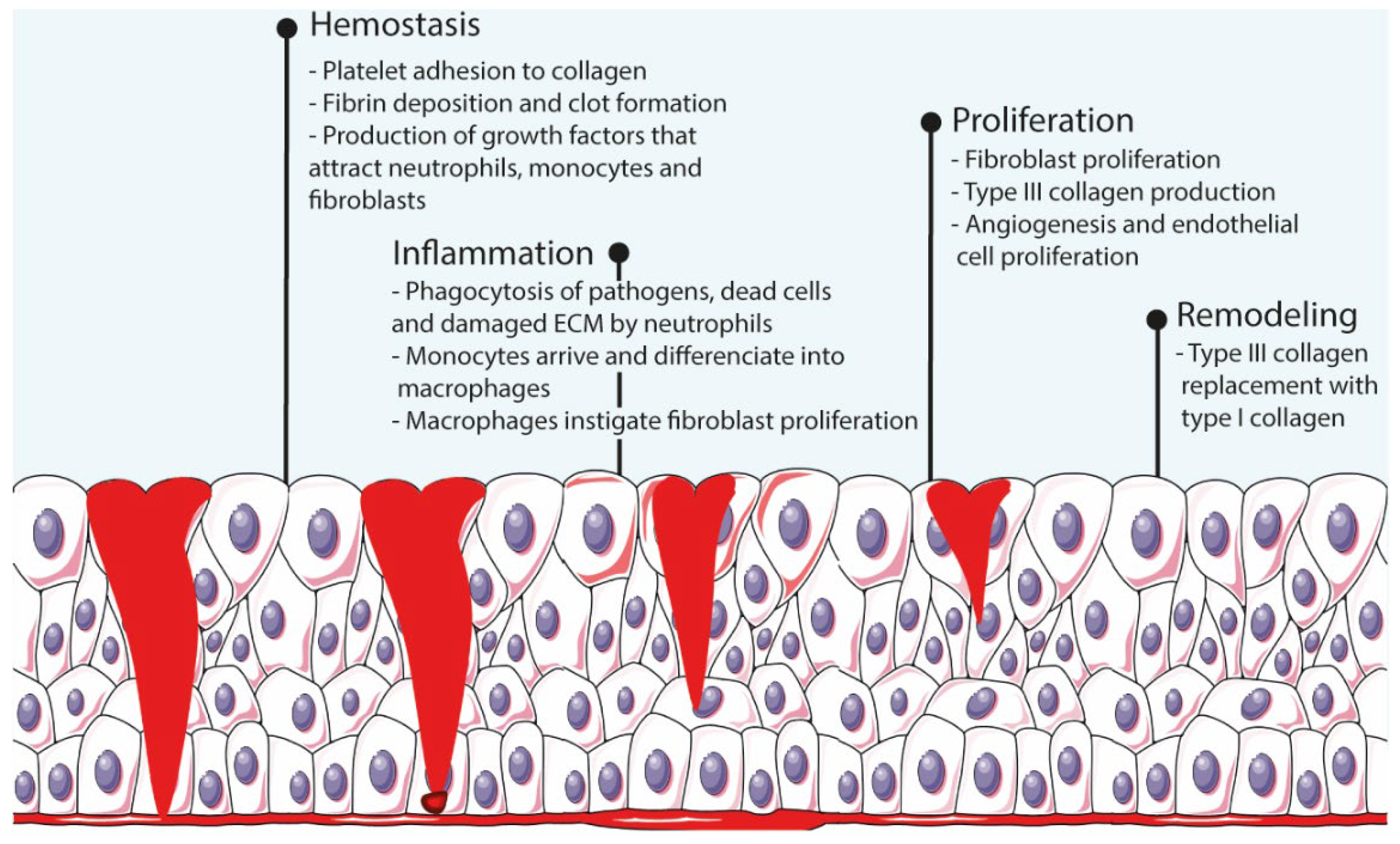
:1. Introduction—How Wound Care Is Still Relevant Nowadays
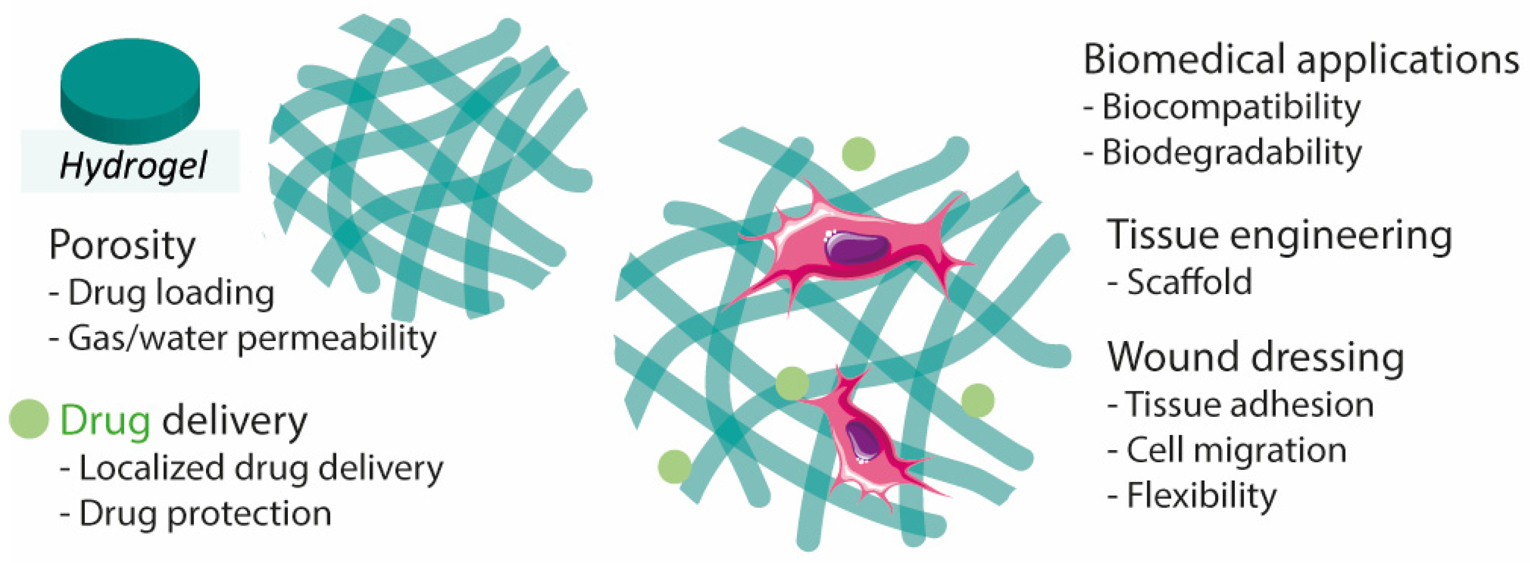
2. Hydrogels
3. Nitric Oxide and Its Donors
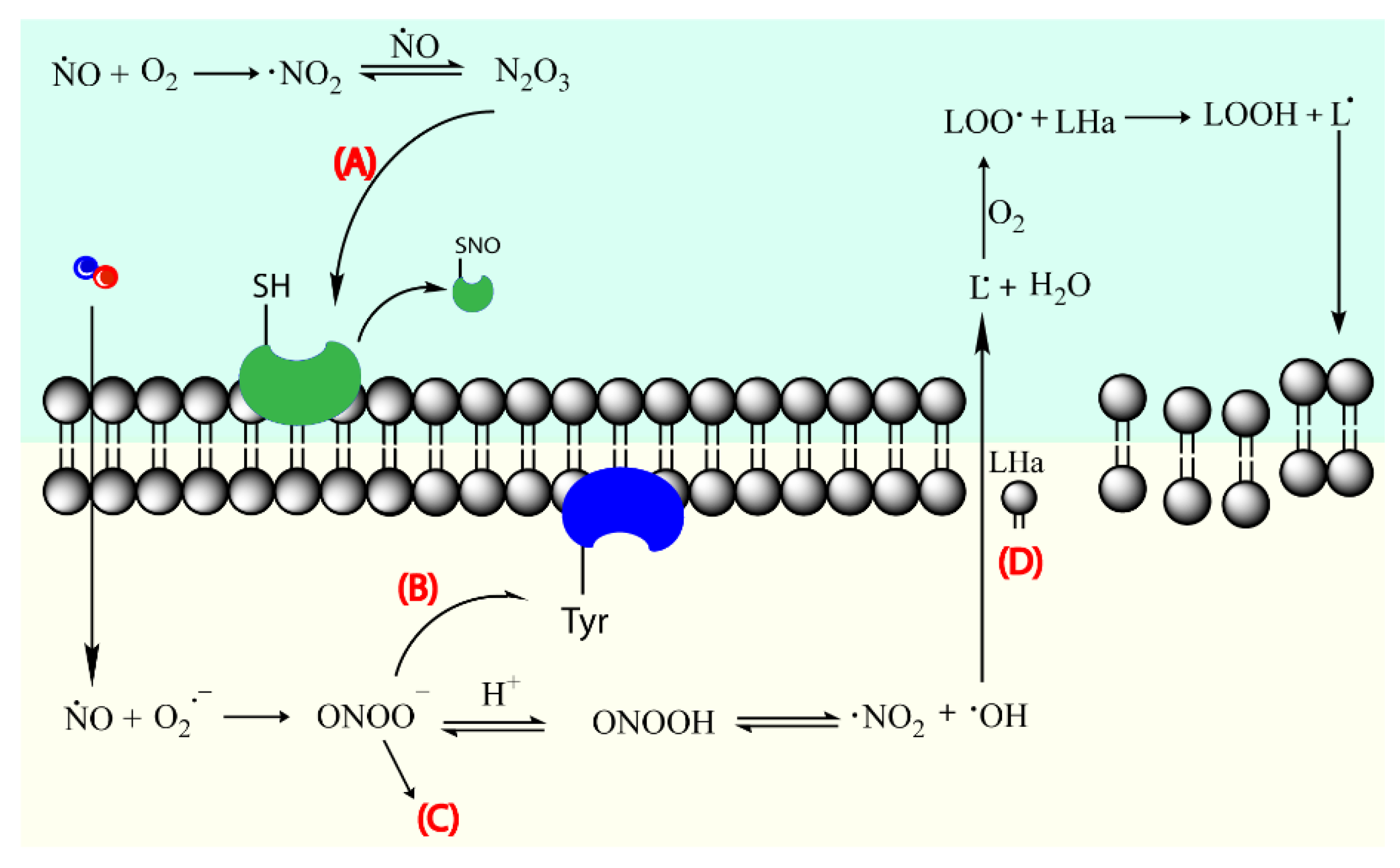
3.1. The Tiniest Antimicrobial Agent
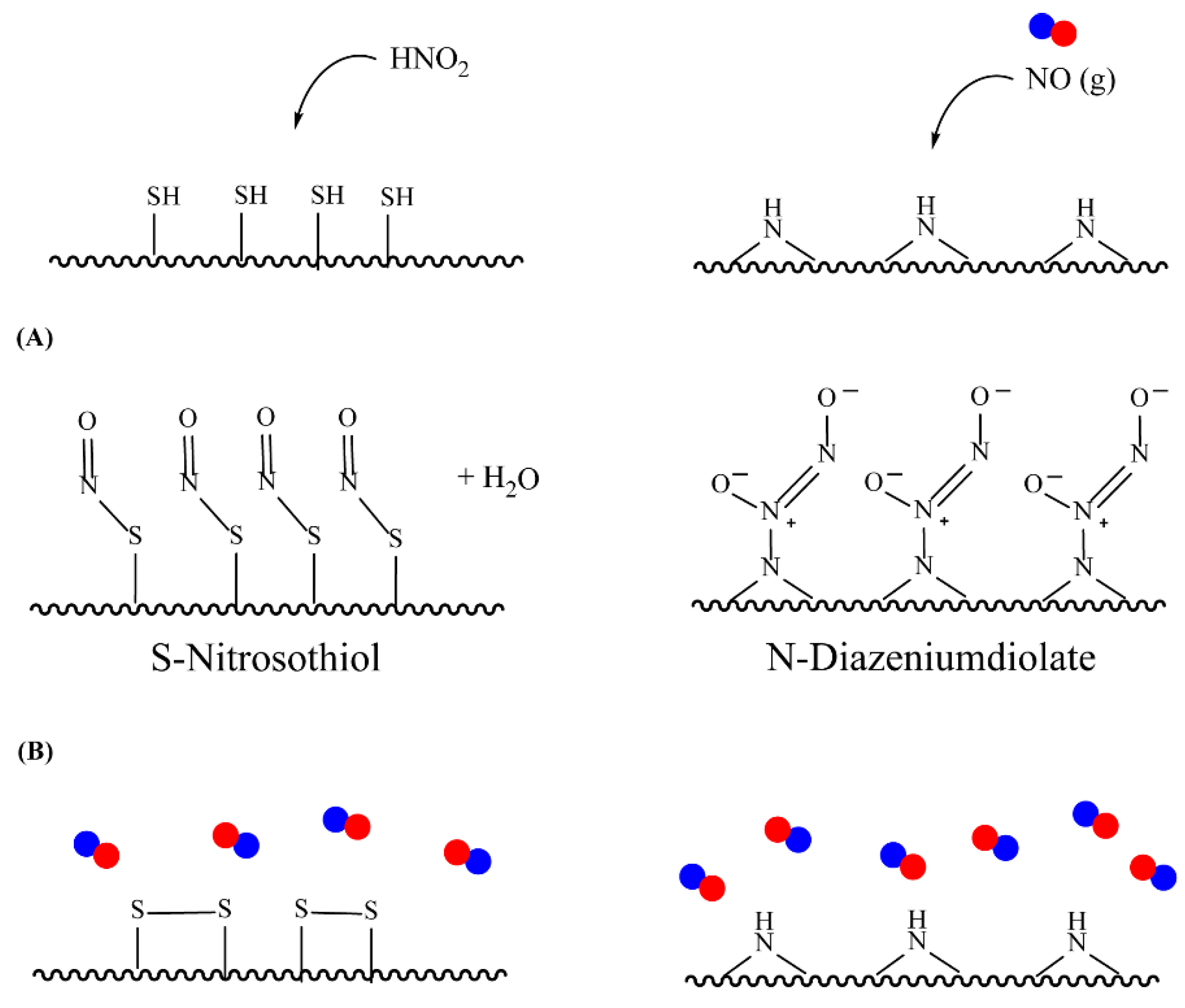
3.2. Nitric Oxide Donors
4. NO-Releasing Hydrogel-Based Systems
4.1. Physically Adsorbed NO Donors
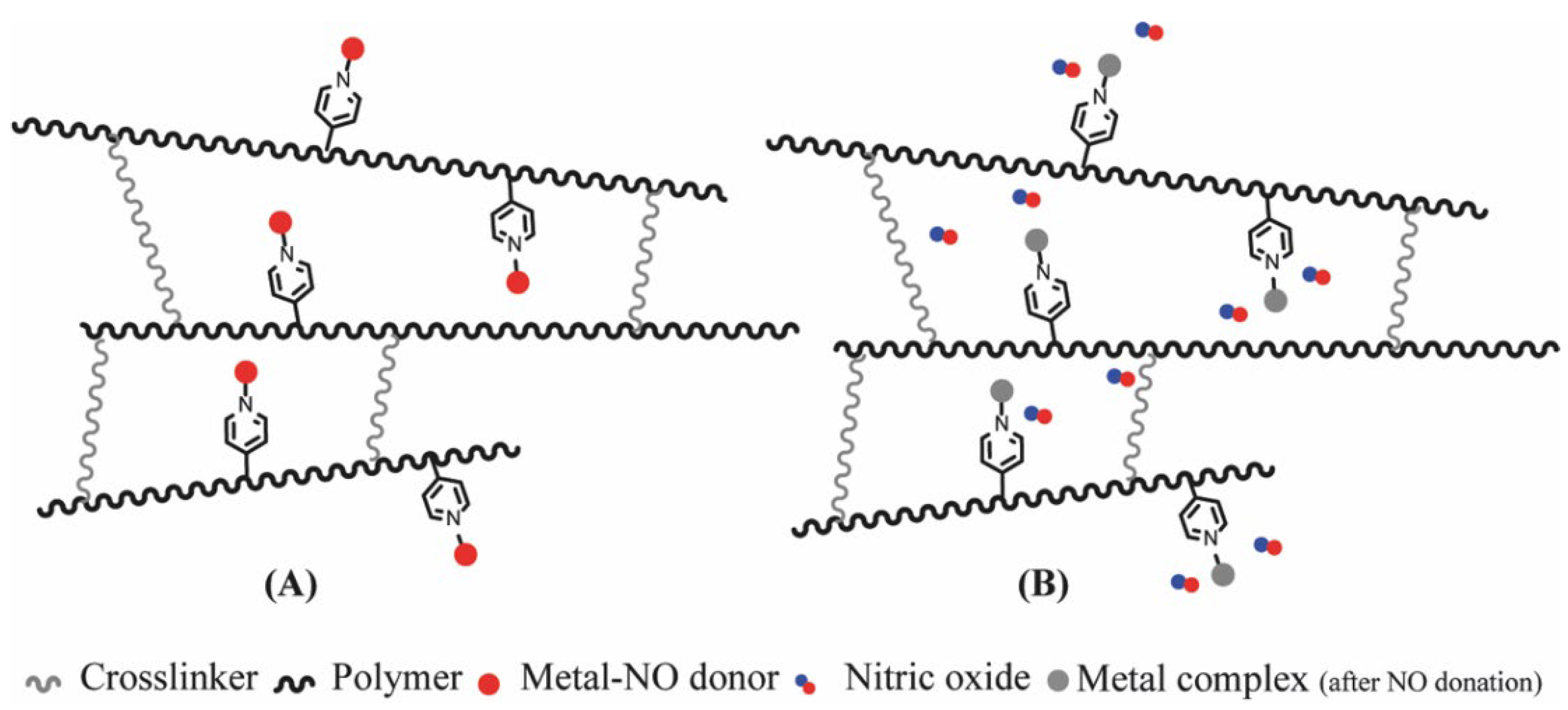
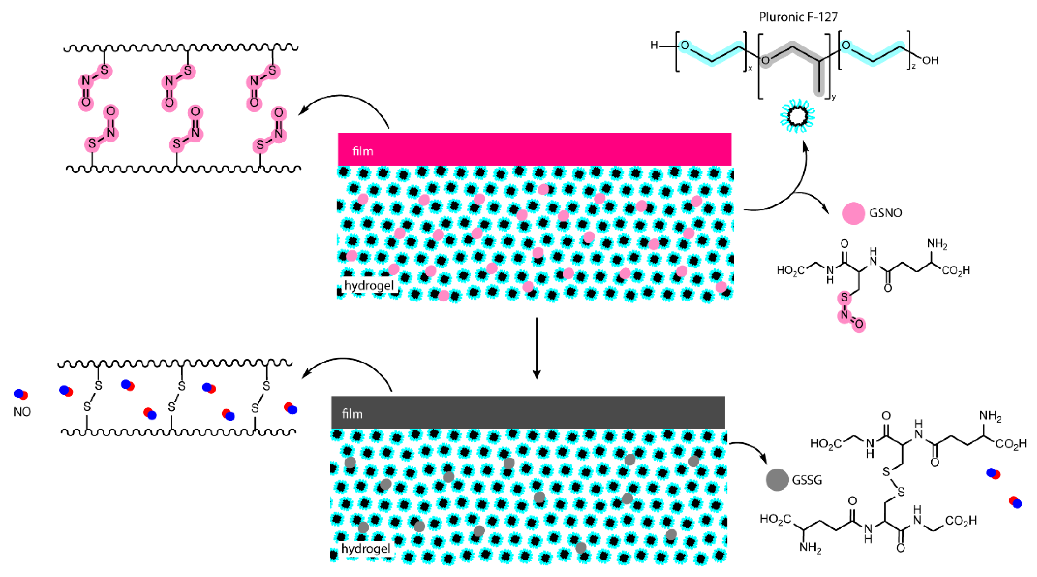
4.2. Chemically Attached NO Donors

| Hydrogel | NO Donor | NO Release Features | Reference |
|---|---|---|---|
| Poly(vinyl alcohol) | N-Diazeniumdiolate | ~48 h | [123] |
| Poly(vinyl alcohol) | RSNO | Photochemical release | [39] |
| Pluronic F-127 | RSNO | [124] | |
| Pluronic F-127 and branched PEI | N-Diazeniumdiolate | Burst release in first hours, sustained up to 50 h | [77,125] |
| Chitosan | N-Diazeniumdiolate | Enzymatic deprotection by glycosidase | [126,127] |
| NapFFGEE peptide | N-Diazeniumdiolate | Enzymatic deprotection by glutathione/ glutathione S-transferase | [96] |
| Naphthalene-terminated FFGGG peptide | N-Diazeniumdiolate | Enzymatic deprotection | [94] |
| Fmoc-Pexiganan and Pexiganan | N-Diazeniumdiolate | ~400 h | [128] |
| Gelatin | SNAP | Burst release in first 2 h, sustained up to 72 h | [95] |
| Chitosan and hyaluronic acid | SNAC | Burst release in first 2 h, sustained up to 48 h | [41] |
| Chitosan | N-Diazeniumdiolate | ~48 h Enzymatic deprotection | [65] |
| Fibrin | SNAP | Light exposure | [92] |
| Laponite-poly(pentaethylenehexamine) composite | N-diazeniumdiolate | Burst release | [129] |
| Alginate modified with DETA | N-Diazeniumdiolate | ~4 days | [80] |
| PEG | S-nitrocysteine | ~24 h | [130] |
| Poly(caprolactone)/Poly(sulfhydrylated polyester) | RSNO | [64] | |
| Nap-FFKEGG | N-Diazeniumdiolate | No burst release | [131] |
| Alginate and branched PEI | N-Diazeniumdiolate | Addition of Cu (II) increases NO release rate | [27] |
| Chitosan, PEG, and glucose | Nitrite SNAC | [132] | |
| Poly(ε-lysine) | N-Diazeniumdiolate | ~15 h | [76] |
| Poly(2-hydroxyethyl methacrylate) | Ruthenium nitrosyl | Photochemical release | [122] |
4.3. Antibacterial Activity and Wound Healing of NO-Releasing Hydrogels
5. Conclusions
6. Future Perspective
Funding
Conflicts of Interest
References
- Ambekar, R.S.; Kandasubramanian, B. Advancements in nanofibers for wound dressing: A review. Eur. Polym. J. 2019, 117, 304–336. [Google Scholar] [CrossRef]
- Wang, W.; Lu, K.J.; Yu, C.H.; Huang, Q.L.; Du, Y.Z. Nano-drug delivery systems in wound treatment and skin regeneration. J. Nanobiotechnol. 2019, 17, 82. [Google Scholar] [CrossRef] [PubMed]
- Xiang, J.; Zhu, R.; Lang, S.; Yan, H.; Liu, G.; Peng, B. Mussel-inspired immobilization of zwitterionic silver nanoparticles toward antibacterial cotton gauze for promoting wound healing. Chem. Eng. J. 2021, 409, 128291. [Google Scholar] [CrossRef]
- Han, G.; Ceilley, R. Chronic Wound Healing: A Review of Current Management and Treatments. Adv. Ther. 2017, 34, 599–610. [Google Scholar] [CrossRef] [PubMed] [Green Version]
- Demidova-Rice, T.N.; Hamblin, M.R.; Herman, I.M. Acute and impaired wound healing: Pathophysiology and current methods for drug delivery, part 1: Normal and chronic wounds: Biology, causes, and approaches to care. Adv. Skin Wound Care 2012, 25, 304–314. [Google Scholar] [CrossRef] [Green Version]
- El-Aassar, M.R.; Ibrahim, O.M.; Fouda, M.M.G.; Fakhry, H.; Ajarem, J.; Maodaa, S.N.; Allam, A.A.; Hafez, E.E. Wound dressing of chitosan-based-crosslinked gelatin/ polyvinyl pyrrolidone embedded silver nanoparticles, for targeting multidrug resistance microbes. Carbohydr. Polym. 2021, 255, 117484. [Google Scholar] [CrossRef]
- Saghazadeh, S.; Rinoldi, C.; Schot, M.; Kashaf, S.S.; Sharifi, F.; Jalilian, E.; Nuutila, K.; Giatsidis, G.; Mostafalu, P.; Derakhshandeh, H.; et al. Drug delivery systems and materials for wound healing applications. Adv. Drug Deliv. Rev. 2018, 127, 138–166. [Google Scholar] [CrossRef]
- Suetens, C.; Latour, K.; Kärki, T.; Ricchizzi, E.; Kinross, P.; Moro, M.L.; Jans, B.; Hopkins, S.; Hansen, S.; Lyytikäinen, O.; et al. Prevalence of healthcare-associated infections, estimated incidence and composite antimicrobial resistance index in acute care hospitals and long-term care facilities: Results from two European point prevalence surveys, 2016 to 2017. Eurosurveillance 2018, 23, 1800516. [Google Scholar] [CrossRef] [Green Version]
- Monnet, D.L.; Harbarth, S. Will coronavirus disease (COVID-19) have an impact on antimicrobial resistance? Eurosurveillance 2020, 25, 2001886. [Google Scholar] [CrossRef]
- Cheng, W.; Wang, M.; Chen, M.; Niu, W.; Li, Y.; Wang, Y.; Luo, M.; Xie, C.; Leng, T.; Lei, B. Injectable antibacterial antiinflammatory molecular hybrid hydrogel dressing for rapid MDRB-infected wound repair and therapy. Chem. Eng. J. 2021, 409, 128140. [Google Scholar] [CrossRef]
- Ketabchi, N.; Dinarvand, R.; Adabi, M.; Gholami, M.; Firoozi, S.; Amanzadi, B.; Faridi-Majidi, R. Study of Third-Degree Burn Wounds Debridement and Treatment by Actinidin Enzyme Immobilized on Electrospun Chitosan/PEO Nanofibers in Rats. Biointerface Res. Appl. Chem. 2021, 11, 10358–10370. [Google Scholar] [CrossRef]
- Matsliah, L.; Goder, D.; Giladi, S.; Zilberman, M. In vitro characterization of novel multidrug-eluting soy protein wound dressings. J. Biomater. Appl. 2021, 35, 978–993. [Google Scholar] [CrossRef]
- Karakaya, P.S.; Oktay, A.; Seventekin, N.; Yesil-Celiktas, O. Design of a new generation wound dressing with pine bark extract. J. Ind. Text. 2021, 50, 1193–1204. [Google Scholar] [CrossRef]
- Khan, T.A.; Peh, K.K.; Ch’ng, H.S. Mechanical, bioadhesive strength and biological evaluations of chitosan films for wound dressing. J. Pharm. Pharm. Sci. 2000, 3, 303–311. [Google Scholar]
- Hajikhani, M.; Emam-Djomeh, Z.; Askari, G. Fabrication and characterization of mucoadhesive bioplastic patch via coaxial polylactic acid (PLA) based electrospun nanofibers with antimicrobial and wound healing application. Int. J. Biol. Macromol. 2021, 172, 143–153. [Google Scholar] [CrossRef]
- Eriksson, E.; Liu, P.Y.; Schultz, G.S.; Martins-Green, M.M.; Tanaka, R.; Weir, D.; Gould, L.J.; Armstrong, D.G.; Gibbons, G.W.; Wolcott, R.; et al. Chronic wounds: Treatment consensus. Wound Repair Regen. 2022, 30, 156–171. [Google Scholar] [CrossRef]
- Pan, H.; Fan, D.; Cao, W.; Zhu, C.; Duan, Z.; Fu, R.; Li, X.; Ma, X. Preparation and Characterization of Breathable Hemostatic Hydrogel Dressings and Determination of Their Effects on Full-Thickness Defects. Polymers 2017, 9, 727. [Google Scholar] [CrossRef] [Green Version]
- Park, J.W.; Hwang, S.R.; Yoon, I.-S. Advanced Growth Factor Delivery Systems in Wound Management and Skin Regeneration. Molecules 2017, 22, 1259. [Google Scholar] [CrossRef] [Green Version]
- Jimi, S.; Jaguparov, A.; Nurkesh, A.; Sultankulov, B.; Saparov, A. Sequential Delivery of Cryogel Released Growth Factors and Cytokines Accelerates Wound Healing and Improves Tissue Regeneration. Front. Bioeng. Biotechnol. 2020, 8, 345. [Google Scholar] [CrossRef]
- Moeini, A.; Pedram, P.; Makvandi, P.; Malinconico, M.; Gomez d’Ayala, G. Wound healing and antimicrobial effect of active secondary metabolites in chitosan-based wound dressings: A review. Carbohydr. Polym. 2020, 233, 115839. [Google Scholar] [CrossRef]
- Qu, J.; Zhao, X.; Liang, Y.; Zhang, T.; Ma, P.X.; Guo, B. Antibacterial adhesive injectable hydrogels with rapid self-healing, extensibility and compressibility as wound dressing for joints skin wound healing. Biomaterials 2018, 183, 185–199. [Google Scholar] [CrossRef] [PubMed]
- Montaser, A.S.; Rehan, M.; El-Senousy, W.M.; Zaghloul, S. Designing strategy for coating cotton gauze fabrics and its application in wound healing. Carbohydr. Polym. 2020, 244, 116479. [Google Scholar] [CrossRef] [PubMed]
- Mirmajidi, T.; Chogan, F.; Rezayan, A.H.; Sharifi, A.M. In vitro and in vivo evaluation of a nanofiber wound dressing loaded with melatonin. Int. J. Pharm. 2021, 596, 120213. [Google Scholar] [CrossRef] [PubMed]
- Anjum, S.; Arora, A.; Alam, M.S.; Gupta, B. Development of antimicrobial and scar preventive chitosan hydrogel wound dressings. Int. J. Pharm. 2016, 508, 92–101. [Google Scholar] [CrossRef] [PubMed]
- Zhang, M.; Yang, M.; Woo, M.W.; Li, Y.; Han, W.; Dang, X. High-mechanical strength carboxymethyl chitosan-based hydrogel film for antibacterial wound dressing. Carbohydr. Polym. 2021, 256, 117590. [Google Scholar] [CrossRef]
- Porto, I.C.C.M. Polymer Biocompatibility. In Polymerization; Gomes, A.S., Ed.; IntechOpen: London, UK, 2012. [Google Scholar] [CrossRef] [Green Version]
- Jeong, H.; Kim, T.; Earmme, T.; Hong, J. Acceleration of Nitric Oxide Release in Multilayer Nanofilms through Cu(II) Ion Intercalation for Antibacterial Applications. Biomacromolecules 2021, 22, 1312–1322. [Google Scholar] [CrossRef]
- Contardi, M.; Kossyvaki, D.; Picone, P.; Summa, M.; Guo, X.; Heredia-Guerrero, J.A.; Giacomazza, D.; Carzino, R.; Goldoni, L.; Scoponi, G.; et al. Electrospun polyvinylpyrrolidone (PVP) hydrogels containing hydroxycinnamic acid derivatives as potential wound dressings. Chem. Eng. J. 2021, 409, 128144. [Google Scholar] [CrossRef]
- Champeau, M.; Seabra, A.B.; de Oliveira, M.G. Hydrogels for Topical Nitric Oxide Delivery. In Nitric Oxide Donors: Novel Biomedical Applications and Perspectives; Seabra, A.B., Ed.; Elsevier: London, UK, 2017; pp. 313–330. [Google Scholar] [CrossRef]
- Pal, A.; Bajpai, J.; Bajpai, A.K. Poly (acrylic acid) grafted gelatin nanocarriers as swelling controlled drug delivery system for optimized release of paclitaxel from modified gelatin. J. Drug Deliv. Sci. Technol. 2018, 45, 323–333. [Google Scholar] [CrossRef]
- Gupta, A.; Briffa, S.M.; Swingler, S.; Gibson, H.; Kannappan, V.; Adamus, G.; Kowalczuk, M.; Martin, C.; Radecka, I. Synthesis of Silver Nanoparticles Using Curcumin-Cyclodextrins Loaded into Bacterial Cellulose-Based Hydrogels for Wound Dressing Applications. Biomacromolecules 2020, 21, 1802–1811. [Google Scholar] [CrossRef]
- Ling, Z.; Chen, Z.; Deng, J.; Wang, Y.; Yuan, B.; Yang, X.; Lin, H.; Cao, J.; Zhu, X.; Zhang, X. A novel self-healing polydopamine-functionalized chitosan-arginine hydrogel with enhanced angiogenic and antibacterial activities for accelerating skin wound healing. Chem. Eng. J. 2021, 420, 130302. [Google Scholar] [CrossRef]
- Hoang Thi, T.T.; Pilkington, E.H.; Nguyen, D.H.; Lee, J.S.; Park, K.D.; Truong, N.P. The Importance of Poly(ethylene glycol) Alternatives for Overcoming PEG Immunogenicity in Drug Delivery and Bioconjugation. Polymers 2020, 12, 298. [Google Scholar] [CrossRef] [Green Version]
- Pelegrino, M.T.; Lima, B.D.; do Nascimento, M.H.M.; Lombello, C.B.; Brocchi, M.; Seabra, A.B. Biocompatible and Antibacterial Nitric Oxide-Releasing Pluronic F-127/Chitosan Hydrogel for Topical Applications. Polymers 2018, 10, 452. [Google Scholar] [CrossRef] [Green Version]
- Wei, Q.Y.; Xu, Y.M.; Lau, A.T.Y. Recent progress of nanocarrier-based therapy for solid malignancies. Cancers 2020, 12, 2783. [Google Scholar] [CrossRef]
- Zheng, Z.; Bian, S.; Li, Z.; Zhang, Z.; Liu, Y.; Zhai, X.; Pan, H.; Zhao, X. Catechol modified quaternized chitosan enhanced wet adhesive and antibacterial properties of injectable thermo-sensitive hydrogel for wound healing. Carbohydr. Polym. 2020, 249, 116826. [Google Scholar] [CrossRef]
- Drury, J.L.; Mooney, D.J. Hydrogels for tissue engineering: Scaffold design variables and applications. Biomaterials 2003, 24, 4337–4351. [Google Scholar] [CrossRef]
- Han, W.; Zhou, B.; Yang, K.; Xiong, X.; Luan, S.; Wang, Y.; Xu, Z.; Lei, P.; Luo, Z.; Gao, J.; et al. Biofilm-inspired adhesive and antibacterial hydrogel with tough tissue integration performance for sealing hemostasis and wound healing. Bioact. Mater. 2020, 5, 768–778. [Google Scholar] [CrossRef]
- Lourenco, S.D.; de Oliveira, M.G. Topical photochemical nitric oxide release from porous poly(vinyl alcohol) membrane for visible light modulation of dermal vasodilation. J. Photochem. Photobiol. A Chem. 2017, 346, 548–558. [Google Scholar] [CrossRef]
- Lee, J.; Hlaing, S.P.; Cao, J.F.; Hasan, N.; Ahn, H.J.; Song, K.W.; Yoo, J.W. In Situ Hydrogel-Forming/Nitric Oxide-Releasing Wound Dressing for Enhanced Antibacterial Activity and Healing in Mice with Infected Wounds. Pharmaceutics 2019, 11, 496. [Google Scholar] [CrossRef] [Green Version]
- Yang, Y.; Zhou, Y.T.; Li, Y.L.; Guo, L.Y.; Zhou, J.; Chen, J.H. Injectable and self-healing hydrogel containing nitric oxide donor for enhanced antibacterial activity. React. Funct. Polym. 2021, 166, 10. [Google Scholar] [CrossRef]
- Liu, H.L.; Zhu, X.L.; Guo, H.M.; Huang, H.L.; Huang, S.H.; Huang, S.S.; Xue, W.; Zhu, P.; Guo, R. Nitric oxide released injectable hydrogel combined with synergistic photothermal therapy for antibacterial and accelerated wound healing. Appl. Mater. Today 2020, 20, 12. [Google Scholar] [CrossRef]
- Joseph, C.A.; McCarthy, C.W.; Tyo, A.G.; Hubbard, K.R.; Fisher, H.C.; Altscheffel, J.A.; He, W.L.; Pinnaratip, R.; Liu, Y.; Lee, B.P.; et al. Development of an Injectable Nitric Oxide Releasing Poly(ethylene) Glycol-Fibrin Adhesive Hydrogel. ACS Biomater. Sci. Eng. 2019, 5, 959–969. [Google Scholar] [CrossRef]
- Friedman, A.J.; Han, G.; Navati, M.S.; Chacko, M.; Gunther, L.; Alfieri, A.; Friedman, J.M. Sustained release nitric oxide releasing nanoparticles: Characterization of a novel delivery platform based on nitrite containing hydrogel/glass composites. Nitric Oxide 2008, 19, 12–20. [Google Scholar] [CrossRef]
- Miao, H.; Hao, W.; Liu, H.; Liu, Y.; Fu, X.; Huang, H.; Ge, M.; Qian, Y. Highly Flexibility, Powder Self-Healing, and Recyclable Natural Polymer Hydrogels. Gels 2022, 8, 89. [Google Scholar] [CrossRef]
- Kim, J.O.; Noh, J.K.; Thapa, R.K.; Hasan, N.; Choi, M.; Kim, J.H.; Lee, J.H.; Ku, S.K.; Yoo, J.W. Nitric oxide-releasing chitosan film for enhanced antibacterial and in vivo wound-healing efficacy. Int. J. Biol. Macromol. 2015, 79, 217–225. [Google Scholar] [CrossRef]
- Wo, Y.; Brisbois, E.J.; Bartlett, R.H.; Meyerhoff, M.E. Recent advances in thromboresistant and antimicrobial polymers for biomedical applications: Just say yes to nitric oxide (NO). Biomater. Sci. 2016, 4, 1161–1183. [Google Scholar] [CrossRef] [Green Version]
- Fontana, K.; Mutus, B. Nitric Oxide-Donating Devices for Topical Applications. In Nitric Oxide Donors: Novel Biomedical Applications and Perspectives; Seabra, A.B., Ed.; Elsevier: London, UK, 2017; pp. 55–74. [Google Scholar] [CrossRef]
- Yang, C.; Jeong, S.; Ku, S.; Lee, K.; Park, M.H. Use of gasotransmitters for the controlled release of polymer-based nitric oxide carriers in medical applications. J. Control. Release 2018, 279, 157–170. [Google Scholar] [CrossRef]
- Rong, F.; Tang, Y.; Wang, T.; Feng, T.; Song, J.; Li, P.; Huang, W. Nitric oxide-releasing polymeric materials for antimicrobial applications: A review. Antioxidants 2019, 8, 556. [Google Scholar] [CrossRef] [Green Version]
- Malone-Povolny, M.J.; Maloney, S.E.; Schoenfisch, M.H. Nitric Oxide Therapy for Diabetic Wound Healing. Adv. Healthc. Mater. 2019, 8, e1801210. [Google Scholar] [CrossRef]
- Zaja-Milatovic, S.; Gupta, R.C. Handbook of Toxicology of Chemical Warfare Agents, 2nd ed.; Academic Press: Cambridge, MA, USA, 2015. [Google Scholar] [CrossRef]
- Rouillard, K.R.; Novak, O.P.; Pistiolis, A.M.; Yang, L.; Ahonen, M.J.R.; McDonald, R.A.; Schoenfisch, M.H. Exogenous Nitric Oxide Improves Antibiotic Susceptibility in Resistant Bacteria. ACS Infect. Dis. 2021, 7, 23–33. [Google Scholar] [CrossRef]
- Hetrick, E.M.; Shin, J.H.; Stasko, N.A.; Johnson, C.B.; Wespe, D.A.; Holmuhamedov, E.; Schoenfisch, M.H. Bactericidal Efficacy of Nitric Oxide-Releasing Silica Nanoparticles. ACS Nano 2008, 2, 235–246. [Google Scholar] [CrossRef]
- Puca, V.; Marulli, R.Z.; Grande, R.; Vitale, I.; Niro, A.; Molinaro, G.; Prezioso, S.; Muraro, R.; Di Giovanni, P. Microbial Species Isolated from Infected Wounds and Antimicrobial Resistance Analysis: Data Emerging from a Three-Years Retrospective Study. Antibiotics 2021, 10, 1162. [Google Scholar] [CrossRef] [PubMed]
- Nguyen, T.-K.; Selvanayagam, R.; Ho, K.K.K.; Chen, R.; Kutty, S.K.; Rice, S.A.; Kumar, N.; Barraud, N.; Duong, H.T.T.; Boyer, C. Co-delivery of nitric oxide and antibiotic using polymeric nanoparticles. Chem. Sci. 2016, 7, 1016–1027. [Google Scholar] [CrossRef] [PubMed] [Green Version]
- Ravikumar, G.; Chakrapani, H. Synergistic activities of Nitric Oxide and Various Drugs. In Nitric Oxide Donors: Novel Biomedical Applications and Perspectives; Seabra, A.B., Ed.; Elseivier: London, UK, 2017; pp. 293–312. [Google Scholar] [CrossRef]
- Yang, L.; Feura, E.S.; Ahonen, M.J.R.; Schoenfisch, M.H. Nitric Oxide–Releasing Macromolecular Scaffolds for Antibacterial Applications. Adv. Healthc. Mater. 2018, 7, 1800155. [Google Scholar] [CrossRef] [PubMed]
- Maillard, J.-Y.; Kampf, G.; Cooper, R. Antimicrobial stewardship of antiseptics that are pertinent to wounds: The need for a united approach. JAC-Antimicrob. Resist. 2021, 3, dlab027. [Google Scholar] [CrossRef]
- Castro, J.; Lima, Â.; Sousa, L.G.V.; Rosca, A.S.; Muzny, C.A.; Cerca, N. Crystal Violet Staining Alone Is Not Adequate to Assess Synergism or Antagonism in Multi-Species Biofilms of Bacteria Associated With Bacterial Vaginosis. Front. Cell. Infect. Microbiol. 2022, 11, 1375. [Google Scholar] [CrossRef]
- Vazquez, N.M.; Mariani, F.; Torres, P.S.; Moreno, S.; Galván, E.M. Cell death and biomass reduction in biofilms of multidrug resistant extended spectrum β-lactamase-producing uropathogenic Escherichia coli isolates by 1,8-cineole. PLoS ONE 2020, 15, e0241978. [Google Scholar] [CrossRef]
- Junka, A.F.; Żywicka, A.; Szymczyk, P.; Dziadas, M.; Bartoszewicz, M.; Fijałkowski, K. A.D.A.M. test (Antibiofilm Dressing’s Activity Measurement)—Simple method for evaluating anti-biofilm activity of drug-saturated dressings against wound pathogens. J. Microbiol. Methods 2017, 143, 6–12. [Google Scholar] [CrossRef]
- Wan, X.Z.; Liu, S.; Xin, X.X.; Li, P.F.; Dou, J.; Han, X.; Kang, I.K.; Yuan, J.; Chi, B.; Shen, J. S-nitrosated keratin composite mats with NO release capacity for wound healing. Chem. Eng. J. 2020, 400, 10. [Google Scholar] [CrossRef]
- Baldim, V.; de Oliveira, M.G. Poly-epsilon-caprolactone/polysulfhydrylated polyester blend: A platform for topical and degradable nitric oxide-releasing materials. Eur. Polym. J. 2018, 109, 143–152. [Google Scholar] [CrossRef]
- Nie, Y.; Zhang, K.; Zhang, S.Q.; Wang, D.; Han, Z.; Che, Y.; Kong, D.; Zhao, Q.; Han, Z.; He, Z.-X.; et al. Nitric oxide releasing hydrogel promotes endothelial differentiation of mouse embryonic stem cells. Acta Biomater. 2017, 63, 190–199. [Google Scholar] [CrossRef]
- Yang, S.; Zheng, X.; Qian, M.; Wang, H.; Wang, F.; Wei, Y.; Midgley, A.C.; He, J.; Tian, H.; Zhao, Q. Nitrate-Functionalized poly(ε-Caprolactone) Small-Diameter Vascular Grafts Enhance Vascular Regeneration via Sustained Release of Nitric Oxide. Front. Bioeng. Biotechnol. 2021, 9, 770121. [Google Scholar] [CrossRef]
- Napoli, C.; Paolisso, G.; Casamassimi, A.; Al-Omran, M.; Barbieri, M.; Sommese, L.; Infante, T.; Ignarro, L.J. Effects of Nitric Oxide on Cell Proliferation: Novel Insights. J. Am. Coll. Cardiol. 2013, 62, 89–95. [Google Scholar] [CrossRef]
- Wu, Y.; Liang, T.Z.; Hu, Y.; Jiang, S.A.; Luo, Y.S.; Liu, C.; Wang, G.; Zhang, J.; Xu, T.; Zhu, L. 3D bioprinting of integral ADSCs-NO hydrogel scaffolds to promote severe burn wound healing. Regen. Biomater. 2021, 8, rbab014. [Google Scholar] [CrossRef]
- Ramadass, S.K.; Nazir, L.S.; Thangam, R.; Perumal, R.K.; Manjubala, I.; Madhan, B.; Seetharaman, S. Type I collagen peptides and nitric oxide releasing electrospun silk fibroin scaffold: A multifunctional approach for the treatment of ischemic chronic wounds. Colloids Surf. B Biointerfaces 2019, 175, 636–643. [Google Scholar] [CrossRef]
- Frost, M.C. Improving the Performance of Implantable Sensors with Nitric Oxide Release. In Nitric Oxide Donors: Novel Biomedical Applications and Perspectives; Seabra, A.B., Ed.; Elseivier: London, UK, 2017; pp. 191–220. [Google Scholar] [CrossRef]
- Zhang, C.; Biggs, T.D.; Devarie-Baez, N.O.; Shuang, S.; Dong, C.; Xian, M. S-Nitrosothiols: Chemistry and reactions. Chem. Commun. 2017, 53, 11266–11277. [Google Scholar] [CrossRef]
- Marazzi, M.; López-Delgado, A.; Fernández-González, M.A.; Castaño, O.; Frutos, L.M.; Temprado, M. Modulating nitric oxide release by S-nitrosothiol photocleavage: Mechanism and substituent effects. J. Phys. Chem. A 2012, 116, 7039–7049. [Google Scholar] [CrossRef]
- Singh, R.J.; Hogg, N.; Joseph, J.; Kalyanaraman, B. Mechanism of nitric oxide release from S-nitrosothiols. J. Biol. Chem. 1996, 271, 18596–18603. [Google Scholar] [CrossRef] [Green Version]
- de Oliveira, M.G. S-Nitrosothiols as Platforms for Topical Nitric Oxide Delivery. Basic Clin. Pharmacol. Toxicol. 2016, 119, 49–56. [Google Scholar] [CrossRef] [Green Version]
- Gur, S.; Chen, A.L.; Kadowitz, P.J. Nitric Oxide Donors and Penile Erectile Function. In Nitric Oxide Donors: Novel Biomedical Applications and Perspectives; Seabra, A.B., Ed.; Elseivier: London, UK, 2017; pp. 121–140. [Google Scholar] [CrossRef]
- Aveyard, J.; Deller, R.C.; Lace, R.; Williams, R.L.; Kaye, S.B.; Kolegraff, K.N.; Curran, J.M.; D’Sa, R.A. Antimicrobial Nitric Oxide Releasing Contact Lens Gels for the Treatment of Microbial Keratitis. ACS Appl. Mater. Interfaces 2019, 11, 37491–37501. [Google Scholar] [CrossRef]
- Kim, J.; Lee, Y.; Singha, K.; Kim, H.W.; Shin, J.H.; Jo, S.; Han, D.K.; Kim, W.J. NONOates-Polyethylenimine Hydrogel for Controlled Nitric Oxide Release and Cell Proliferation Modulation. Bioconjugate Chem. 2011, 22, 1031–1038. [Google Scholar] [CrossRef]
- Kashfi, K.; Duvalsaint, P.L. Nitric Oxide Donors and Therapeutic Applications in Cancer. In Nitric Oxide Donors: Novel Biomedical Applications and Perspectives; Seabra, A.B., Ed.; Elseivier: London, UK, 2017; pp. 75–120. [Google Scholar] [CrossRef]
- Fu, J.; Han, J.; Meng, T.; Hu, J.; Yin, J. Novel α-ketoamide based diazeniumdiolates as hydrogen peroxide responsive nitric oxide donors with anti-lung cancer activity. Chem. Commun. 2019, 55, 12904–12907. [Google Scholar] [CrossRef]
- Hasan, N.; Lee, J.; Kwak, D.; Kim, H.; Saparbayeva, A.; Ahn, H.J.; Yoon, I.S.; Kim, M.S.; Jung, Y.; Yoo, J.W. Diethylenetriamine/NONOate-doped alginate hydrogel with sustained nitric oxide release and minimal toxicity to accelerate healing of MRSA-infected wounds. Carbohydr. Polym. 2021, 270, 118387. [Google Scholar] [CrossRef]
- Keefer, L.K. Nitric oxide (NO)- and nitroxyl (HNO)-generating diazeniumdiolates (NONOates): Emerging commercial opportunities. Curr. Top. Med. Chem. 2005, 5, 625–636. [Google Scholar] [CrossRef]
- Halpenny, G.M.; Steinhardt, R.C.; Okialda, K.A.; Mascharak, P.K. Characterization of pHEMA-based hydrogels that exhibit light-induced bactericidal effect via release of NO. J. Mater. Sci.-Mater. Med. 2009, 20, 2353–2360. [Google Scholar] [CrossRef] [Green Version]
- Dave, R.N.; Joshi, H.M.; Venugopalan, V.P. Biomedical evaluation of a novel nitrogen oxides releasing wound dressing. J. Mater. Sci.-Mater. Med. 2012, 23, 3097–3106. [Google Scholar] [CrossRef]
- Shishido, S.M.; Seabra, A.B.; Loh, W.; De Oliveira, M.G. Thermal and photochemical nitric oxide release from S-nitrosothiols incorporated in Pluronic F127 gel: Potential uses for local and controlled nitric oxide release. Biomaterials 2003, 24, 3543–3553. [Google Scholar] [CrossRef]
- Pelegrino, M.T.; de Araújo, D.R.; Seabra, A.B. S-nitrosoglutathione-containing chitosan nanoparticles dispersed in Pluronic F-127 hydrogel: Potential uses in topical applications. J. Drug Deliv. Sci. Technol. 2018, 43, 211–220. [Google Scholar] [CrossRef]
- Wo, Y.; Li, Z.; Colletta, A.; Wu, J.; Xi, C.; Matzger, A.J.; Brisbois, E.J.; Bartlett, R.H.; Meyerhoff, M.E. Study of crystal formation and nitric oxide (NO) release mechanism from S-nitroso-N-acetylpenicillamine (SNAP)-doped CarboSil polymer composites for potential antimicrobial applications. Compos. Part B Eng. 2017, 121, 23–33. [Google Scholar] [CrossRef]
- Namivandi-Zangeneh, R.; Sadrearhami, Z.; Bagheri, A.; Sauvage-Nguyen, M.; Ho, K.K.K.; Kumar, N.; Wong, E.H.H.; Boyer, C. Nitric Oxide-Loaded Antimicrobial Polymer for the Synergistic Eradication of Bacterial Biofilm. ACS Macro Lett. 2018, 7, 592–597. [Google Scholar] [CrossRef]
- Kamaruzzaman, N.F.; Tan, L.P.; Hamdan, R.H.; Choong, S.S.; Wong, W.K.; Gibson, A.J.; Chivu, A.; Pina, M.d.F. Antimicrobial Polymers: The Potential Replacement of Existing Antibiotics? Int. J. Mol. Sci. 2019, 20, 2747. [Google Scholar] [CrossRef] [Green Version]
- Nguyen, T.-K.; Lam, S.J.; Ho, K.K.K.; Kumar, N.; Qiao, G.G.; Egan, S.; Boyer, C.; Wong, E.H.H. Rational Design of Single-Chain Polymeric Nanoparticles That Kill Planktonic and Biofilm Bacteria. ACS Infect. Dis. 2017, 3, 237–248. [Google Scholar] [CrossRef]
- Yu, Y.T.; Shi, S.W.; Wang, Y.; Zhang, Q.L.; Gao, S.H.; Yang, S.P.; Liu, J.G. A Ruthenium Nitrosyl-Functionalized Magnetic Nanoplatform with Near-Infrared Light-Controlled Nitric Oxide Delivery and Photothermal Effect for Enhanced Antitumor and Antibacterial Therapy. ACS Appl. Mater. Interfaces 2020, 12, 312–321. [Google Scholar] [CrossRef]
- Kandoth, N.; Mosinger, J.; Gref, R.; Sortino, S. A NO photoreleasing supramolecular hydrogel with bactericidal action. J. Mater. Chem. B 2013, 1, 3458–3463. [Google Scholar] [CrossRef]
- VanWagner, M.; Rhadigan, J.; Lancina, M.; Lebovsky, A.; Romanowicz, G.; Holmes, H.; Brunette, M.A.; Snyder, K.L.; Bostwick, M.; Lee, B.P.; et al. S-Nitroso-N-acetylpenicillamine (SNAP) Derivatization of Peptide Primary Amines to Create Inducible Nitric Oxide Donor Biomaterials. ACS Appl. Mater. Interfaces 2013, 5, 8430–8439. [Google Scholar] [CrossRef]
- Mohamed, N.A.; Ahmetaj-Shala, B.; Duluc, L.; Mackenzie, L.S.; Kirkby, N.S.; Reed, D.M.; Lickiss, P.D.; Davies, R.P.; Freeman, G.R.; Wojciak-Stothard, B.; et al. A New NO-Releasing Nanoformulation for the Treatment of Pulmonary Arterial Hypertension. J. Cardiovasc. Transl. Res. 2016, 9, 162–164. [Google Scholar] [CrossRef] [Green Version]
- Yao, X.P.; Liu, Y.; Gao, J.; Yang, L.; Mao, D.; Stefanitsch, C.; Li, Y.; Zhang, J.; Ou, L.L.; Kong, D.L.; et al. Nitric oxide releasing hydrogel enhances the therapeutic efficacy of mesenchymal stem cells for myocardial infarction. Biomaterials 2015, 60, 130–140. [Google Scholar] [CrossRef]
- Xing, Q.; Yates, K.; Bailey, A.; Vogt, C.; He, W.L.; Frost, M.C.; Zhao, F. Effects of local nitric oxide release on human mesenchymal stem cell attachment and proliferation on gelatin hydrogel surface. Surf. Innov. 2013, 1, 224–232. [Google Scholar] [CrossRef]
- Zhang, J.M.; Deng, M.G.; Shi, X.G.; Zhang, C.N.; Qu, X.W.; Hu, X.L.; Wang, W.W.; Kong, D.L.; Huang, P.S. Cascaded amplification of intracellular oxidative stress and reversion of multidrug resistance by nitric oxide prodrug based-supramolecular hydrogel for synergistic cancer chemotherapy. Bioact. Mater. 2021, 6, 3300–3313. [Google Scholar] [CrossRef]
- Kandoth, N.; Malanga, M.; Fraix, A.; Jicsinszky, L.; Fenyvesi, É.; Parisi, T.; Colao, I.; Sciortino, M.T.; Sortino, S. A host-guest supramolecular complex with photoregulated delivery of nitric oxide and fluorescence imaging capacity in cancer cells. Chem.-Asian J. 2012, 7, 2888–2894. [Google Scholar] [CrossRef]
- Sung, Y.-C.; Jin, P.-R.; Chu, L.-A.; Hsu, F.-F.; Wang, M.-R.; Chang, C.-C.; Chiou, S.-J.; Qiu, J.T.; Gao, D.-Y.; Lin, C.-C.; et al. Delivery of nitric oxide with a nanocarrier promotes tumour vessel normalization and potentiates anti-cancer therapies. Nat. Nanotechnol. 2019, 14, 1160–1169. [Google Scholar] [CrossRef]
- Mintz, J.; Vedenko, A.; Rosete, O.; Shah, K.; Goldstein, G.; Hare, J.M.; Ramasamy, R.; Arora, H. Current Advances of Nitric Oxide in Cancer and Anticancer Therapeutics. Vaccines 2021, 9, 94. [Google Scholar] [CrossRef]
- Holmes, A.J. The Role of L-Ascorbic Acid in S-Nitrosothiol Decomposition and Aspects of the Nitrosation of Thiones. Ph.D. Thesis, Durham University, Durham, UK, 2000. [Google Scholar]
- Huang, S.S.; Liu, H.L.; Liao, K.D.; Hu, Q.Q.; Guo, R.; Deng, K.X. Functionalized GO Nanovehicles with Nitric Oxide Release and Photothermal Activity-Based Hydrogels for Bacteria-Infected Wound Healing. ACS Appl. Mater. Interfaces 2020, 12, 28952–28964. [Google Scholar] [CrossRef]
- Zahid, A.A.; Augustine, R.; Dalvi, Y.B.; Reshma, K.; Ahmed, R.; Rehman, S.R.U.; Marei, H.E.; Alfkey, R.; Hasan, A. Development of nitric oxide releasing visible light crosslinked gelatin methacrylate hydrogel for rapid closure of diabetic wounds. Biomed. Pharmacother. 2021, 140, 111747. [Google Scholar] [CrossRef]
- Champeau, M.; Povoa, V.; Militao, L.; Cabrini, F.M.; Picheth, G.F.; Meneau, F.; Jara, C.P.; de Araujo, E.P.; de Oliveira, M.G. Supramolecular poly(acrylic acid)/F127 hydrogel with hydration-controlled nitric oxide release for enhancing wound healing. Acta Biomater. 2018, 74, 312–325. [Google Scholar] [CrossRef]
- Vercelino, R.; Cunha, T.M.; Ferreira, E.S.; Cunha, F.Q.; Ferreira, S.H.; de Oliveira, M.G. Skin vasodilation and analgesic effect of a topical nitric oxide-releasing hydrogel. J. Mater. Sci.-Mater. Med. 2013, 24, 2157–2169. [Google Scholar] [CrossRef]
- Georgii, J.L.; Amadeu, T.P.; Seabra, A.B.; de Oliveira, M.G.; Monte-Alto-Costa, A. Topical S-nitrosoglutathione-releasing hydrogel improves healing of rat ischaemic wounds. J. Tissue Eng. Regen. Med. 2011, 5, 612–619. [Google Scholar] [CrossRef]
- Amadeu, T.P.; Seabra, A.B.; de Oliveira, M.G.; Costa, A.M.A. S-nitrosoglutathione-containing hydrogel accelerates rat cutaneous wound repair. J. Eur. Acad. Dermatol. Venereol. 2007, 21, 629–637. [Google Scholar] [CrossRef]
- Amadeu, T.P.; Seabra, A.B.; de Oliveira, M.G.; Monte-Alto-Costa, A. Nitric oxide donor improves healing if applied on inflammatory and proliferative phase. J. Surg. Res. 2008, 149, 84–93. [Google Scholar] [CrossRef]
- Seabra, A.B.; Fitzpatrick, A.; Paul, J.; De Oliveira, M.G.; Weller, R. Topically applied S-nitrosothiol-containing hydrogels as experimental and pharmacological nitric oxide donors in human skin. Br. J. Dermatol. 2004, 151, 977–983. [Google Scholar] [CrossRef]
- Seabra, A.B.; Pankotai, E.; Feher, M.; Somlai, A.; Kiss, L.; Biro, L.; Szabo, C.; Kollai, M.; de Oliveira, M.G.; Lacza, Z. S-nitrosoglutathione-containing hydrogel increases dermal blood flow in streptozotocin-induced diabetic rats. Br. J. Dermatol. 2007, 156, 814–818. [Google Scholar] [CrossRef]
- Cao, J.F.; Su, M.Z.; Hasan, N.; Lee, J.; Kwak, D.; Kim, D.Y.; Kim, K.; Lee, E.H.; Jung, J.H.; Yoo, J.W. Nitric Oxide-Releasing Thermoresponsive Pluronic F127/Alginate Hydrogel for Enhanced Antibacterial Activity and Accelerated Healing of Infected Wounds. Pharmaceutics 2020, 12, 926. [Google Scholar] [CrossRef]
- Parisi, C.; Seggio, M.; Fraix, A.; Sortino, S. A High-Performing Metal-Free Photoactivatable Nitric Oxide Donor with a Green Fluorescent Reporter. ChemPhotoChem 2020, 4, 742–748. [Google Scholar] [CrossRef]
- Urzedo, A.L.; Goncalves, M.C.; Nascimento, M.H.M.; Lombello, C.B.; Nakazato, G.; Seabra, A.B. Cytotoxicity and Antibacterial Activity of Alginate Hydrogel Containing Nitric Oxide Donor and Silver Nanoparticles for Topical Applications. ACS Biomater. Sci. Eng. 2020, 6, 2117–2134. [Google Scholar] [CrossRef] [PubMed]
- Hasan, S.; Thomas, N.; Thierry, B.; Prestidge, C.A. Controlled and Localized Nitric Oxide Precursor Delivery From Chitosan Gels to Staphylococcus aureus Biofilms. J. Pharm. Sci. 2017, 106, 3556–3563. [Google Scholar] [CrossRef]
- Zahid, A.A.; Ahmed, R.; Rehman, S.R.U.; Augustine, R.; Tariq, M.; Hasan, A. Nitric oxide releasing chitosan-poly (vinyl alcohol) hydrogel promotes angiogenesis in chick embryo model. Int. J. Biol. Macromol. 2019, 136, 901–910. [Google Scholar] [CrossRef]
- Najafi, H.; Abolmaali, S.S.; Heidari, R.; Valizadeh, H.; Jafari, M.; Tamaddon, A.M.; Azarpira, N. Nitric oxide releasing nanofibrous Fmoc-dipeptide hydrogels for amelioration of renal ischemia/reperfusion injury. J. Control. Release 2021, 337, 1–13. [Google Scholar] [CrossRef]
- Fraix, A.; Gref, R.; Sortino, S. A multi-photoresponsive supramolecular hydrogel with dual-color fluorescence and dual-modal photodynamic action. J. Mat. Chem. B 2014, 2, 3443–3449. [Google Scholar] [CrossRef]
- Fraix, A.; Kandoth, N.; Gref, R.; Sortino, S. A Multicomponent Gel for Nitric Oxide Photorelease with Fluorescence Reporting. Asian J. Org. Chem. 2015, 4, 256–261. [Google Scholar] [CrossRef]
- Gutierrez Cisneros, C.; Bloemen, V.; Mignon, A. Synthetic, Natural, and Semisynthetic Polymer Carriers for Controlled Nitric Oxide Release in Dermal Applications: A Review. Polymers 2021, 13, 760. [Google Scholar] [CrossRef]
- Chen, G.Q.; Li, J.L.; Song, M.C.; Wu, Z.Y.; Zhang, W.Z.; Wang, Z.Y.; Gao, J.; Yang, Z.M.; Ou, C.W. A Mixed Component Supramolecular Hydrogel to Improve Mice Cardiac Function and Alleviate Ventricular Remodeling after Acute Myocardial Infarction. Adv. Funct. Mater. 2017, 27, 1701798. [Google Scholar] [CrossRef]
- Bruschi, M.L. Mathematical models of drug release. In Strategies to Modify the Drug Release from Pharmaceutical Systems; Bruschi, M.L., Ed.; Woodhead Publishing: Sawston, UK, 2015; pp. 63–86. [Google Scholar] [CrossRef]
- Fu, K.; Wu, H.; Su, Z. Self-assembling peptide-based hydrogels: Fabrication, properties, and applications. Biotechnol. Adv. 2021, 49, 107752. [Google Scholar] [CrossRef] [PubMed]
- Halpenny, G.M.; Olmstead, M.M.; Mascharak, P.K. Incorporation of a designed ruthenium nitrosyl in PolyHEMA hydrogel and light-activated delivery of NO to myoglobin. Inorg. Chem. 2007, 46, 6601–6606. [Google Scholar] [CrossRef] [PubMed]
- Masters, K.S.B.; Leibovich, S.J.; Belem, P.; West, J.L.; Poole-Warren, L.A. Effects of nitric oxide releasing poly(vinyl alcohol) hydrogel dressings on dermal wound healing in diabetic mice. Wound Repair Regen. 2002, 10, 286–294. [Google Scholar] [CrossRef] [PubMed] [Green Version]
- Picheth, G.F.; da Silva, L.C.E.; Giglio, L.P.; Plivelic, T.S.; de Oliveira, M.G. S-nitrosothiol-terminated Pluronic F127: Influence of microstructure on nitric oxide release. J. Colloid Interface Sci. 2020, 576, 457–467. [Google Scholar] [CrossRef]
- Kang, Y.; Kim, J.; Lee, Y.M.; Im, S.; Park, H.; Kim, W.J. Nitric oxide-releasing polymer incorporated ointment for cutaneous wound healing. J. Control. Release 2015, 220, 624–630. [Google Scholar] [CrossRef]
- Zhang, K.Y.; Chen, X.N.; Li, H.F.; Feng, G.W.; Nie, Y.; Wei, Y.Z.; Li, N.N.; Han, Z.B.; Han, Z.C.; Kong, D.L.; et al. A nitric oxide-releasing hydrogel for enhancing the therapeutic effects of mesenchymal stem cell therapy for hindlimb ischemia. Acta Biomater. 2020, 113, 289–304. [Google Scholar] [CrossRef]
- Zhao, Q.; Zhang, J.M.; Song, L.J.; Ji, Q.; Yao, Y.; Cui, Y.; Shen, J.; Wang, P.G.; Kong, D.L. Polysaccharide-based biomaterials with on-demand nitric oxide releasing property regulated by enzyme catalysis. Biomaterials 2013, 34, 8450–8458. [Google Scholar] [CrossRef]
- Durao, J.; Vale, N.; Gomes, S.; Gomes, P.; Barrias, C.C.; Gales, L. Nitric Oxide Release from Antimicrobial Peptide Hydrogels for Wound Healing. Biomolecules 2019, 9, 4. [Google Scholar] [CrossRef] [Green Version]
- Park, K.; Dawson, J.I.; Oreffo, R.O.C.; Kim, Y.H.; Hong, J. Nanoclay-Polyamine Composite Hydrogel for Topical Delivery of Nitric Oxide Gas via Innate Gelation Characteristics of Laponite. Biomacromolecules 2020, 21, 2096–2103. [Google Scholar] [CrossRef]
- Masters, K.S.B.; Lipke, E.A.; Rice, E.E.H.; Liel, M.S.; Myler, H.A.; Zygourakis, C.; Tulis, D.A.; West, J.L. Nitric oxide-generating hydrogels inhibit neointima formation. J. Biomater. Sci.-Polym. Ed. 2005, 16, 659–672. [Google Scholar] [CrossRef] [Green Version]
- Deng, Y.; Chen, G.Q.; Ye, M.; He, Y.Y.; Li, Z.H.; Wang, X.B.; Ou, C.W.; Yang, Z.M.; Chen, M.S. Bifunctional Supramolecular Hydrogel Alleviates Myocardial Ischemia/Reperfusion Injury by Inhibiting Autophagy and Apoptosis. J. Biomed. Nanotechnol. 2018, 14, 1458–1470. [Google Scholar] [CrossRef]
- Nacharaju, P.; Tuckman-Vernon, C.; Maier, K.E.; Chouake, J.; Friedman, A.; Cabrales, P.; Friedman, J.M. A nanoparticle delivery vehicle for S-nitroso-N-acetyl cysteine: Sustained vascular response. Nitric Oxide-Biol. Chem. 2012, 27, 150–160. [Google Scholar] [CrossRef] [Green Version]
- Schanuel, F.S.; Santos, K.S.R.; Monte-Alto-Costa, A.; de Oliveira, M.G. Combined nitric oxide-releasing poly(vinyl alcohol) film/F127 hydrogel for accelerating wound healing. Colloid Surf. B-Biointerfaces 2015, 130, 182–191. [Google Scholar] [CrossRef]
- Breijyeh, Z.; Jubeh, B.; Karaman, R. Resistance of Gram-Negative Bacteria to Current Antibacterial Agents and Approaches to Resolve It. Molecules 2020, 25, 1340. [Google Scholar] [CrossRef] [Green Version]


| S-Nitrosothiol | Chemical Structure |
|---|---|
| GSNO S-nitrosogluthathione | 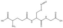 |
| SNAC S-nitroso-N-acetylcysteine | 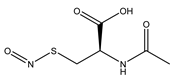 |
| SNAP S-nitroso-N-acetylpenicillamine | 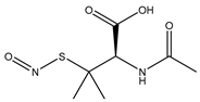 |
| SNMSA S-nitroso-mercaptosuccinic acid | 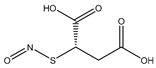 |
| N-Diazeniumdiolate | Chemical Structure | t1/2 |
|---|---|---|
| PROLI/NO 1-[-2-(-carboxylate)pyrrolidine-1-yl] NONOate |  | 2 s |
| MAHMA/NO Methylamine hexamethylene methylamine NONOate |  | 1 min |
| DEA/NO Diethylamine NONOate | 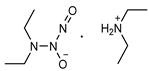 | 2 min |
| SPER/NO Spermine NONOate | 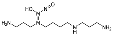 | 6 min |
| PAPA/NO Propylamine propylamine NONOate | 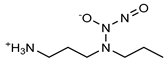 | 15 min |
| DPTA/NO Dipropylentriamine NONOate |  | 3 h |
| Hydrogel | NO Donor | NO Release Features | References |
|---|---|---|---|
| pHEMA | Manganese nitrosyl | Light-activated | [82] |
| Methacrylate-modified gelatin /hyaluronic acid graft dopamine | N,N′-di–sec–butyl–N,N′-dinitroso-1,4-phenylenediamine (BNN6) | [101] | |
| Gelatin methacrylate and oxide dextran | BNN6 | Near-infrared release | [42] |
| Gelatin | Sodium nitrite | [83] | |
| Gelatin methacrylate | SNAP (S-nitroso-N-acetylpenicillamine) | [102] | |
| Gelatin and sodium alginate | SNAP | Burst release in first 4 h, sustained up to 120 h | [68] |
| F-127/PAA | GSNO | ~200 min constant ~5 days | [103] |
| Pluronic F-127 | GSNO | -------- | [34,104,105,106,107] |
| Pluronic F-127 | GSNO SNAC (S-nitroso-N-acetylcysteine) | Thermal or photochemical release | [84,108,109] |
| Pluronic F-127 | GSNO | [85] | |
| Pluronic F-127 and alginate | GSNO | [110] | |
| Pluronic F-127 Pluronic P-123 | Nitroso-derivative of 4-amino-7-nitrobenzofurazan | Photochemical release | [111] |
| Alginate, pectin and PEG | GSNO | Release for at least 18 h of GSNO | [40] |
| Alginate | S-nitroso-mercaptosuccinic acid | Burst release in first 5 h, sustained in following hours (tested up to 18 h) | [112] |
| Chitosan | Isosorbide mononitrate (ISMN) | [113] | |
| Chitosan | GSNO | Sustained for over 48 h | [46] |
| Chitosan, PEG, sugar | Sodium nitrite | Sustained for at least 24 h | [44] |
| Chitosan, PVP, PEG | Nitrite | Burst release for 120 min followed by sustained up to 8 h | [93] |
| Chitosan, PVA | SNAP | Continuous release for at least 120 h | [114] |
| Chitosan and Poly(vinyl alcohol) | Ruthenium nitrosyl | NIR-induced release | [90] |
| PEG, fibrinogen | SNAP | Photolytic and thermal activation | [43] |
| Fmoc-FF | SNAP | Burst release in the first 12 h, sustained over 7 days | [115] |
| Poly(β-cyclodextrin) and modified dextran | Nitro compound | Photochemical | [91,97,116,117] |
| Advantages | Limitations | |
|---|---|---|
| Mechanism | NO Donor Incorporation | |
| Physical adsorption | Simple, no reactions or modifications required Any NO donor can be incorporated | Possible leaching Storage, stability, and release depend on hydrogel–donor interactions |
| Chemical attachment | No leaching Ease to create RSNOs and NONOates | Requires complex reactions |
| NO release | ||
| Hydrolysis | Uncomplicated release triggers | Undesired release in water containing environments |
| Enzymatic catalysis | Not subject to uncontrolled release due to specific triggers | Release rate depends on enzyme kinetics |
| Photocatalysis | Limited application, requires direct irradiation | |
| Gram + | Gram − | Effect | NO Donor/Hydrogel | References |
|---|---|---|---|---|
| Antibacterial activity assessed in vitro | ||||
| S. aureus | E. coli | Bactericidal | Metal-NO complex/Chitosan, PVA | [90] |
| E. coli | Bactericidal | Nitro compound/Poly(cyclodextrin) | [91] | |
| S. epidermis | E. coli | Bactericidal | NONOate/Chitosan, Hyaluronic acid | [41] |
| S. mutans S. aureus | E. coli | Bactericidal | RSNO/Alginate | [112] |
| P. aeruginosa | Bactericidal | GSNO/Chitosan, Pluronic F-127 | [34] | |
| S. aureus | P. aeruginosa | Bactericidal | NONOate/Alginate, PEI | [27] |
| P. aeruginosa | Bactericidal Biofilm dispersal | NONOate/antimicrobial polymer | [87] | |
| With enhanced wound healing tested in vivo | ||||
| S. aureus | E. coli | Bactericidal | BNN6/GelMA | [42] |
| S. aureus | P. aeruginosa | Bactericidal | GSNO/Chitosan | [46] |
| MRSA | P. aeruginosa | Bactericidal | GSNO/Alginate, Pectin, PEG | [40] |
| MRSA | MRPA | Bactericidal | GSNO/Alginate | [110] |
Publisher’s Note: MDPI stays neutral with regard to jurisdictional claims in published maps and institutional affiliations. |
© 2022 by the authors. Licensee MDPI, Basel, Switzerland. This article is an open access article distributed under the terms and conditions of the Creative Commons Attribution (CC BY) license (https://creativecommons.org/licenses/by/4.0/).
Share and Cite
Tavares, G.; Alves, P.; Simões, P. Recent Advances in Hydrogel-Mediated Nitric Oxide Delivery Systems Targeted for Wound Healing Applications. Pharmaceutics 2022, 14, 1377. https://doi.org/10.3390/pharmaceutics14071377
Tavares G, Alves P, Simões P. Recent Advances in Hydrogel-Mediated Nitric Oxide Delivery Systems Targeted for Wound Healing Applications. Pharmaceutics. 2022; 14(7):1377. https://doi.org/10.3390/pharmaceutics14071377
Chicago/Turabian StyleTavares, Gina, Patrícia Alves, and Pedro Simões. 2022. "Recent Advances in Hydrogel-Mediated Nitric Oxide Delivery Systems Targeted for Wound Healing Applications" Pharmaceutics 14, no. 7: 1377. https://doi.org/10.3390/pharmaceutics14071377
APA StyleTavares, G., Alves, P., & Simões, P. (2022). Recent Advances in Hydrogel-Mediated Nitric Oxide Delivery Systems Targeted for Wound Healing Applications. Pharmaceutics, 14(7), 1377. https://doi.org/10.3390/pharmaceutics14071377