
Associations of GNAS and RGS Gene Polymorphisms with the Risk of Ritodrine-Induced Adverse Events in Korean Women with Preterm Labor: A Cohort Study


Abstract
1. Introduction
2. Methods
2.1. Study Patients
2.2. Genotyping Methods
2.3. Statistical Analyses
3. Results
4. Discussion
5. Limitation
6. Conclusions
Author Contributions
Funding
Institutional Review Board Statement
Informed Consent Statement
Data Availability Statement
Conflicts of Interest
References
- Romero, R.; Dey, S.K.; Fisher, S.J. Preterm labor: One syndrome, many causes. Science 2014, 345, 760–765. [Google Scholar] [CrossRef] [PubMed]
- Waitzman, N.J.; Jalali, A.; Grosse, S.D. Preterm birth lifetime costs in the United States in 2016: An update. Semin. Perinatol. 2021, 45, 151390. [Google Scholar] [CrossRef] [PubMed]
- World Health Organization. Preterm Birth: Fact Sheet 2018. Available online: https://www.who.int/news-room/fact-sheets/detail/preterm-birth (accessed on 1 August 2021).
- Li, C.; Cao, M.; Zhou, X. Role of epigenetics in parturition and preterm birth. Biol. Rev. Camb. Philos. Soc. 2022, 97, 851–873. [Google Scholar] [CrossRef] [PubMed]
- Gilman-Sachs, A.; Dambaeva, S.; Salazar Garcia, M.D.; Hussein, Y.; Kwak-Kim, J.; Beaman, K. Inflammation induced preterm labor and birth. J. Reprod. Immunol. 2018, 129, 53–58. [Google Scholar] [CrossRef]
- Garfield, L.; Chin, E. Pharmacology for Preterm Labor. J. Perinat. Neonatal Nurs. 2020, 34, 155–161. [Google Scholar] [CrossRef]
- Patel, S.S.; Ludmir, J. Drugs for the Treatment and Prevention of Preterm Labor. Clin. Perinatol. 2019, 46, 159–172. [Google Scholar] [CrossRef]
- Haas, D.M.; Caldwell, D.M.; Kirkpatrick, P.; McIntosh, J.J.; Welton, N.J. Tocolytic therapy for preterm delivery: Systematic review and network meta-analysis. BMJ 2012, 345, e6226. [Google Scholar] [CrossRef]
- Lamont, C.D.; Jørgensen, J.S.; Lamont, R.F. The safety of tocolytics used for the inhibition of preterm labour. Expert Opin. Drug Saf. 2016, 15, 1163–1173. [Google Scholar] [CrossRef]
- Sameshima, H.; Saito, S.; Matsuda, Y.; Kamitomo, M.; Makino, S.; Ohashi, M.; Kino, E.; Kanayama, N.; Takeda, S. Annual Report of the Perinatology Committee, Japan Society of Obstetrics and Gynecology, 2016: Overall report on a comprehensive retrospective study of obstetric management of preterm labor and preterm premature rupture of the membranes. J. Obstet. Gynaecol. Res. 2018, 44, 5–12. [Google Scholar] [CrossRef]
- Lam, F.; Gill, P. Beta-Agonist tocolytic therapy. Obstet. Gynecol. Clin. N. Am. 2005, 32, 457–484. [Google Scholar] [CrossRef]
- Canadian Preterm Labor Investigators Group. Treatment of preterm labor with the beta-adrenergic agonist ritodrine. N. Engl. J. Med. 1992, 327, 308–312. [Google Scholar] [CrossRef] [PubMed]
- Modzelewska, B. Beta-adrenoceptors in obstetrics and gynecology. Dev. Period Med. 2016, 20, 93–98. [Google Scholar] [PubMed]
- Shigemi, D.; Aso, S.; Yasunaga, H. Inappropriate use of ritodrine hydrochloride for threatened preterm birth in Japan: A retrospective cohort study using a national inpatient database. BMC Prgenancy Childbirth 2019, 19, 204. [Google Scholar] [CrossRef]
- Chung, J.E.; Choi, S.A.; Hwang, H.S.; Park, J.Y.; Lee, K.E.; Yee, J.; Kim, Y.J.; Gwak, H.S. Association between B2-adrenergic receptor gene polymorphisms and adverse events of ritodrine in the treatment of preterm labor: A prospective observational study. BMC Genet. 2017, 18, 96. [Google Scholar] [CrossRef]
- Seo, H.; Kwon, E.J.; You, Y.A.; Park, Y.; Min, B.J.; Yoo, K.; Hwang, H.S.; Kim, J.H.; Kim, Y.J. Deleterious genetic variants in ciliopathy genes increase risk of ritodrine-induced cardiac and pulmonary side effects. BMC Med. Genom. 2018, 11, 4. [Google Scholar] [CrossRef] [PubMed]
- Lee, S.M.; Park, Y.; Kim, Y.J.; Hwang, H.S.; Seo, H.; Min, B.J.; Lee, K.H.; Kim, S.Y.; Jung, Y.M.; Lee, S.; et al. Identifying genetic variants associated with ritodrine-induced pulmonary edema. PLoS ONE. 2020, 15, e0241215. [Google Scholar] [CrossRef] [PubMed]
- Thompson, M.D.; Cole, D.E.; Jose, P.A.; Chidiac, P. G protein-coupled receptor accessory proteins and signaling: Pharmacogenomic insights. Methods Mol. Biol. 2014, 1175, 121–152. [Google Scholar]
- Masuho, I.; Balaji, S.; Muntean, B.S.; Skamangas, N.K.; Chavali, S.; Tesmer, J.J.G.; Babu, M.M.; Martemyanov, K.A. A Global Map of G protein Signaling Regulation by RGS Proteins. Cell 2020, 183, 503–521.e19. [Google Scholar] [CrossRef]
- Hao, J.; Michalek, C.; Zhang, W.; Zhu, M.; Xu, X.; Mende, U. Regulation of cardiomyocyte signaling by RGS proteins: Differential selectivity towards G proteins and susceptibility to regulation. J. Mol. Cell Cardiol. 2006, 41, 51–61. [Google Scholar] [CrossRef]
- Purcell, S.; Neale, B.; Todd-Brown, K.; Thomas, L.; Ferreira, M.A.R.; Bender, D.; Maller, J.; Sklar, P.; De Bakker, P.I.; Daly, M.J.; et al. PLINK: A toolset for whole-genome association and population-based linkage analysis. Am. J. Hum. Genet. 2007, 81, 559–575. [Google Scholar] [CrossRef]
- Hall, J.A.; Petch, M.C.; Brown, M.J. Intracoronary injections of salbutamol demonstrate the presence of functional beta 2-adrenoceptors in the human heart. Circ. Res. 1989, 65, 546–553. [Google Scholar] [CrossRef] [PubMed]
- Brodde, O.E.; Michel, M.C. Adrenergic and muscarinic receptors in the human heart. Pharmacol. Rev. 1999, 51, 651–690. [Google Scholar] [PubMed]
- Brodde, O.E. Beta 1- and beta 2-adrenoceptors in the human heart: Properties, function, and alterations in chronic heart failure. Pharmacol. Rev. 1991, 43, 203–242. [Google Scholar] [PubMed]
- Ali, D.C.; Naveed, M.; Gordon, A.; Majeed, F.; Saeed, M.; Ogbuke, M.I.; Atif, M.; Zubair, H.M.; Changxing, L. B-adrenergic receptor, an essential target in cardiovascular diseases. Heart Fail. Rev. 2020, 25, 343–354. [Google Scholar] [CrossRef] [PubMed]
- Gaudin, C.; Ishikawa, Y.; Wight, D.C.; Mahdavi, V.; Nadal-Ginard, B.; Wagner, T.E.; Vatner, C.J. Overexpression of Gs alpha protein in the hearts of transgenic mice. J. Clin. Investig. 1995, 95, 1676–1683. [Google Scholar] [CrossRef]
- Iwase, M.; Uechi, M.; Vatner, D.E.; Asai, K.; Shannon, R.P.; Kudej, R.K.; Wagner, T.E.; Wight, D.C.; Patrick, T.A.; Ishikawa, Y.; et al. Cardiomyopathy induced by cardiac Gs alpha overexpression. Am. J. Physiol. 1997, 272, H585–H589. [Google Scholar] [CrossRef]
- Iwase, M.; Bishop, S.P.; Uechi, M.; Vatner, D.E.; Shannon, R.P.; Kudej, R.K.; Wight, D.C.; Wagner, T.E.; Ishikawa, Y.; Homcy, C.J.; et al. Adverse effects of chronic endogenous sympathetic drive induced by cardiac GS alpha overexpression. Circ. Res. 1996, 78, 517–524. [Google Scholar] [CrossRef]
- Hollinger, S.; Hepler, J.R. Cellular regulation of RGS proteins: Modulators and integrators of G protein signaling. Pharmacol. Rev. 2002, 54, 527–559. [Google Scholar] [CrossRef]
- Wieland, T.; Mittmann, C. Regulators of G-protein signalling: Multifunctional proteins with impact on signalling in the cardiovascular system. Pharmacol. Ther. 2003, 97, 95–115. [Google Scholar] [CrossRef]
- Tsang, S.; Woo, A.Y.; Zhu, W.; Xiao, R.P. Deregulation of RGS2 in cardiovascular diseases. Front. Biosci. 2010, 2, 547–557. [Google Scholar]
- Tuomi, J.M.; Chidiac, P.; Jones, D.L. Evidence for enhanced M3 muscarinic receptor function and sensitivity to atrial arrhythmia in the RGS2-deficient mouse. Am. J. Physiol. Heart Circ. Physiol. 2010, 298, H554–H561. [Google Scholar] [CrossRef] [PubMed][Green Version]
- Heximer, S.P.; Knutsen, R.H.; Sun, X.; Kaltenbronn, K.M.; Rhee, M.H.; Peng, N.; Oliveira-dos-Santos, A.; Penninger, J.M.; Muslin, A.J.; Steinberg, T.H.; et al. Hypertension and prolonged vasoconstrictor signaling in RGS2-deficient mice. J. Clin. Investig. 2003, 111, 445–452. [Google Scholar] [CrossRef] [PubMed]
- Zhang, W.; Anger, T.; Su, J.; Hao, J.; Xu, X.; Zhu, M.; Gach, A.; Cui, L.; Liao, R.; Mende, U. Selective loss of fine tuning of Gq/11 signaling by RGS2 protein exacerbates cardiomyocyte hypertrophy. J. Biol. Chem. 2006, 281, 5811–5820. [Google Scholar] [CrossRef] [PubMed]
- McNabb, H.J.; Zhang, Q.; Sjogren, B. Emerging Roles for Regulator of G protein Signaling 2 in (Patho)physiology. Mol. Pharmacol. 2020, 98, 751–760. [Google Scholar] [CrossRef] [PubMed]
- Jie, L.; Owens, E.A.; Plante, L.A.; Fang, Z.; Rensing, D.T.; Moeller, K.D.; Osei-Owusu, P. RGS2 squelches vascular Gi/o and Gq signaling to modulate myogenic tone and promote uterine blood flow. Physiol. Rep. 2016, 4, e12692. [Google Scholar] [CrossRef]
- Qin, M.; Huang, H.; Wang, T.; Hu, H.; Liu, Y.; Gu, Y.; Cao, H.; Li, H.; Huang, C. Atrial tachyarrhythmia in Rgs5-null mice. PLoS ONE 2012, 7, e46856. [Google Scholar] [CrossRef]
- Perschbacher, K.J.; Deng, G.; Fisher, R.A.; Gibson-Corley, K.N.; Santillan, M.K.; Grobe, J.L. Regulators of G protein signaling in cardiovascular function during pregnancy. Physiol. Genom. 2018, 50, 590–604. [Google Scholar] [CrossRef]
- Mary, C. Regulation by 3’-Untranslated Regions. Annu. Rev. Genet. 2017, 51, 171–194. [Google Scholar] [CrossRef]
- Rose, A.B. Nuclear Pre-mRNA Processing in Plants; Springer: Berlin/Heidelberg, Germany, 2008; pp. 277–290. [Google Scholar]
- Carithers, L.J.; Ardlie, K.; Barcus, M.; Branton, P.A.; Britton, A.; Buia, S.A.; Compton, C.C.; DeLuca, D.S.; Peter-Demchok, J.; Gelfand, E.T.; et al. A Novel Approach to High-Quality Postmortem Tissue Procurement: The GTEx Project. Biopreserv. Biobank. 2015, 13, 311–319. [Google Scholar] [CrossRef]


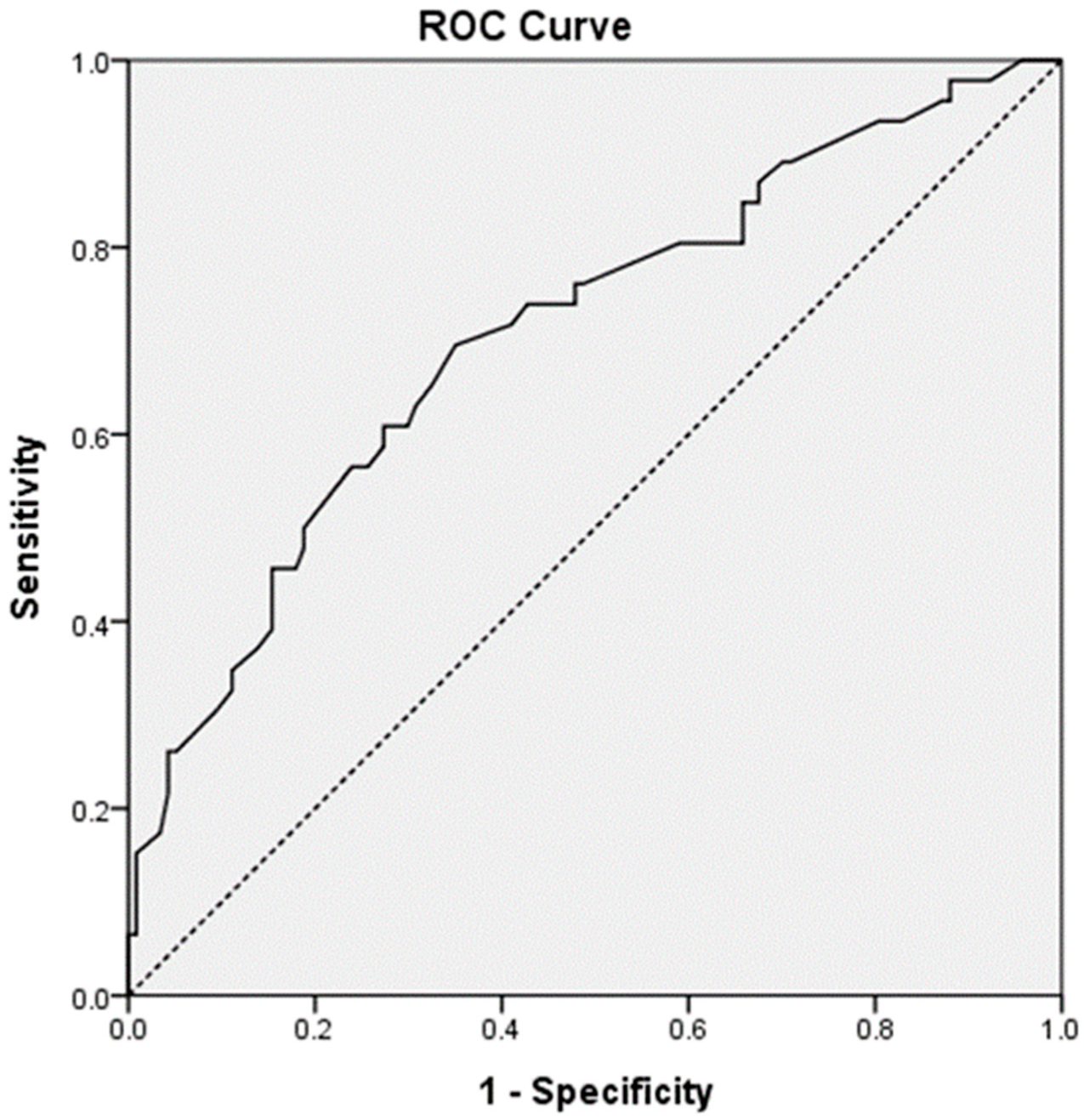
| Parameters | AE | No AE | p-Value |
|---|---|---|---|
| Age (years) | 0.429 | ||
| <35 | 37 (80.4) | 100 (85.5) | |
| ≥35 | 9 (19.6) | 17 (14.5) | |
| Gestational age (weeks) | 0.322 | ||
| <30 | 24 (52.2) | 51 (43.6) | |
| ≥30 | 22 (47.8) | 66 (56.4) | |
| Weight (kg) | 0.057 | ||
| <60 | 24 (52.2) | 42 (35.9) | |
| ≥60 | 22 (47.8) | 75 (64.1) | |
| Height (cm) | 0.009 | ||
| <160 | 22 (47.8) | 31 (26.5) | |
| ≥160 | 24 (52.2) | 86 (73.5) | |
| Maximum infusion rate (cc/hr) | 0.799 | ||
| <60 | 21 (45.7) | 56 (47.9) | |
| ≥60 | 25 (54.3) | 61 (52.1) |
| Gene and SNP | Chromosomal Location | Allele A; Allele B a | AE (AA/AB/BB) | No AE (AA/AB/BB) | Odds Ratio (95% CI) | p-Value |
|---|---|---|---|---|---|---|
| GNAS | ||||||
| rs12625436 | Chr20:58870158 | G *; A | 7/27/12 | 23/56/38 | 0.96 (0.59–1.57) | 0.871 |
| rs13831 | Chr20:58900136 | A *; G | 6/25/15 | 12/50/55 | 0.68 (0.41–1.13) | 0.138 |
| rs6128461 | Chr20:58902035 | T *; C | 8/24/14 | 19/49/49 | 0.78 (0.48–1.26) | 0.309 |
| rs7121 | Chr20:58903752 | C *; T | 13/23/10 | 18/60/39 | 0.60 (0.36–0.99) | 0.044 |
| rs3730168 | Chr20:58903884 | G; A * | 17/21/8 | 63/50/4 | 2.18 (1.26–3.79) | 0.006 |
| rs6026593 | Chr20:58904078 | A; G * | 36/9/1 | 81/32/3 | 0.70 (0.34–1.43) | 0.322 |
| RGS2 | ||||||
| rs1856840 | Chr1:192842157 | T; C * | 15/25/6 | 47/57/13 | 1.25 (0.74–2.10) | 0.405 |
| rs4606 | Chr1:192812042 | C *; G | 10/30/6 | 30/59/27 | 0.87 (0.52–1.46) | 0.601 |
| rs1152746 | Chr1:192827775 | C *; T | 0/8/38 | 3/36/78 | 2.39 (1.05–5.41) | 0.038 |
| RGS5 | ||||||
| rs3806366 | Chr1:163145531 | A; G * | 22/18/6 | 59/49/9 | 1.20 (0.72–2.02) | 0.484 |
| rs2815276 | Chr1:163155478 | A*; G | 8/19/19 | 14/66/37 | 1.10 (0.66–1.86) | 0.711 |
| rs2662776 | Chr1:163195239 | A; G * | 23/20/3 | 65/45/7 | 1.18 (0.68–2.04) | 0.566 |
| rs1509018 | Chr1:163218794 | G; C * | 22/18/5 | 52/54/11 | 0.94 (0.553–1.59) | 0.812 |
| rs6698367 | Chr1:163226647 | C; T * | 29/17/0 | 58/53/6 | 0.54 (0.28–1.03) | 0.063 |
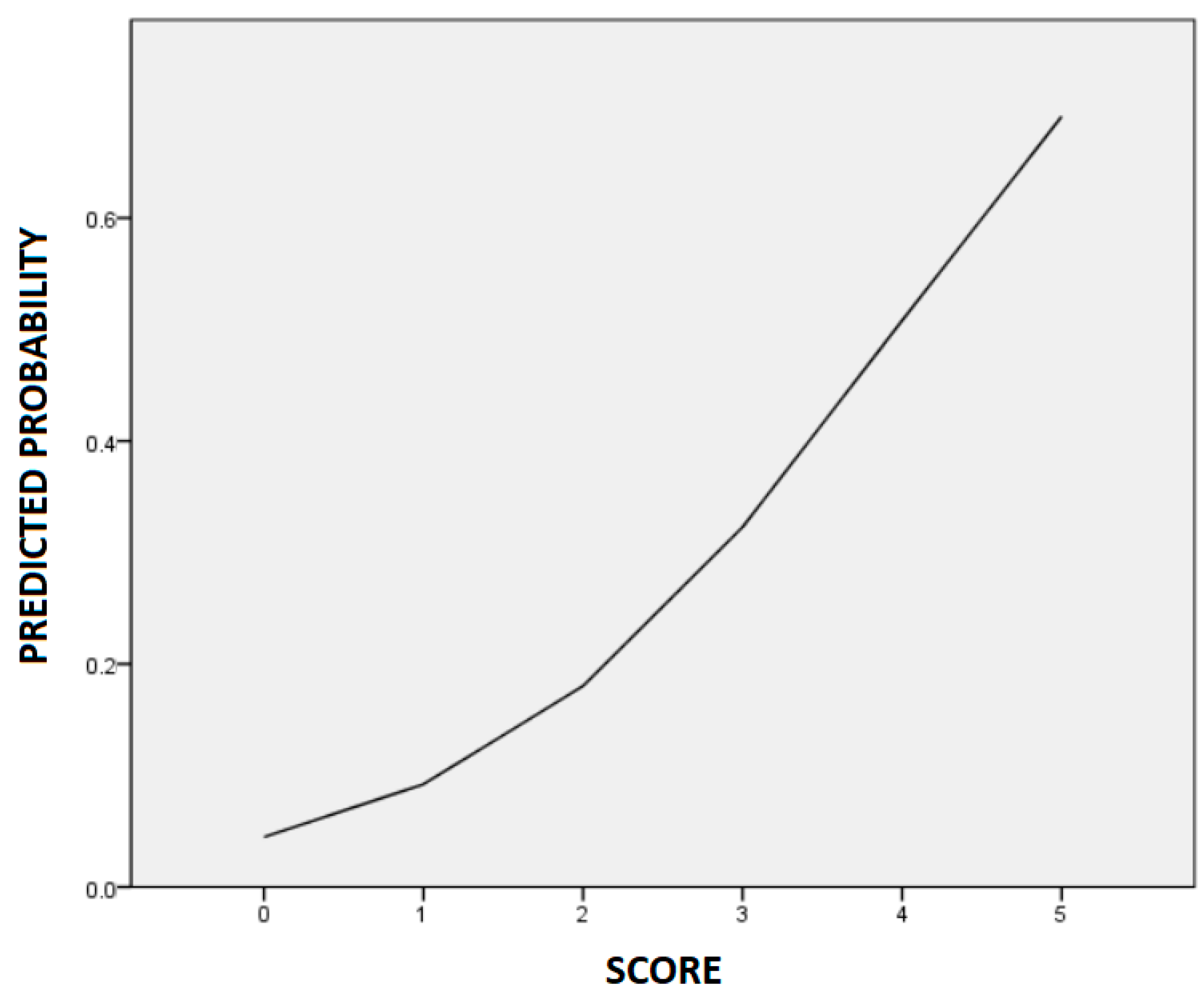
| Parameters | Crude Odds Ratio | Adjusted Odds Ratio | Score |
|---|---|---|---|
| Age ≥ 35 years | 1.43 (0.59–3.49) | 1.90 (0.69–5.27) | |
| Gestational age < 30 weeks | 1.41 (0.71–2.80) | 1.66 (0.79–3.49) | |
| Height < 160 cm | 2.54 (1.25–5.17) * | 2.41 (1.13–5.12) * | 0, 1 a |
| GNAS rs7121 | 0.60 (0.36–0.99) * | 1.00 (0.51–1.97) | |
| GNAS rs3730168 | 2.18 (1.26–3.79) ** | 2.10 (1.03–4.30) * | 0, 1, 2 b |
| RGS2 rs1152746 | 2.39 (1.05–5.41) * | 2.63 (1.07–6.49) * | 0, 1, 2 b |
| Risk Score | AE | No AE |
|---|---|---|
| 0 | 0 (0.0) | 2 (100.0) |
| 1 | 2 (13.3) | 13 (86.7) |
| 2 | 12 (18.5) | 53 (81.5) |
| 3 | 15 (30.6) | 34 (69.4) |
| 4 | 13 (46.4) | 15 (53.6) |
| 5 | 4 (100.0) | 0 (0.0) |
Publisher’s Note: MDPI stays neutral with regard to jurisdictional claims in published maps and institutional affiliations. |
© 2022 by the authors. Licensee MDPI, Basel, Switzerland. This article is an open access article distributed under the terms and conditions of the Creative Commons Attribution (CC BY) license (https://creativecommons.org/licenses/by/4.0/).
Share and Cite
Jang, E.-J.; Kim, Y.-J.; Hwang, H.-S.; Yee, J.; Gwak, H.-S. Associations of GNAS and RGS Gene Polymorphisms with the Risk of Ritodrine-Induced Adverse Events in Korean Women with Preterm Labor: A Cohort Study. Pharmaceutics 2022, 14, 1220. https://doi.org/10.3390/pharmaceutics14061220
Jang E-J, Kim Y-J, Hwang H-S, Yee J, Gwak H-S. Associations of GNAS and RGS Gene Polymorphisms with the Risk of Ritodrine-Induced Adverse Events in Korean Women with Preterm Labor: A Cohort Study. Pharmaceutics. 2022; 14(6):1220. https://doi.org/10.3390/pharmaceutics14061220
Chicago/Turabian StyleJang, Eun-Jeong, Young-Ju Kim, Han-Sung Hwang, Jeong Yee, and Hye-Sun Gwak. 2022. "Associations of GNAS and RGS Gene Polymorphisms with the Risk of Ritodrine-Induced Adverse Events in Korean Women with Preterm Labor: A Cohort Study" Pharmaceutics 14, no. 6: 1220. https://doi.org/10.3390/pharmaceutics14061220
APA StyleJang, E.-J., Kim, Y.-J., Hwang, H.-S., Yee, J., & Gwak, H.-S. (2022). Associations of GNAS and RGS Gene Polymorphisms with the Risk of Ritodrine-Induced Adverse Events in Korean Women with Preterm Labor: A Cohort Study. Pharmaceutics, 14(6), 1220. https://doi.org/10.3390/pharmaceutics14061220