Next Article in Journal
Manipulating Macrophage/Microglia Polarization to Treat Glioblastoma or Multiple Sclerosis
Previous Article in Journal
Delivery of Oligonucleotides: Efficiency with Lipid Conjugation and Clinical Outcome
 
 
Retraction published on 30 January 2024, see Pharmaceutics 2024, 16(2), 199.
Article

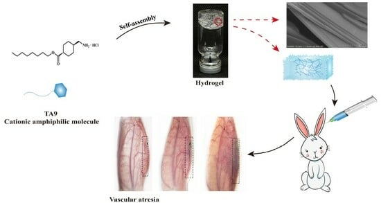
RETRACTED: A Novel Drug Self-Delivery System from Fatty Alcohol Esters of Tranexamic Acid for Venous Malformation Sclerotherapy

1
School of Pharmacy, Shenyang Pharmaceutical University, Shenyang 110016, China
2
School of Life Science and Biopharmaceutics, Shenyang Pharmaceutical University, Shenyang 110016, China
3
Department of Basic Pharmaceutical Sciences, School of Pharmacy, Husson University, Bangor, ME 13802, USA
*
Authors to whom correspondence should be addressed.
Pharmaceutics 2022, 14(2), 343; https://doi.org/10.3390/pharmaceutics14020343
Submission received: 22 December 2021 / Revised: 25 January 2022 / Accepted: 29 January 2022 / Published: 1 February 2022 / Retracted: 30 January 2024

Abstract

:
Venous malformation (VM), which causes severe damage to patients’ appearance and organ function, is one of the most common vascular malformations. At present, many drugs in clinical treatment cause various adverse reactions. Herein, we synthesized cationic amphiphilic gelators (TA6, TA8, and TA9) by introducing saturated carbon chains of different lengths to tranexamic acid (TA), which could self-assemble into low-molecular-weight gels (LMWGs) as drug delivery carriers by hydrogen bonds, van der Waals forces, and hydrophobic interactions. The rheological properties, gelation driving force and drug release profiles of TA6, TA8, and TA9 hydrogels were characterized, and the results indicated that the hydrogels prepared in this study possessed the typical characteristics of a gel and could release drugs slowly. More importantly, the TA9 gelator showed significant pharmacological activity, in that it served as both an active drug compound and a drug carrier. The in vitro experiments demonstrated that TA9 induced HUVECs death and hemolysis by destroying cell membranes in a dose-dependent manner, and caused cell death and hemolysis at a concentration of 0.09 µM/mL. Meanwhile, we found TA9 could interact not only with fibrinogen, but also with other endogenous molecules in the blood. After the administration of TA9 hydrogel for 15 days, macroscopic imaging and histological evaluation in mice and rabbits displayed obvious thrombi, inflammatory reactions, and venous embolization, indicating that the mechanism of the TA9 hydrogel in treating VM was involved in two processes. Firstly, the TA9 hydrogel relied on its mechanical strength to physically block veins and continuously release TA9, in situ, for targeted therapy. Then, TA9 destroyed endothelial cells and damaged venous walls critically, causing thrombi. Most excitingly, TA9 was hydrolyzed to TA by enzymes that inhibited the degradation of thrombi by plasmin to prolong the embolization time and to promote venous fibrosis. Compared with other clinically available sclerosants, the degradation of TA9 also empowered a better biocompatibility and biodegradability for the TA9 hydrogel. In conclusion, we synthesized a potentially safe and effective derivative of TA and developed a low-molecular-weight gel as a self-delivery system for TA in treating VM.

Graphical Abstract

1. Introduction

The International Society for the Study of Vascular Anomalies (ISSVA) classifies vascular abnormalities into hemangioma and vascular malformation based on the biological characteristics of vascular endothelial cells proposed by John B. Mulliken [1,2]. VM is a type of vascular malformation, caused by abnormal development of the venous system during embryonic growth, and is the most common vascular malformation in the clinic [2]. Its pathological manifestations are an abnormal expansion of the venous sinus, reduction of the smooth muscle of the venous wall, and degeneration of adventitia fibers. These abnormally dilated veins proliferate in child development and, generally, without self-healing, eventually causing abnormal appearance, organ dysfunction, and other chronic diseases [3].
Standard treatment methods for VM are surgical resection, sclerotherapy, laser embolotherapy, and limb compression [2]. Among them, minimally invasive intravascular sclerotherapy performs excellently in the treatment of slow-flow VM. Sclerotherapy is the injection of sclerosants into malformed veins to damage endothelial cells, peel off the intima, and expose the collagen fibers under the membrane, thickening the venous wall and making the abnormal veins occlude or atrophy. More recently, anhydrous ethanol [4], sodium tetradecyl sulfate (STS), polidocanol (POL) [5], pingyangmycin, and bleomycin have been clinically frequently utilized, but they still have significant problems [6]. For example, high-concentration absolute ethanol, with its poor retention, will be quickly removed by blood flow when directly exposed to veins, so the dosage or administration times have to be increased, leading to adverse reactions, such as nasty aches, vasospasm, and tissue necrosis [7,8]. Pingyangmycin powder injection induces systemic toxicity, such as severe lung injury, because of poor retention, and foam sclerosants are limited by their preparation methods, unstable foam volume, and short foam half-life [9]. Therefore, accurate embolization, increasing the retention of active drugs in the lesions, prolonging the half-life, and reducing systemic side effects are the key to relevant drug research and development for the treatment of VM [10].
New formulations that accurately embolize and achieve sustained drug release are favored in treating VM. For instance, modified foam sclerosants are formed by adding other surfactants to prolong the half-life of the foam. Injectable in situ gels are available to be applied directly at the lesion site to control drugs released locally and avoid many adverse reactions [11,12]. Traditional polymer gels with covalent bonds are commonly used but difficult to degrade in vivo [13]. LMWGs, which refer to the formation of low-molecular gelators through non-covalent bonds, have greater advantages [14]. Compared with polymer gels, LMWGs have considerable mechanical properties; meanwhile, reversible non-covalent bonds and the functional groups of gelators confer to the gels more stimulus responsiveness (pH, light, magnetism, enzyme, ion, etc.) and better biodegradability [15,16]. Researchers have attempted to systematically modify and optimize existing gelators or to introduce diverse functional groups to form new compounds and construct progressive LMWGs [17]. The derivatives of amino acids [18], peptides [19], carbohydrates, fatty acids, pyridines, and urea bonds have been used as LMWGs carriers to deliver drugs, proteins, and genes in vivo and have fully demonstrated the outstanding drug-loading and drug release capabilities, biodegradability, and biocompatibility thereof [20,21]. There are two potential delivery methods for LMWGs. Firstly, the LMWGs act as an inert carrier to physically load the active drug compounds, and then the drugs are released through diffusion and erosion of the gel by a medium, such as physiological fluids. Secondly, the introduction of functional groups into the active drugs are designed as self-assembled gelators, and the active drugs are released through hydrolysis or enzymolysis, which significantly increases the drug load while reducing the toxicity and side effects caused by inactive substances. It has been suggested that it is necessary to determine the pharmacological activity of the gelator since the pharmacological effect of the gelator may be equivalent to that of the active drug [22,23,24].
TA, also known as trans-4-aminomethyl-cyclohexane-carboxylic acid, is commonly used clinically to stanch bleeding during surgery, as a tyrosinase inhibitor [25]. As a hydrophilic lysine analog, TA has amino and carboxyl groups that can be modified. We used acylation to introduce saturated carbon chains of different lengths to synthesize a series of TA analogs (TA6, TA8, and TA9) and found that these derivatives could self-assemble in water to form LMWGs quickly. Based on this, we have further studied the properties and gel-forming mechanism of these hydrogels. TA9, as a cationic amphiphilic molecule, possesses similar pharmacological activity as STS for the treatment of VM. Afterward, we studied the mechanism of action of the TA9 hydrogels through in vitro experiments and evaluated its efficacy and biocompatibility using mice tail-vein and rabbit ear-vein models. TA9 hydrogel, as a novel sclerosant, may have a dual mechanism of “coagulation initiation—fibrinolysis inhibition” in the treatment of VM.

2. Materials and Methods

2.1. Materials

TA was purchased from Wuhan Lullaby Pharmaceutical Chemical Co., Ltd. (Wuhan, China). The chemicals 1-hexanol, 1-octanol, and 1-nonanol were bought from Shanghai Yuanye Bio-Technology Co., Ltd. (Shanghai, China). Dulbecco’s Modified Eagle’s Medium (DMEM) and cell-culture Petri dishes were obtained from Corning (New York, NY, USA). Penicillin–streptomycin was obtained from Gibco (Waltham, MA, USA). Trypsin was obtained from Sigma-Aldrich (St. Louis, MO, USA). Fetal bovine serum (FBS) was obtained from Zhejiang Tianhang Biotechnology Co., Ltd. (Hangzhou, China). A Cell Counting Kit-8 (CCK-8) and a Lactate Dehydrogenase (LDH) Cytotoxicity Assay Kit were purchased from Shanghai Beyotime Biotechnology Co., Ltd. (Shanghai, China). All of the other reagents were of analytical grade and used without further purification. Human umbilical vein endothelial cells (HUVECs) were provided by ATCC (Rockefeller, MD, USA). Fluorescent Cy7 was obtainedd from Beijing Solarbio Life Sciences Co., Ltd. (Beijing, China). Kunming (KM) mice and New Zealand rabbits were provided by the Animal Center of Shenyang Pharmaceutical University. All animal experiments and procedures were reviewed and approved by the Animal Care and Use Committee at the Shenyang Pharmaceutical University (Ethical Committee approval number: SYPU-IACUC-C2019-12-14-105; date of approval: 14 December 2019).

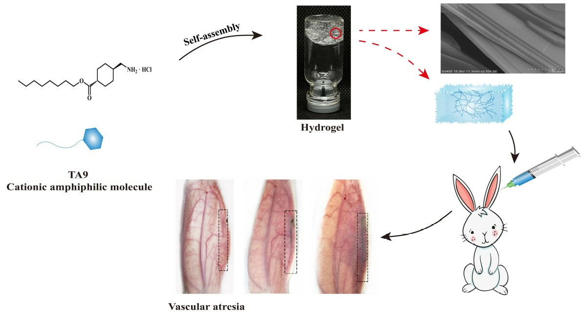
2.2. Synthesis and Characterization of TA6, TA8, and TA9

TA (3.00 g) was dissolved in dichloromethane (100 mL), then 1.66 ml of thionyl chloride was added dropwise at 4 °C. The reaction mixture was heated at 25 °C for 12 h. Then alcohols (2.84 mL 1-hexanol, 3.59 mL 1-octanol, 3.97 mL 1-nonanol) were added dropwise and reacted for another 24 h. The solvent was removed under reduced pressure and white powder was obtained. The powder was then washed with methyl tert-butyl ether and dried under vacuum for 24 h. In the end, three types of TA compounds with different molecular weights (TA6, TA8, and TA9) were produced. The synthetic route is shown in Figure 1A. When n was five, the compound was named TA6, n was seven, the compound was TA8, and n was eight, the compound was TA9. The 1H-NMR and 13C-NMR spectra of TA6, TA8, and TA9 were measured using a NMR spectrometer (Bruker AV-400, Karlsruhe Germany) with D2O and CD3OD as solvents. In addition, a mass spectrometer (MS, Thermo Fisher Scientific, Bremen, Germany) was used to obtain the mass spectra to characterize the structure of products. A differential calorimetry scanner (DSC, Netzsch 3500 Sirius, Hanau Germany) was used to determine the melting points of the synthesized TA derivatives.

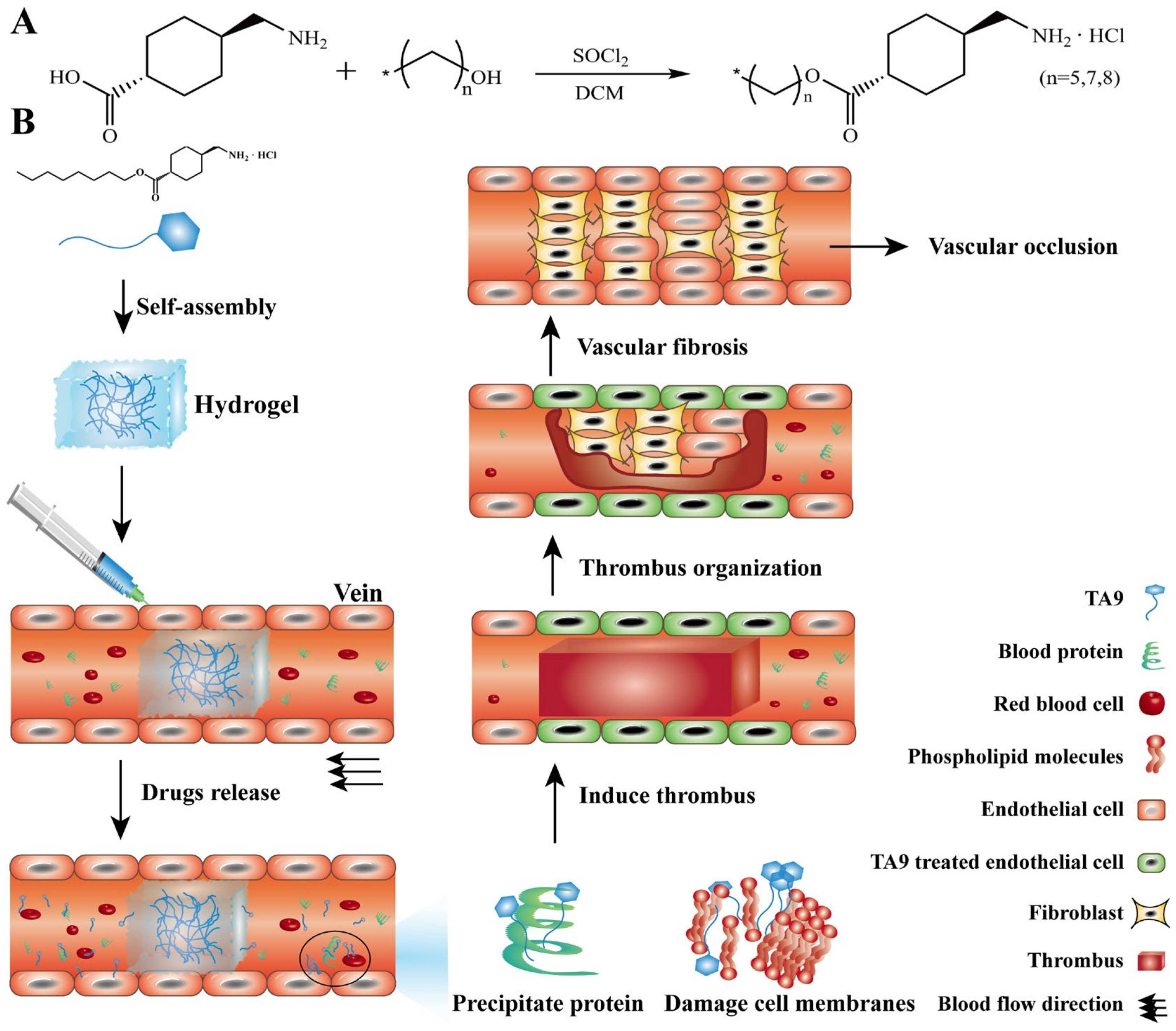
2.3. Metabolism Studies of TA6, TA8, and TA9 In Vitro

The metabolic kinetics of ester bonds of TA6, TA8, and TA9, in normal saline (NS), phosphate-buffer solution (PBS, pH7.4), and rat plasma were assessed. TA6, TA8, or TA9 (0.1 mM) were dissolved in 1 mL NS or PBS and then incubated at 37 °C, respectively. Then, 50 μL of the incubated solutions were collected at 4 h, 12 h, and 24 h for further analysis of TA6, TA8, and TA9. For the metabolic study in rat plasma, 0.1 mL of the TA6, TA8, or TA9 solutions (1 mM/L) were incubated with 1.9 mL of fresh rat plasma at 37 °C. Afterward, 20 μL of the mixtures were collected and added into 40 μL glacial acetonitrile to terminate reactions at the specified time. The resulting solutions were centrifuged at 15,000 g for 10 min at 4 °C, and the supernatants were analyzed.
The collected samples from NS, PBS, and rat plasma metabolic incubation mixtures were analyzed using an LC-MS system with a heated electrospray ionization (HESI) source (Thermo Fisher, Germany). The samples were separated with an Acquity HSS T3 column (100 mm × 2.1 mm, 1.8 mm, Waters, Ireland), which was maintained at 45 °C. The mobile phase was 0.2% formic acid aqueous solution and acetonitrile, and the flow rate was set as 0.4 mL/min.

2.4. Preparation of TA6, TA8, and TA9 Hydrogel

A weighed amount of the TA6 gelators and 1 g of purified water were transferred to vials for a period of time until a transparent hydrogel was formed. Inverting the gelator-containing vial verified whether the hydrogel was obtained. TA8 and TA9 hydrogels were obtained in the same way. The concentrations of all hydrogels were expressed in w/w (%).

2.5. Study of the Self-Assembly Behaviors of TA6, TA8, and TA9 Hydrogels

2.5.1. Gelation Mechanism

The driving forces of gelation of TA hydrogels were determined by FTIR (Thermo Fisher Nicolet iS5, Waltham, MA, USA and a NMR spectrometer (Bruker AV-400, Karlsruhe, Germany). The TA6, TA8, and TA9 powders and corresponding dry gels were pressed with potassium bromide to obtain FTIR spectra with a scanning resolution of 4 cm−1 and scanning range of 4000–400 cm−1, respectively. 1H-NMR spectra of the TA6, TA8, and TA9 solutions (1% and 10%) and the individually formed hydrogels (TA6: 56.5%, TA8: 47.4%, TA9: 41.2%) were obtained using D2O as a solvent at 25 °C [26].

2.5.2. Microscopic Study

The microstructures of TA6, TA8, and TA9 hydrogels were examined by scanning electron microscope (SEM, HITACHI S-800, Tokyo Japan). The lyophilized samples were further sputter-coated with gold for SEM observation.

2.6. Characterization of TA6, TA8, and TA9 Hydrogel

2.6.1. Determination of CGC

Critical gelation concentration (CGC) of TA6, TA8, and TA9 were determined at 25 °C by vial inversion method. Each gelator was continually added to vials containing 1 g of purified water. When the vial was turned over with no liquid flowing, the gelator concentration was recorded, and defined as the CGC% (w/w) [27]. Based on the CGC of each hydrogel, four concentrations (56.5%, 58.3%, 60.0%, and 61.5% were selected for the TA6 hydrogel; 47.4%, 50.0%, 52.4%, and 54.5% were selected for the TA8 hydrogel; 41.2%, 44.4%, 47.4%, and 50.0% were selected for the TA9 hydrogel) were selected for subsequent hydrogel characterization studies.

2.6.2. Determination of tgel and TG-S

Each gelator was accurately weighed, and purified water (1 g) was added. The time required for the gel–water mixtures to change from turbidity to clarification was recorded, and defined as the critical gelation time (tgel) [27].
The gel–sol transition temperatures (TG-S) of the TA6, TA8, and TA9 hydrogels were determined by the dropping-ball method. The prepared hydrogels were placed in an oil bath at a heating rate of 1 °C/min. When a steel ball with a diameter of 0.28 mm and a weight of 0.096 g could fall from the top to the bottom of the gel, the temperature was recorded, and defined as TG-S [18].

2.6.3. Rheology Studies

The rheological properties of the TA6, TA8, and TA9 hydrogels were measured with a rheometer (AR 2000, Newcastle DE, USA) using parallel-plate geometry with a diameter of 20 mm at 25 °C. TA6, TA8, and TA9 hydrogels (1 g) were prepared. The viscosity variation was conducted with shear rates from 0 to 120 s−1. In a stress-sweep experiment, the storage and loss moduli (G’, G”) of the stress from 0.01% to 100% were evaluated, and a constant strain of 1 Hz was applied to determine the linear viscoelastic region (LVR). A frequency sweep from 0.1 to 100 Hz was conducted at a constant strain of 0.01% [28].

2.6.4. Erosion Behavior and Syringeability Studies

The in vitro drug release behavior of the TA6, TA8, and TA9 hydrogels was evaluated in NS [29]. TA6, TA8, and TA9 hydrogels (1 g) were placed in 5-mL test tubes. Test tubes with a known weight and of 1.00-cm diameters were used. Then, 1 mL of NS was added as a release medium, and the mixture was gently shaken at 75 rpm and 37 °C. All the release media were withdrawn at specified times. Then, test tubes were weighed, after drying the test tube wall with filter paper. One milliliter of fresh NS was added to the tube to continue the study, until the weight of the remaining hydrogel in the test tube was less than 90% of the initial weight. The initial weights of the gels and test tubes were set as M0. The weights of the gels and test tubes at specified times were set as Mt. The weight of the test tubes was set as M.
Erosion (%) = (M0 − Mt)/(M0 − M) * 100%
TA8 and TA9 hydrogels were drawn into a 2-mL syringe with a needle-type of 25 G to determine their syringeability [29]. We used the same force (180 G) to push the syringe to observe the hydrogels passing through the 25-G needle.

2.7. Study on the Mechanism of TA9 in Treating VM In Vitro

2.7.1. Cytotoxicity Assay

The cytotoxicity of TA9 was analyzed via a standard CCK-8 assay. HUVECs (1 × 10⁴ cells per well) were seeded into a 96-well cell-culture plate for 12 h and grown in DMEM at 37 °C under 5% CO2. TA9 at various concentrations was prepared in DMEM, added into HUVECs, and incubated for another 24 h. Then, 10 μL of CCK-8 reagent was added to each well and incubated for 2 h. After that, the absorbances of all samples were measured at 450 nm by a microplate reader (Infinite F200 PRO, TECAN, Männedorf Switzerland). Cells without any treatments were taken as a control, and the cell viability was presented as the absorbance percentage of samples relative to the control. The cytotoxicity of TA and 1-nonanol were analyzed in the same way.

2.7.2. Cell Membrane Integrity Assay

A lactate dehydrogenase (LDH) assay was used to measure the integrity of cell membranes [30]. HUVECs (1 × 10⁴ cells per well) were seeded into a 96-well cell culture plate for 12 h and grown in DMEM at 37 °C under 5% CO2. Cells were then washed with PBS (pH 7.4). TA9 at various concentrations was prepared in DMEM, added into HUVECs, and incubated for another 24 h. A LDH release assay was performed using a Beyotime Luciferase Assay kit (Beyotime, China). The absorbances of all samples were measured at 490 nm by a microplate reader (Infinite F200 PRO, TECAN, Männedorf Switzerland). Cells without any treatments were taken as a control, and the percent of LDH release was presented as the absorbance percentage of samples relative to the control.

2.7.3. Hemolysis Assay

The damaging effect of TA9 on erythrocytes was determined by the rat erythrocyte hemolysis method. Erythrocytes from a healthy SD rat were collected in a vacuum tube to which sodium citrate was added as an anticoagulant. Then, the erythrocytes were harvested by centrifugation for 10 min at 720 g at 4 °C. The erythrocytes were rinsed and diluted with NS to reach a final cell concentration of 4%. Equal volumes of the cell suspension and TA9 solutions at different concentrations were mixed together and incubated at 37 °C for 2 h. After the mixtures were centrifuged, the supernatants were determined at 540 nm by a microplate reader (Infinite F200 PRO, TECAN, Männedorf Switzerland). Equal volumes of red blood cell suspension, in only NS and in distilled water, were used as a negative control and a positive control, respectively.

2.7.4. Effect of TA9 on Fibrinogen

A volume of 47.4% TA9 hydrogel (0.1 g) was added into 10 mg/mL bovine fibrinogen solution. The light transmittance of the solution was determined at 450 nm by an ultraviolet spectrophotometer (Unico, UV 2000, Shanghai, China), and the turbidity (%) was calculated.
Turbidity (%) = 100% − transmittance (%)

2.7.5. Evaluation of TA Inhibition on Plasmin

We determined the inhibitory effect of TA9 hydrolysate TA on plasmin by the fibrin plate method [31]. Firstly, fibrin plates were prepared according to the methods of a previous report. Agarose solution (2%) was prepared in PBS (pH 7.4) and incubated at 65 °C for 30 min. Bovine fibrinogen solution (30 mg/mL) was mixed with the 2% agarose solution in equal volume, then 200 µL of 1 mg/mL thrombin solution was added into the mixture and blended evenly. The mixture was poured into dishes and holes were made after agarose solidification at room temperature.
Equal volumes of PBS, TA, and hydrolyzed TA9 solutions were incubated with 50 U/ml of plasmin (PLA) solution at 37 °C for 10 min. Then, 10 µL of the above solutions was added to the prepared agarose wells and incubated at 37 °C. The radius (r) of the dissolving circles was measured with a Vernier caliper after 18 h, then the dissolution area was calculated. The PBS solution was used as a control.
Dissolution area (mm2) = π * r2

2.8. Pharmacodynamic Evaluation In Vivo

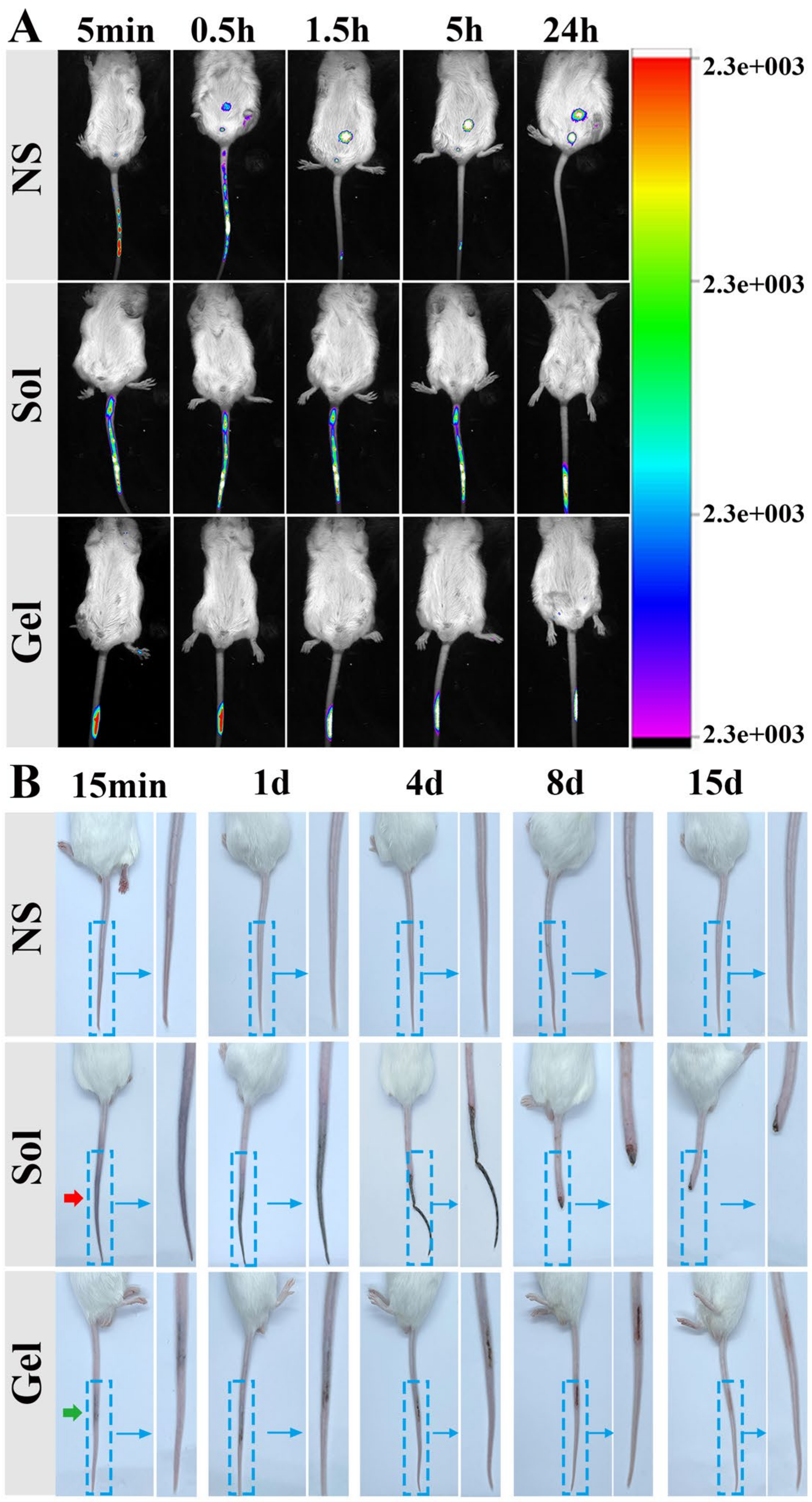
2.8.1. Study on Retention of TA9 Hydrogel In Vivo

Nine KM mice were used for in vivo retention studies of the hydrogels. The mice were divided randomly into three treatment groups, including NS, 8% TA9 solution, and 47.4% TA9 hydrogel. Then, 30 µL of the above three treatments containing the same amount of the fluorescent substance Cy7 were slowly injected into mouse tail veins. The distribution of Cy7 was recorded and quantified by an in vivo imaging system (IVIS, Bruker In-Vivo Xtreme, Karlsruhe Germany) at 5 min, 0.5 h, 1.5 h, 5 h, and 24 h.

2.8.2. Evaluation of Embolus in Mouse Tail Vein

Venous embolus in mouse tail vein was evaluated. Fifteen KM mice were divided into three treatment groups, NS, 8% TA9 solution, and 47.4% TA9 hydrogel, for in vivo evaluation of venous damage. The above treatment formulations (30 µL of each) were injected into individual mouse tail veins. The changes in each tail vein were inspected macroscopically, and photos were taken at 15 min, 1 day, 4 days, 8 days, and 15 days.

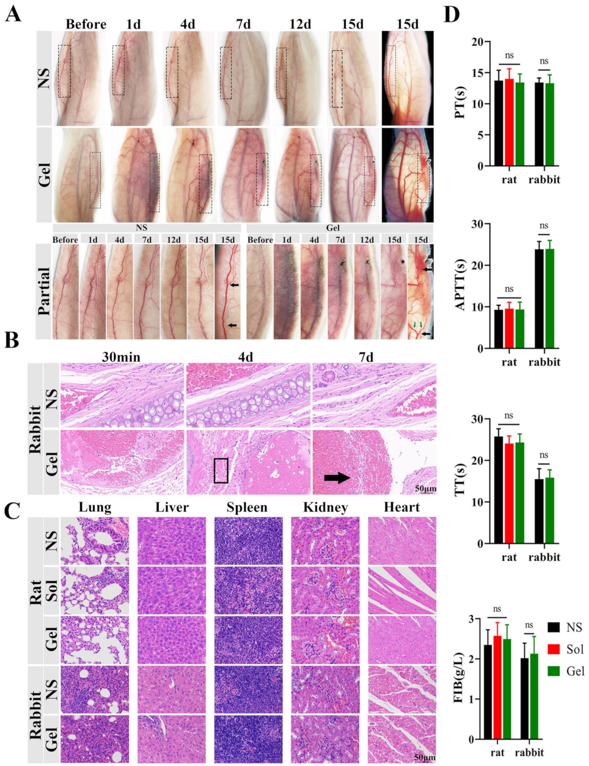
2.8.3. Evaluation of Embolus in Rabbit Ear Vein

Venous embolus in rabbit ear vein was evaluated according to the methods of a previous report [32]. The blood circulation system of the rabbit ear has abundant arteriovenous anastomosis and an interconnected venous network, the structure of which is comparable to the sinusoidal distribution of venous malformations, so it has been adopted as an animal model. Ten healthy New Zealand rabbits were divided randomly into two groups for in vivo evaluation of embolus. NS and 47.4% TA9 hydrogel (50 mL each) were injected into the marginal ear veins of individual testing rabbits. The macroscopic changes in the ear veins were observed, and photos were taken at 1 day, 4 days, 7 days, 12 days, and 15 days.
At 30 min, 4 days and 7 days after injection, one rabbit of each group was euthanized. The ear veins were collected, and fixed with 4% paraformaldehyde, paraffin sectioned, and H and E staining was performed to assess histological changes according to a previous report’s methods.

2.8.4. Evaluation of the Systemic Safety Profiles of TA Hydrogels

The biocompatibility of a drug is one of the critical factors determining whether it can be practically used in clinics. So, we determined the prothrombin time (PT), activated partial thromboplastin time (APTT), thrombin time (TT), and fibrinogen content (FIB) of the tested mice and rabbits to evaluate the effects of TA9 on coagulation function after treatment and recorded the life activities and survival status of animals to assess the safety of TA9. At the end of the experiments, rabbits and mice were euthanized and their hearts, livers, spleens, lungs, and kidneys were taken for histological observation by H and E staining.

2.9. Statistical Analysis

All data were presented as mean ± standard deviation (SD). Statistical analysis was done using one-way analyses of variance (ANOVA) with Graphpad Prism 8 (Graphpad Software, San Diego CA, USA). In all experiments, *p < 0.05, **p < 0.01, ***p < 0.001, ****p < 0.0001 were considered statistically significant.

3. Results

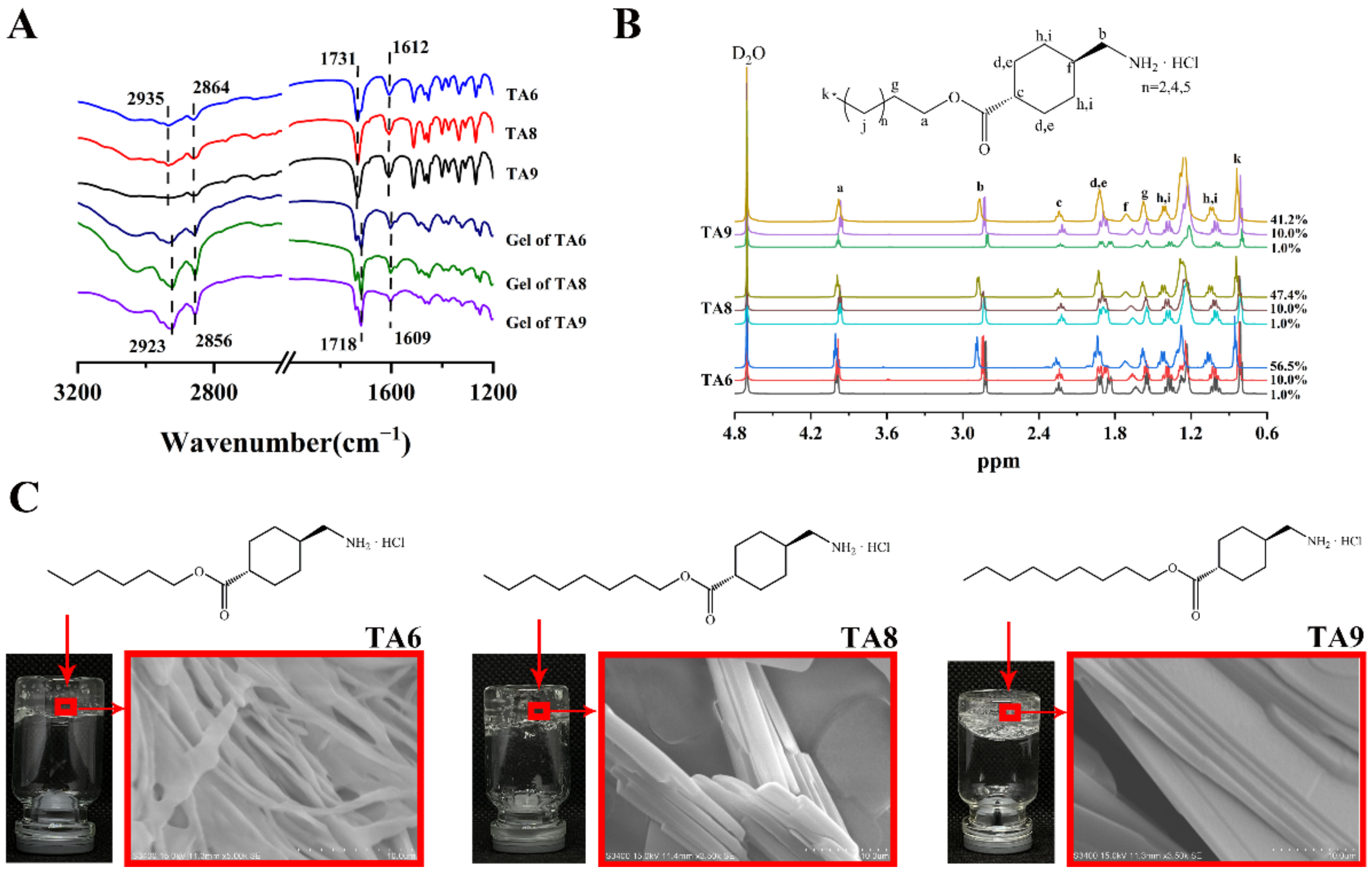
3.1. Synthesis and Characterization of TA6, TA8, and TA9

TA6 was synthesized as a white crystal. The yield of TA6 was 96.83% and the melting point was 125–126 °C.
1H-NMR (600 MHz, D2O) δ 3.99 (t, J = 6.7 Hz, 2H), 2.82 (d, J = 7.1 Hz, 2H), 2.31–2.17 (m, 1H), 1.92 (d, J = 5.4 Hz, 2H), 1.84 (d, J = 12.2 Hz, 2H), 1.67–1.60 (m, 1H), 1.58–1.49 (m, 2H), 1.37 (qd, J = 13.0, 3.2 Hz, 2H), 1.32–1.08 (m, 6H), 1.01 (qd, J = 13.0, 3.3 Hz, 2H), 0.81 (t, J = 6.8 Hz, 3H). 13C-NMR (151 MHz, MeOD) δ 175.79, 64.12, 44.64, 42.60, 35.24, 31.07, 28.70, 28.21, 27.85, 25.20, 22.11, 12.83. MS (ESI) m/z: [M+H]+ calcd for C14H28NO2: 241.38, founded 242.20.
TA8 was synthesized as a white crystal. The yield of TA6 was 94.52% and the melting point was 129–130 °C.
1H-NMR (600 MHz, D2O) δ 3.98 (t, J = 6.8 Hz, 2H), 2.81 (d, J = 7.1 Hz, 2H), 2.22 (dd, J = 9.6, 6.2 Hz, 1H), 1.91 (d, J = 12Hz, 2H), 1.84 (d, J = 11.2 Hz, 2H), 1.63 (dd, J = 7.3, 3.7 Hz, 1H), 1.54 (dd, J = 14.1, 6.9 Hz, 2H), 1.36 (tt, J = 13.0, 6.5 Hz, 2H), 1.29–1.10 (m, 10H), 0.99 (dt, J = 12.8, 9.8 Hz, 2H), 0.80 (t, J = 6.9 Hz, 3H). 13C-NMR (151 MHz, MeOD) δ 175.78, 64.11, 44.65, 42.61, 35.25, 31.45, 28.82, 28.79, 28.71, 28.24, 27.86, 25.52, 22.19, 12.92. MS (ESI) m/z: [M+H]+ calcd for C16H32NO2: 269.43, founded 270.20.
TA9 was synthesized as a white crystal. The yield of TA6 was 97.09% and the melting point was 130–131 °C.
1H-NMR (600 MHz, D2O) δ 3.97 (t, J = 7.0 Hz, 2H), 2.84 (d, J = 7.0 Hz, 2H), 2.21 (dd, J = 19.9, 7.7 Hz, 1H), 1.90 (t, J = 13.9 Hz, 4H), 1.75–1.61 (m, 1H), 1.55 (dd, J = 13.4, 6.6 Hz, 2H), 1.47–1.32 (m, 2H), 1.14–1.31 (m, J = 18.1 Hz, 12H), 1.08–0.93 (m, 2H), 0.81 (t, J = 6.8 Hz, 3H). 13C-NMR (151 MHz, MeOD) δ 175.78, 64.10, 44.65, 42.61, 35.25, 31.52, 29.11, 28.85, 28.83, 28.71, 28.24, 27.86, 25.51, 22.23, 12.93. MS (ESI) m/z: [M+H]+ calcd for C17H34NO2: 283.46, founded 284.30.

3.2. Metabolism Study of TA6, TA8, and TA9 In Vitro

The synthesized TA6, TA8, and TA9 were stable in both NS and PBS (pH 7.4) at 37 °C for 24 h (Figure 2A). So, the influence on subsequent related experiments due to the degradation of esters in TA compounds could be excluded. However, these TA compounds were rapidly cleaved in plasma after approximately 20 min (Figure 2B), clearly indicating that the esters could be completely hydrolyzed by enzymes in plasma, which also confirmed the possibility of the esters being degraded after entering the blood.

3.3. Study on Self-Assembly Behaviors of TA6, TA8, and TA9 Hydrogels

Herein, to understand the molecular self-assembly process in the hydrogels of TA6, TA8, and TA9, we performed FTIR studies and concentration-dependent 1H-NMR experiments. In the FTIR spectra (Figure 3A), we monitored the stretching frequency of hydrogen-bond functionalities. The typical bands at 1731 cm−1 and 1609 cm−1 of the TA6, TA8, and TA9 powders were assigned to νC=O and δN-H, respectively. In contrast, the shift of wavenumber to 1718 cm−1 and 1602 cm−1 showed that both C=O and N–H in the gelators were involved in hydrogen-bonding interactions in the corresponding hydrogels. Additionally, for the original powders of compounds, absorption bands appearing at 2935 cm−1 and 2864 cm−1 were ascribed to νasC-H and νsC-H of the methylene group on the alkyl chain, while these bands were shifted slightly to 2923 cm−1 and 2856 cm−1 in the dried gels of compounds, respectively, indicating that van der Waals forces and hydrophobic forces led to the formation of strong aggregation of alkyl chains, such that the mobility of hydrophobic chains decreased. Studies suggest that the strength of hydrogen bonds in water is weak, so it has been inferred that the hydrophobic force plays a more significant role in the anisotropic aggregation process of water [33].
As shown in the 1H-NMR spectra (Figure 3B), with the concentration of different gelators increasing between solution and hydrogel, the proton chemical shift of C–H in the alkyl chain shifted to a lower field, which implied the presence of van der Waals forces and hydrophobic forces in the hydrogel system. At the same time, the protons in cyclohexane group (d, e) split from double peaks to multiple peaks, which showed that the free vibration of molecules was reduced [34]. The network structures of the three hydrogels composed of fibers with evidently distinct thicknesses, widths, and morphologies were observed by SEM. With the increase of molecular carbon chains, the widths and thicknesses of the hydrogel fibers increased obviously (Figure 3C). This difference may be due to the joint action of the above molecular structure and driving forces.

3.4. Characterization of TA6, TA8, and TA9 Hydrogels

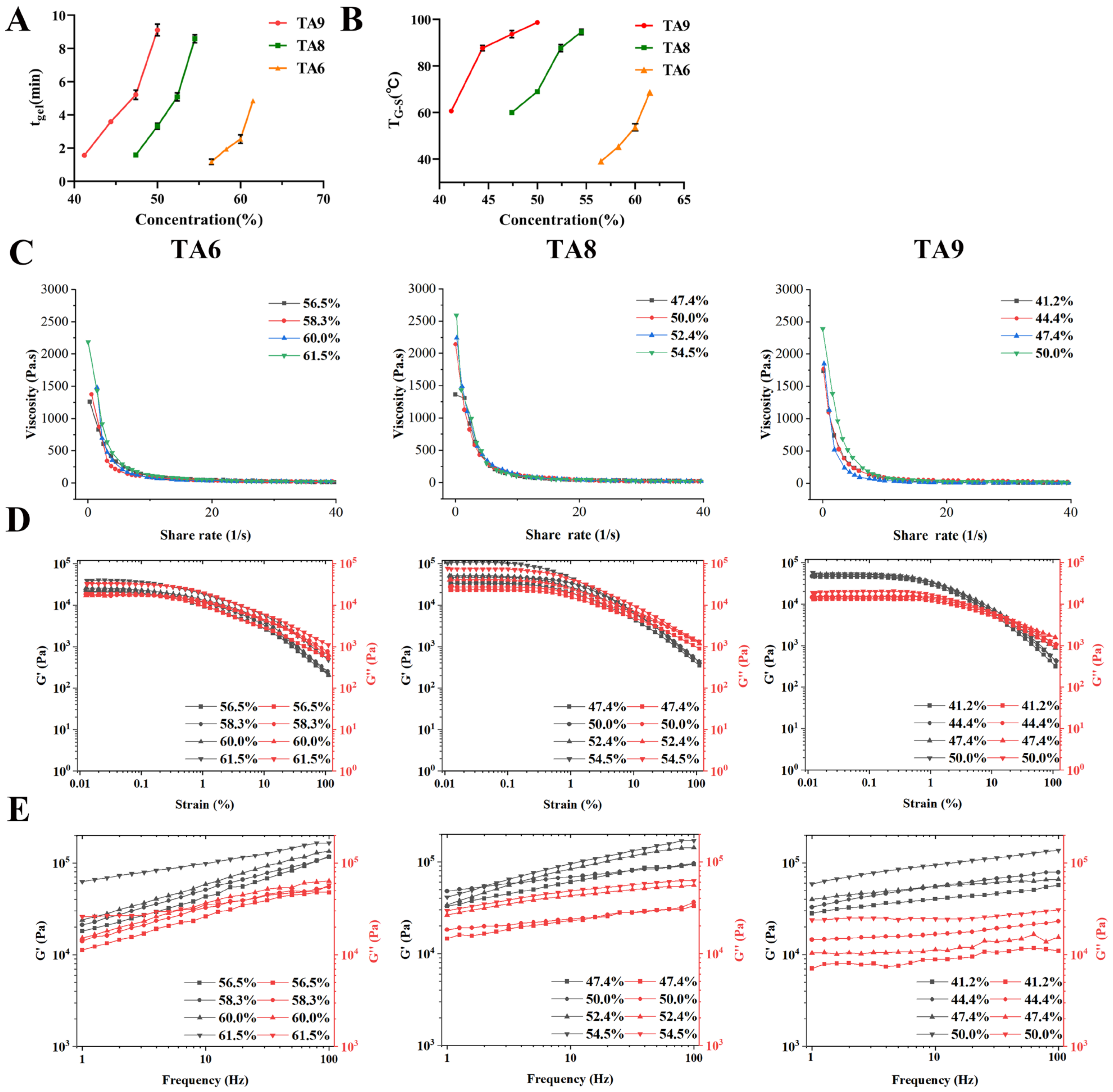
3.4.1. Determination of CGC, tgel, and TG-S

The CGCs of TA6, TA8, and TA9 were determined to be 55.9%, 46.8%, and 38.3%, respectively. The lower value of CGC represented the stronger gelation ability. The CGC of TA6 was higher than that of TA9, which reflected the vital influence of carbon chain length on gelation ability. tgel and TG-S were increased with the increase of gelator concentration and carbon chain length (Figure 4A, B). The results were consistent with other previous reports [18]. These phenomena could be attributed to the increase of van der Waals forces and hydrophobic forces induced by longer carbon chains, which made the hydrogel structure undergo slight but complex changes, requiring a longer time to stabilize and a higher temperature to function. The three kinds of hydrogels were thermally reversible, that is, they could be heated repeatedly and cooled without destroying the gel properties. All the TG-S of the hydrogels were higher than 37 °C, which was conducive to the stability of the hydrogel in vivo. The tgel of all hydrogels were within 10 min, which is convenient for the hydrogels to form when administered to avoid other problems caused by storage. However, hydrogels can be stable for several months at room temperature under sealed conditions.

3.4.2. Rheology Studies

The rheology of the TA6, TA8, and TA9 hydrogels at several concentrations was investigated. The results showed that, as the shear rate increased, the viscosity of all the hydrogels decreased (Figure 4C), demonstrating that they were shear-thinning, non-Newtonian fluids, which facilitated the injection and administration of the hydrogels and the filling of irregular vascular cavities. There were differences in the rheology profiles of the three hydrogels. The initial viscosity increased with the increase of the concentration, but the viscosity decrease rate was the opposite, indicating that the increase in concentration enhances the capacity of the hydrogels to withstand shear, which could be explained by the augmented intermolecular force. The stress-sweep experiment showed that the linear viscoelastic regions (LVR) of the TA6, TA8, and TA9 hydrogels were 0.1%, 0.3%, and 0.6%, respectively. The increased LVR indicated that the longer carbon chains enhanced the ability of the hydrogels to resist external stress, which could be related to the magnitude of the intermolecular force. However, the differences in LVR data among different concentrations of the same TA gels were not distinct (Figure 4D). After determining the LVR of all hydrogels, we adopted 0.01% as the strain concentration in subsequent experimental conditions. The frequency dependences of G’ of the TA6, TA8, and TA9 hydrogels were higher than their corresponding G”, which demonstrated the typical viscoelastic properties of gels [35], and gelator strength varied with concentration and by species. Furthermore, as the frequency increased, the G’ and G’’ of the TA9 hydrogel increased less, indicating that the structure of the TA9 hydrogel was more stable compared with those of TA6 and TA8 (Figure 4E). In addition, the mechanical properties of the TA9 hydrogel injected by injector were not affected, which showed the steady mechanical properties of the hydrogel as a self-delivery carrier for intravenous administration.

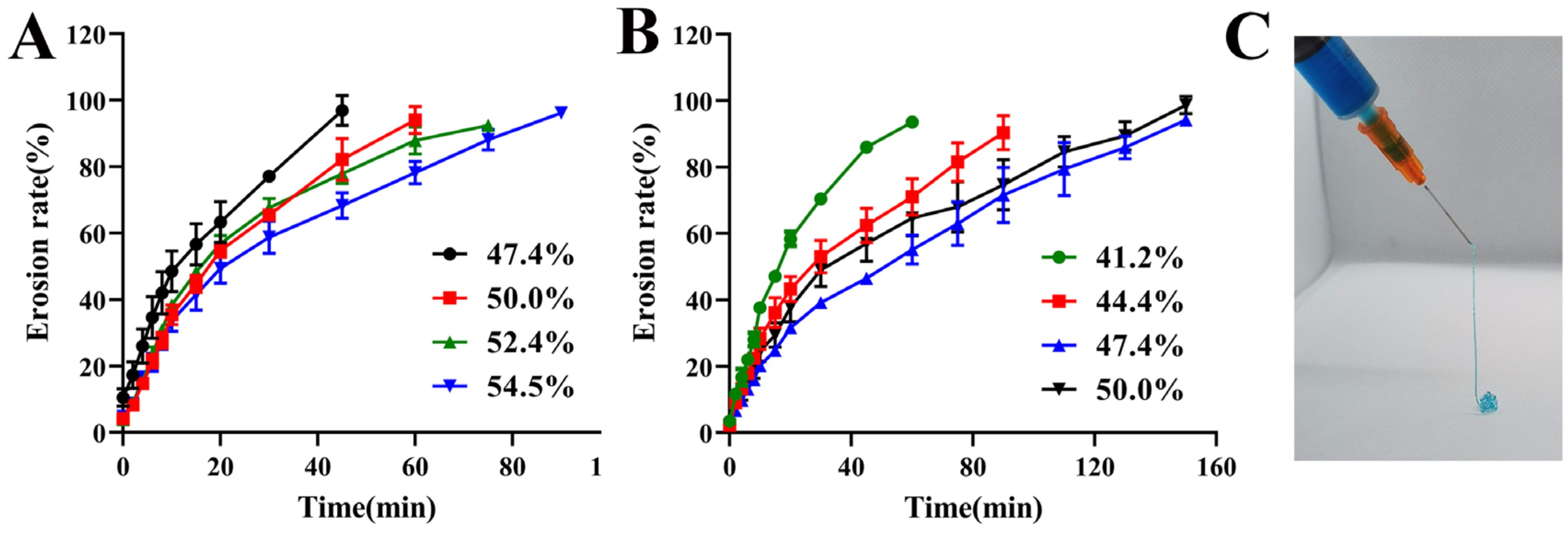
3.4.3. Erosion Behavior and Syringeability Studies

In vitro erosion experiments simulated the erosion profiles of the hydrogels in the blood after entering the veins. It has been reported that the release behavior of drugs is consistent with the erosion behavior of a hydrogel when the drugs act as a carrier, such that erosion rate can represent drug release rate. In a preliminary experiment, it was found that the TA6 hydrogel was completely eroded in a very short period of time at the highest concentration, indicating that its poor ability to oppose erosion by the dissolution medium could lead to rapid drug release. Therefore, the TA6 hydrogel was eliminated in later studies. The erosion behaviors of the TA8 and TA9 hydrogels in NS showed a sustained release of drugs. The erosion rate decreased at higher concentrations of the same kind of hydrogel and at the same concentrations of the hydrogel with longer alkyl chains, implying that higher concentrations of the TA9 hydrogel were more instrumental in slowing the release of the drugs and prolonging treatment time (Figure 5A, B).
That a hydrogel can pass through syringe needles smoothly during administration is a prerequisite for successful intravenous administration. The viscosity of hydrogels is affected by the concentrations of TA. Hence, the appropriate concentrations were selected according to whether the hydrogels could smoothly pass-through syringes with 25-G needles. The results showed that the TA8 hydrogel at concentrations of 47.4% and 50.0%, and the TA9 hydrogel at concentrations of 41.2%, 44.4%, and 47.4% could pass through syringes smoothly (Figure 5C). Overall, based on the rheological properties and in vitro erosion experiments, we selected the TA9 hydrogel at a concentration of 47.4% for the subsequent in vivo study.

3.5. Study on the Mechanism of TA9 in Treating VM In Vitro

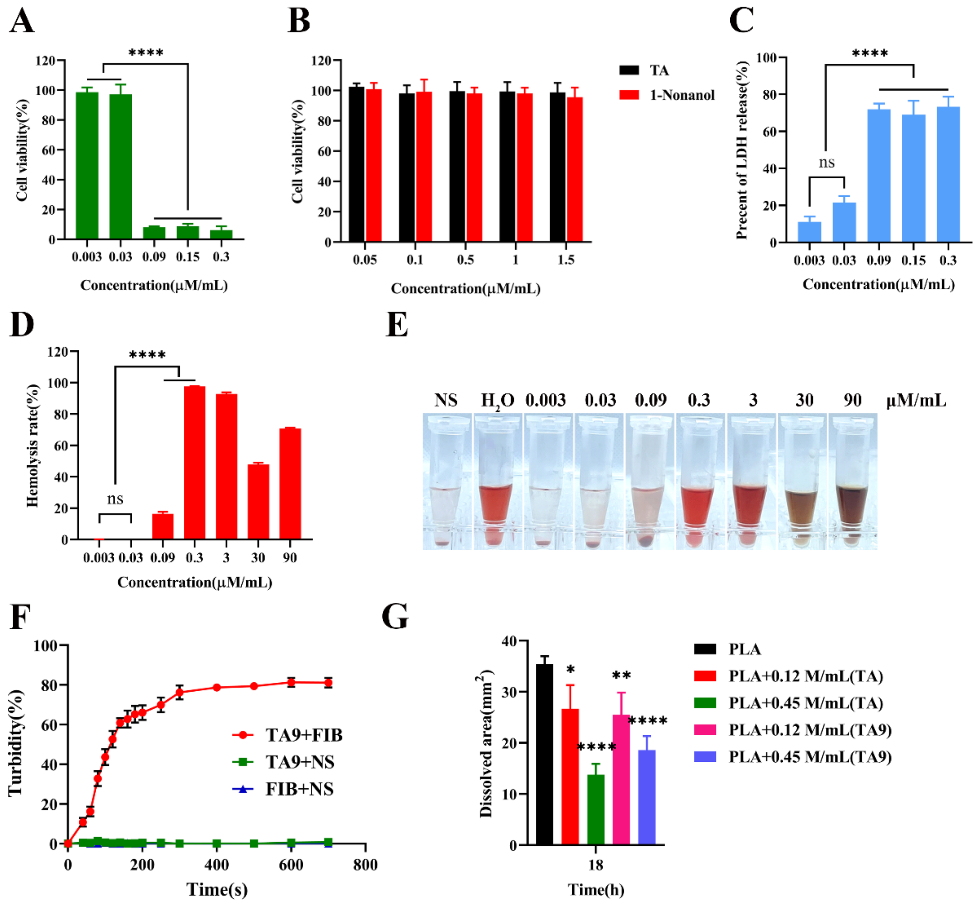
3.5.1. Cytotoxicity Study and Cell Membrane Integrity Assay

The cytotoxicity caused by TA9 was dose-dependent. Cell viability was higher than 95% at the concentration of 0.03 mM/mL. In comparison, it was only about 8% at a concentration of 0.09 mM/mL, suggesting that the cytotoxicity of TA9 was concentration-dependent, and that TA9 had a significant toxicity towards HUVECs after reaching a certain concentration (Figure 6A). Afterward, to ensure the cytotoxicity was induced by TA9 rather than by its metabolites, TA and 1-nonanol were incubated with HUVECs. Specifically, the results revealed that cell viability was approximately 98%, even at 1.5 mM/mL. Therefore, we determined that the cytotoxicity originated from TA9 and proved the safety of TA9 after metabolism (Figure 6B). Considering the molecular structure of TA9, we hypothesized that its toxicity might be derived from its cationic amphiphilicity and that the cause of cell death might be the destruction of cell membranes and the leakage of cell contents. Thus, we indirectly evaluated the degree of cell membrane damage by the release of LDH. The percent of LDH release was also concentration-dependent; it was about 23% at a concentration of 0.03 mM/mL, while it was more than 70% at a concentration of 0.09 M/mL (Figure 6C). The results demonstrated TA9 had a remarkable effect on the integrity of the cell membranes and seemed consistent with the cytotoxicity results.

3.5.2. Hemolysis Assay and Effect of TA9 on Fibrinogen

The hemolysis experiment showed that TA9 at a concentration above 0.06 mM/mL caused hemolysis, which further proved that TA9 could destroy cell membranes. When the concentration was higher than 0.3 mM/mL, the hemolysis rate could not be determined accurately, as the color of hemoglobin changed (Figure 6D,E). It was speculated that TA9 might also change the structure of hemoglobin through electrostatic and hydrophobic effects after cell membrane disruption. Beyond that, we added TA9 hydrogel to the fibrinogen solution, and found that the system was drastically changed under turbidity, and flocculent precipitates continued to precipitate out, indicating that TA9 was able to interact with fibrinogen and that the mechanism of action might be similar to that of hemoglobin (Figure 6F).

3.5.3. Evaluation of TA Inhibition on Plasmin

TA9, as a gelator, can be degraded into TA, a plasmin inhibitor. Therefore, we determined the inhibitory effect of TA9 on plasmin after its alkaline hydrolysis using the fibrin plate method. Plasmin will dissolve fibrin and make the fibrin plate appear as a transparent dissolving circle. The dissolution areas represent the degree of fibrinolysis by plasmin, and indirectly indicate the activity of plasmin (Figure 6G). The results confirmed that TA and the hydrolyzed TA9 could significantly inhibit the activity of plasmin in a concentration-dependent manner. This inhibitory effect probably could maintain the stability of a thrombus that had formed in the body, slowing down the degradation of the thrombus by plasmin and extending the embolization time in veins.

3.6. Pharmacodynamic Evaluation In Vivo

3.6.1. Study on the Retention of TA9 Hydrogel In Vivo

The retention and target of the TA9 hydrogel were investigated by analyzing the spread of the fluorescent marker Cy7 in vivo (Figure 7A). The fluorescent marker, in the NS group, was distributed in the whole rat tail vein at 30 min, then disappeared at 5 h, whereas most of fluorescence in the TA solution group and TA hydrogel group was still concentrated in the tail vein, even after 24 h. The difference was that the fluorescent marker in the TA hydrogel group only stayed at the administration site, and that of the TA solution group was distributed in the whole rat tail vein, illustrating that the TA hydrogel had better retention and could achieve the target delivery of TA in malformed veins.

3.6.2. Evaluation of Embolus in Mouse Tail Vein

The efficacy of TA9 was studied in rat tail vein after intravascular administration (Figure 7B). There was no significant change in the tail in the NS group upon observation. However, 15 min after injection of the 8% TA9 solution, the mouse tail was blackened from the injection site to the tail tip (shown with a red arrow), and the tail tips became necrotic 4 days later. This might be because the 8% TA9 solution could not be contained at the administration site and diffused, resulting in a large area of uncontrollable thrombus, and necrosis of the tail tip. In the TA hydrogel group, evident thrombus (shown with a green arrow) and occlusion were observed at the administration site within 15 min and at 15 days after administration, respectively, but no thrombus or other negative effects were found at other sites, demonstrating the targeting ability of TA9 hydrogel to induce venous occlusion. Compared with the solution group, TA9 hydrogel had better biocompatibility. Owing to the possible side effects of the 8% TA9 solution, we decided not to test it in rabbits.

3.6.3. Evaluation of Embolus in Rabbit Ear Vein

The rabbit auricular vein system was utilized as a model of slow blood flow velocity to monitor the efficacy of TA9 hydrogel in vivo (Figure 8A). There were no other damages incurred to rabbit ears in the NS group. However, in the TA9 hydrogel group, upon injecting TA9 hydrogel, local thrombus and inflammation evidently took place in the vein and surrounding tissues. At 15 d, inflammation and thrombus disappeared, and no blood was found flowing through the vein treated with the TA9 hydrogel. The occlusion appeared to be irreversible over one month, and no tissue necrosis was observed. From the beginning of venous embolization, the blood flow in marginal ear veins was mainly dependent on small collateral veins to preserve the average blood circulation of rabbit ears, and the collateral veins were observed to become thicker (shown with a green arrow), ensuring the everyday life activities of the rabbit ears. It was worth noting that the vascular occlusion site was consistent with the administration position (shown with a black arrow), and no other adverse reactions, such as tissue necrosis and distal thrombosis. were observed.
Micrographs of H-and-E-stained rabbit ear veins at 30 min, 4 d, and 7 d after NS and TA9 hydrogel treatments were also taken to provide more histological information about the efficacies of the therapies (Figure 8B). No thrombus or other lesions were discovered in the NS group. In the TA9 hydrogel group, necrosis of endothelial cells, along with the formation of mixed thrombus in the vein, were observed at 30 min after administration. In other words, the white thrombus formed by the adhesion and aggregation of platelets adhered to the venous wall, and extended inward, slowing down or blocking blood flow, causing the central red blood cells to aggregate into clusters and be bound by the fibrin network to form a red thrombus, which made the vein embolized. On day 4, the venous wall was seriously damaged, resulting in the leakage of red blood cells into the surrounding tissues (shown in the black box). After 7 days, the inflammation of the surrounding tissue was alleviated, and endothelial cells and fibroblasts appeared around and inside the thrombus, indicating the proliferation of fibrous tissue (shown with a black arrow). In fact, after thrombosis, TA gel could activate the fibrinolytic system to dissolve the thrombus. It was found that the thrombus could be maintained at the administration site for a long time, which might be because TA could retain stability after TA9 hydrolysis. To sum up, both macroscopic and histological investigations showed that TA9 caused vein fibrosis by destroying endothelial cells and damaging venous walls. These results correlated very well with the macroscopic observation presented above.

3.6.4. Evaluation of Systemic Safety Profiles of TA Hydrogels

H and E staining showed no thrombus or lesions in the examined organs after treatment (Figure 8C). There was no significant difference in the data of PT, APTT, TT and FIB in rabbits and mice after treatments with NS, 8% solution, or TA hydrogel (p > 0.05), indicating that the therapeutic dose of TA9 did not affect their coagulation function (Figure 8D). Rabbits and mice could survive for one month without abnormal symptoms, such as weight loss, anorexia, or hemiplegia, which convincingly proved that TA9 had high biocompatibility.

4. Discussion

Vascular malformation includes capillary malformation (CM), arteriovenous malformation (AVM), venous malformation (VM), lymphatic malformation (LM), and so forth. VM is the most common type of congenital vascular malformation. About 40% of VMs occur in the mouth, head, neck, or face with complex structure and vascular plexus [36,37]. Surgical resection is challenging in curing VM entirely because of the particular location of the lesion and the infiltration and expansion of adjacent tissues, and, in severe cases, will lead to severe complications, such as bleeding and nerve injury [3]. As the primary treatment method, sclerotherapy has reached expert consensus [38]. In this study, we synthesized a novel type of cationic amphiphilic molecule with TA as the basic structure, which could be driven by hydrogen bond, van der Waals forces and hydrophobic forces to form a transparent hydrogel as a self-delivery carrier. According to previous reports, gelators usually contain both hydrophilic and hydrophobic groups to successfully self-assemble in water or organic solvents [39]. However, much work is needed in the development of gelators, as strictly controlling the hydrophilic and hydrophobic equilibrium of gelators is pivotal. In hydrogels, hydrophilic groups (such as carboxylic acids, amino acids, sugars, phosphates, and quaternary amines) with rigid segments and flexible tails are considered the essential elements for the stable self-assembly of most gelators. The hydrophilic groups advance dissolution, while the hydrophobic carbon chain promotes aggregation [40]. Scientists introduce hydrophilic or hydrophobic groups into compounds that are easily modified or can provide a self-assembly driving force, then empower them with a balanced amphipathic ability to achieve self-installation [18,19]. As for TA derivatives, the groups of amino, cyclohexyl, and carbon chains can better balance their hydrophilic and hydrophobic properties. TA6, TA8, and TA9 could gelatinize not only in purified water but also in NS and PBS (pH 7.4). Therefore, hydrogels could stably exist in the physiological environment of the body and become an effective drug delivery system. From the results of the hydrogel characterization, it was found that the increased length and concentration of carbon chains affected the thermal stability and mechanical properties of the hydrogels [26], which was attributed to the slight changes (of length, thickness, and width) in the hydrogel fibers caused by the change of intermolecular van der Waals forces and hydrophobic forces, and this change enhanced the mechanical strength of the hydrogel. The augmented mechanical strength was conducive to the rapid displacement of blood to block veins after the hydrogels were injected, which could allow fully contacting the venous wall and resisting the erosion of the gels by the blood, enhancing the retention and prolonging the action time of loaded drugs. Furthermore, good retention and slow release of hydrogels can reduce many side effects caused by TA9, therefore improving its safety. Considering all of the desirable properties of hydrogels, we chose the 47.4%-TA9 hydrogel as the most appropriate drug self-assembly carrier in this study.
The treatment of VM with surfactant sclerosants (such as STS) depends on the disruption of endothelial cells by surfactant sclerosants. As a cationic amphiphilic molecule, TA9 probably has a similar mechanism to those of surfactant sclerosants to induce cytotoxicity and hemolysis [41]. The positively charged TA9 can be adsorbed on the negatively charged cell membrane surface by electrostatic interaction [42]. Amphiphilicity causesTA9 to be arranged on the membrane surface or to be directly inserted into the membrane in the form of monomers or an aggregate. After reaching saturation on the membrane or reaching the critical micelle concentration (CMC) in the medium, TA9 reduces the surface tension of the cell membrane, increases the membrane permeability, or forms an aggregate directly with phospholipids and proteins on the membrane, resulting in the rupture or dissolution of the cell membrane [43]. The interactions between TA9 and protein could be applied in explaining the mechanisms of the TA9 hydrogel. The polar head group and non-polar alkyl chain of TA9 act on the hydrophilic and hydrophobic parts of the protein with electrostatic and hydrophobic interactions, respectively, changing the protein structure to promote protein aggregation. For example, commonly used sclerosing agents (e.g., STS and POL), were found to interact with blood proteins (including albumin, fibrinogen, apolipoprotein, etc.) [44]. When TA9 hydrogel was added to plasma, flocculent precipitates were observed, suggesting that TA9 might act not only upon fibrinogen and hemoglobin but also on other proteins in the blood. Whether TA9 forms a reversible complex with a protein or destroys the protein structure to inactivate it needs to be further researched. Particularly, some researchers believe that complexation with proteins can reduce drug toxicity and the possibility of thrombosis or other adverse reactions in deep veins, due to drug diffusion [45].
An apparent thrombus emerged at the administration site within 15 min after administration, suggesting that TA9 hydrogel took effect quickly. H and E staining displayed a white thrombus formed by platelet aggregation and a red thrombus formed by the accumulation of red blood cells, from which it could be inferred that the hydrogel or new thrombus slowed down blood flow. This thrombus was formed due to the severe destruction of intravascular cells and proteins by TA9, resulting in serious damage to the vascular endothelium and activation of the coagulation system. Perhaps the formation of thrombi was capable of reducing the erosion rate of blood to the hydrogel. On the other hand, part of the TA9 released from the hydrogel could be rapidly metabolized to TA, in vivo. TA is capable of blocking lysine-binding sites on plasminogen molecules and, thereby, inhibiting the interaction of plasminogen and the heavy chain of plasmin with lysine residues on the surface of fibrin, enhancing the tightness of thrombi and the persistence of embolus. So, TA9 hydrogel, as a sclerosant, may have a dual mechanism of “coagulation initiation—fibrinolysis inhibition”.
Rapid degradation of TA9 prevented the destruction of normal veins and tissues after TA9 diffusion, and endowed the hydrogel with excellent biodegradability and biocompatibility, since the metabolites TA and 1-nonanol had no apparent toxicity on cells and did not affect the coagulation systems of the animals. Previous studies have shown that TA could improve platelet function and inhibit plasmin-induced platelet activation to promote the stability of existing clots rather than increase the risk of thrombosis [25]. Besides, plasminogen and plasmin played a vital role in the process of connecting fibrinolysis and the inflammatory system. TA might reduce the inflammatory response after thrombosis induced by other substances by inhibiting its mediated inflammation [46,47]. These, probably, are the most prominent differences between TA9 and other sclerosing agents, which greatly benefit the treatment of vascular malformations. We propose to further explore the mechanisms of action and efficacy of TA9 in a follow-up study.

5. Conclusions

We reported three kinds of cationic amphiphilic molecules (TA6, TA8, and TA9) synthesized from tranexamic acid and medium-chain alcohol in this study. These TA6, TA8, and TA9 molecules could self-assemble into hydrogels to enhance drug retention and release drugs in a continuous manner. Both in vivo and in vitro experiments showed that TA9 damaged the venous wall and caused thrombosis and fibrotic veins for the treatment of VM. More importantly, TA9 could be degraded into TA by enzymes to inhibit thrombus degradation and its degradation products had no apparent toxicity, indicating that TA9 might become a drug self-delivery carrier with positive biodegradability and biocompatibility for the treatment of venous malformations.

6. Patent

The patent application related to the study in the paper has been filed in China (application number 2021111549351.4).

Author Contributions

Conceptualization, P.D. and H.X.; methodology, Y.C.; software, D.S.; validation, M.M.; formal analysis, X.Z.; investigation, Q.H.; resources, D.S.; data curation, Q.H.; writing—original draft preparation, Y.C.; writing—review and editing, T.Y. All authors have read and agreed to the published version of the manuscript.

Funding

The research was supported by School Seeding Grant of Shenyang Pharmaceutical University (Grant No. 43210078).

Institutional Review Board Statement

All animal experiments and procedures were reviewed and approved by the Animal Care and Use Committee at the Shenyang Pharmaceutical University (Ethical Committee approval number: SYPU-IACUC-C2019-12-14-105; date of approval: 14 December 2019).

Informed Consent Statement

Not applicable.

Data Availability Statement

The data presented in this study are available on request from the corresponding author.

Conflicts of Interest

The authors declare no conflict of interest.

References

  1. Mulliken, J.; Glowacki, J. Hemangiomas and vascular malformations in infants and children: A classification based on endothelial characteristics. Plast. Reconstr. Surg. 1982, 69, 412–422. [Google Scholar] [CrossRef] [PubMed]
  2. Lee, B.; Baumgartner, I.; Berlien, P.; Bianchini, G.; Burrows, P.; Gloviczki, P.; Huang, Y.; Laredo, J.; Loose, D.; Markovic, J.; et al. Guideline. Diagnosis and treatment of venous malformations. consensus document of the international union of phlebology (iup): Updated-2013. Int. Angiol. J. Int. Union Angiol. 2014, 34, 97–149. [Google Scholar]
  3. Wetzel-Strong, S.; Detter, M.; Marchuk, D. The pathobiology of vascular malformations: Insights from human and model organism genetics. J. Pathol. 2017, 241, 281–293. [Google Scholar] [CrossRef] [PubMed]
  4. El-Hakim, I.; Alyamani, A. Management of palatal vascular malformation using absolute ethanol sclerotherapy. Clin. Pract. 2011, 1, e86. [Google Scholar] [CrossRef] [PubMed]
  5. Eckmann, D. Polidocanol for endovenous microfoam sclerosant therapy. Expert Opin. Investig. Drugs 2009, 18, 1919–1927. [Google Scholar] [CrossRef] [PubMed]
  6. Jin, Y.; Lin, X.; Chen, H.; Hu, X.; Fan, X.; Li, W.; Ma, G.; Yang, C.; Wang, W. Auricular arteriovenous malformations: Potential success of superselective ethanol embolotherapy. J. Vasc. Interv. Radiol. JVIR 2009, 20, 736–743. [Google Scholar] [CrossRef]
  7. Hayden, M.R. Developmental Biology: Frontiers for Clinical Genetics. Clin. Genet. 2014, 86. [Google Scholar] [CrossRef]
  8. Horbach, S.; Lokhorst, M.; Saeed, P.; de Goüyon Matignon de Pontouraude, C.; Rothová, A.; van der Horst, C. Sclerotherapy for low-flow vascular malformations of the head and neck: A systematic review of sclerosing agents. J. Plast. Reconstr. Aesthetic Surg. 2016, 69, 295–304. [Google Scholar] [CrossRef]
  9. Chen, F.; Song, S.; Wang, H.; Zhang, W.; Lin, C.; Ma, S.; Ye, T.; Zhang, L.; Yang, X.; Qin, X.; et al. Injectable chitosan thermogels for sustained and localized delivery of pingyangmycin in vascular malformations. Int. J. Pharm. 2014, 476, 232–240. [Google Scholar] [CrossRef]
  10. Van Damme, A.; Seront, E.; Dekeuleneer, V.; Boon, L.M.; Vikkula, M. New and Emerging Targeted Therapies for Vascular Malformations. Am. J. Clin. Dermatol. 2020, 21, 657–668. [Google Scholar] [CrossRef]
  11. Zhang, L.; Chen, F.; Zheng, J.; Wang, H.; Qin, X.; Pan, W. Chitosan-based liposomal thermogels for the controlled delivery of pingyangmycin: Design, optimization and in vitro and in vivo studies. Drug Deliv. 2018, 25, 690–702. [Google Scholar] [CrossRef] [PubMed]
  12. Del Castillo-Santaella, T.; Yang, Y.; Martínez-González, I.; Gálvez-Ruiz, M.; Cabrerizo-Vílchez, M.; Holgado-Terriza, J.; Selles-Galiana, F.; Maldonado-Valderrama, J. Effect of Hyaluronic Acid and Pluronic-F68 on the Surface Properties of Foam as a Delivery System for Polidocanol in Sclerotherapy. Pharmaceutics 2020, 12, 1039. [Google Scholar] [CrossRef] [PubMed]
  13. Xie, L.; Yue, W.; Ibrahim, K.; Shen, J. A Long-Acting Curcumin Nanoparticle/In Situ Hydrogel Composite for the Treatment of Uveal Melanoma. Pharmaceutics 2021, 13, 1335. [Google Scholar] [CrossRef] [PubMed]
  14. Pierre, T.; Weiss, R.G. Low Molecular Mass Gelators of Organic Liquids and the Properties of Their Gels. Chem. Rev. 1997, 97, 3133–3160. [Google Scholar]
  15. Majumder, J.; Dastidar, P. An Easy Access to Organic Salt-Based Stimuli-Responsive and Multifunctional Supramolecular Hydrogels. Chem. Eur. J. 2016, 22, 9267–9276. [Google Scholar] [CrossRef] [PubMed]
  16. Li, X.; Bian, S.; Zhao, M.; Han, X.; Liang, J.; Wang, K.; Jiang, Q.; Sun, Y.; Fan, Y.; Zhang, X. Stimuli-responsive biphenyl-tripeptide supramolecular hydrogels as biomimetic extracellular matrix scaffolds for cartilage tissue engineering. Acta Biomater. 2021, 131, 128–137. [Google Scholar] [CrossRef] [PubMed]
  17. Ghosh, D.; Mulvee, M.T.; Damodaran, K.K. Tuning Gel State Properties of Supramolecular Gels by Functional Group Modification. Molecules 2019, 24, 3472. [Google Scholar] [CrossRef] [PubMed]
  18. Roy, S.; Dasgupta, A.; Das, P. Alkyl chain length dependent hydrogelation of L-tryptophan-based amphiphile. Langmuir ACS J. Surf. Colloids 2007, 23, 11769–11776. [Google Scholar] [CrossRef]
  19. Cinar, G.; Ozdemir, A.; Hamsici, S.; Gunay, G.; Dana, A.; Tekinay, A.B.; Guler, M.O. Local delivery of doxorubicin through supramolecular peptide amphiphile nanofiber gels. Biomater. Sci. 2016, 5, 67–76. [Google Scholar] [CrossRef]
  20. Wu, C.; Wang, X.; Shi, Y.; Wang, B.; Xue, W.; Zhang, Y. Transforming sustained release into on-demand release: Self-healing guanosine-borate supramolecular hydrogels with multiple responsiveness for Acyclovir delivery. Biomater. Sci. 2020, 8, 6190–6203. [Google Scholar] [CrossRef]
  21. Guo, Q.; Liu, Y.; Mu, G.; Yang, L.; Wang, W.; Liu, J.; Liu, J. A peptide–drug hydrogel to enhance the anti-cancer activity of chlorambucil. Biomater. Sci. 2020, 8, 5638–5646. [Google Scholar] [CrossRef] [PubMed]
  22. Zhi, K.; Wang, J. Retraction: A self-assembled supramolecular natural product gel from liquidambaric acid in traditional Chinese medicine with inherent anti-inflammatory activity for drug delivery. J. Mater. Chem. B 2020, 8, 11109. [Google Scholar] [CrossRef] [PubMed]
  23. Zhou, Y.; Lei, L.; Zhang, Z.; Zhang, R.; Song, Q.; Li, X. Cation instructed steroidal prodrug supramolecular hydrogel. J. Colloid Interface Sci. 2018, 528, 10–17. [Google Scholar] [CrossRef] [PubMed]
  24. Vemula, P.K.; Wiradharma, N.; Ankrum, J.; Miranda, O.R.; John, G.; Karp, J.M. Prodrugs as self-assembled hydrogels: A new paradigm for biomaterials. Curr. Opin. Biotechnol. 2013, 24, 1174–1182. [Google Scholar] [CrossRef] [PubMed]
  25. Hanley, C.; Callum, J.; Jerath, A. Tranexamic acid and trauma coagulopathy: Where are we now? Br. J. Anaesth. 2021, 126, 12–17. [Google Scholar] [CrossRef]
  26. Delbecq, F.; Tsujimoto, K.; Ogue, Y.; Endo, H.; Kawai, T. N-stearoyl amino acid derivatives: Potent biomimetic hydro/organogelators as templates for preparation of gold nanoparticles. J. Colloid Interface Sci. 2013, 390, 17–24. [Google Scholar] [CrossRef]
  27. Komba, S.; Iwaura, R. NDevelopment of Low-Molecular-Mass Organogelators: Synthesis and Physical Properties of Linear Saturated Fatty Acyl-GABAs and Their Ester Derivatives. ACS omega 2021, 6, 20912–20923. [Google Scholar] [CrossRef]
  28. Miller, B.; Hansrisuk, A.; Highley, C.B.; Caliari, S.R. Guest–Host Supramolecular Assembly of Injectable Hydrogel Nanofibers for Cell Encapsulation. ACS Biomater. Sci. Eng. 2021, 7, 4164–4174. [Google Scholar] [CrossRef]
  29. Fan, J.; Zhong, H.; Zhang, X.; Yuan, T.; Chen, H.; Peng, H. Preparation and Characterization of Oleanolic Acid-Based Low-Molecular-Weight Supramolecular Hydrogels Induced by Heating. ACS Appl. Mater. Interfaces 2021, 13, 29130–29136. [Google Scholar] [CrossRef]
  30. Liu, H.; Sun, Y.; Lang, L.; Yang, T.; Zhao, X.; Cai, C.; Liu, Z.; Ding, P. Nuclear localization signal peptide enhances transfection efficiency and decreases cytotoxicity of poly(agmatine/N,N′-cystamine-bis-acrylamide)/pDNA complexes. J. Cell. Biochem. 2019, 120, 16967–16977. [Google Scholar] [CrossRef]
  31. Millar, W.T.; Smith, J.F.B. The comparison of solid phase and fibrin plate methods for the measurement of plasminogen activators. Thromb. Res. 1983, 30, 431–439. [Google Scholar] [CrossRef]
  32. Gao, Z.; Ding, P.; Zhang, L.; Shi, J.; Yuan, S.; Wei, J.; Chen, D. Study of a Pingyangmycin delivery system: Zein/Zein–SAIB in situ gels. Int. J. Pharm. 2007, 328, 57–64. [Google Scholar] [CrossRef]
  33. Xu, L.; Liang, Y.; Sun, C.; Hao, N.; Yan, J.; Gao, W.; He, B. Substitution of Percutaneous Ethanol Injection with a Low Molecular Weight Peptide Gel Mimicking Chemoembolization for Cancer Therapy. Nanotheranostics 2017, 1, 313. [Google Scholar] [CrossRef] [PubMed]
  34. Skilling, K.J.; Citossi, F.; Bradshaw, T.D.; Ashford, M.; Kellam, B.; Marlow, M. Insights into low molecular mass organic gelators: A focus on drug delivery and tissue engineering applications. Soft Matter 2014, 10, 237–256. [Google Scholar] [CrossRef] [PubMed]
  35. Alonci, G.; Mocchi, R.; Sommatis, S.; Capillo, M.; Liga, E.; Janowska, A.; Nachbaur, L.; Zerbinati, N. Physico-Chemical Characterization and In Vitro Biological Evaluation of a Bionic Hydrogel Based on Hyaluronic Acid and l-Lysine for Medical Applications. Pharmaceutics 2021, 13, 1194. [Google Scholar] [CrossRef] [PubMed]
  36. Brouillard, P.; Vikkula, M. Vascular malformations: Localized defects in vascular morphogenesis. Clin. Genet. 2003, 63, 340–351. [Google Scholar] [CrossRef]
  37. Prasetyono, T.; Kreshanti, P. Efficacy of intra-lesional alcohol injection as alternative and/or complementary treatment of vascular malformations: A systematic review. J. Plast. Reconstr. Aesthetic Surg. JPRAS 2010, 63, 1071–1079. [Google Scholar] [CrossRef]
  38. Heit, J.; Do, H.; Prestigiacomo, C.; Delgado-Almandoz, J.; English, J.; Gandhi, C.; Albuquerque, F.; Narayanan, S.; Blackham, K.; Abruzzo, T.; et al. Guidelines and parameters: Percutaneous sclerotherapy for the treatment of head and neck venous and lymphatic malformations. J. Neurointerv. Surg. 2017, 9, 611–617. [Google Scholar] [CrossRef]
  39. Bansode, N.D.; Sindhu, K.R.; Morel, C.; Rémy, M.; Verget, J.; Boiziau, C.; Barthélémy, P. A disulfide based low molecular weight gel for the selective sustained release of biomolecules. Biomater. Sci. 2020, 8, 3186–3192. [Google Scholar] [CrossRef]
  40. Estroff, L.; Hamilton, A. Water gelation by small organic molecules. Chem. Rev. 2004, 104, 1201–1218. [Google Scholar] [CrossRef]
  41. Khan, A.B.; Khan, J.M.; Ali, M.S.; Khan, R.H.; Din, K.U. Interaction of amphiphilic drugs with human and bovine serum albumins. Spectrochim. Acta Part A: Mol. Biomol. Spectrosc. 2012, 97, 119–124. [Google Scholar] [CrossRef]
  42. Torres-Luna, C.; Hu, N.; Koolivand, A.; Fan, X.; Zhu, Y.; Domszy, R.; Yang, J.; Yang, A.; Wang, N.S. Effect of a Cationic Surfactant on Microemulsion Globules and Drug Release from Hydrogel Contact Lenses. Pharmaceutics 2019, 11, 262. [Google Scholar] [CrossRef] [PubMed]
  43. Schreier, S.; Malheiros, S.V.; de Paula, E. Surface active drugs: Self-association and interaction with membranes and surfactants. Physicochemical and biological aspects. Biochim. Biophys. Acta (BBA) Biomembr. 2000, 1508, 210–234. [Google Scholar] [CrossRef]
  44. Parsi, K.; Exner, T.; Connor, D.; Herbert, A.; Ma, D.; Joseph, J. The lytic effects of detergent sclerosants on erythrocytes, platelets, endothelial cells and microparticles are attenuated by albumin and other plasma components in vitro. Eur. J. Vasc. Endovasc. Surg. Off. J. Eur. Soc. Vasc. Surg. 2008, 36, 216–223. [Google Scholar] [CrossRef] [PubMed]
  45. Parsi, K.; Exner, T.; Low, J.; Fung Ma, D.; Joseph, J. In vitro effects of detergent sclerosants on clot formation and fibrinolysis. Eur. J. Vasc. Endovasc. Surg. Off. J. Eur. Soc. Vasc. Surg. 2011, 41, 267–277. [Google Scholar] [CrossRef]
  46. Franchini, M.; Mannucci, P. The never ending success story of tranexamic acid in acquired bleeding. Haematologica 2020, 105, 1201–1205. [Google Scholar] [CrossRef]
  47. Renckens, R.; Weijer, S.; de Vos, A.; Pater, J.; Meijers, J.; Hack, C.; Levi, M.; van der Poll, T. Inhibition of plasmin activity by tranexamic acid does not influence inflammatory pathways during human endotoxemia. Arterioscler. Thromb. Vasc. Biol. 2004, 24, 483–488. [Google Scholar] [CrossRef] [PubMed]
Figure 1. (A) Synthetic routes of TA6 (n = 5), TA8 (n = 7), and TA9 (n = 8). (B) The mechanism of self-assembled TA9 hydrogel for treating VM. The TA9 hydrogel is injected to block the malformed veins and the TA9 is released in situ at the same time. Amphiphilic TA9 damages red blood cells and endothelial cells by destroying cell membranes and depositing proteins, causing serious damage to the venous wall and inducing thrombosis. Endothelial cells and fibroblasts grow from the venous wall to the thrombus to cause vein fibrosis. Finally, hydrogel blockage and thrombus embolus, together, induce the long-term occlusion of local veins.
Pharmaceutics 14 00343 g001
Figure 2. Metabolic kinetics of TA6, TA8, and TA9 in (A) NS and PBS (pH 7.4) and (B) rat plasma at 37 °C. All data are presented as the mean ± SD (n = 3).
Pharmaceutics 14 00343 g002
Figure 3. Study of the self-assembly behaviors of TA6, TA8, and TA9 hydrogel. (A) FITR spectra of powders and hydrogels of TA6 (56.5%), TA8 (47.4%), and TA9 (41.2%). (B) 1H-NMR spectra of TA6, TA8, and TA9 solutions (1% and 10%) and hydrogels of TA6 (56.5%), TA8 (47.4%), and TA9 (41.2%). (C) Chemical structure of TA6, TA8, and TA9; photographs and SEM images of TA6 (56.5%), TA8 (47.4%), and TA9 (41.2%) hydrogels. The scale bars represents 10 µm.
Pharmaceutics 14 00343 g003
Figure 4. Characterization of TA6, TA8, and TA9 hydrogels. (A) tgel, (B) TG-S of TA6, TA8, and TA9 hydrogels at different concentrations (n = 3). (C) Continuous flow, (D) stress sweep, and (E) frequency sweep analyses of the TA6, TA8, and TA9 hydrogels at four concentrations.
Pharmaceutics 14 00343 g004
Figure 5. (A,B) Erosion behaviors of the TA8 and TA9 hydrogels at different concentrations (n = 3); (C) 47.4% TA9 hydrogels could pass through a syringe with a 25-G needle smoothly (methylene blue staining was used for easy visualization).
Pharmaceutics 14 00343 g005
Figure 6. Study of the mechanism of TA9 in treating VM in vitro. (A) Cytotoxicity assay of HUVECs in various concentrations of TA9 solutions for 24 h (n = 5). (B) Cytotoxicity assay of HUVECs in different concentrations of TA and 1-nonanol solutions for 24 h (n = 5). (C) The release of LDH after incubating HUVECs with different concentrations of TA9 solutions for 24 h (n = 5). (D,E) Hemolysis assay of different concentrations of TA9 solutions for 2 h (n = 3). (F) Turbidity changes after TA9 hydrogel was added into fibrinolysin solution. (G) Inhibitory effects of different concentrations of TA and TA9 after alkaline hydrolysis on plasmin for 18 h. The data are presented as the mean ± SD (n = 3). * p < 0.05, ** p < 0.01, **** p < 0.0001.
Pharmaceutics 14 00343 g006
Figure 7. Embolus study on mouse tail vein. (A) IVIS in vivo images of a mice tail after injection of NS, 8% TA9 solution, and 47.4% TA9 hydrogel containing Cy7 at 24 h. (B) Embolus study on mouse tail vein for 15 d.
Pharmaceutics 14 00343 g007
Figure 8. (A) Embolus study on rabbit ear marginal vein for 15 d. (B) Histological analysis of rabbit ear marginal vein at 30 min, 4 d, and 7 d. (C) Histological analysis of hearts, livers, spleens, lungs, and kidneys in mice and rabbits after treatments. (D) Determination of PT, APTT, TT, and FIB in mice and rabbits after treatment. The data are presented as the mean ± SD (n = 5).
Pharmaceutics 14 00343 g008
Publisher’s Note: MDPI stays neutral with regard to jurisdictional claims in published maps and institutional affiliations.

