Article

Synthesis, Physicochemical and Biological Study of Gallium-68- and Lutetium-177-Labeled VEGF-A165/NRP-1 Complex Inhibitors Based on Peptide A7R and Branched Peptidomimetic

by
Katarzyna Masłowska
1,*,
Ewa Witkowska
2,
Dagmara Tymecka
2,
Paweł Krzysztof Halik
1,
Aleksandra Misicka
2,* and
Ewa Gniazdowska
1
1
Centre of Radiochemistry and Nuclear Chemistry, Institute of Nuclear Chemistry and Technology, Dorodna 16, 03-195 Warsaw, Poland
2
Faculty of Chemistry, University of Warsaw, Pasteura 1, 02-093 Warsaw, Poland
*
Authors to whom correspondence should be addressed.
Pharmaceutics 2022, 14(1), 100; https://doi.org/10.3390/pharmaceutics14010100
Submission received: 26 October 2021 / Revised: 22 December 2021 / Accepted: 28 December 2021 / Published: 1 January 2022
(This article belongs to the Special Issue Cancer Therapy Resistance: Choosing Kinase Inhibitors)

Abstract

:
Neuropilin-1 (NRP-1) is a surface receptor found on many types of cancer cells. The overexpression of NRP-1 and its interaction with vascular endothelial growth factor-165 (VEGF165) are associated with tumor growth and metastasis. Therefore, compounds that block the VEGF165/NRP-1 interaction represent a promising strategy to image and treat NRP-1-related pathologies. The aim of the presented work was to design and synthesize radioconjugates of two known peptide-type inhibitors of the VEGF165/NRP-1 complex: A7R peptide and its shorter analog, the branched peptidomimetic Lys(hArg)-Dab-Pro-Arg. Both peptide-type inhibitors were coupled to a radionuclide chelator (DOTA) via a linker (Ahx) and so radiolabeled with Ga-68 and Lu-177 radionuclides, for diagnostic and therapeutic uses, respectively. The synthesized radioconjugates were tested for their possible use as theranostic-like radiopharmaceuticals for the imaging and therapy of cancers that overexpress NRP-1. The obtained results indicate good efficiency of the radiolabeling reaction and satisfactory stability, at least 3t1/2 for the 68Ga- and 1t1/2 for the 177Lu-radiocompounds, in solutions mimicking human body fluids. However, enzymatic degradation of both the studied inhibitors caused insufficient stability of the radiocompounds in human serum, indicating that further modifications are needed to sufficiently stabilize the peptidomimetics with inhibitory properties against VEGF165/NRP-1 complex formation.

1. Introduction

NRP-1 plays one of the most important roles in the development of angiogenesis, which is the formation of new blood vessels from existing ones. One of the main stages of angiogenesis is the interaction between the pro-angiogenic factor (VEGF-A165), its receptor (VEGFR-2), and the NRP-1 co-receptor [1,2,3,4,5,6,7]. NRP-1 acts in two ways: it binds to the pro-angiogenic ligand VEGF-A165 through its b1/b2 subdomains, and in parallel, it acts as a co-receptor for VEGFR-2. The resulting ternary VEGF-A165/VEGFR-2/NRP-1 complex induces autophosphorylation of the VEGFR-2 tyrosine kinase domains, which influences cell proliferation, differentiation, migration, gene expression, and apoptotic survival of endothelial cells, which ultimately induces angiogenesis [8,9,10]. Many reports also indicate that NRP-1, which is found on many types of cancer cells, lacks catalytic activity and may also serve as a separate receptor for VEGF-A165, stimulating tumor growth and metastasis. NRP-1 overexpression may also increase tumor growth and is often associated with poor prognosis, especially in tumors of epithelial origin [3,11,12,13,14,15,16,17,18]. Due to the significant role of NRP-1 in angiogenesis and the relatively well-known mechanisms of the formation and action of the VEGF-A165/NRP-1 complex, the design of compounds that block the formation of this complex is an interesting direction in the search for anti-angiogenic and anti-cancer drugs [4,19,20,21,22,23,24,25]. Such angiogenesis inhibitors that target the VEGF-A165/VEGFR-2/NRP-1 complex constitute a wide variety of compounds, including anti-VEGF or anti-VEGFR monoclonal antibodies [26,27,28], VEGFR-binding peptides and proteins [23,29,30], small molecular inhibitors of receptor tyrosine kinases of VEGF receptors [22,31,32,33], and various NRP-1-targeting substances such as peptides and peptidomimetics [8,34,35,36,37,38,39,40,41,42,43,44,45,46]. A significant achievement was the identification (by a mutated phage library screening) of a heptapeptide Ala-Thr-Trp-Leu-Pro-Pro-Arg (A7R), which selectively inhibits VEGF165 binding to NRP-1 and decreases breast cancer angiogenesis and growth in vivo [35,47]. Further studies of the shortest active fragment of A7R led to a more elaborate branched peptidomimetic, Lys(hArg)-Dab-Pro-Arg, which was a stronger inhibitor of VEGF-A165 binding with NRP-1, as determined by an in vitro ELISA assay, and was more stable in human serum compared to A7R [43].
At the same time, angiogenesis inhibitors labeled with diagnostic radionuclides (emitters of gamma or beta plus radiation) or therapeutic radionuclides (emitters of Auger electrons and alpha or beta minus radiation) can serve as diagnostic or therapeutic radiopharmaceuticals, respectively. The diagnostic methods of nuclear medicine can detect diseases at an early stage, much earlier than the accompanying morphological changes that could be detected by classical medicinal diagnosis. Such early and apposite diagnoses strongly promote the effectiveness of consecutive therapy. Importantly, radiocompounds designed from these compounds must maintain their inhibitory activity despite modifications to their chemical structure, such as appending a chelator.
In recent years, targeting overexpressed receptors on tumor cells with radiolabeled peptides has become very important in anticancer therapy [48], so we decided to investigate two novel radioconjugates based on VEGF-A165/NRP-1 inhibitors: the A7R peptide and Lys(hArg)-Dab-Pro-Arg peptidomimetic.
The aim of the presented research was the design and synthesis of novel radioconjugates as well as physicochemical characterization of the obtained radiocompounds in terms of the requirements for receptor radiopharmaceuticals.

2. Materials and Methods

2.1. Materials

Unless otherwise specified, reagents and solvents were obtained from commercial sources and used without further purification. Fmoc-Arg(Pbf)-Wang resin was obtained from Activotec (Cambridge, UK). Amino acids and coupling reagents were purchased from Iris Biotech (Marktredwitz, Germany). DOTA-tris(tBu)-NHS was purchased from CheMatech (Dijon, France). Pooled human serum (HS) was obtained from Innovative Research (Novi, MI, USA).
Ga-68 radionuclide (emitter β+, t1/2 = 67.7 min, Eβmax = 1.92 MeV) in the form of [68Ga]GaCl3 in 0.1 M HCl was obtained from a 68Ge/68Ga generator (Eckert & Ziegler, Germany). Lu-177 radionuclide (emitter β (76%), t1/2 = 6.65 d, Eβmax = 0.497 MeV) in the form of [177Lu]LuCl3 in 0.04 M HCl was purchased from National Centre for Nuclear Research Radioisotope Centre POLATOM, Świerk-Otwock, Poland, at a specific activity ≥ 370 GBq/mg Lu.
Conjugate analyses were performed on a KNAUER RP-HPLC on an analytical Eurospher-100-C-18 column (5 μm, 250 × 4.6 mm). Radioconjugate analyses were performed on a Shimadzu RP-HPLC on a semi-preparative Phenomenex Jupiter Proteo 90Å column (4 μm, 250 × 10 mm) with a Jupiter Proteo precolumn (20 × 2.1 mm) and on an analytical Phenomenex Jupiter 4u Proteo 90Å column (4 μm, 250 × 4.6 mm).
Deionized water was prepared in a Hydrolab water purification system (Hydrolab, Straszyn, Poland).

2.2. Methods

2.2.1. Analytical Methods
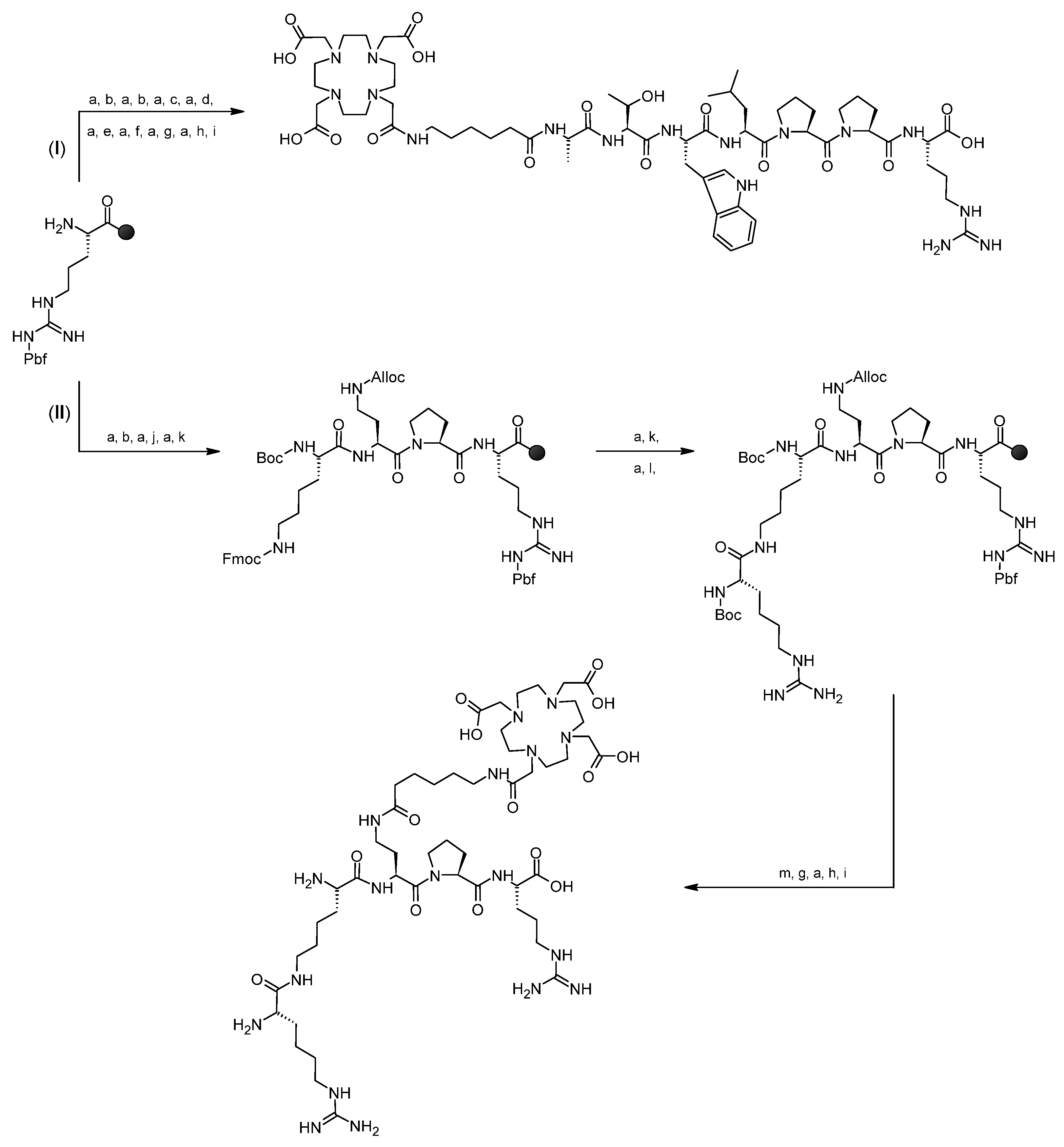
Conjugates 1 and 2 (compounds with a DOTA chelator) were analyzed by reverse-phase high-pressure liquid chromatography (RP-HPLC) in System 1 or 2.
System 1: RP-HPLC analytical Eurospher-100-C-18 column, 5 μm, 250 × 4.6 mm, solvent A: water with 0.1% trifluoroacetic acid (TFA, v/v), solvent B: acetonitrile/water (80:20, v/v) with 0.1% TFA (v/v), UV/Vis detection at 220 nm, gradient elution: 0–20 min 20 to 70% B, flow 1 mL/min.
System 1a: RP-HPLC semi-preparative Nucleosil-300-C18 column, 5 μm, 250 × 8 mm, solvent A: water with 0.1% trifluoroacetic acid (TFA, v/v), solvent B: acetonitrile/water (80:20, v/v) with 0.1% TFA (v/v), UV/Vis detection at 220 nm, gradient elution: 0–5 min 8% B, 5–15 min 8 to 18% B, 15–25 min 18% B, 25–35 min 18 to 35% B, 35–55 min 35% B, 55–60 min 35 to 40% B, 60–65 min 40 to 100% B, flow 2 mL/min.
System 2: RP-HPLC analytical Eurospher-100-C-18 column, 5 μm, 250 × 4.6 mm, solvent A: water with 0.1% TFA (v/v), solvent B: acetonitrile/water (80:20, v/v) with 0.1% TFA (v/v), UV/Vis detection at 220 nm, gradient elution: 0–20 min 5 to 60% B, flow 1 mL/min.
System 2a: RP-HPLC semi-preparative Nucleosil-300-C18 column, 5 μm, 250 × 8 mm, solvent A: water with 0.1% trifluoroacetic acid (TFA, v/v), solvent B: acetonitrile/water (80:20, v/v) with 0.1% TFA (v/v), UV/Vis detection at 220 nm, gradient elution: 0–15 min 5 to 15% B, 15–45 min 15% B, 45–55 min 15 to 100% B, flow 2 mL/min.
Analyses and purification of the radiopreparations and their cold reference compounds were performed in Systems 3 or 4 using the RP-HPLC method with gamma or UV/Vis detection, respectively. Radioactivity of the collected samples were measured using Wizard2 2-Detector Gamma Counter (PerkinElmer) and/or Atomlab 500 Dose Calibrator (BIODEX). Electrospray ionization mass spectrometry analyses (ESI-MS) were performed to confirm the presence of the proper compounds.
System 3: RP-HPLC semi-preparative Phenomenex Jupiter Proteo 90Å column, 4 μm, 250 × 10 mm, with Jupiter Proteo precolumn, 20 × 2.1 mm, gamma or UV/VIS detection (220 nm), solvent A: acetonitrile with 0.1% TFA (v/v), solvent B: water with 0.1% TFA (v/v), gradient elution: 0–20 min 20 to 80% A, 20–30 min 80% A, 30–32 min 80 to 20% A, flow 2 mL/min.
System 4: RP-HPLC analytical Phenomenex Jupiter 4u Proteo 90Å column, 4 μm, 250 × 4.6 mm, gamma or UV/VIS detection (220 nm), solvent A: acetonitrile with 0.1% TFA (v/v), solvent B: water with 0.1% TFA (v/v), gradient elution: gradient elution: 0–20 min 1 to 50% A, 20–25 min 50 to 95% A, 25–31 min 95% A, 31–35 min 95 to 1% A. flow 1 mL/min.

2.2.2. Syntheses

Synthesis of conjugates 1 and 2
The synthesis of conjugates DOTA-Ahx-A7R (1, Figure 1A) and Lys(hArg)-Dab(Ahx-DOTA)-Pro-Arg (2, Figure 1B), based on A7R (Figure 1A black) as a parent peptide and its shorter analogue Lys(hArg)-Dab-Pro-Arg (Figure 1B black), respectively, were carried out manually, on the preloaded Fmoc-Arg(Pbf)-Wang resin with a capacity of 0.32 mmol/g (0.48 mmol scale), following the Fmoc chemistry. Coupling of 3 eq. amino acids was done using 3 eq. DIC and 3 eq. HOBt in DMF (3 mL) [49]. Completion of coupling was checked using a Kaiser [50] or chloranil test [51]. Fmoc deprotection step was done using 30% piperidine in DMF. Guanylation reaction was performed for 4 days using di-Boc-S-methylisothiourea (3 eq. in 3 mL DCM) [52]. The Alloc deprotection step was done using tetrakis(triphenylphosphine)palladium(0) (0.06 mmol; 0.25 eq.) in the presence of 24 eq. PhSiH3 (5.76 mmol) in DCM [53]. The coupling of the chelator was performed using the active ester method with 1 eq. DOTA-tris(tBu)-NHS and 3 eq. Et3N for about 20 h.
The cleavage of the final conjugates were performed using the mixture of TFA:PhOH:H2O:TIPS (88:5:5:2, v/v/v/v) for 2 h. Crude conjugates were precipitated by a dropwise addition into a cold diethyl ether, and then were purified using semi-preparative KNAUER RP-HPLC in System 1a for conjugate 1 and System 2a for conjugate 2. Purified conjugates were analyzed by mass spectrometry method (ESI-MS).
Preparation of [68Ga]Ga-DOTA-Ahx-A7R (68Ga-1) and [68Ga]Lys(hArg)-Dab(Ahx-DOTA-Ga)-Pro-Arg (68Ga-2) radioconjugates
[68Ga]Ga-DOTA-Ahx-A7R (68Ga-1) and [68Ga]Lys(hArg)-Dab(Ahx-DOTA-Ga)-Pro-Arg (68Ga-2) radioconjugates were synthesized according to the following procedure: into a vial containing about 10–20 nmol of lyophilized conjugate 1 or conjugate 2, we added 300–400 μL of 0.2 M acetate buffer (pH 4.5), and 200–300 μL of the [68Ga]GaCl3 (10–30 MBq) solution from the 68Ge/68Ga generator. The reaction mixture at pH 3.0 was heated for 10 min at 95 °C. The resulting radioconjugates were then purified by RP-HPLC in System 3 with gamma detection for radioconjugate 68Ga-1 (RCY 93.3 ± 0.3%, n = 4, molar activity 1.2 MBq/nmol, pH 3.0) and in System 4 with gamma detection for radioconjugate 68Ga-2 (RCY 91.5 ± 0.8%, n = 4, molar activity 1.2 MBq/nmol, pH 3.0). Pure fractions of 68Ga-1 and68Ga-2 radioconjugates were evaporated under N2 and dissolved on PBS.
Preparation of [177Lu]Lu-DOTA-Ahx-A7R (177Lu-1) and [177Lu]Lys(hArg)-Dab(Ahx-DOTA-Lu)-Pro-Arg (177Lu-2) radioconjugates
[177Lu]Lu-DOTA-Ahx-A7R (177Lu-1) and [177Lu]Lys(hArg)-Dab(Ahx-DOTA-Lu)-Pro-Arg (177Lu-2) radioconjugates were synthesized according to the following procedure: into a vial containing about 2.5–20 nmol of lyophilized conjugate 1 or conjugate 2, we added 200–300 μL of 0.2 M acetate buffer (pH 4.5), 150–200 μL H2O, and 5–15 μL of the [177Lu]LuCl3 (1–15 MBq) solution. In some cases, we also added 0.5–2.0 µL 0.1 M HCl to obtain a desired pH. The reaction mixture at pH 4.5 was heated for 10 min at 95 °C. The resulting conjugates were then purified by RP-HPLC in System 3 with gamma detection for radioconjugate 177Lu-1 (RCY 95.5 ± 1.2%, n = 4, molar activity 0.3 MBq/nmol, pH 4.5) and in System 4 with gamma detection for radioconjugate 177Lu-2 (RCY 96.2 ± 2.6%, n = 4, molar activity 0.3 MBq/nmol, pH 4.5). Pure fractions of 177Lu-1 and 177Lu-2 radioconjugates were evaporated under N2 and dissolved on PBS.
All radioconjugates were purified before the use in further experiments, namely, the lipophilicity and stability studies.
Preparation of cold reference compounds Ga/Lu-DOTA-Ahx-A7R (Ga/Lu-1) and Lys(hArg)-Dab(Ahx-DOTA-Ga/Lu)-Pro-Arg (Ga/Lu-2)
To verify the identity of the 68Ga-1, 177Lu-1, 68Ga-2, and 177Lu-2 radioconjugates, the analogues with stable gallium and lutetium isotopes under the same reaction conditions were synthesized and analyzed by the RP-HPLC method (System 3 or 4 with UV/Vis detection) and ESI-MS methods.
Ga-DOTA-Ahx-A7R (Ga-1) and Lys(hArg)-Dab(Ahx-DOTA-Ga)-Pro-Arg (Ga-2) cold reference compounds were synthesized according to the following procedure: into a vial containing approximately 70 nmol of conjugate 1 or 2 dissolved in 300 μL of 0.2 M acetate buffer (pH 4.5), we added the 60 μL of 1.34 mg/mL GaCl3 solution in 0.065 M HCl. The reaction mixture at pH 4.0 was heated for 10 min at 95 °C. The reaction progress was checked by RP-HPLC in System 3 for Ga-1 and in System 4 for Ga-2.
Lu-DOTA-Ahx-A7R (Lu-1) and Lys(hArg)-Dab(Ahx-DOTA-Lu)-Pro-Arg (Lu-2) cold reference compounds were synthesized according to the following procedure: to a vial containing approximately 70 nmol of conjugate 1 or 2 dissolved in 600 μL of 0.2 M acetate buffer (pH 4.5), we added the 3.5 μL of concentrated LuCl3 solution in 14 M HCl. The reaction mixture at pH 4.0 was heated for 10 min at 95 °C. The reaction progress was checked by RP-HPLC in System 3 for Lu-1 and in System 4 for Lu-2.

2.2.3. Physicochemical Properties Study of the Radioconjugates

All studies of the physicochemical properties of the synthesized radiopreparations were carried out using radioconjugates previously isolated from the reaction mixture.
Radioconjugates lipophilicity test
The lipophilicity (L) of tested radiocompounds, defined as the decimal logarithm (logP) of the partition coefficient (P) of the compound between the two immiscible phases, was determined in the system PBS solution (aqueous phase, polar phase, pH 7.4) and n-octanol (organic, non-polar phase) according to the following formula:
L = logP = log A o A w
where A o —organic phase radioactivity; A w —aqueous phase radioactivity.
Radioactivity of both phases, resulting from the concentration of the tested radiocompound in each of them, was determined by measuring the gamma radiation with a Wizard counter in three independent experiments.
Before starting the research, both liquid phases were saturated with each other to avoid errors resulting from the mutual solubility of both liquids. The partition coefficient (P) result is shown as the mean ± SD. After the end of the experiment, we performed RP-HPLC analysis of the aqueous phase to confirm the tested radioconjugate remained intact during the experiment.
Radioconjugates stability tests
In accordance with requirements for potentially novel radiopharmaceuticals, we tested newly designed radiopreparations for their stability in solutions that act as human body fluids and also in human serum [54,55].
Stability studies in PBS buffer, cysteine and histidine
Stability studies in PBS buffer were performed in order to investigate the possible influence of buffer components on the radiocompound decomposition process. We isolated 68Ga-1, 177Lu-1, 68Ga-2, and 177Lu-2 radioconjugates from the reaction mixture and incubated each in PBS buffer. After about 3 h, we analyzed aliquots by RP-HPLC in System 3 with gamma detection for 68Ga-1 and 177Lu-1 and in System 4 with gamma detection for 68Ga-2 and 177Lu-2.
Stability studies of the isolated from the reaction mixture 68Ga-1, 177Lu-1, 68Ga-2, and 177Lu-2 radioconjugates in solutions containing excess amounts of strongly competing natural ligands with chemically reactive groups, e.g., -NH2, -SH, and -COOH (the so-called challenge experiments). For this purpose, each radioconjugate was incubated at 37 °C in 1 mM cysteine (Cys) or histidine (His) solutions in PBS buffer (the ligand concentration was about 1000 times higher than the concentration of tested radioconjugate). After the specified incubation time, which was up to 4 h for the 68Ga-radiocompounds and up to 6 days for the 177Lu-radiocompounds, we analyzed solutions by RP-HPLC in System 3 with gamma detection for 68Ga-1 and 177Lu-1 and in System 4 with gamma detection for 68Ga-2 and 177Lu-2.
Stability studies in human serum
Stability studies of the 68Ga-1, 177Lu-1, 68Ga-2, and 177Lu-2 radioconjugates in human serum (HS) were performed according to the following procedure: into a vial containing 900 µL of HS we added 100 µL of the tested radioconjugate and incubated the mixture at 37 °C. After the specified incubation times (10 min–4.5 h for 68Ga-radiocompounds and 10 min–4 days for 177Lu-radiocompounds), we withdrew the aliquots, mixed with ethanol to precipitate the proteins, and centrifuged (14,000 rpm, 5–15 min) to separate the protein compounds from the supernatant. The gamma radioactivity of the obtained precipitate and supernatant was then measured using a Wizard detector. Using the formula below, the amount of radioconjugate remaining in the supernatant (and also bound by serum protein components) was calculated.
  liquid   phase   radioactivity   liquid   phase   radioactivity +   percipitate   radioactivity   × 100 %
We also analyzed the supernatant at each time by RP-HPLC in System 3 with gamma detection for 68Ga-1 and 177Lu-1 and System 4 with gamma detection for 68Ga-2 and 177Lu-2 to check whether the studied radiocompound remained unchanged during the experiment.

3. Results

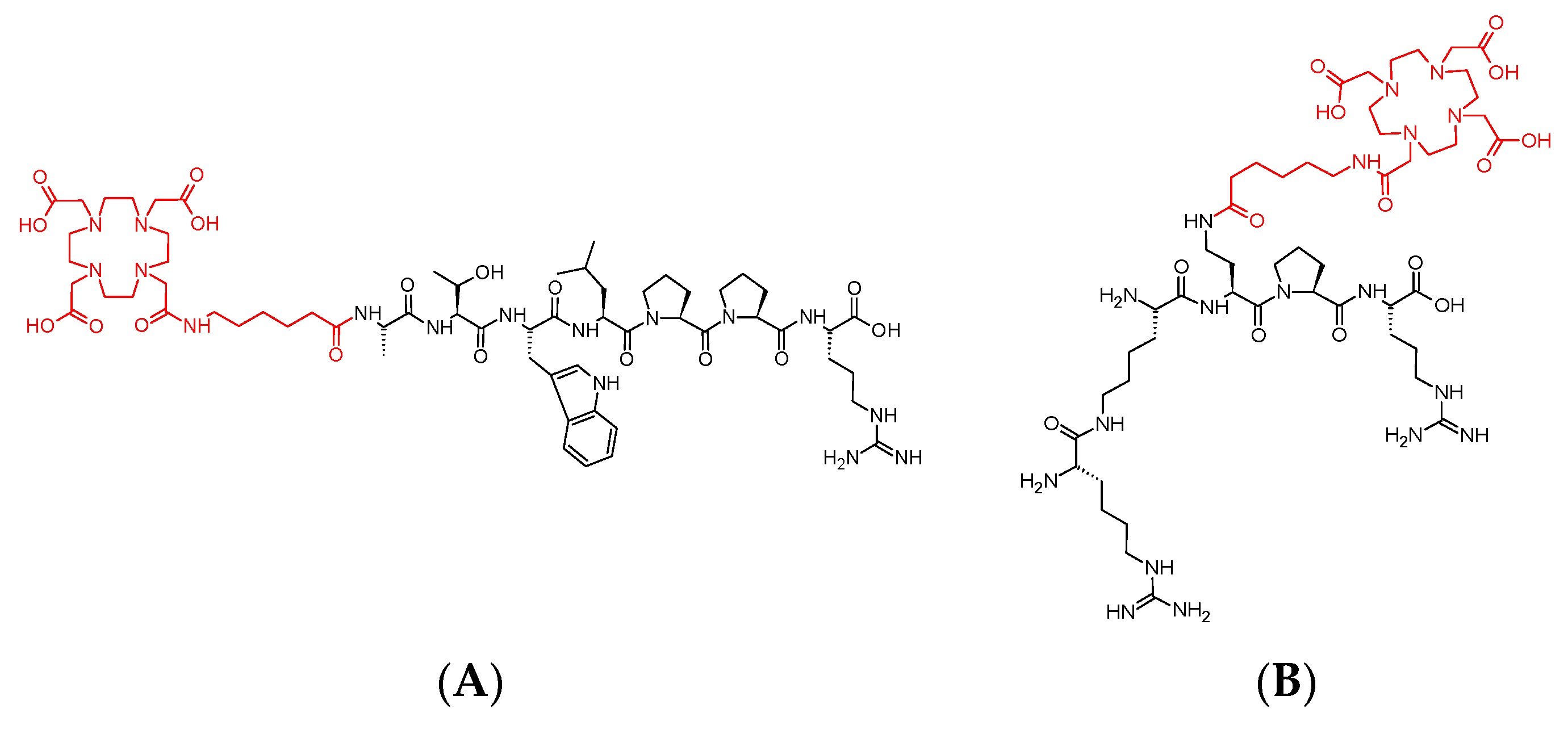
3.1. Syntheses of Conjugates 1 and 2

The synthesis of conjugates 1 and 2 (Scheme 1) was carried out by Solid Phase Peptide Synthesis (SPPS) on the preloaded Fmoc-Arg(Pbf)-Wang resin following the Fmoc chemistry according to the standard coupling DIC/HOBt protocol. In the case of synthesis of conjugate 1, after building a linear peptide on the resin and coupling of the Ahx spacer, the chelator DOTA was attached by the active ester method using DOTA-tris(tBu)-NHS.
The synthesis of conjugate 2 required a few additional steps, as shown in Scheme 1. After the synthesis of fully protected, linear tripeptide (Fmoc-Dab(Alloc)-Pro-Arg(Pbf)-Wang), we attached the N-terminal lysine using Boc-Lys(Fmoc), which enabled the attachment of another Boc-Lys(Fmoc) to the side chain after removal of the Fmoc group from the epsilon amino group. After Fmoc deprotection of the epsilon amino group of the second lysine, we carried out the guanylation reaction for 4 days to lead to hArg creation using di-Boc-S-methylisothiourea, until the Kaiser test was negative. Next, after Alloc deprotection of the amino group of the side chain of the Dab residue, we coupled Fmoc-Ahx. Following the coupling sequence, Fmoc deprotection was performed as the next step, and then the DOTA chelator was attached by the active ester method using DOTA-tris(tBu)-NHS. The cleavage of the synthetized compounds from the resin was performed using the TFA:PhOH:H2O:TIPS mixture simultaneously removing all Boc protections. We purified the crude compounds by RP-HPLC and analyzed by ESI-MS (Table 1).

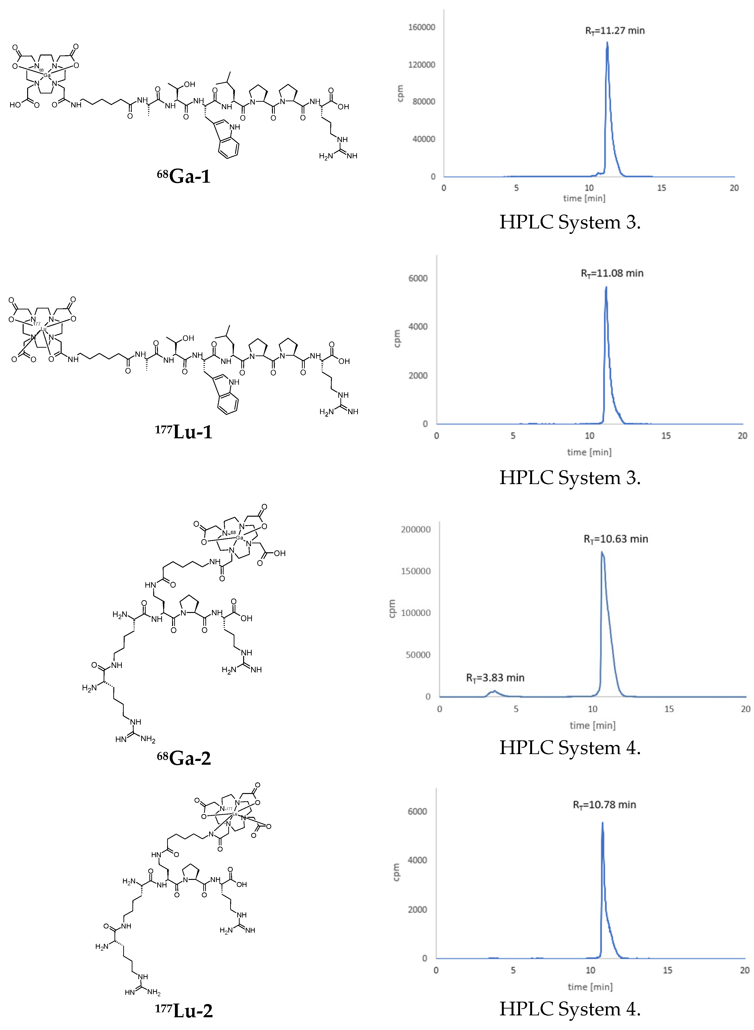
3.2. Syntheses of Radioconjugates

All obtained radiocompounds were formed with high radiochemical yield (>90% for 68Ga-1 and 68Ga-2, and >95% for 177Lu-1 and 177Lu-2) and high radiochemical purity (>90%). The chemical formulas of the 68Ga-1, 177Lu-1, 68Ga-2, and 177Lu-2 radioconjugates as well as the RP-HPLC radiochromatograms of the labeling reaction mixtures are presented in Figure 2. The statistical data of the radiochemical yield (RCY) and radiochemical purity (RCP) (presented as the mean ± SD, n = the number of repetitions) of the obtained 68Ga-1, 177Lu-1, 68Ga-2, and 177Lu-2 radioconjugates are presented in Table 2.

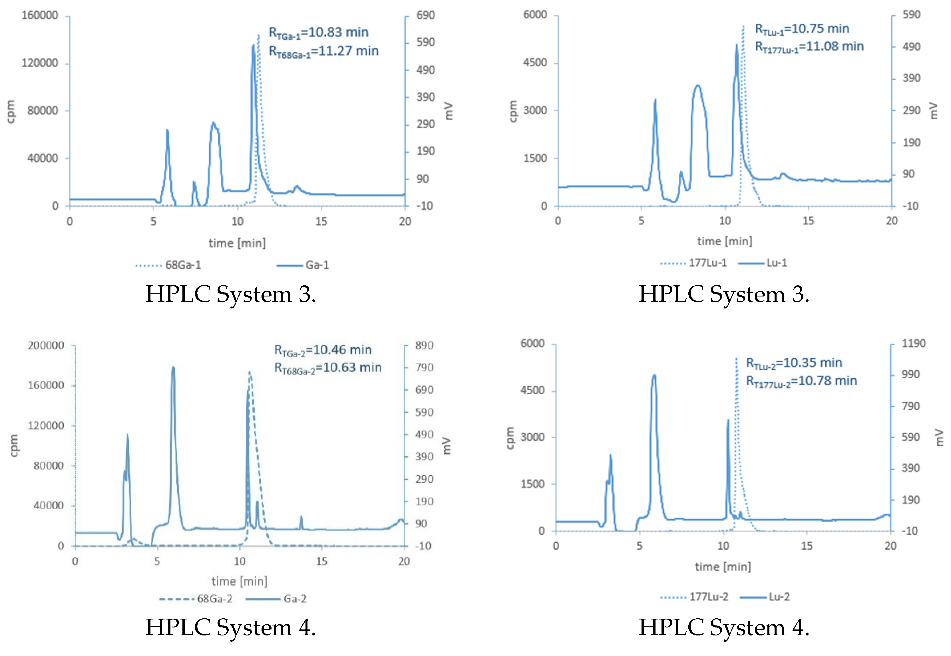
3.3. Synthesis of Cold References Compounds

RP-HPLC analyses of the reaction mixtures of the cold reference compounds are presented in Figure 3. The compounds characterized with RT values as 10.83 min, 10.75 min, 10.46 min, and 10.35 min were isolated from the reaction mixture and identified by ESI-MS as Ga-DOTA-Ahx-A7R (Ga-1), Lu-DOTA-Ahx-A7R (Lu-1), Lys(hArg)-Dab(Ahx-DOTA-Ga)-Pro-Arg (Ga-2), and Lys(hArg)-Dab(Ahx-DOTA-Lu)-Pro-Arg (Lu-2), respectively. The RT values of these reference compounds coincided with the corresponding retention times of the appropriate radiocompounds (68Ga-1, 177Lu-1, 68Ga-2, and 177Lu-2; Figure 3), which confirmed the stability of both the A7R and Lys(hArg)-Dab-Pro-Arg molecules under these synthesis conditions and confirmed that we obtained the desired radiocompounds.
The results of ESI-MS analyses confirming the cold reference compounds were obtained are shown in Table 3.

3.4. Lipophilicity Studies

Lipophilicity is one of the most important physicochemical parameters affecting the absorption, distribution, metabolism, and excretion of drug molecules in the organism, the so-called ADME profile [56,57,58,59]. Preparations characterized by high lipophilicity show a high affinity for fats and a low affinity for water; they are also able to effectively penetrate cell membranes. From the point of view of pharmacology, lipophilicity is an important prognostic factor for drugs and other medical preparations to predict toxic activity and to characterize biological activity, the ability to accumulate in organisms, and the metabolism of substances [60,61,62,63,64,65]. The lipophilicity values obtained for tested radioconjugates 68Ga-1, 177Lu-1, 68Ga-2, and 177Lu-2 (presented as the mean ± SD) are shown in Table 4. The main reason for the low lipophilicity value is that the tested radiocompounds are based on peptides and peptidomimetics, which are usually characterized by a low lipophilicity value. Moreover, conjugation of the macrocyclic DOTA chelator with the biomolecule also results in a lower lipophilicity value of the conjugate compared to the lipophilicity of the biomolecule itself.
Radiochromatograms of the aqueous phases after the lipophilicity tests of the radiopreparations showed, in all cases, single peaks at RT values that corresponded to the tested radiocompounds, which proves the stability of the radiopreparations during the experiment.

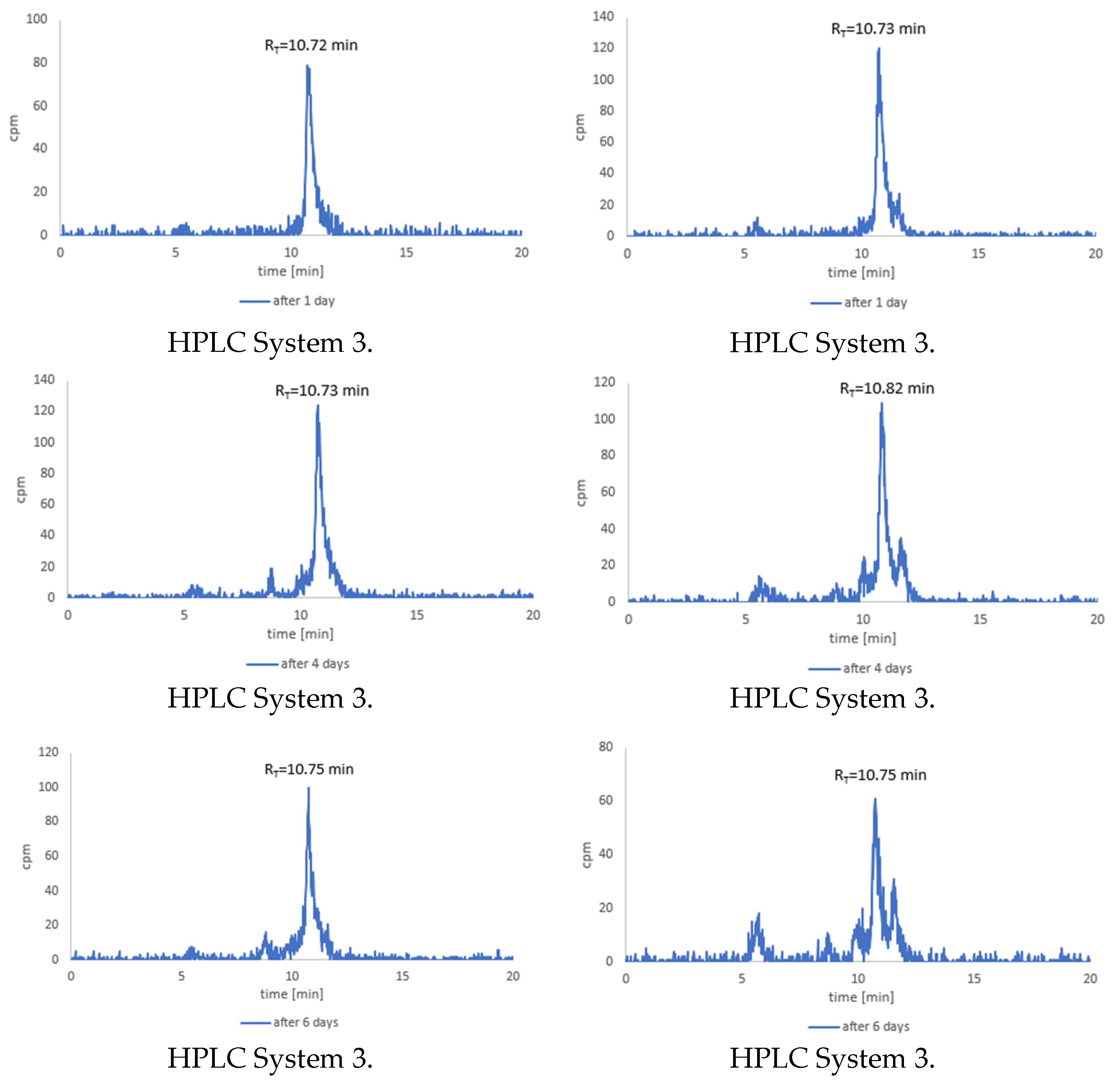
3.5. Stability Studies of Radioconjugates in PBS Buffer, Cysteine and Histidine

Stability tests of all radioconjugates incubated in PBS solution for about 3 h showed no decomposition of radioconjugates. In all the RP-HPLC radiochromatograms only single peaks were recorded, with RT values that corresponded to the appropriate radioconjugate, which confirms the stability of radioconjugates under such conditions.

3.6. Stability Tests for 68Ga-1 and 68Ga-2 Radioconjugates

Stability tests of the 68Ga-1 radioconjugate in the Cys (Figure 4 left) and His (Figure 4 right) solutions showed satisfactory stability in the period of about 3 h, corresponding to the three half-lives of the radionuclide gallium-68. In the RP-HPLC radiochromatograms recorded after 3 h, we observed only traces of the products of radioconjugate decomposition (Figure 4).
Similarly, stability studies of the 68Ga-2 radioconjugate in Cys (Figure 5 left) and His (Figure 5 right) solutions carried out over the same time interval demonstrated the complete stability of the radioconjugate.

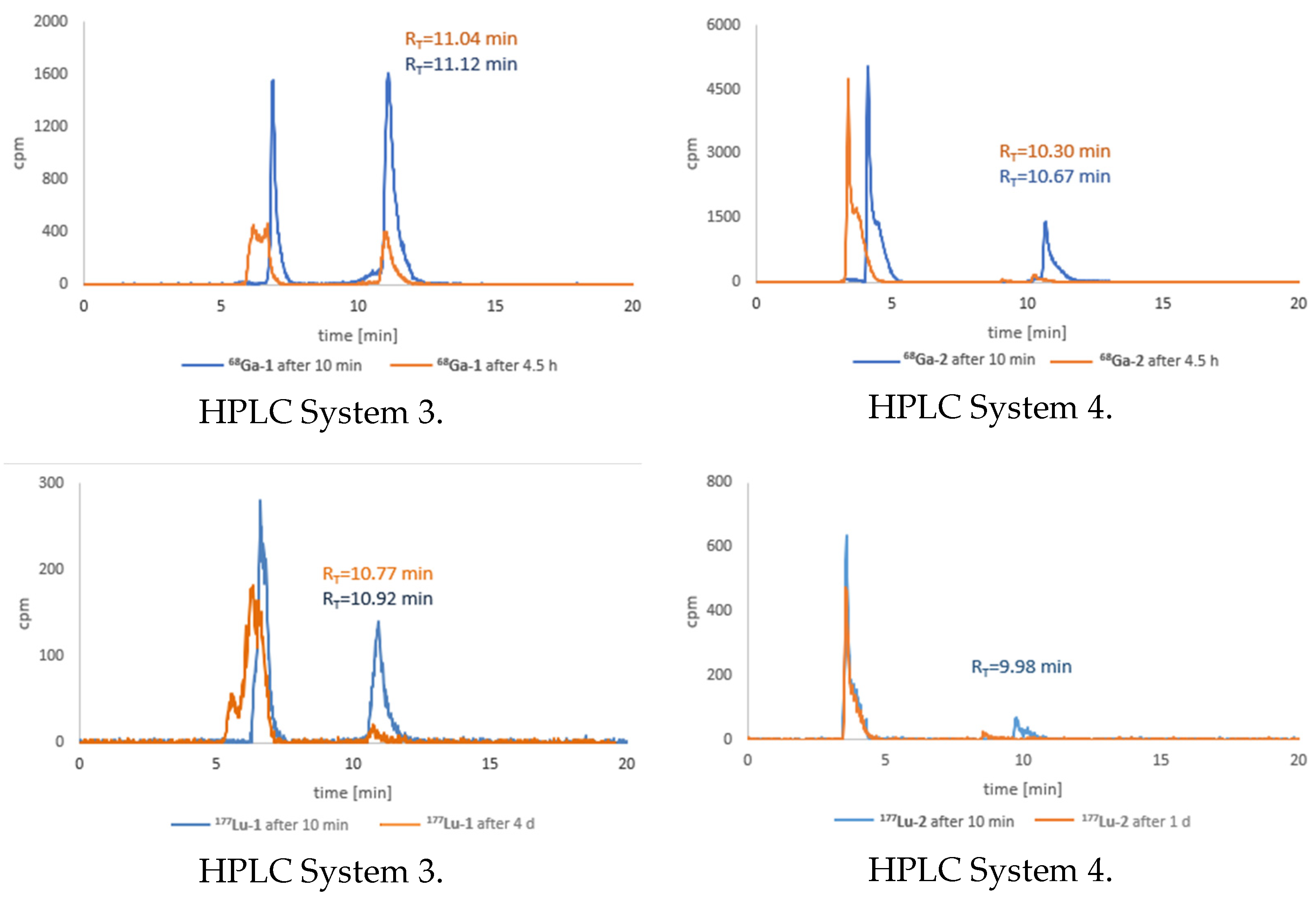
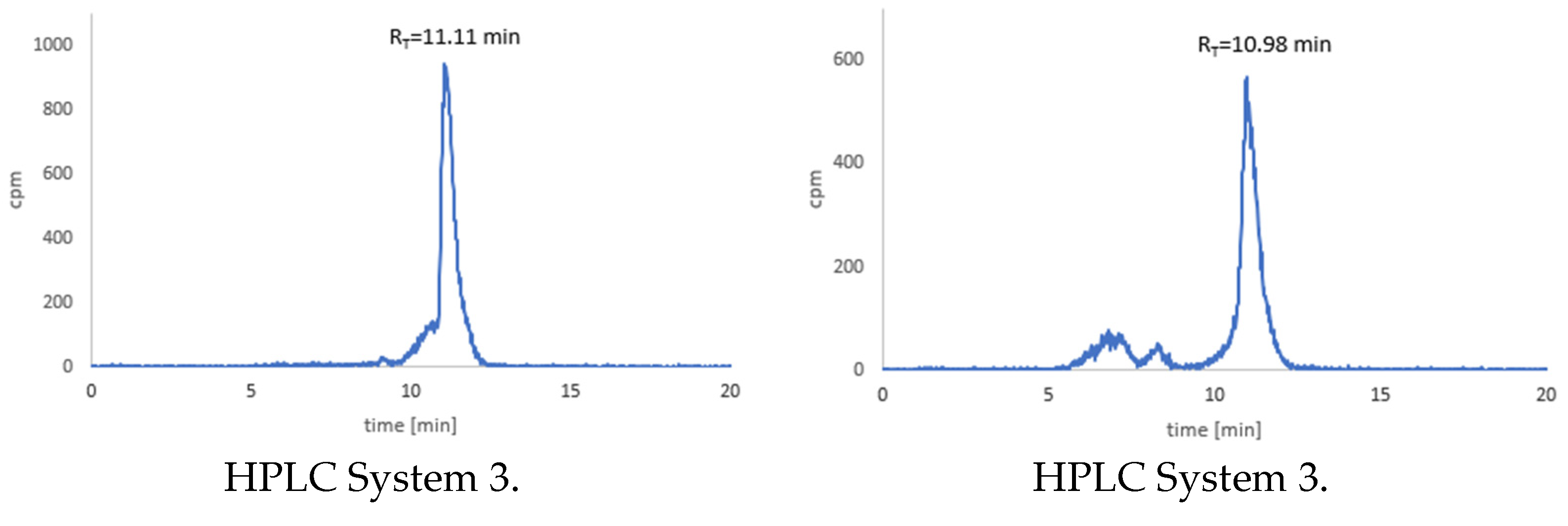
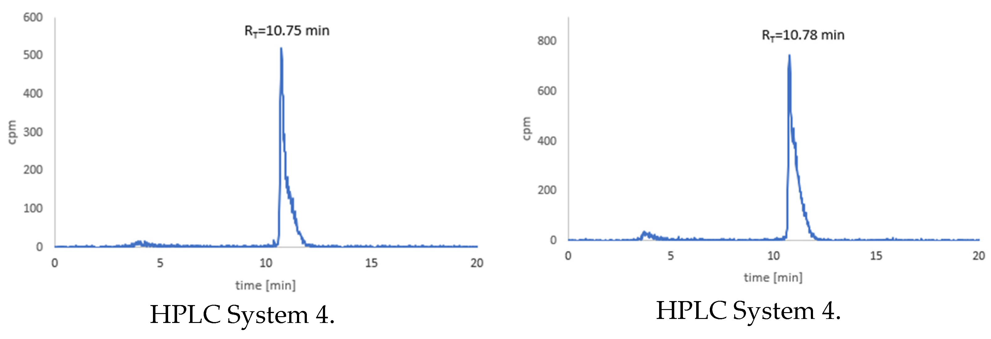
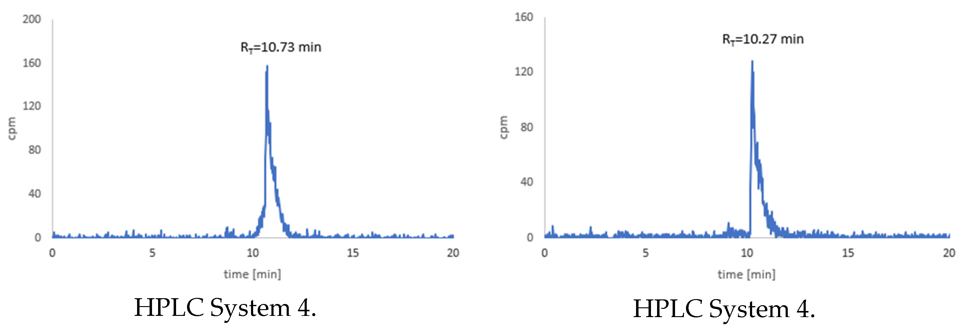
3.7. Stability Tests for 177Lu-1 and 177Lu-2 Radioconjugates

Stability studies of the radioconjugates containing lutetium-177 radionuclide in Cys and His solutions were carried out in the period of about 6 days, corresponding to the one half-live of the radionuclide lutetium-177. Incubation of the 177Lu-1 radioconjugate in Cys and His solutions for a longer period, more than 4 days, showed slow degradation of this radiopreparation in both solutions (Figure 6). The radioconjugate was completely stable for 1 day, but after 6 days, traces of the products of radioconjugate decomposition were already visible, and the degradation of the radioconjugate was noticeably higher during incubation in the His solution (Figure 6 right).
In contrast, the 177Lu-2 radioconjugate proved to be completely stable in both solutions throughout the incubation period (Figure 7).

3.8. Stability Studies of Radioconjugates in Human Serum

Radiochromatograms illustrating the results of the stability tests of the 68Ga-1, 177Lu-1 and 68Ga-2, 177Lu-2 radioconjugates in human serum are shown in Figure 8. The statistical data of the radiochemical purity (presented as the mean ± SD, n = the number of repetitions) of the obtained 68Ga-1, 177Lu-1, 68Ga-2, and 177Lu-2 radioconjugates in the human serum stability tests are presented in Table 5.
As can be seen, all radioconjugates were unstable in HS due to biodegradation of the A7R peptide and Lys(hArg)-Dab-Pro-Arg peptidomimetic by endogenous enzymes. After 10 min of incubation, the radiochromatograms already showed additional peaks corresponding to the enzymatic degradation products. During incubation, the height of these additional peaks increased, and the peak heights corresponding to the tested radioconjugates decreased. After about 4 h in the case of the 68Ga-1 and 68Ga-2 radioconjugates and about 4 days in the case of the 177Lu-1 and 177Lu-2 radioconjugates, the peaks corresponding to the tested radiopreparations were often hardly visible, indicating almost complete biodegradation of the radioconjugates. Radioactivity measurements of the precipitated protein serum components and the supernatants indicated that, in the case of the 68Ga-1 and 177Lu-1 radioconjugates, about 15% of the radiocompounds was bound to the protein serum components. In the case of the 68Ga-2 and 177Lu-2 radioconjugates, about 28% of the radiocompounds was bound with protein serum components.

4. Discussion

The aim of the present study was to synthesize and to characterize the physicochemical properties of radiolabeled A7R peptide and Lys(hArg)-Dab-Pro-Arg peptidomimetic for potential application as diagnostic or therapeutic receptor radiopharmaceuticals. According to the literature data, the heptapeptide A7R is known to be an effective antagonist of the VEGF-A165 binding with NRP-1 and to show in vivo anti-angiogenic properties [35], whereas the Lys(hArg)-Dab-Pro-Arg peptidomimetic is much more active (IC50 = 0.2 μM) than the heptapeptide A7R (IC50 = 5.9 μM [42]), with a stability half-life in human serum that is nearly 2 days [41,43]. To be able to synthesize radioconjugates, we attached a DOTA chelator to the initial compounds—A7R or its shorter analogue—via an Ahx linker.
All the designed radioconjugates were synthesized with high radiochemical yield and high radiochemical purity (Figure 2 right), and the methods for their synthesis were relatively simple and cheap. Identity of the radioconjugates was confirmed by ESI-MS analysis of their cold reference compounds.
All radioconjugates turned out to be highly hydrophilic compounds with logP values in the range of −3.40 to −4.57 (Table 4). Moreover, the radioconjugates with Ga-68 turned out to be visibly less lipophilic due to a free carboxyl group of the DOTA chelator. These values were much lower than those preferred for radiopharmaceuticals, in the ranges of 1 to 4 and 1.5 to 2.5, which are suitable for crossing the blood–tissue [66] and blood–brain barriers [67], respectively. However, the ability to cross the blood–tissue barrier is not a crucial parameter in this case due to the presence of VEGF165 in the endothelium, the single layer of squamous endothelial cells that lines the interior surface of blood vessels, and its ability to interact directly with blood.
Radioconjugates 68Ga-1 and 68Ga-2 and radioconjugate 177Lu-2 were sufficiently stable in solutions acting as human body fluids, PBS buffer, and Cys or His solutions. However, the stability of the 177Lu-1 radioconjugate that contained therapeutic radionuclide Lu-177 and based on the A7R peptide turned out to be rather insufficient. After 6 days of incubation, which is just one half-life of the radionuclide Lu-177, the radioconjugate decomposition products were already visible (Figure 6). Because this is a potential therapeutic radiopharmaceutical containing radionuclide with a half-life of 6.65 days, such stability in these solutions is rather low.
Stability studies of the radioconjugates in human serum showed relatively rapid decomposition of all radiocompounds due to enzymatic biodegradation of A7R peptide and Lys(hArg)-Dab-Pro-Arg peptidomimetic (Figure 8). Comparing the RP-HPLC radiochromatograms recorded after a 10 min incubation, it is apparent that the radioconjugates 68Ga-2 and 177Lu-2 were more readily biodegraded in HS compared to the radioconjugates 68Ga-1 and 177Lu-1. In the case of 177Lu-1 radiopreparation after 4 days of incubation, the radioconjugate was still visible in the sample, while in the case of 177Lu-2 after 1 day of incubation, only traces of radioconjugate were present in the sample. Moreover, during the incubation of 68Ga-1 and 177Lu-1 in HS, about 15% of the radiopreparations were bound to the serum peptide components, while in the case of radioconjugates 68Ga-2 and 177Lu-2, almost twice as much, about 28%. This difference can be explained by the less-polar nature of the A7R (Ala1-Thr2-Trp3-Leu4-Pro5-Pro6-Arg7-OH) peptide and the polar nature of the Lys1(hArg)-Dab2-Pro3-Arg4 peptidomimetic. The former compound had three reactive functional groups—the guanidine group of Arg7, the amine group of Ala1, and the carboxyl group of Arg—while the peptidomimetic had six reactive functional groups—the three amino groups of Lys1, hArg, and Dab2, the two guanidine groups of hArg and Arg4, and one carboxyl group of Arg4. These reactive groups, depending on the pH and under the influence of external conditions, can assume, respectively, positive or negative charges, and interact with the protein components of the human serum. Despite the fact that one amine group was used to attach the DOTA chelator via the Ahx linker in both conjugates, the radioconjugates produced by the labeling reactions of conjugate 1 and 2 should still differ in charge, polarity, and chemical reactivity because of differences in the physicochemical properties of their parent compounds, the A7R peptide and Lys(hArg)-Dab-Pro-Arg peptidomimetic.
Based on the above data, it should be assessed that, unfortunately, radioconjugates 68Ga-2 and 177Lu-2 that were based on the more biologically active biomolecule, Lys(hArg)-Dab-Pro-Arg peptidomimetic, were less stable in HS than those based on the A7R peptide, 68Ga-1 and 177Lu-1. Such a low stability of 68Ga-2 and 177Lu-2 was initially surprising because, according to the literature data, the determined half-life of the Lys(hArg)-Dab-Pro-Arg peptidomimetic in HS was nearly 2 days (41 h), and the period of its complete decomposition in HS estimated from the graph was over 4 days [43]. As determined from our experiments, the period of almost complete enzymatic biodegradation of radioconjugate 177Lu-2, resulting from enzymatic biodegradation of the Lys(hArg)-Dab-Pro-Arg peptidomimetic, was less than 1 day. These two seemingly contradictory results were due to the different conditions of the stability studies. In the first case, the peptidomimetic concentration in the sample was 0.9 µmol/mL, which allowed us to detect the biodegradation using the UV/VIS detection [43]. In the second case, the peptidomimetic concentration was equal to the concentration of the radioconjugate and was incomparably lower, i.e., about 2 nmol/mL; experiments with such a low radioconjugate concentration were possible due to using gamma detection. Consequently, in the first case, the test was carried out with saturation of the enzymes present in HS sample, while in the second case, the amount of the peptidomimetic was much smaller than the amount of enzyme. Hence, in the first case, intact peptidomimetic was visible in the sample for about 4 days, while this period was only 1 day in the present study.
To conclude, our study showed that all radioconjugates obtained in the presented work were not sufficiently stable in HS, and so they unfortunately do not fully meet the requirements for radiopharmaceuticals. Nevertheless, the structure of the peptidomimetics can be modified easily, which makes it possible to design a new formulation that is more stable in HS. In the near future, we plan to modify them by using D-amino acids or replacing one or more peptide bonds with their bioisosteres (e.g., N-methyl peptide bond, or reduced bond -CH2-NH-). For radiopreparations sufficiently stable in human serum and suitable for in vivo applications, we plan more advanced biological tests (using NRP-1 overexpressing cell lines) and finally tests on living organisms (a biodistribution study in mice or rats).

Author Contributions

Conceptualization, E.G. and A.M.; methodology, E.G., A.M., E.W., K.M. and P.K.H. software, K.M., D.T., E.W. and P.K.H.; validation, E.G., A.M. and D.T.; investigation, K.M., D.T., E.W. and P.K.H.; resources, K.M., D.T. and P.K.H.; writing—K.M., D.T., E.G. and A.M.; writing—review and editing, E.G., A.M., D.T. and K.M.; visualization, K.M., D.T., E.G. and A.M.; project administration, E.G. and A.M. All authors have read and agreed to the published version of the manuscript.

Funding

This research was carried out within grant 2019/33/B/NZ7/02818, supported by the National Science Centre (Poland).

Institutional Review Board Statement

Not applicable.

Informed Consent Statement

Not applicable.

Data Availability Statement

Not applicable.

Acknowledgments

The contributions of students Katarzyna Masłowska and Paweł Krzysztof Halik were done in the frame of the National Centre for Research and Development Project No. POWR.03.02.00-00-I009/17 (Radiopharmaceuticals for molecularly targeted diagnosis and therapy, RadFarm, Operational Project Knowledge Education Development 2014–2020, co-financed by European Social Fund).

Conflicts of Interest

The authors declare no conflict of interest.

Abbreviations

A7RAla-Thr-Trp-Leu-Pro-Pro-Arg-OH peptide
ACNAcetonitrile
ADMEAbsorption, distribution, metabolism, and excretion
Ahx6-aminohexanoic acid
AllocAllyloxycarbonyl group
AoOrganic phase radioactivity
AwAqueous phase radioactivity
Boc
COOH
Tert-butyloxycarbonyl group
Carboxyl group
CysCysteine
Dab2,4-Diaminobutyric Acid
DCMDichloromethane
DICN,N′-Di(propan-2-yl)methanediimine
DMFN,N-Dimethylformamide
DOTA2,2′,2″,2‴-(1,4,7,10-Tetraazacyclododecane-1,4,7,10-tetrayl)
tetraacetic acid
DOTA-tris(tBu)-NHS tri-tert-butyl 2,2′,2″-(10-(2-((2,5-dioxopyrrolidin-1-yl)oxy)-2-oxoethyl)
-1,4,7,10-tetraazacyclododecane-1,4,7-triyl)triacetate
ESI-MSElectrospray ionization mass spectrometry analyses
Et3NTriethylamine
EβmaxBeta emitter with a maximum energy
Fmoc9-fluorenylmethoxycarbonyl group
hArgHomoarginine
HClHydrogen chloride
HisHistidine
HOBt 1H-1,2,3-Benzotriazol-1-ol
HSHuman Serum
IC50
L
Half maximal inhibitory concentration
Lipophilicity
logPDecimal logarithm of partition coefficient
m/zMass-to-charge ratio
mAbMonoclonal antibodies
NH2Amino group
NRP-1Neuropilin-1
PPartition coefficient
PbfPentamethyl-2,3-dihydrobenzofuran-5-sulfonyl group
PBS
PhOH
Phosphate-buffered saline
Phenol
PhSiH3Phenylsilane
RP-HPLCReverse-Phase High Pressure Liquid Chromatography
RTRetention time
RTKsReceptor Tyrosine Kinases
SDStandard deviation
SHThiol group
SPPSSolid Phase Peptide Synthesis
t1/2Half-life time
TFATrifluoroacetic acid
TIPSTri(propan-2-yl)silane
VEGFVascular Endothelial Growth Factor
VEGF-A165Vascular Endothelial Growth Factor-A165
VEGFRVascular Endothelial Growth Factor Receptor
VEGFR-2Vascular Endothelial Growth Factor Receptor 2
sigma; operator for summation

References

  1. Ferrara, N. Role of vascular endothelial growth factor in regulation of physiological angiogenesis. Am. J. Physiol. Cell Physiol. 2001, 280, 1358–1366. [Google Scholar] [CrossRef]
  2. Ferrara, N.; Gerber, H.P.; LeCouter, J. The biology of VEGF and its receptors. Nat. Med. 2003, 9, 669–676. [Google Scholar] [CrossRef] [PubMed]
  3. Grünewald, F.S.; Prota, A.E.; Giese, A.; Ballmer-Hofer, K. Structure–function analysis of VEGF receptor activation and the role of coreceptors in angiogenic signaling. BBA—Proteins Proteom. 2010, 1804, 567–580. [Google Scholar] [CrossRef] [PubMed]
  4. Djordjevic, S.; Driscoll, P.C. Targeting VEGF signalling via the neuropilin co-receptor. Drug Discov. Today 2013, 18, 447–455. [Google Scholar] [CrossRef] [PubMed] [Green Version]
  5. Roskoski, R., Jr. Vascular endothelial growth factor (VEGF) signaling in tumor progression. Crit. Rev. Oncol. Hematol. 2007, 62, 179–213. [Google Scholar] [CrossRef]
  6. Soker, S.; Fidder, H.; Neufeld, G.; Klagsbrun, M. Characterization of Novel Vascular Endothelial Growth Factor (VEGF) Receptors on Tumor Cells That Bind VEGF165 via Its Exon 7-encoded Domain. J. Biol. Chem. 1996, 271, 5761–5767. [Google Scholar] [CrossRef] [Green Version]
  7. Goel, H.L.; Mercurio, A.M. VEGF targets the tumour cell. Nat. Rev. Cancer 2013, 13, 871–882. [Google Scholar] [CrossRef]
  8. Masłowska, K.; Halik, P.K.; Tymecka, D.; Misicka, A.; Gniazdowska, E. The Role of VEGF receptors as molecular target in nuclear medicine for cancer diagnosis and combination therapy. Cancers 2021, 13, 1072. [Google Scholar] [CrossRef]
  9. Guo, H.F.; Vander Kooi, C.W. Neuropilin functions as an essential cell surface receptor. J. Biol. Chem. 2015, 290, 29120–29126. [Google Scholar] [CrossRef] [Green Version]
  10. Soker, S.; Takashima, S.; Miao, H.Q.; Neufeld, G.; Klagsbrun, M. Neuropilin-1 is expressed by endothelial and tumor cells as an isoform-specific receptor for vascular endothelial growth factor. Cell 1998, 92, 735–745. [Google Scholar] [CrossRef] [Green Version]
  11. Latil, A.; Bieche, I.; Pesche, S.; Valeri, A.; Fournier, G.; Cussenot, O.; Lidereau, R. VEGF overexpression in clinically localized prostate tumors and neuropilin-1 overexpression in metastatic forms. Int. J. Cancer 2000, 89, 167–171. [Google Scholar] [CrossRef]
  12. Stephenson, J.M.; Banerjee, S.; Saxena, N.K.; Cherian, R.; Banerjee, S.K. Neuropilin-1 is differentially expressed in myoepithelial cells and vascular smooth muscle cells in preneoplastic and neoplastic human breast: A possible marker for the progression of breast cancer. Int. J. Cancer 2002, 101, 409–414. [Google Scholar] [CrossRef] [PubMed]
  13. Parikh, A.A.; Liu, W.B.; Fan, F.; Stoeltzing, O.; Reinmuth, N.; Bruns, C.J.; Bucana, C.D.; Evans, D.B.; Ellis, L.M. Expression and regulation of the novel vascular endothelial growth factor receptor neuropilin-1 by epidermal growth factor in human pancreatic carcinoma. Cancer 2003, 98, 720–729. [Google Scholar] [CrossRef]
  14. Parikh, A.A.; Fan, F.; Liu, W.B.; Ahmad, S.A.; Stoeltzing, O.; Reinmuth, N.; Bielenberg, D.; Bucana, C.D.; Klagsburn, M.; Ellis, L.M. Neuropilin-1 in human colon cancer: Expression, regulation, and role in induction of angiogenesis. Am. J. Pathol. 2004, 164, 2139–2151. [Google Scholar] [CrossRef]
  15. Jubb, A.M.; Strickland, L.A.; Liu, S.D.; Mak, J.; Schmidt, M.; Koeppen, H. Neuropilin-1 expression in cancer and development. J. Pathol. 2012, 226, 50–60. [Google Scholar] [CrossRef] [PubMed]
  16. Grandclement, C.; Borg, C. Neuropilins: A new target for cancer therapy. Cancers 2011, 3, 1899–1928. [Google Scholar] [CrossRef] [PubMed] [Green Version]
  17. Puszko, A.K.; Sosnowski, P.; Pułka-Ziach, K.; Hermine, O.; Hopfgartner, G.; Lepelletier, Y.; Misicka, A. Urea moiety as amide bond mimetic in peptide-like inhibitors of VEGF-A165/NRP-1 complex. Bioorganic Med. Chem. Lett. 2019, 29, 2493–2497. [Google Scholar] [CrossRef]
  18. Bagri, A.; Tessier-Lavigne, M.; Watts, R.J. Neuropilins in tumor biology. Clin. Cancer Res. 2009, 15, 1860–1864. [Google Scholar] [CrossRef] [Green Version]
  19. Soker, S.; Gollamudi-Payne, S.; Fidder, H.; Charmahelli, H.; Klagsbrun, M. Inhibition of vascular endothelial growth factor (VEGF)-induced endothelial cell proliferation by a peptide corresponding to the exon 7-encoded domain of VEGF165. J. Biol. Chem. 1997, 272, 31582–31588. [Google Scholar] [CrossRef] [Green Version]
  20. Cook, K.M.; Figg, W.D. Angiogenesis inhibitors: Current strategies and future prospects. CA Cancer J. Clin. 2010, 60, 222–243. [Google Scholar] [CrossRef] [Green Version]
  21. Kiselyov, A.; Balakin, K.V.; Tkachenko, S.E. VEGF/VEGFR signalling as a target for inhibiting angiogenesis. Expert Opin. Investig. Drugs 2007, 16, 83–107. [Google Scholar] [CrossRef] [PubMed]
  22. Tortora, G.; Melisi, D.; Ciardiello, F. Angiogenesis: A target for cancer therapy. Curr. Pharm. Des. 2004, 10, 11–26. [Google Scholar] [CrossRef] [PubMed]
  23. Rüegg, C.; Hasmim, M.; Lejeune, F.J.; Alghisi, G.C. Antiangiogenic peptides and proteins: From experimental tools to clinical drugs. Biochim. Biophys. Acta 2006, 1765, 155–177. [Google Scholar] [CrossRef] [PubMed]
  24. Murukesh, N.; Dive, C.; Jayson, G.C. Biomarkers of angiogenesis and their role in the development of VEGF inhibitors. Br. J. Cancer 2010, 102, 8–18. [Google Scholar] [CrossRef] [Green Version]
  25. Peng, K.; Bai, Y.; Zhu, Q.; Hu, B.; Xu, Y. Targeting VEGF–neuropilin interactions: A promising antitumor strategy. Drug Discov. Today 2019, 24, 656–664. [Google Scholar] [CrossRef] [PubMed]
  26. Ferrara, N.; Hillan, K.J.; Gerber, H.P.; Novotny, W. Discovery and development of bevacizumab, an anti-VEGF antibody for treating cancer. Nat. Rev. Drug Discov. 2004, 3, 391–400. [Google Scholar] [CrossRef]
  27. Zhao, Y.; Adjei, A.A. Targeting angiogenesis in cancer therapy: Moving beyond vascular endothelial growth factor. Oncologist 2015, 20, 660–673. [Google Scholar] [CrossRef] [PubMed] [Green Version]
  28. Jayson, G.C.; Zweit, J.; Jackson, A.; Mulatero, C.; Julyan, P.; Ranson, M.; Broughton, L.; Wagstaff, J.; Hakannson, L.; Groenewegen, G.; et al. Molecular imaging and biological evaluation of HuMV833 anti-VEGF antibody: Implications for trial design of antiangiogenic antibodies. J. Natl. Cancer Inst. 2002, 94, 1484–1493. [Google Scholar] [CrossRef] [PubMed] [Green Version]
  29. Lu, X.; Fu Wang, R. A concise review of current radiopharmaceuticals in tumor angiogenesis imaging. Curr. Pharm. Des. 2012, 18, 1032–1040. [Google Scholar] [CrossRef] [PubMed]
  30. Christoforidis, J.B.; Briley, K.; Binzel, K.; Bhatia, P.; Wei, L.; Kumar, K.; Knopp, M.V. Systemic biodistribution and intravitreal pharmacokinetic properties of bevacizumab, ranibizumab, and aflibercept in a nonhuman primate model. Invst. Ophthalmol. Vis. Sci. 2017, 58, 5636–5645. [Google Scholar] [CrossRef] [PubMed] [Green Version]
  31. Backer, M.V.; Backer, J.M. Imaging key biomarkers of tumor angiogenesis. Theranostics 2012, 2, 502–515. [Google Scholar] [CrossRef] [PubMed] [Green Version]
  32. Féliz, L.R.; Tsimberidou, A.M. Anti-vascular endothelial growth factor therapy in the era of personalized medicine. Cancer Chemother. Pharmacol. 2013, 72, 1–12. [Google Scholar] [CrossRef]
  33. Kniess, T. Radiolabeled small molecule inhibitors of VEGFR-recent advances. Curr. Pharm. Des. 2012, 18, 2867–2874. [Google Scholar] [CrossRef]
  34. Binétruy-Tournaire, R.; Demangel, C.; Malavaud, B.; Vassy, R.; Rouyre, S.; Kraemer, M.; Plouët, J.; Derbin, C.; Perret, G.; Mazie, J.C. Identification of a peptide blocking vascular endothelial growth factor (VEGF)-mediated angiogenesis. EMBO J. 2000, 19, 1525–1533. [Google Scholar] [CrossRef] [PubMed] [Green Version]
  35. Starzec, A.; Vassy, R.; Martin, A.; Lecouvey, M.; Di Benedetto, M.; Crépin, M.; Perret, G.Y. Antiangiogenic and antitumor activities of peptide inhibiting the vascular endothelial growth factor binding to neuropilin-1. Life Sci. 2006, 79, 2370–2381. [Google Scholar] [CrossRef]
  36. Teesalu, T.; Sugahara, K.N.; Kotamraju, V.R.; Ruoslahti, E. C-end rule peptides mediate neuropilin-1-dependent cell, vascular, and tissue penetration. Proc. Natl. Acad. Sci. USA 2009, 106, 16157–16162. [Google Scholar] [CrossRef] [Green Version]
  37. Jarvis, A.; Allerston, C.K.; Jia, H.; Herzog, B.; Garza-Garcia, A.; Winfield, N.; Ellard, K.; Aqil, R.; Lynch, R.; Chapman, C.; et al. Small molecule inhibitors of the neuropilin-1 vascular endothelial growth factor A (VEGF-A) interaction. J. Med. Chem. 2010, 53, 2215–2226. [Google Scholar] [CrossRef] [PubMed]
  38. Fedorczyk, B.; Lipiński, P.F.; Puszko, A.K.; Tymecka, D.; Wilenska, B.; Dudka, W.; Perret, G.Y.; Misicka, A. Triazolopeptides inhibiting the interaction between neuropilin-1 and vascular endothelial growth factor-165. Molecules 2019, 24, 1756. [Google Scholar] [CrossRef] [PubMed] [Green Version]
  39. Puszko, A.K.; Sosnowski, P.; Rignault-Bricard, R.; Hermine, O.; Hopfgartner, G.; Pułka-Ziach, K.; Lepelletier, Y.; Misicka, A. Urea-Peptide Hybrids as VEGF-A165/NRP-1 Complex Inhibitors with Improved Receptor Affinity and Biological Properties. Int. J. Mol. Sci. 2021, 22, 72. [Google Scholar] [CrossRef]
  40. Puszko, A.K.; Sosnowski, P.; Raynaud, F.; Hermine, O.; Hopfgartner, G.; Lepelletier, Y.; Misicka, A. Does Cysteine Rule (CysR) Complete the CendR Principle? Increase in Affinity of Peptide Ligands for NRP-1 Through the Presence of N-Terminal Cysteine. Biomolecules 2020, 10, 448. [Google Scholar] [CrossRef] [Green Version]
  41. Puszko, A.K.; Sosnowski, P.; Tymecka, D.; Raynaud, F.; Hermine, O.; Lepelletier, Y.; Misicka, A. Neuropilin-1 peptide-like ligands with proline mimetics, tested using the improved chemiluminescence affinity detection method. Medchemcomm 2019, 10, 332–340. [Google Scholar] [CrossRef]
  42. Grabowska, K.; Puszko, A.K.; Lipiński, P.F.; Laskowska, A.K.; Wileńska, B.; Witkowska, E.; Misicka, A. Design, synthesis and in vitro biological evaluation of a small cyclic peptide as inhibitor of vascular endothelial growth factor binding to neuropilin-1. Bioorganic Med. Chem. Lett. 2016, 26, 2843–2846. [Google Scholar] [CrossRef] [PubMed]
  43. Tymecka, D.; Puszko, A.K.; Lipiński, P.F.; Fedorczyk, B.; Wilenska, B.; Sura, K.; Perret, G.Y.; Misicka, A. Branched pentapeptides as potent inhibitors of the vascular endothelial growth factor 165 binding to Neuropilin-1: Design, synthesis and biological activity. Eur. J. Med. Chem. 2018, 158, 453–462. [Google Scholar] [CrossRef]
  44. Tymecka, D.; Lipiński, P.F.; Fedorczyk, B.; Puszko, A.; Wileńska, B.; Perret, G.Y.; Misicka, A. Structure-activity relationship study of tetrapeptide inhibitors of the Vascular Endothelial Growth Factor A binding to Neuropilin-1. Peptides 2017, 94, 25–32. [Google Scholar] [CrossRef] [PubMed]
  45. Grabowska, K.; Puszko, A.K.; Lipiński, P.F.; Laskowska, A.K.; Wileńska, B.; Witkowska, E.; Perret, G.Y.; Misicka, A. Structure-activity relationship study of a small cyclic peptide Hc[Lys-Pro-Glu]-Arg-OH: A potent inhibitor of Vascular Endothelial Growth Factor interaction with Neuropilin-1. Bioorganic Med. Chem. 2017, 25, 597–602. [Google Scholar] [CrossRef]
  46. Fedorczyk, B.; Lipiński, P.F.; Tymecka, D.; Puszko, A.K.; Wilenska, B.; Perret, G.Y.; Misicka, A. Conformational latitude–activity relationship of KPPR tetrapeptide analogues toward their ability to inhibit binding of vascular endothelial growth factor 165 to neuropilin-1. J. Pept. Sci. 2017, 23, 445–454. [Google Scholar] [CrossRef]
  47. Starzec, A.; Ladam, P.; Vassy, R.; Badache, S.; Bouchemal, N.; Navaza, A.; du Penhoat, C.H.; Perret, G.Y. Structure-function analysis of the antiangiogenic ATWLPPR peptide inhibiting VEGF165 binding to neuropilin-1 and molecular dynamics simulations of the ATWLPPR/neuropilin-1 complex. Peptides 2007, 28, 2397–2402. [Google Scholar] [CrossRef]
  48. Maleki, F.; Farahani, A.M.; Rezazedeh, F.; Sadeghzadeh, N. Structural modifications of amino acid sequences of radiolabeled peptides for targeted tumor imaging. Bioorganic Chem. 2020, 99, 103802. [Google Scholar] [CrossRef]
  49. Rich, D.H.; Singh, J. The carbodiimide method. In The Peptides: Analysis, Synthesis, Biology, Volume 1: Major Methods of Peptide; Gross, E., Meienhofer, J., Eds.; Academic Press: New York, NY, USA, 1979; Volume 1, pp. 241–261. [Google Scholar] [CrossRef]
  50. Kaiser, E.; Colescott, R.L.; Bossinger, C.D.; Cook, P.I. Color test for detection of free terminal amino groups in the solid-phase synthesis of peptides. Anal. Biochem. 1970, 34, 595–598. [Google Scholar] [CrossRef]
  51. Christensen, T. Qualitative test for monitoring coupling completeness in solid phase peptide synthesis using chloranil. Acta Chem. Scand. B 1979, 33, 763–766. [Google Scholar] [CrossRef]
  52. Izdebski, J.; Witkowska, E.; Kunce, D.; Orłowska, A.; Baranowska, B.; Radzikowska, M.; Smoluch, M. New potent hGH-RH analogues with increased resistance to enzymatic degradation. J. Pept. Sci. 2002, 8, 289–296. [Google Scholar] [CrossRef] [PubMed]
  53. Thieriet, N.; Alsina, J.; Giralt, E.; Guibé, F.; Albericio, F. Use of Alloc-amino Acids in Solid-Phase Peptide Synthesis. Tandem Deprotection-Coupling Reactions Using Neutral Conditions. Tetrahedron Lett. 1997, 38, 7275–7278. [Google Scholar] [CrossRef]
  54. Gillings, N.; Hjelstuen, O.; Ballinger, J.; Behe, M.; Decristoforo, C.; Elsinga, P.; Ferrari, V.; Peitl, P.K.; Koziorowski, J.; Laverman, P.; et al. Guideline on current good radiopharmacy practice (cGRPP) for the small-scale preparation of radiopharmaceuticals. EJNMMI Radiopharm. Chem. 2021, 6, 8. [Google Scholar] [CrossRef] [PubMed]
  55. European Medicines Agency Guideline on the Non-Clinical Requirements for Radiopharmaceuticals. Available online: https://www.ema.europa.eu/en/documents/scientific-guideline/draft-guideline-non-clinical-requirements-radiopharmaceuticals-first-version_en.pdf (accessed on 14 December 2021).
  56. Balani, S.K.; Miwa, G.T.; Gan, L.-S.; Wu, J.-T.; Lee, F.W. Strategy of Utilizing In Vitro and In Vivo ADME Tools for Lead Optimization and Drug Candidate Selection. Curr. Top. Med. Chem. 2005, 5, 1033–1038. [Google Scholar] [CrossRef] [PubMed]
  57. Tibbitts, J.; Canter, D.; Graff, R.; Smith, A.; Khawil, L.A. Key factors influencing ADME properties of therapeutic proteins: A need for ADME characterization in drug discovery and development. mAbs 2016, 8, 229–245. [Google Scholar] [CrossRef] [Green Version]
  58. Doogue, M.P.; Polasek, T.M. The ABCD of clinical pharmacokinetics. Ther. Adv. Drug Saf. 2013, 4, 5–7. [Google Scholar] [CrossRef] [Green Version]
  59. Nelson, E. Kinetics of drug absorption, distribution, metabolism, and excretion. J. Pharm. Sci. 1961, 50, 181–192. [Google Scholar] [CrossRef]
  60. Hansch, C.; Clayton, J.M. Lipophilic character and biological activity of drugs II: The parabolic case. J. Pharm. Sci. 1973, 62, 1–21. [Google Scholar] [CrossRef]
  61. Waterhouse, R.N. Determination of lipophilicity and its use as a predictor of blood–brain barrier penetration of molecular imaging agents. Mol. Imaging Biol. 2003, 5, 376–389. [Google Scholar] [CrossRef]
  62. Waring, M.J. Lipophilicity in drug discovery. Expert Opin. Drug Discov. 2010, 5, 235–248. [Google Scholar] [CrossRef] [PubMed]
  63. Arnott, J.A.; Planey, S.L. The influence of lipophilicity in drug discovery and design. Expert Opin. Drug Discov. 2012, 7, 863–875. [Google Scholar] [CrossRef]
  64. Rutkowska, E.; Pajak, K.; Jóźwiak, K. Lipophilicity--methods of determination and its role in medicinal chemistry. Acta Pol. Pharm. 2013, 70, 3–18. [Google Scholar] [PubMed]
  65. Chung, T.D.Y.; Terry, D.B.; Smith, L.H.; Markossian, S.; Grossman, A.; Brimacombe, K.; Arkin, M.; Auld, D.; Austin, C.P.; Baell, J.; et al. In Vitro and In Vivo Assessment of ADME and PK Properties During Lead Selection and Lead Optimization—Guidelines, Benchmarks and Rules of Thumb; Eli Lilly & Company and the National Center for Advancing Translational Sciences: Bethesda, ML, USA, 2004. Available online: https://www.ncbi.nlm.nih.gov/books/NBK326710/ (accessed on 17 December 2021).
  66. Pike, V. Radiotracers: Crossing the blood-brain barrier and surviving metabolism. Trends Pharmacol. Sci. 2009, 30, 431–440. [Google Scholar] [CrossRef] [PubMed] [Green Version]
  67. Misra, A.; Ganesh, S.; Shahiwala, A.; Shah, S.P. Drug delivery to the central nervous system: A review. J. Pharm Pharm. Sci. 2003, 6, 252–273. [Google Scholar] [PubMed]
Figure 1. Chemical structure of molecules: (A) DOTA-Ahx-A7R (conjugate 1); (B) Lys(hArg)-Dab(Ahx-DOTA)-Pro-Arg (conjugate 2). The DOTA chelator and Ahx linker that were attached to the parent compounds (in black) are shown in red.
Pharmaceutics 14 00100 g001
Scheme 1. Synthesis strategy of conjugate 1 (path I) and 2 (path II) by SPPS method. a—30% piperidine in DMF; b—Fmoc-Pro-OH:DIC:HOBt; c—Fmoc-Leu-OH:DIC:HOBt; d—Fmoc-Trp(Boc)-OH:DIC:HOBt; e—Fmoc-Thr(tBu)-OH:DIC:HOBt; f—Fmoc-Ala-OH:DIC:HOBt; g—Fmoc-Ahx-OH:DIC:HOBt; h—DOTA-tris(tBu)-NHS:triethylamine; i—TFA/PhOH/H2O/TIPS; j—Fmoc-L-Dab(Alloc)-OH:DIC:HOBt; k—Boc-Lys(Fmoc)-OH:DIC:HOBt; l—di-Boc-S-methylisothiourea:n-butylamine:DCM; m—phenylsilane:tetrakis(triphenylphosphine)-palladium:DCM.
Pharmaceutics 14 00100 sch001
Figure 2. The chemical structures (left) and RP-HPLC radiochromatograms (right) of the 68Ga-1, 177Lu-1, 68Ga-2, and 177Lu-2 radioconjugates.
Pharmaceutics 14 00100 g002
Figure 3. RP-HPLC chromatograms of the reaction mixtures of the cold reference compounds Ga-1, Lu-1, Ga-2, and Lu-2—solid lines—and their corresponding radioconjugates 68Ga-1, 177Lu-1, 68Ga-2, and 177Lu-2—dotted lines.
Pharmaceutics 14 00100 g003
Figure 4. RP-HPLC radiochromatograms (System 3) of the stability studies of the 68Ga-1 radioconjugate in Cys (left, RCP 95.2 ± 1.5%, n = 3) and His (right, RCP 76.2 ± 2.7%, n = 3) solutions recorded after 3.5 h of incubation.
Pharmaceutics 14 00100 g004
Figure 5. RP-HPLC radiochromatograms (System 4) of the stability studies of the 68Ga-2 radioconjugate in Cys (left, RCP 98.1 ± 1.2%, n = 3) and His (right, RCP 95.3 ± 2.0%, n = 3) solutions recorded after 3.5 h of incubation.
Pharmaceutics 14 00100 g005
Figure 6. RP-HPLC radiochromatograms (System 3) of the stability studies of the 177Lu-1 radioconjugate in Cys (left, after 1 day RCP 98.2 ± 2.5%, n = 3; after 4 days RCP 94.5 ± 3.0, n = 3; after 6 days RCP 93.0 ± 3.6%, n = 3) and His (right, after 1 day RCP 95.6 ± 2.5%, n = 3; after 4 days RCP 82.2 ± 3.2%, n = 3; after 6 days RCP 64.5 ± 4.1%, n = 3) solutions recorded after different time intervals.
Pharmaceutics 14 00100 g006
Figure 7. RP-HPLC radiochromatograms (System 4) of the stability studies of the 177Lu-2 radioconjugate in Cys (left, RCP 98.3 ± 2.5%, n = 3) and His (right, RCP 98.3 ± 2.8%, n = 3) solutions recorded after 6 days.
Pharmaceutics 14 00100 g007
Figure 8. RP-HPLC radiochromatograms illustrating the results of the stability tests of the 68Ga-1 and 177Lu-1 (left, System 3) and 68Ga-2, 177Lu-2 (right, System 4) radioconjugates in human serum recorded after different time intervals.
Pharmaceutics 14 00100 g008
Table 1. Results of the ESI-MS analyses of synthesized conjugates 1 and 2.
ConjugateRT (min)
(RP-HPLC System)
Signal Found (m/z)Signal Calculated (m/z)
110.9
(1)
[M+3H]3+: 447.5[M+3H]3+: 447.0
[M+2H]2+: 670.7M+2H]2+: 670.0
[M−2H]2−: 668.6[M−2H]2−:668.0
[M−H]: 1337.9[M−H]: 1337.9
29.3
(2)
[M+4H]4+: 293.2[M+4H]4-: 293.0
[M+3H]3+: 390.6[M+3H]3+: 390.3
[M+2H]2+: 585.4[M+2H]2+: 585.0
[M−H]: 1167.7[M−H]: 1167.0
Table 2. Results of radiochemical yield and radiochemical purity of the synthesized 68Ga-1, 177Lu-1, 68Ga-2, and 177Lu-2 radioconjugates.
RadioconjugateRCY ± SD, n = 4
(%)
RCP ± SD, n = 4
(%)
68Ga-193.3 ± 0.397.1 ± 0.9
177Lu-195.5 ± 1.299.8 ± 0.9
68Ga-291.5 ± 0.893.4 ± 0.3
177Lu-296.2 ± 2.699.3 ± 1.9
Table 3. Results of ESI-MS analyses of the synthesized cold reference compounds Ga-1, Lu-1, Ga-2, Lu-2.
CompoundRT (min)
(RP-HPLC System)
Signal Found (m/z)Signal Calculated (m/z)
Ga-110.83
(3)
[M]+: 1405.60 and 1407.61 [M]+: 1405.64 and 1407.64
Lu-110.75
(3)
[M+H]+: 1511.63[M+H]+: 1511.66
Ga-210.46
(4)
[M]+: 1235.60 and 1237.63 [M]+: 1235.62 and 1237.62
Lu-210.35
(4)
[M+H]+: 1341.59[M+H]+: 1341.62
Table 4. RT and logP values of the tested radioconjugates.
RadiocompoundRP-HPLC SystemRT (min)logP ± SD
68Ga-1311.27−3.92 ± 0.03
177Lu-1311.08−3.40 ± 0.14
68Ga-2410.63−4.57 ± 0.05
177Lu-2410.78−3.75 ± 0.08
Table 5. Statistical data of the 68Ga-1, 177Lu-1, 68Ga-2, and 177Lu-2 radioconjugates radiochemical purity corresponding to the radiochromatograms presented in Figure 8.
RadioconjugateRCP ± SD, n = 3
(%)
after 10 minafter 4.5 hafter 1 dayAfter 4 days
68Ga-165.3 ± 3.736.6 ± 1.4n/an/a
68Ga-227.7 ± 2.03.6 ± 0.2n/an/a
177Lu-139.2 ± 2.0------2.6 ± 0.2
177Lu-213.8 ± 1.5---2.7 ± 0.1---
Publisher’s Note: MDPI stays neutral with regard to jurisdictional claims in published maps and institutional affiliations.

