Abstract
Due to the unwanted toxic properties of some drugs, new efficient methods of protection of the organisms against that toxicity are required. New materials are synthesized to effectively disseminate the active substance without affecting the healthy cells. Thus far, a number of polymers have been applied to build novel drug delivery systems. One of interesting polymers for this purpose is povidone, pVP. Contrary to other polymeric materials, the synthesis of povidone nanoparticles can take place under various condition, due to good solubility of this polymer in several organic and inorganic solvents. Moreover, povidone is known as nontoxic, non-carcinogenic, and temperature-insensitive substance. Its flexible design and the presence of various functional groups allow connection with the hydrophobic and hydrophilic drugs. It is worth noting, that pVP is regarded as an ecofriendly substance. Despite wide application of pVP in medicine, it was not often selected for the production of drug carriers. This review article is focused on recent reports on the role povidone can play in micro- and nano drug delivery systems. Advantages and possible threats resulting from the use of povidone are indicated. Moreover, popular biomedical aspects are discussed.
1. Introduction
In recent years, the treatment of many diseases is based on conventional and simple methods of drug dosage. Simple tablets, pills, capsules, creams, liquids, aerosols, suppositories, injectables, and ointments are often used [1]. Many drugs were approved despite their toxic properties and unwanted activities. This happened because the use of them was more beneficial for the organism than the potential risk posed by them [2]. Due to this fact, after the application of a specific drug, some side effects may occur. An unwanted response of the organism is often enhanced because of the long circulation time of the drug in the bloodstream and the possibility of penetration of various organs before the drug reaches the destination. Unspecified bio-distribution and lack of controllability of the drug release are serious limitations of conventional methods of treatment. It should also be noted that the death of a patient may be a result of a side effect of the anticancer treatment [3]. Moreover, the problems associated with administering conventional drugs are uncontrolled release, insufficient efficiency of the absorption process, and small bioavailability of drugs that lead to the use of higher doses. Because of this danger, the demand for improved safety of pharmacological treatment increased in recent years. Particularly intensive work was focused on drug delivery systems (DDSs). DDSs are defined as devices or preparations that enable the controlled introduction and distribution of the drug in the body [4]. The following steps can be identified in the process of construction and administration of a DDS: (a) synthesis of the carrier, (b) immobilization of the drug inside the carrier, (c) injection of the drug-currier products into the bloodstream, and (d) release of the active substance at a particular place, time, and at a specified rate. The transport of the drug-loaded carriers directly to the destination minimizes their influence on the healthy cells and the appearance of side effects. Generally, the application of DDS may eliminate/neutralize not only the toxicity to healthy cells and the attack on other organs, but also low drug solubility and the lack of biocompatibility [5]. DDSs were most commonly used to treat cancer. For this reason, in our review, we limit ourselves to this kind of illness. Nevertheless, in the literature, reports concerning the application of DDSs in other diseases can also be found.
Recently the use of micro- and nanoparticles as potential carriers for anticancer drugs has gained in popularity. The synthesized nanoparticles are inorganic or organic, such as metallic particles, magnetic particles, quantum dots, and micelles, liposomes, dendrimers and polymers (including hydrogels) [6]. Among others, hydrogels appeared to be attractive materials for drug delivery carriers. They are made of polymers and form three-dimensional networks filled with aqueous media. The substantial water-absorption capacity of hydrogels is connected with the presence of hydrophilic groups in the monomers and crosslinkers that are used in the synthesis of the polymer chains [7,8]. The interesting ability of hydrogels is the extensive change in their volume during the phase transformation, which occurs in response to a change in the environmental factors. These factors include temperature, pH, ionic strength, and concentration of specific ions [9,10]. The very small size of nanohydrogels enables their injection, distribution in the body through the bloodstream, and penetration into the cells by endocytosis. Thus far, a number of polymers have been applied to build drug delivery systems. The natural polymers, including chitosan [11,12], gelatin [13,14], polylactic acid [15,16], polysiloxane [17,18], collagen [19,20], and alginate [21,22] appeared to be good and useful materials. In addition to these compounds, the synthetic polymers were also used. They include poly(N-isopropylacrylamide) [23,24], poly(acrylic acid) [25,26], poly(N-vinylcaprolactam) [27,28], polyethylene glycol [29,30], polyurethane [31,32] and some more.
One of the very interesting materials belonging to the hydrogel class is poly (N-vinyl pyrrolidone) (pVP); it is also called povidone and polyvidone. The biomedical properties and applications of povidone are well described in the literature. Povidone entered the list of materials approved by the U.S. Food and Drug Administration and is listed in the issues of Pharmacopoeia [33]. An apparent advantage of pVP is the presence of the hydrophobic alkyl group and the polar amide group in the pyrrolidone ring. As a result, pVP is well soluble in cold water and many organic solvents, which allows the synthesis of micro- and nanoparticles under various conditions [34]. A solution of pVP is really stable; nevertheless, mold may appear in it after some time. pVP can be sterilized with steam; this does not affect its properties [35]. In solutions, pVP exhibits a high degree of compatibility/complementarity with many inorganic salts and other chemicals, which is very useful in the process of inserting drugs into the carrier particles [36]. It is an odorless and non-flammable material [37]. Moreover, pVP exhibits hygroscopic and adhesive properties [38,39]. Due to rising ecological awareness, absolutely safe materials should be selected for the pharmaceuticals. pVP is considered to be an environmentally friendly substance [40]. Therefore, it can be effectively used in the chemical and pharmaceutical industry. From the perspective of the design of DDSs and subsequent examination of the ability of the carrier to deliver the drug to the cells, another important ability of pVP is its fluorescence [41]. pVP exhibits two excitation peaks: At 285 and 330 nm, and one emission peak, at either 400 or 408 nm [42]. This ability can be employed to study the effectiveness of the drug delivery to the cells. Finally, pVP is a nontoxic, non-carcinogenic, and temperature-insensitive substance, and is well biocompatible with living tissue [43]. Despite this, it is still not very popular as the material for micro- and nano DDSs. However, recently, several reports on employing unique abilities of pVP in the design of drug carriers have appeared. Due to this fact, in this review, in addition to presenting the main properties and biomedical applications of povidone, we focus on the roles pVP can play in drug delivery systems.
2. Popular Biomedical Applications of pVP
Due to its antitoxic properties, pVP may be used in medicine as the coating for potentially dangerous compounds and the reducer of their unwanted activities [44]. The coating was applied in the drug called povidone-iodine (pVP-I), a formulation usually used by surgeons as a preoperative antiseptic [45]. The antibacterial, antiviral, and antifungal activities of pVP-I contributed to the selection of this drug as the first-line preventive aid to assisting the treatment of COVID-19. At present, the ongoing clinical trials demonstrate the benefits of PVP-I in intranasal and gargling use against COVID-19 [46]. In addition, pVP is a component of eye drops, epidermal, and vaginal preparations, etc. [47,48,49]. Taking into account good solubility of pVP in different solvents, it can be combined with an active substance to improve its absorbability [50,51]. pVP’s ability of swelling or its hygroscopic properties made possible the application of povidone as tablet disintegrant [52,53]. Contrary, considering the adhesive ability of pVP, it appeared to be useful as the binder for tablets [33,54]. Moreover, it was used as an ingredient of several lubricants [55]. Since pVP can absorb water, it also plays the role of a countermeasure against diarrhea in medicine [56]. As pVP has the ability of easily forming a film, it is used in contact lenses [57,58]. It has also been used as blood plasma substitute, but finally was replaced by another substance [59,60]. pVP has several synonyms. In Table 1, some clinically approved drugs and preparations with pVP as ingredient are shown. In micro- and nanoscale, povidone may be potentially useful in drug delivery systems; this will be addressed in the next section.

Table 1.
List of selected clinically approved drugs containing pVP.
3. pVP in Micro- and Nano Drug Delivery Systems
Due to the diversity of properties of povidone, it can play several roles in micro and nano drug delivery systems. Povidone functions can be analyzed from two different perspectives. First, pVP can serve in drug delivery systems as a modifying component in the copolymer, base material, and the shell (see Figure 1). On the other hand, the tasks of povidone can be identified by analyzing the prism of activity it evokes. All major pVP activities are presented in Figure 2 and Figure 3. They include: Unwanted-toxicity reduction, stabilization of solutions, enhancement of drug bioavailability, improvement in film formation, solubilization of sparingly soluble substances, lyophilization, and generally drug delivery [61,62].
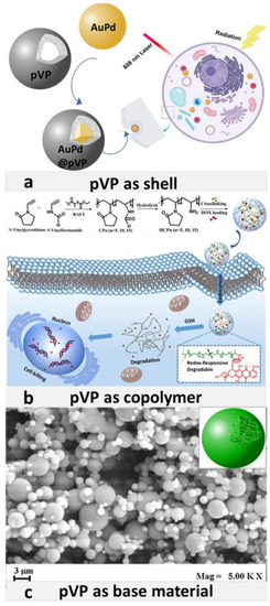
Figure 1.
(a) Scheme of the AuPd core and pVP shell in AuPd@pVP nanoparticles, and their application in photo-thermal therapy. Adapted with permission from [93], Elsevier, 2020. (b) Scheme of synthesis of pVP and N-vinylformamide copolymer and way of its operation against cancer cells. Adapted with permission from [99], Elsevier, 2019. (c) Field Emission Scanning Electron Microscope image of pVP microparticles precipitated from DMSO at 90 bar, 40 °C, 30 mg/mL and obtained using SAS method. Adapted with permission [100], Elsevier, 2016.
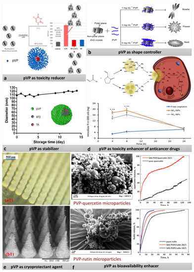
Figure 2.
(a) Scheme of reduction of 5-fluorouracil of neutrophil extracellular traps by closing the drug into Amph-pVP. Adapted with permission from [82], Elsevier, 2020. (b) Influence of different concentration of pVP on the shape of ZnO-PVP nanoparticles. Adapted with permission from [76], Elsevier, 2019. (c) Antidegradation action of pVP. Adapted with permission from [83], Springer, 2020. (d) Scheme of the action of nanoparticles obtained by two different methods, and graph showing enhancement of cisplatin toxicity through increased intercellular accumulation of drug bonded with NP compared to free cisplatin (*—statistically significant difference (p < 0.05) from free cisplatin, **—statistically significant difference from M2-NPs). Reproduced with permission from [92], Elsevier, 2020. (e) Digital microscope (a1) and SEM (b1) images showing formulation of microneedles prepared from carvacrol- poly(caprolactone) nanoparticles with pVP as cryoprotectant agent. Reproduced with permission from [79], Elsevier, 2020. (f) Increased bioavailability of quercitin and rutin via covering drug with pVP. Reproduced with permission from [87], Elsevier, 2019.
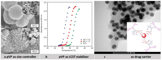
Figure 3.
(a) Scanning electron micrographs of iron oxide powders synthesized in ethylene glycol at 140 °C for 2 h without pVP (top picture) and with pVP (bottom picture). Adapted with permission from [72], Elsevier, 2018. (b) LCST profiles of poly(NIPAAm-co-NVP) nanoparticles under simulated physiological conditions showing influence of pVP on LCST of polymers. Reproduced with permission from [96], Budapest University of Technology and Economics, 2019 (c) TEM images of pVP functionalized gold nanoparticles with curcumin and scheme of possible conjugation of Cur with pVP coated Au-Nps. Reproduced with permission from [62], Elsevier, 2012.
However, the building role is usually related to the activity function. Moreover, pVP can be added to the synthesis mixture due to its ability to limit the particle size and to control the particle shape during the polymerization. In Table 2 the correlation between pVP functions and potential biomedical application of drugs containing pVP in DDSs is shown.
Table 2.
Examples of application of pVP as nanocarriers.
3.1. pVP Coating
One type of drug nanocarriers is core-shell nanoparticles. Metal-, magnetic-, and polymeric materials are mainly used as cores. The core is primarily a magazine for the active substance. The coating/shell is usually made from polymers, including pVP. Still, the shell can fulfill various functions [63]. In the next paragraphs, the application of pVP coating was clearly described.
3.1.1. pVP as Stabilizer and Size Limiter
The stabilizing properties of pVP can be seen in three different perspectives. The first one is the prevention of aggregation. For example, Foldbjerg et al. prepared the Ag-NPs solution by suspending Ag-NPs powder in ELGA water. pVP was used as the stabilizer. The toxicity was examined by means of the flowcytometric annexin V/propidium iodide (PI) assay. The results showed that both Ag+ and pVP-Ag-NPs induced necrosis and apoptosis in THP-1 cells. Moreover, during the exposure, the ROS were produced. The results proved that oxidative stress was an important indicator of cytotoxicity caused by Ag-NPs and Ag+ [64]. In another research, Guo et al. investigated the influence of pVP coated Ag-NPs on inhibition of the growth of acute myeloid leukemia (AML). Additionally, the experiment was repeated with Ag+. Compared to their previous research, they specified the cytotoxicity mechanism of Ag-NPs. The nanoparticles were prepared by a continuous flow electrochemical process in which only polyvinylpyrrolidone (pVP) and two silver rods were used as the stable agent and the electrodes, respectively. The influence of vitamin C and N-acetyl-l-cysteine (NAC) as protection against ROS was also investigated. The results showed that Ag-NPs were capable of bypassing typical barriers and then releasing silver ions that damaged the cell machinery. Moreover, the toxicity of Ag+ was much higher than that of pVP-coated AgNPs against THP-1 cells. In the case of NAC and vitamin C, the research showed that both compounds were capable of completely reversing the generation of ROS after the treatment with Ag-NPs. However, only NAC could protect the cells from more serious damages such as apoptosis [65]. In view of the increase of ROS level after the use of Ag-NPs, Fahrenholtz et al. investigated its influence on three ovarian cancer cell lines: SKOV3, A2780, and OVCAR3. The main choice for the treatment of ovarian cancer is cis-platin (cis-diamminedichloroplatinum(II), CDDP). The authors, besides the examinations with alone pVP-coated Ag-Nps, as the first ones inspected heft combined therapy with the use of prepared nanoparticles and CDDP. The results showed that Ag-Nps were highly cytotoxic against A2780 and SKOV3 cells, whereas OVCAR3 cells were less sensitive to Ag-NPs. The level of ROS was higher in A2780 and SKOV3 compared to OVCAR3. Due to that, it was possible that DNA damage occurred in A2780 and SKOV3. Moreover, the combination of CDDC with pVP- coated Ag-Nps, had the ability to reduce the number of cytostatic doses. However, the authors emphasized that the in vivo examination of the combined therapy should be done in the future [66]. The literature reports indicated that Ag-NPs can be used in tumor therapies and sensing applications and became an encouraging tool for in-vivo imaging. However, after the injection to an organism, they may be coated by protein corona pretty fast, which might lead to the deactivation of the nanoparticle before the start of its therapeutic action [67]. Due to this fact, Batista et al. used pVP as one of the coating ingredients to limit protein aggregation. The authors investigated protein absorption on the Ag-NPs surface stabilized with a mixture of pVP, poly(ethylene oxide)—PEO, and PEO-b-P2VP (poly(ethylene oxide)-b-poly(2-vinyl pyridine). The results were satisfactory [68]. pVP was also used to coat other metal particles, such as gold [69,70].
Regarding the second property, in addition to their stabilizing function, pVP shell can be useful as a size limiter [71,72]. In particular, in paper [73], Au-NPs were synthesized via the addition of Cetyl trimethylammonium bromide (CTAB) solution to HAuCl4. Next, Au-NPs were functionalized through the dynamic stirring method with pVP. Both Au-Nps and pVP coated Au-NPs were characterized using HRTEM and zeta potential. The result showed that zeta potential increased from −34.3 mV for free Au-Nps to −18.7 mV for pVP-coated nanoparticle. HRTEM analysis showed a slight increase in the size of the nanoparticles from 13 to 14.2 nm that was associated with the presence of the polymer. Despite the magnification of nanoparticles, at the same time, the reduction of the size of the metallic core was observed. The results confirmed the size-limiting ability of pVP. The obtained nanoparticles were connected with doxorubicin (Dox@pVP-AuNPs) and used in the treatment of human lung cancer. It appeared that Dox@PVP-Au-NPs were more cytotoxic compared to free DOX. It was caused by the highly effective cellular entry of Dox@PVP-Au-NP. Moreover, Dox@ PVP-AuNPs increased ROS generation, induced both early and late apoptosis in the lung cancer cells, and sensitized mitochondrial membrane potential.
3.1.2. pVP as Shape Controller
Apart from the anti-aggregation and size-limiting ability of pVP, its stabilizing properties involve shape control of the synthesized nanoparticles [74,75]; this is the third property of pVP. For this reason, Yang et al. [76] developed a facile approach to the synthesis of ZnO in different shapes via the chemical precipitation method in water. Contrary to the previous reports, their method was eco-friendly and eliminated the use of organic solvents. For the synthesis, a mixture of zinc nitrate, urea, and pVP was used. The achievement of three different shapes, such as flower, bowtie, and nest, was possible thanks to the use of different concentrations of pVP. With the smallest concentration of pVP, the bowtie-shaped particles were formed. The process involved three distinct phases. Firstly, star-like particles were formed. Next, the formation of densely packed sharp branches on each side of the ZnO-particles was observed. Finally, the branches merged into flakes, leading to bowtie-shaped particles. In the case of using an intermediate concentration of pVP the particles formed the flower-like shape. At first, the porous spheres were produced. Then, the obtained particle served as a template for the growth of petals. Finally, using the greatest concentration of pVP, the nest particles were observed. The obtained nanoparticles were toxic against E. coli and S. aureus. The antibacterial activity depended not only on the morphology of the nanoparticles but also on their surface area, pore size and the used components. However, the killing-bacteria ability appeared mainly due to the release of Zn2+ and the generation of corresponding ROS-independent oxidative stress.
3.1.3. pVP as Cryoprotective Agent
The interesting property of pVP was noticed by Linde Laboratories. They proposed to use this polymer as a substance able to protect cells from freezing injury. This can be possible through plugging the pores in the cells with their coating. This action prevented the cells from overdehydration, ice formation, and changes in the intracellular electrolyte concentration. In addition, pVP allowed lyophilization of biological materials when glycerol was used [77]. This research was groundbreaking; pVP’s ability was used in many clinical applications, among others, in the lyophilization of cells. The cryoprotection abilities were also used in micro and nano drug-delivery systems. Dorati et al. studied the lyophilization process of nanoparticles based on polylactide-co-glycolide (PLGA-H) and polylactide-co-glycolide-co-polyethylenglycol (PLGA-PEG) in a ratio of 30:70. The nanoparticles were obtained via the solid-oil-in-water technique. As cryoprotection agents, pVP-K17, pVP-K32 and sodium carboxymetylcellulose were used. The results showed that the use of polymers stabilized the nanoparticles during the freeze-drying process. In consequence, the aggregation did not occur. All formulations containing cryoprotectants exhibited desired properties after their lyophilization, such as correct size and shape. In addition, in the samples with pVP-K17 and pVP-K32, the collapse phenomenon was not present. The authors suggested that the best protection of the particles against freeze destruction was a blend with pVP-K17 and pVP-K32. In another work, before the lyophilization process, the nanoparticles were loaded with gentamycin—an antibiotic used to treat several types of bacterial infections. One of the important aspects concerning the stability of nanoparticles is zeta potential. The solution of gentamicin sulfate loaded NPs is characterized by zeta potential close to the neutral value (from 0.5 to 3.58 mV), which corresponds to unstable suspensions. After lyophilization, the redispersed gentamycin sulfate-loaded NPs had a slightly negative zeta potential, which was caused by cryoprotectant interactions. However, the neutral zeta potential has a positive impact on antimicrobial activity [78]. Mir et al. examined the influence of pVP concentration (0.5 ÷ 2% w/v) on the size of the carvacrol-poly(caprolactone) nanoparticles after the lyophilization process. The results indicated that with a high pVP concentration (1 ÷ 2%), the size of the nanoparticles decreased; however, the NPs aggregated. For a small pVP concentration (0.5%), the size of NPs was the same as before the lyophilization, and the NPs were freely flowing [79]. Gaber et al. compared three types of cryoprotectant agents, such as hydroxypropyl-β-cyclodextrin (HPβCD), pVP, and trehalose, in the lyophilization process of albumin nanocapsules (NC) and nanoemulsion (NE). The obtained results indicated that the presence of 5% pVP led to the formation of fluffy powders of both NC and NE. However, after the lyophilization process, the size of NE diminished, while the size of NC increased. For 2.5% concentration of pVP the fluffy powders were not observed. The products, after lyophilization, were non-dispersible and viscous [80]. The mentioned above research data confirmed the cryoprotective abilities of pVP. Finally, depending on the substances used, the concentration of pVP should be either elevated or decreased.
3.1.4. pVP as Unwanted-Toxicity Reducer
The antitoxic activity of pVP can be useful for reducing the adverse influence of the drugs and other substances on the patient body. As was already mentioned, some metal-oxide nanoparticles are exploited for the delivery of medicines. However, their use led to significantly lowered cell viability. İşçi et al. checked the influence of pVP on the toxicity of iron(III) oxide nanoparticles. For this purpose, they synthesized Fe3O4 nanoparticles with pVP shells. The obtained results showed that, compared to non-covered Fe3O4, the PVP coating of the nanoparticles increased the protection of the healthy osteoblast cells. Moreover, the addition of polymeric shells did not affect the superparamagnetic character of the iron oxide [81]. One complication occurring in patients suffering from neoplastic diseases is venous thromboembolism. The main cause of venous diseases is neutrophil extracellular traps (NETs). They are also an important factor in the development of the tumor growth process. The literature reports say that some anticancer drugs, e.g., 5-fluorouracil (5FU), can escalate the problem with creating NETs. For this reason, Basyreva et al. developed protection of patients against side effects of 5FU chemotherapy. They prepared amphiphilic poly-N-vinylpyrrolidone (Amph-pVP) and used it as a 5-fluorouracil (5FU) carrier. The obtained data showed that 5FU alone increased two to three times the total amount of NETs in the blood. However, when Amph-pVP was used, the adverse effect of 5FU was completely blocked, and the formation of NETs was not observed [82]. The pVP coating can play several roles. For example, Chen et al. prepared the bortezomib (BTZ) nanoparticles with tannic acid covered by pVP shell. Firstly, it seemed that polymer only stabilized nanoparticles. The results showed that the addition of pVP made them stable over the entire time of the experiment (15 days). This system also appeared to be useful in antitumor treatment. The cytotoxicity examinations indicated that the modified BTZ nanoparticles were more cytotoxic than BTZ alone. Moreover, the analysis of the distribution of the drug in model tumor-bearing mouse led to a conclusion that a higher concentration of the drug in the tumor tissue was found when BTZ was covered with pVP. Correspondingly, the modified BTZ nanoparticles caused superior therapeutic effects. Next, in vitro examination indicated pVP as a substance for improving the bioavailability of BTZ. However, in vivo experiments showed that mice treated with BTZ alone were characterized by weight loss while the pVP coated BTZ-NPs led to weight gain in mice. This phenomenon indicated the reduced systemic toxicity of the BTZ-NPs [83].
3.1.5. pVP for Protection and Solubilization of Substances and for Enhancing Solubility
The pVP polymer can protect healthy tissues from toxic substances, improve the solubility of the drug and enhance its bioavailability. For example, variety of structures/compositions of pVP is responsible for its solubility in many different substances. This property can be used to increase the bioavailability of some drugs after loading them into the polymer net. The drug solubility in aqueous solutions is enhanced through the formation of the drug-polymer complex [84]. This feature can be employed in building specific drug delivery systems acting against, e.g., neoplastic cells. In this respect, Tiwari et al., in their research, used graphene nanoparticles coated with pVP as quercetin (QSR)- and gefitinib (GEF) drug carriers. Moreover, the influence of the combination of these drugs on ovarian cancer cells was investigated. GO-NPs were connected with pVP in a carbodiimide-activated esterification reaction. The results showed that the functionalization of GO-NPs surface with the selected polymer, pVP, increased drug solubility, and biocompatibility. In addition, the combined drugs improved cytotoxicity against the ovarian cell [85]. On the other hand, it was shown that the addition of pVP to GO did not influence the cytotoxicity of graphene oxide significantly, contrary to the popular belief that pVP acts as the toxicity reducer [86]. In another paper, Ozkan et al. prepared pVP particles connected with the drug through the supercritical antisolvent process (SAS). Quercetin and rutin were used as the model substances. These compounds belong to the class of flavonols and are characterized by anti-inflammatory, antioxidant, and antiviral activities. The main disadvantage of flavonols is their poor solubility in water, which results in their weak bioavailability. The obtained results proved that the use of the polymer led to an increase in the dissolution rates by circa 10 times for quercetin and 3.19 times for rutin, compared to flavonols alone. In addition, high effectiveness of connection of the drug with the polymer was achieved [87]. Homayouni, using pVP-K30 and HPMC (hydroxypropyl methylcellulose), obtained curcumin (Cur) nanoparticles by antisolvent crystallization. Cur is anticancer, antioxidant, anti-inflammatory, and antimicrobial. However, it just, as flavonols, is characterized by poor water solubility, which in consequence limits its oral bioavailability. It appeared that by using pVP the solubility of curcumin was increased. The time of dissolution was shortened from circa 20 min for curcumin alone to circa 7 min for the nanoparticles with pVP. Moreover, the examinations revealed that pVP inhibited the aggregation of the particles and that the connection between Cur and pVP is realized by hydrogen bonding [88]. The literature papers report that anticancer curcumin acts like inhibitor for STAT3 and NF-κB signaling pathways, which plays the main role in cancer development. In addition, Cur can counteract the activation of Sp-1 and its downstream genes such as EPHB2, ADEM10, HDAC4, calmodulin, and SEPP1 [89]. Moreover, Cur solubility can be improved when one of its chains is connected with a metallic nanoparticle while another chain is attached to one of the side chains of such soluble polymers as inter ilia pVP. For this purpose, the folate–curcumin-loaded gold–polyvinylpyrrolidone nanoparticles (FA–CurAu-PVP NPs) were prepared. The drug and the targeting substance were connected with Au-NPs using the layer by layer assembling. FA–CurAu-PVP NPs were characterized with UV–Vis spectra, FT IR spectroscopy, X-ray powder diffraction, and thermogravimetric analysis. In vitro analysis was done using the MTT assay. Additionally, the in vivo test of NPs against mouse-model breast cancer was examined. The results suggested that the obtained nanoparticles seemed to be a promising candidate for Cur chemotherapy to destroy the tumor cells without harming normal cells [90].
From the drug-delivery perspective, one of the most important properties of polymeric carriers is their biocompatibility with the tissue. Thus far, a rather big number of reports on compliance of pVP have appeared. However, there are only a few research papers on the performance of this polymeric carrier in the blood. Tsatsakis et al. checked the behavior of Amph-pVP nanoparticles in the presence of whole blood, blood plasma, and blood cells. Moreover, they evaluated the effects of these nanoparticles on endothelium cell growth/viability. Amph-pVP was obtained via self-assembling in an aqueous medium. pVP formed a hydrophilic shell placed over the hydrophobic alkyl core. It was found that Amph–pVP had no effect on the coagulation process, complement activation, and platelet aggregation in the tested concentration range of 0.05 ÷ 0.5 mg/mL. In addition, no considerable hemolytic or inflammatory effect was observed [91]. Pornpitchanarong et al. prepared two types of nanoparticles based on pVP and acrylic acid: Copolymer and core-shell structure. The surfactant-free emulsion polymerization was used. The size of nanoparticles was in the range 138 ÷ 183 nm. The obtained nanoparticles were loaded with cis-platin via a coordinate covalent bond, and relatively high drug loading was achieved. The cytotoxic experiments showed higher toxicity of cis-platin loaded nanoparticles against the cancer cells compared to the drug alone. Since the nanoparticles guaranteed sustained, moderate release of the drug, they can be used in long-term therapy, which is safer for the patients [92]. Xiang et el. developed the reduction method to synthesize AuPd@pVP NPs in the presence of pVP. The polymer was used as both surfactant and stabilizing agent. They reported that thanks to the formation of pVP shell, good biocompatibility both in vivo and in vitro were possible, and the obtained nanoparticles were useful in photothermal therapy (PTT) under NIR laser irradiation of low power [93]. Mahdavi et al. prepared gadolinium oxide nanoparticles covered with pVP. This radiosensitized core-shell system could be combined with a cytostatic drug. In consequence, it was possible to use this multifunctional drug delivery system in tumor treatment. The authors confirmed that the surface modification of Gd2O3 NPs with PVP could increase their biocompatibility. The obtained results showed that the viability and surviving fraction of B16F10 melanoma cells were significantly inhibited when Gd2O3 @ PVP-DOX NPs under radiation were used compared with treating with the free DOX or PVP-Gd2O3 NPs alone under radiation [94].
Thenmozhi concluded that the functionalized iron oxide nanoparticles induced apoptosis in MCF-7 breast cancer cell lines. Iron oxide nanoparticles were coated with PVP by adding PVP to a suspension of nanoparticles, sonicating the solution for 30 min, and thermal annealing the separated product at 600 °C for 30 min. Finally, the PVP coated iron nanoparticles were biofunctionalized with Syzygium aromaticum flower bud’s (clove) extract. The author found that a significant quantity of the nanoparticles loaded with S. aromaticum extract was in the cells during the sub-G1 state. In addition, the nanoparticles influenced the caspase–3, caspase–8 and caspase–9 responses. The oxidative stress caused by ROS formed after the treatment with nanoparticles was also observed. The pVP coating played not only the role of the stabilizer of iron oxide nanoparticles but also improved the bioavailability and therapeutic effect of plant extract [95].
3.2. pVP in Copolymers
3.2.1. pVP as LCST Stabilizer
pVP often worked as a copolymer. From the point of view of drug delivery, in the case of temperature-sensitive systems, the LCST (lower critical solution temperature) close to the physiological level (37 °C) should be achieved. Effective change of LCST can be obtained after adding a hydrophilic monomer to the reaction mixture. For example, a copolymer poly(N-isopropylacrylamide-co-N-vinylpyrrolidone) was synthesized. The best results were obtained for a monomer molar ratio (NIPAAm/NVP) of 91.5:8.5 in the copolymer. Next, the polymerization suspension was used to form the microspheres. Finally, the microspheres were loaded with a model drug—diclofenac. The release of that drug through the pulsatile mechanism was investigated [96]. In another try, a new microgel based on acrylamide (AAM), 1-vinyl-2-pyrrolidone (NVP), and 2-(diethylamino)ethyl methacrylate (DEAEMA) was prepared. The free-radical precipitation polymerization was applied. The obtained microparticles increased the LCST temperature from 32 °C to circa 38 °C (physiological level). 5-FU was loaded into the microgel with efficiency of 32.47 mg/g. The obtained results indicated that 5-Fu was released slightly faster at pH 7.4, and the microgel did not exhibit visible cytotoxicity [97].
3.2.2. pVP as Solubilizing Agent
An interesting application of pVP copolymer was proposed by Swilem et al. They synthesized nanogels based on poly(acrylic acid)-co-N-vinylpyrrolidone using the ionizing radiation method. The obtained material was used as a tear substitute; it could be useful in treatment of dry-eye syndrome. Poly(acrylic acid) is a major component of various brands of artificial tears and is characterized by mucoadhesive property and high water absorbance. However, there were disadvantages of using this kind of polymer; viscous films could be formed and they caused transient blurring of vision and sticky sensations. The addition of pVP reduced viscosity of tears significantly and, therefore, increased the comfort of the patient. Moreover, the use of the copolymer enhanced markedly the effectiveness of dry eye curing [98].
3.2.3. pVP as Base Material
pVP can also be useful as the base material. As already mentioned, pVP is a hydrophilic polymer, which can improve biocompatibility and solubility of many drugs. However, only several reports on pVP as the main ingredient of the nanoparticles appeared in the literature. For example, Peng et al. synthesized, with the addition of N-vinylformamide to the main monomer, the pVP material using the reversible addition–fragmentation chain-transfer (RAFT) polymerization. The obtained material was sensitive to redox actions and degraded in the presence of GSH. The nanogel could be used to carry doxorubicin (DOX). A decent level of drug loading was achieved. The in vitro examinations indicated that blank nanogels were biocompatible, and DOX-loaded nanoparticles exhibited medium antitumor activity [99]. Prosapio et al. proposed a method of obtaining drug/polymer microparticles, which was based on coprecipitation of the active substance with a hydrophilic polymer (pVP) using a supercritical antisolvent. Nimesulide was used as a model drug. The size of microparticles was in a range from 1.7 to 4 μm. The drug-release examination indicated that by using pVP the dissolution rate could be enhanced by circa 2.5 times [100].
4. Limitations of pVP
There have been only several reports on rare cases where pVP caused side effects in the patients. It acted as an allergen and caused urticaria, dyspnoea, and anaphylaxis [101,102]. For this reason, the particles of high molecular weight, multiple doses, and injections of large amounts of pVP in the same place should be avoided. Regarding the use of pVP during intracytoplasmic sperm injection as the improver of viscosity of the sperm solution, pVP may cause changes in the sperm structure and its visibility in the injected embryos during the early development period [103]. pVP, in general, is classified as a biodegradable material. However, there are several reports in the literature on the degradation problem and in consequence the limitation for parenteral administration. Because of that, low molecular weight pVP should be useful for parental administration, due to its possibility of being excreted through the kidneys [104].
5. Conclusions and Future Perspectives
Despite many advantages and a wide use in medicine, compared to other polymers, pVP is not a very popular material in drug delivery systems. Thus far, pVP is mainly used in drug delivery systems as the coating or shell ingredient (see Table 2), the protection from aggregation and the shape and size controller. Few studies were concerned with nano- and microhydrogels, including pVP as a copolymer and its ability to change LCST of the main polymer. However, due to an increase in demand for new biomaterials and the use of micro- and nano drug delivery systems, the situation should change soon. In addition, pVP has other abilities, which can be used in the production of new copolymers with new properties. Thus far, just a few reports on the application of pVP as the main component in the carriers have appeared. A pending challenge is to use pVP in micro- and nanomedical devices as the main building material. The literature reports are focused mainly on the nanoparticle synthesis employing the electron beam method; this method requires rather expensive, specific equipment. Moreover, there is some danger related to the emitted X-ray radiation. In the future, development of simpler and more efficient methods of synthesis of the pVP particles may lead to a significant progress in the construction of pVP micro- and nano drug-carriers.
Author Contributions
Conceptualization, E.W., Z.S., M.K.; methodology, E.W., Z.S., M.K.; writing—original draft preparation, E.W.; writing—review and editing, Z.S., M.K.; visualization, E.W.; supervision, M.K.; funding acquisition, M.K. All authors have read and agreed to the published version of the manuscript.
Funding
This work was supported by the National Science Center of Poland under grant number 2018/31/B/ST5/02859. E.W. work was implemented as a part of the Operational Project Knowledge Education Development 2014–2020 financed by the European Social Fund.
Institutional Review Board Statement
Not applicable.
Informed Consent Statement
Not applicable.
Data Availability Statement
Not applicable.
Conflicts of Interest
The authors declare no conflict of interest.
Abbreviations
| Dox@pVP-AuNPs | doxorubicin loaded povidone coated gold nanoparticles |
| 5FU | 5-fluorouracil |
| AAM | acrylamide |
| Ag-NPs | silver nanoparticles |
| Amph-pVP | amphiphilic poly-N-vinylpyrrolidone |
| Au-NPs | gold nanoparticles |
| AuPd@pVP NP | povidone coated gold-palladium nanoparticles |
| BTZ | bortezomib |
| CDDP | cis-diamminedichloroplatinum II |
| CTAB | cetyl trimethylammonium bromide |
| Cur | curcumin |
| DDSs | drug delivery systems |
| DEAEMA | 2-(diethylamino)ethyl methacrylate |
| FA–CurAu-PVP NPs | folate–curcumin-loaded gold–polyvinylpyrrolidone nanoparticles |
| Gd2O3 NPs | gadolin oxide nanoparticles |
| GEF | gefitnib |
| GO-NPs | graphene oxide nanoparticles |
| HPMC | hydroxypropyl methylcellulose |
| HPβCD | hydroxypropyl-β-cyclodextrin |
| LCST | lower critical solution temperature |
| NaBH4 | sodium borohydride |
| NAC | N-acetyl-l-cysteine |
| NC | nanocapsules |
| NE | nanoemulsion |
| NETs | neutrophil extracellular traps |
| NIPAAm/NVP | poly(N-isopropylacrylamide-co-N-vinylpyrrolidon |
| NVP | 1-vinyl-2-pyrrolidone |
| PEO | poly(ethylene oxide) |
| PEO-b-P2VP | 2-vinyl pyridine poly(ethylene oxide)-b-poly(2-vin(2-vinyl pyridine |
| PLGA-H | polylactide-co-glycolide |
| PLGA-PEG | polylactide-co-glycolide-co-polyethylenglycol |
| PTT | photothermal therapy |
| pVP | poly (N-vinyl pyrrolidone) |
| pVP-I | povidone iodine |
| pVP-Ag-NPs | povidone coated silver nanoparticles |
| QSR | quercetin |
| RAFT | the reversible addition–fragmentation chain-transfer polymerization |
| SAS | supercritical antisolvent process |
| THP-1 | human monocytic cell line |
References
- Laffleur, F.; Keckeis, V. Advances in drug delivery systems: Work in progress still needed? Int. J. Pharm. 2020, 2, 100050. [Google Scholar]
- LaSala, R.; LoGreco, A.; Romagnoli, A.; Santoleri, F.; Musicco, F.; Costantini, A. Cancer drugs for solid tumors approved by the EMA since 2014: An overview of pivotal clinical trials. Eur. J. Clin. Pharmacol. 2020, 76, 843–850. [Google Scholar] [CrossRef] [PubMed]
- O’Brien, M.E.R.; Borthwick, A.; Rigg, A.; Leary, A.; Assersohn, L.; Last, K.; Tan, S.; Milan, S.; Tait, D.; E Smith, I. Mortality within 30 days of chemotherapy: A clinical governance benchmarking issue for oncology patients. Br. J. Cancer 2006, 95, 1632–1636. [Google Scholar] [CrossRef] [PubMed]
- Jain, K.K. Drug Delivery Systems; Humana Press: Totowa, NJ, USA, 2008. [Google Scholar]
- Boverhof, D.R.; Bramante, C.M.; Butala, J.H.; Clancy, S.F.; Lafranconi, M.; West, J.; Gordon, S.C. Comparative assessment of na-nomaterial definitions and safety evaluation considerations. Regul. Toxicol. Pharmacol. 2015, 73, 137–150. [Google Scholar] [CrossRef] [PubMed]
- Hossen, S.; Hossain, M.K.; Basher, M.; Mia, M.; Rahman, M.; Uddin, M.J. Smart nanocarrier-based drug delivery systems for cancer therapy and toxicity studies: A review. J. Adv. Res. 2019, 15, 1–18. [Google Scholar] [CrossRef]
- Hamidi, M.; Azadi, A.; Rafiei, P. Hydrogel nanoparticles in drug delivery. Adv. Drug Deliv. Rev. 2008, 60, 1638–1649. [Google Scholar] [CrossRef]
- Peppas, N.A.; Khare, A.R. Preparation, structure and diffusional behavior of hydrogels in controlled release. Adv. Drug Deliv. Rev. 1993, 11, 1–35. [Google Scholar] [CrossRef]
- Constantin, M.; Bucatariu, S.; Ascenzi, P.; Butnaru, M.; Fundueanu, G. Smart drug delivery system activated by specific biomole-cules. Mater. Sci. Eng. C 2020, 108, 110466. [Google Scholar] [CrossRef]
- Karbarz, M.; Mackiewicz, M.; Kaniewska, K.; Marcisz, K.; Stojek, Z. Recent developments in design and functionalization of micro- and nanostructural environmentally-sensitive hydrogels based on N-isopropylacrylamide. Appl. Mater. Today 2017, 9, 516–532. [Google Scholar] [CrossRef]
- Rosso, A.P.; Martinelli, M. Preparation and characterization of dendronized chitosan/gelatin-based nanogels. Eur. Polym. J. 2020, 124, 109506. [Google Scholar] [CrossRef]
- Mahmoodzadeh, F.; Ghorbani, M.; Jannat, B. Glutathione and pH-responsive chitosan-based nanogel as an efficient nanoplatform for controlled delivery of doxorubicin. J. Drug Deliv. Sci. Technol. 2019, 54, 101315. [Google Scholar] [CrossRef]
- Rao, K.M.; Ramanjaneyulu, G.; Ha, C.-S. Curcumin encapsulated pH sensitive gelatin based interpenetrating polymeric network nanogels for anti cancer drug delivery. Int. J. Pharm. 2015, 478, 788–795. [Google Scholar] [CrossRef]
- Suraiya, A.B.; Hun, M.L.; Truong, V.X.; Forsythe, J.S.; Chidgey, A.P. Gelatin-based 3D microgels for in vitro T lineage cell generation. ACS Biomater. Sci. Eng. 2020, 6, 2198–2208. [Google Scholar] [CrossRef] [PubMed]
- Pandey, K.; Saha, S. Microencapsulated zero valent iron nanoparticles in polylactic acid matrix for in situ remediation of contaminated water. J. Environ. Chem. Eng. 2020, 8, 103909. [Google Scholar] [CrossRef]
- Lu, T.; Hu, F.; Yue, H.; Yang, T.; Ma, G. The incorporation of cationic property and immunopotentiator in poly (lactic acid) microparticles promoted the immune response against chronic hepatitis B. J. Control. Release 2020, 321, 576–588. [Google Scholar] [CrossRef]
- Zhao, X.; Li, Y.; Li, B.; Hu, T.; Yang, Y.; Li, L.; Zhang, J. Environmentally benign and durable superhydrophobic coatings based on SiO2 nanoparticles and silanes. J. Colloid Interface Sci. 2019, 542, 8–14. [Google Scholar] [CrossRef] [PubMed]
- Wei, M.; Han, J.-Y.; Gao, Y.-J.; Jiang, S.-L.; Sun, F. Surface morphology and property of UV-cured film containing photopolymerizable polysiloxane-based nanogels with initiating capability. Int. J. Ind. Chem. 2019, 10, 281–289. [Google Scholar] [CrossRef]
- Ghorbani, M.; Nezhad-Mokhtari, P.; Ramazani, S. Aloe vera-loaded nanofibrous scaffold based on Zein/Polycaprolactone/Collagen for wound healing. Int. J. Biol. Macromol. 2020, 153, 921–930. [Google Scholar] [CrossRef]
- Lazurko, C.; Khatoon, Z.; Goel, K.; Sedlakova, V.; Cimenci, C.E.; Ahumada, M.; Zhang, L.; Mah, T.-F.; Franco, W.; Suuronen, E.J.; et al. Multifunctional nano and collagen-based therapeutic materials for skin repair. ACS Biomater. Sci. Eng. 2020, 6, 1124–1134. [Google Scholar] [CrossRef]
- Manuja, A.; Raguvaran, R.; Kumar, B.; Kalia, A.; Tripathi, B. Accelerated healing of full thickness excised skin wound in rabbits using single application of alginate/acacia based nanocomposites of ZnO nanoparticles. Int. J. Biol. Macromol. 2020, 155, 823–833. [Google Scholar] [CrossRef]
- Singh, I.; Lacko, C.S.; Zhao, Z.; Schmidt, C.E.; Rinaldi, C. Preparation and evaluation of microfluidic magnetic alginate micropar-ticles for magnetically templated hydrogels. J. Colloid Interface Sci. 2020, 561, 647–658. [Google Scholar] [CrossRef]
- Waleka, E.; Mackiewicz, M.; Romanski, J.; Dybko, A.; Stojek, Z.; Karbarz, M. Degradable nanohydrogel with high doxorubicin loadings exhibiting controlled drug release and decreased toxicity against healthy cells. Int. J. Pharm. 2020, 579, 119188. [Google Scholar] [CrossRef] [PubMed]
- Wang, J.; Huang, N.; Peng, Q.; Cheng, X.; Li, W. Temperature/pH dual-responsive and luminescent drug carrier based on PNIPAM-MAA/lanthanide-polyoxometalates for controlled drug delivery and imaging in HeLa cells. Mater. Chem. Phys. 2020, 239, 121994. [Google Scholar] [CrossRef]
- Mackiewicz, M.; Stojek, Z.; Karbarz, M. Synthesis of cross-linked poly(acrylic acid) nanogels in an aqueous environment using precipitation polymerization: Unusually high volume change. R. Soc. Open Sci. 2019, 6, 190981. [Google Scholar] [CrossRef]
- Kalasin, S.; Sangnuang, P.; Khownarumit, P.; Tang, I.M.; Surareungchai, W. Evidence of Cu(I) coupling with creatinine using cuprous nanoparticles encapsulated with polyacrylic acid (PAA) Gel-Cu (II) in facilitating the determination of advanced kidney dysfunctions. ACS Biomater. Sci. Eng. 2020, 6, 1247–1258. [Google Scholar] [CrossRef]
- Cerda-Sumbarda, Y.D.; Domínguez-González, C.; Zizumbo-López, A.; Licea-Claverie, A. Thermoresponsive nanocomposite hydrogels with improved properties based on poly(N-vinylcaprolactam). Mater. Today Commun. 2020, 24, 10104. [Google Scholar] [CrossRef]
- Xu, F.; Zhu, J.; Lin, L.; Zhang, C.; Sun, W.; Fan, Y.; Yin, F.; Van Hest, J.C.M.; Wang, H.; Du, L.; et al. Multifunctional PVCL nanogels with redox-responsiveness enable enhanced MR imaging and ultrasound-promoted tumor chemotherapy. Theranostics 2020, 10, 4349–4358. [Google Scholar] [CrossRef] [PubMed]
- Fang, Z.; Sun, Y.; Cai, C.; Fan, R.; Guo, R.; Xie, D. Targeted delivery of DOX by transferrin conjugated DSPE-PEG nano-particles in leukemia therapy. Int. J. Polym. Mater. Polym. Biomater. 2021, 70, 27–36. [Google Scholar] [CrossRef]
- Kaldybekov, D.B.; Filippov, S.K.; Radulescu, A.; Khutoryanskiy, V.V. Maleimide-functionalised PLGA-PEG nanoparticles as mucoadhesive carriers for intravesical drug delivery. Eur. J. Pharm. Biopharm. 2019, 143, 24–34. [Google Scholar] [CrossRef] [PubMed]
- Quérette, T.; Fleury, E.; Sintes-Zydowicz, N. Non-isocyanate polyurethane nanoparticles prepared by nanoprecipitation. Eur. Polym. J. 2019, 114, 434–445. [Google Scholar] [CrossRef]
- Zhang, S.; Zhang, D.; Bai, H.; Ming, W. ZnO nanoparticles coated with amphiphilic polyurethane for transparent polyurethane nanocomposites with enhanced mechanical and UV-shielding performance. ACS Appl. Nano Mater. 2019, 3, 59–67. [Google Scholar] [CrossRef]
- Morkhade, D.M. Comparative impact of different binder addition methods, binders and diluents on resulting granule and tablet attributes via high shear wet granulation. Powder Technol. 2017, 320, 114–124. [Google Scholar] [CrossRef]
- Koczkur, K.M.; Mourdikoudis, S.; Polavarapu, L.; Skrabalak, S.E. Polyvinylpyrrolidone (PVP) in nanoparticle synthesis. Dalton Trans. 2015, 44, 17883–17905. [Google Scholar] [CrossRef] [PubMed]
- Wan, L.-S.; Xu, Z.-K.; Huang, X.-J.; Wang, Z.-G.; Wang, J.-L. Copolymerization of acrylonitrile with N-vinyl-2-pyrrolidone to improve the hemocompatibility of polyacrylonitrile. Polymer 2005, 46, 7715–7723. [Google Scholar] [CrossRef]
- Awasthi, R.; Manchanda, S.; Das, P.; Velu, V.; Malipeddi, H.; Pabreja, K.; Pinto, T.D.J.A.; Gupta, G.; Dua, K. Poly(vinylpyrrolidone). In Engineering of Biomaterials for Drug Delivery Systems: Beyond Polyethylene Glycol; Woodhead Publishing Series in Biomaterials: Cambridge, UK, 2018. [Google Scholar]
- Wen, T.; Lu, L.; Dong, C. Enhancing the dehumidification performance of LiCl solution with surfactant PVP-K30. Energy Build. 2018, 171, 183–195. [Google Scholar] [CrossRef]
- Lee, J. Intrinsic adhesion properties of poly(vinyl pyrrolidone) to pharmaceutical materials: Humidity effect. Macromol. Biosci. 2005, 5, 1085–1093. [Google Scholar] [CrossRef]
- Tavlarakis, P.; Urban, J.J.; Snow, N. Determination of total polyvinylpyrrolidone (PVP) in ophthalmic solutions by size exclusion chromatography with ultraviolet-visible detection. J. Chromatogr. Sci. 2011, 49, 457–462. [Google Scholar] [CrossRef]
- Teodorescu, M.; Bercea, M. Poly(vinylpyrrolidone)—A versatile polymer for biomedical and beyond medical applications. Polym. Technol. Eng. 2015, 54, 923–943. [Google Scholar] [CrossRef]
- Song, G.; Lin, Y.; Zhu, Z.; Zheng, H.; Qiao, J.; He, C.; Wang, H. Strong fluorescence of poly(nvinylpyrrolidone) and its oxidized hydrolyzate. Macromol. Rapid Commun. 2015, 26, 278–285. [Google Scholar] [CrossRef]
- Hua, T.I.; Sundari, R.; Lintang, H.O.; Yuliati, L. Polyvinylpyrrolidone as a new fluorescent sensor for nitrate ion. Malays. J. Anal. Sci. 2016, 20, 288–295. [Google Scholar] [CrossRef]
- Sütekin, S.D.; Güven, O. Application of radiation for the synthesis of poly(n-vinyl pyrrolidone) nanogels with controlled sizes from aqueous solutions. Appl. Radiat. Isot. 2019, 145, 161–169. [Google Scholar] [CrossRef] [PubMed]
- Fiscella, R. Ophthalmic Drug Formulations in Clinical Ocular Pharmacology, 5th ed.; Bartlett, J.D., Jaanus, S.D., Eds.; Butter-worth-Heinemann: Oxford, UK, 2008; pp. 17–37. [Google Scholar]
- Darouiche, R.O.; Wall, M.J.; Itani, K.M.F.; Otterson, M.F.; Webb, A.L.; Carrick, M.M.; Miller, H.J.; Award, S.S.; Crosby, C.T.; Mosier, M.C.; et al. Chlorhexidine-alcohol versus povidone-iodine for surgical-site antisepsis. N. Engl. J. Med. 2010, 362, 18–26. [Google Scholar] [CrossRef] [PubMed]
- Kurakula, M.; Rao, G.S.N.K. Pharmaceutical assessment of polyvinylpyrrolidone (pvp): As excipient from conventional to controlled delivery systems with a spotlight on COVID-19 inhibition. J. Drug Deliv. Sci. Technol. 2020, 60, 102046. [Google Scholar] [CrossRef]
- Pinto, R.D.P.; Lira, R.P.C.; Abe, R.Y.; Zacchia, R.S.; Felix, J.P.F.; Pereira, A.V.F.; Arieta, C.E.L.; De Castro, R.S.; Bonon, S.H.A. Dexamethasone/povidone eye drops versus artificial tears for treatment of presumed viral conjunctivitis: A randomized clinical trial. Curr. Eye Res. 2014, 40, 870–877. [Google Scholar] [CrossRef]
- Liccioli, G.; Mori, F.; Barni, S.; Pucci, N.; Novembre, E. Anaphylaxis to polyvinylpyrrolidone in eye drops administered to an adolescent. J. Investig. Allergol. Clin. Immunol. 2018, 28, 263–265. [Google Scholar] [CrossRef] [PubMed]
- Bigliardi, P.L.; Alsagoff, S.A.L.; El-Kafrawi, H.Y.; Pyon, J.-K.; Wa, C.T.C.; Villa, M.A. Povidone iodine in wound healing: A review of current concepts and practices. Int. J. Surg. 2017, 44, 260–268. [Google Scholar] [CrossRef]
- Stupak, E.I.; Bates, T.R. Enhanced absorption and dissolution of reserpine from reserpine-polyvinylpyrrolidone coprecipitates. J. Pharm. Sci. 1972, 61, 400–404. [Google Scholar] [CrossRef]
- Manju, S.; Sreenivasan, K. Synthesis and characterization of a cytotoxic cationic polyvinylpyrrolidone-curcumin conjugate. J. Pharm. Sci. 2011, 100, 504–511. [Google Scholar] [CrossRef]
- Hackl, E.; Ermolina, I. Application of texture analysis technique in formulation development of lyophilized orally disintegrating tablets containing mannitol, polyvinylpyrrolidone and amino acids. AAPS PharmSciTech 2019, 20, 71. [Google Scholar] [CrossRef]
- Khokhar, P.; Shukla, V. Formulation and evaluation of fast dissolving tablets of diclofenac sodium using PVP. Int. J. Pharma Res. Rev. 2014, 3, 12–19. [Google Scholar]
- Arndt, O.-R.; Kleinebudde, P. Influence of binder properties on dry granules and tablets. Powder Technol. 2018, 337, 68–77. [Google Scholar] [CrossRef]
- Guo, Y.; Hao, Z.; Wan, C. Tribological characteristics of polyvinylpyrrolidone (PVP) as a lubrication additive for artificial knee joint. Tribol. Int. 2016, 93, 214–219. [Google Scholar] [CrossRef]
- Singh, B.; Sharma, N. Design of sterculia gum based double potential antidiarrheal drug delivery system. Colloids Surf. B Biointerfaces 2011, 82, 325–332. [Google Scholar] [CrossRef] [PubMed]
- Schafer, J.; Reindel, W.; Steffen, R.; Mosehauer, G.; Chinn, J. Use of a novel extended blink test to evaluate the performance of two polyvinylpyrrolidone-containing, silicone hydrogel contact lenses. Clin. Ophthalmol. 2018, 12, 819–825. [Google Scholar] [CrossRef]
- Xue, Y.; Zhang, W.; Lei, Y.; Dang, M. Novel polyvinyl pyrrolidone-loaded olopatadine HCl-laden doughnut contact lens to treat allergic conjunctivitis. J. Pharm. Sci. 2020, 109, 1714–1724. [Google Scholar] [CrossRef] [PubMed]
- Moffitt, E.A. Blood substitutes. Can. J. Anesth. 1975, 22, 12–19. [Google Scholar] [CrossRef]
- Lecce, J.G.; Matrone, G.; Morgan, D.O. Porcine neonatal nutrition: Absorption of unaltered nonporcine proteins and polyvi-nylpyrrolidone from the gut of piglets and the subsequent effect on the maturation of the serum protein profile. J. Nutr. 1961, 73, 158–166. [Google Scholar] [CrossRef]
- Kadajji, V.G.; Betageri, G.V. Water soluble polymers for pharmaceutical applications. Polymers 2011, 3, 1972–2009. [Google Scholar] [CrossRef]
- Gangwar, R.K.; Dhumale, V.A.; Kumari, D.; Nakate, U.T.; Gosavi, S.; Sharma, R.B.; Kale, S.; Datar, S. Conjugation of curcumin with PVP capped gold nanoparticles for improving bioavailability. Mater. Sci. Eng. C 2012, 32, 2659–2663. [Google Scholar] [CrossRef]
- Kurakula, M.; Naveen, N.R.; Yadav, K.S. Formulations for polymer coatings. Polym. Coat. 2020, 415–443. [Google Scholar] [CrossRef]
- Foldbjerg, R.; Olesen, P.; Hougaard, M.; Dang, D.A.; Hoffmann, H.J.; Autrup, H. PVP-coated silver nanoparticles and silver ions induce reactive oxygen species, apoptosis and necrosis in THP-1 monocytes. Toxicol. Lett. 2009, 190, 156–162. [Google Scholar] [CrossRef]
- Guo, D.; Zhu, L.; Huang, Z.; Zhou, H.; Ge, Y.; Ma, W.; Wu, J.; Zhang, X.; Zhou, X.; Zhang, Y.; et al. Anti-leukemia activity of PVP-coated silver nanoparticles via generation of reactive oxygen species and release of silver ions. Biomaterials 2013, 34, 7884–7894. [Google Scholar] [CrossRef]
- Fahrenholtz, C.D.; Swanner, J.; Ramirez-Perez, M.; Singh, R.N. Heterogeneous responses of ovarian cancer cells to silver nanoparticles as a single agent and in combination with cisplatin. J. Nanomater. 2017, 2017, 5107485. [Google Scholar] [CrossRef] [PubMed]
- Kopp, M.; Kollenda, S.; Epple, M. Nanoparticle-protein interactions: Therapeutic approaches and supramolecular chemistry. Acc. Chem. Res. 2017, 50, 1383–1390. [Google Scholar] [CrossRef]
- Batista, C.C.; Albuquerque, L.J.; Jägerb, A.; Stepánekb, P.; Giacomelli, F.C. Probing protein adsorption onto polymer-stabilized silver nanocolloids towards a better understanding on the evolution and consequences of biomolecular coronas. Mater. Sci. Eng. C 2020, 111, 110850. [Google Scholar] [CrossRef]
- Mohamed, T.; Matou-Nasri, S.; Farooq, A.; Whitehead, D.; Azzawi, M. Polyvinylpyrrolidone-coated gold nanoparticles inhibit endothelial cell viability, proliferation, and ERK1/2 phosphorylation and reduce the magnitude of endothelial-independent dilator responses in isolated aortic vessels. Int. J. Nanomed. 2017, 12, 8813–8830. [Google Scholar] [CrossRef] [PubMed]
- Chen, W.; Zhang, S.; Yu, Y.; Zhang, H.; He, Q. Structural-engineering rationales of gold nanoparticles for cancer theranostics. Adv. Mater. 2016, 28, 8567–8585. [Google Scholar] [CrossRef] [PubMed]
- Wang, H.; Qiao, X.; Chen, J.; Wang, X.; Ding, S. Mechanisms of PVP in the preparation of silver nanoparticles. Mater. Chem. Phys. 2005, 94, 449–453. [Google Scholar] [CrossRef]
- Seo, K.; Sinha, K.; Novitskaya, E.; Graeve, O.A. Polyvinylpyrrolidone (PVP) effects on iron oxide nanoparticle formation. Mater. Lett. 2018, 215, 203–206. [Google Scholar] [CrossRef]
- Ramalingam, V.; Varunkumar, K.; Ravikumar, V.; Rajaram, R. Target delivery of doxorubicin tethered with PVP stabilized gold nanoparticles for effective treatment of lung cancer. Sci. Rep. 2018, 8, 1–12. [Google Scholar] [CrossRef]
- Tao, A.R.; Habas, S.; Yang, P. Shape control of colloidal metal nanocrystals. Small 2008, 4, 310–325. [Google Scholar] [CrossRef]
- Song, Y.-J.; Wang, M.; Zhang, X.-Y.; Wu, J.-Y.; Zhang, T. Investigation on the role of the molecular weight of polyvinyl pyrrolidone in the shape control of high-yield silver nanospheres and nanowires. Nanoscale Res. Lett. 2014, 9, 17. [Google Scholar] [CrossRef] [PubMed]
- Yang, T.; Oliver, S.; Chen, Y.; Boyer, C.; Chandrawati, R. Tuning, crystallization and morphology of zinc oxide with polyvi-nylpyrrolidone: Formation mechanisms and antimicrobial activity. J. Colloid Interface Sci. 2019, 546, 43–52. [Google Scholar] [CrossRef] [PubMed]
- Rajan, R.; Matsumura, K. Development and application of cryoprotectants. Adv. Exp. Med. Biol. 2018, 1081, 339–354. [Google Scholar] [CrossRef] [PubMed]
- Dorati, R.; DeTrizio, A.; Spalla, M.; Migliavacca, R.; Pagani, L.; Pisani, S.; Chiesa, E.; Conti, B.; Modena, T.; Genta, I. Gentamicin sulfate PEG-PLGA/PLGA-H nanoparticles: Screening design and antimicrobial effect evaluation toward clinic bacterial isolates. Nanomaterials 2018, 8, 37. [Google Scholar] [CrossRef]
- Mir, M.; Dian Permana, A.; Ahmed, N.; Majid Khan, G.; ur Rehman, A.; Donnelly, R.F. Enhancement in site-specific delivery of carvacrol for potential treatment of infected wounds using infection responsive nanoparticles loaded into dissolving microneedles: A proof of concept study. Eur. J. Pharm. Biopharm. 2020, 147, 57–68. [Google Scholar] [CrossRef] [PubMed]
- Gaber, M.; Hany, M.; Mokhtar, S.; Helmy, M.W.; Elkodairy, K.A.; Elzoghby, A.O. Boronic-targeted albumin-shell oily-core nanocapsules for synergistic aromatase inhibitor/herbal breast cancer therapy. Mater. Sci. Eng. C 2019, 105, 110099. [Google Scholar] [CrossRef]
- Işçi, S.; Işçi, Y.; Bekaroğlu, M.G. Particle interactions of polyvinylpyrrolidone-coated iron oxide particles as magnetic drug delivery agents. Appl. Phys. A 2017, 123, 534. [Google Scholar] [CrossRef]
- Basyreva, L.Y.; Voinova, E.V.; Gusev, A.A.; Mikhalchik, E.V.; Kuskov, A.N.; Goryachaya, A.V.; Gusev, S.A.; Shtilman, M.I.; Velonia, K.; Tsatsakis, A.M. Fluorouracil neutrophil extracellular traps formation inhibited by polymer nanoparticle shielding. Mater. Sci. Eng. C 2020, 108, 110382. [Google Scholar] [CrossRef]
- Chen, M.-Y.; Xiao, Z.-K.; Lei, X.-P.; Li, J.-X.; Yu, X.-Y.; Zhang, J.-Y.; Ye, G.-D.; Guo, Y.-J.; Mo, G.; Li, C.-W.; et al. Preparation, characterization and in vitro-in vivo evaluation of bortezomib supermolecular aggregation nanovehicles. J. Nanobiotechnol. 2020, 18, 1–12. [Google Scholar] [CrossRef]
- Praphanwittaya, P.; Saokham, P.; Jansook, P.; Loftsson, T. Aqueous solubility of kinase inhibitors: I the effect of hydrophilic polymers on their γ-cyclodextrin solubilization. J. Drug Deliv. Sci. Technol. 2020, 55, 101462. [Google Scholar] [CrossRef]
- Tiwari, H.; Karki, N.; Pal, M.; Basak, S.; Verma, R.K.; Bal, R.; Kandpal, N.D.; Bisht, G.; Sahoo, N.G. Functionalized graphene oxide as a nanocarrier for dual drug delivery applications: The synergistic effect of quercetin and gefitinib against ovarian cancer cells. Colloids Surf. B Biointerfaces 2019, 178, 452–459. [Google Scholar] [CrossRef]
- Zuchowska, A.; Buta, A.; Dabrowski, B.; Jastrzebska, E.; Zukowski, K.; Brzozka, Z. 3D and 2D cell models in a novel microfluidic tool for evaluation of highly chemically and microbiologically pure graphene oxide (GO) as an effective drug carrier. Sens. Actuators B Chem. 2020, 302, 127064. [Google Scholar] [CrossRef]
- Ozkan, G.; Franco, P.; Capanoglu, E.; De Marco, I. PVP/flavonoid coprecipitation by supercritical antisolvent process. Chem. Eng. Process. Process. Intensif. 2019, 146, 107689. [Google Scholar] [CrossRef]
- Homayouni, A.; Sohrabi, M.; Amini, M.; Varshosaz, J.; Nokhodchi, A. Effect of high pressure homogenization on physicochemical properties of curcumin nanoparticles prepared by antisolvent crystallization using HPMC or PVP. Mater. Sci. Eng. C 2019, 98, 185–196. [Google Scholar] [CrossRef] [PubMed]
- Vallianou, N.G.; Evangelopoulos, A.; Schizas, N.; Kazazis, C. Potential anticancer properties and mechanisms of action of curcumin. Anticancer Res. 2015, 35, 645–651. [Google Scholar] [PubMed]
- Mahalunkar, S.; Yadav, A.S.; Gorain, M.; Pawar, V.; Braathen, R.; Weiss, S.; Bogen, B.; Gosavi, S.W.; Kundu, G.C. Functional design of pH-responsive folate-targeted polymer-coated gold nanoparticles for drug delivery and in vivo therapy in breast cancer. Int. J. Nanomed. 2019, 14, 8285–8302. [Google Scholar] [CrossRef]
- Tsatsakis, A.; Stratidakis, A.K.; Goryachaya, A.V.; Tzatzarakis, M.N.; Stivaktakis, P.D.; Docea, A.O.; Berdiaki, A.; Nikitovic, D.; Velonia, K.; Shtilman, M.I.; et al. In vitro blood compatibility and in vitro cytotoxicity of amphiphilic poly-Nvinylpyrrolidone nanoparticles. Food Chem. Toxicol. 2019, 127, 42–52. [Google Scholar] [CrossRef]
- Pornpitchanarong, C.; Rojanarata, T.; Opanasopit, P.; Ngawhirunpat, T.; Patrojanasophon, P. Synthesis of novel N-vinylpyrrolidone/acrylic acid nanoparticles as drug delivery carriers of cisplatin to cancer cells. Colloids Surf. B Biointerfaces 2020, 185, 110566. [Google Scholar] [CrossRef]
- Xiang, Y.; Peng, X.; Kong, X.; Tang, Z.; Quan, H. Biocompatible AuPd@PVP core-shell nanoparticles for enhancement of radiosen-sitivity and photothermal cancer therapy. Colloids Surf. A 2020, 594, 124652. [Google Scholar] [CrossRef]
- Mahdavi, B.; Shokrani, P.; Hejazi, S.H.; Talebi, A.; Taheri, A. Doxorubicin-loaded PVP coated Gd2O3 NPs for effective chemora-diotherapy in melanoma. J. Drug Deliv. Sci. Technol. 2019, 53, 101189. [Google Scholar] [CrossRef]
- Thenmozhi, T. Functionalization of iron oxide nanoparticles with clove extract to induce apoptosis in MCF-7 breast cancer cells. 3 Biotech 2020, 10, 1–13. [Google Scholar] [CrossRef]
- Fundueanu, G.; Constantin, M.; Bucatariu, S.; Ascenzi, P. Poly(N-isopropylacrylamide-co-N-vinylpyrrolidone) thermoresponsive microspheres: The low drug loading ensures the pulsatile release mechanism. Express Polym. Lett. 2020, 14, 63–76. [Google Scholar] [CrossRef]
- Özbaş, Z.; Özkahraman, B.; Bal Öztürk, A. Controlled release profile of 5-fluorouracil loaded P(AAM-co-NVP-co-DEAEMA) microgel prepared via free radical precipitation polymerization. Polym. Bull. 2018, 75, 3053–3067. [Google Scholar] [CrossRef]
- Swilem, A.E.; Elshazly, A.H.; Hamed, A.A.; Hegazy, E.-S.A.; El-Rehim, H.A.A. Nanoscale poly(acrylic acid)-based hydrogels prepared via a green single-step approach for application as low-viscosity biomimetic fluid tears. Mater. Sci. Eng. C 2020, 110, 110726. [Google Scholar] [CrossRef]
- Peng, H.; Huang, X.; Melle, A.; Karperien, M.; Pich, A. Redox-responsive degradable prodrug nanogels for intracellular drug delivery by crosslinking of amine-functionalized poly(N-vinylpyrrolidone) copolymers. J. Colloid Interface Sci. 2019, 540, 612–622. [Google Scholar] [CrossRef]
- Prosapio, V.; Reverchon, E.; De Marco, I. Formation of PVP/nimesulide microspheres by supercritical antisolvent coprecipitation. J. Supercrit. Fluids 2016, 118, 19–26. [Google Scholar] [CrossRef]
- Magee, P. Antiseptic drugs and disinfectants. In Side Effects of Drugs Annual 32; Aronson, J.K., Ed.; Elsevier: Amsterdam, The Netherlands, 2010; pp. 437–443. [Google Scholar]
- Adachi, A.; Fukunaga, A.; Hayashi, K.; Kunisada, M.; Horikawa, T. Anaphylaxis to polyvinylpyrrolidone after vaginal application of povidone-iodine. Contact Dermat. 2003, 48, 133–136. [Google Scholar] [CrossRef] [PubMed]
- Kato, Y.; Nagao, Y. Effect of PVP on sperm capacitation status and embryonic development in cattle. Theriogenology 2009, 72, 624–635. [Google Scholar] [CrossRef]
- Robinson, B.V.; Sullivan, F.M.; Borzelleca, J.F.; Schwartz, S.L. PVP: A Critical Review of the Kinetics and Toxicology of Polyvinylprrolidone (Povidone); Lewis Publishers: New York, NY, USA, 1990. [Google Scholar]


Publisher’s Note: MDPI stays neutral with regard to jurisdictional claims in published maps and institutional affiliations. |
© 2021 by the authors. Licensee MDPI, Basel, Switzerland. This article is an open access article distributed under the terms and conditions of the Creative Commons Attribution (CC BY) license (https://creativecommons.org/licenses/by/4.0/).