
Discovery of the First in Class 9-N-Berberine Derivative as Hypoglycemic Agent with Extra-Strong Action

 ,
,  , ,
, ,
Abstract
:1. Introduction
2. Materials and Methods
2.1. Chemistry
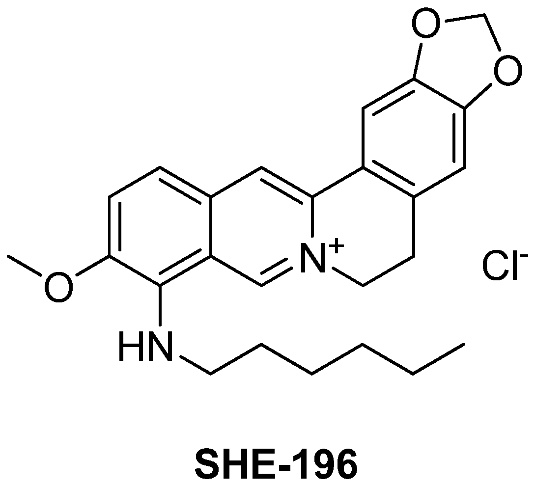
9-(hexylamino)-2,3-methylenedioxy-10-methoxyprotoberberine chloride (SHE-196)
2.2. Biological Experiments
2.2.1. Animals
2.2.2. The OGTT
2.2.3. The Design of the Experiment on AY Mice
2.2.4. Insulin Blood Concentration Evaluation after Single Administration
2.2.5. Insulin ELISA Examination
2.2.6. Biochemical Assays
2.2.7. Toxicology Study
2.2.8. Histological Examination
3. Results
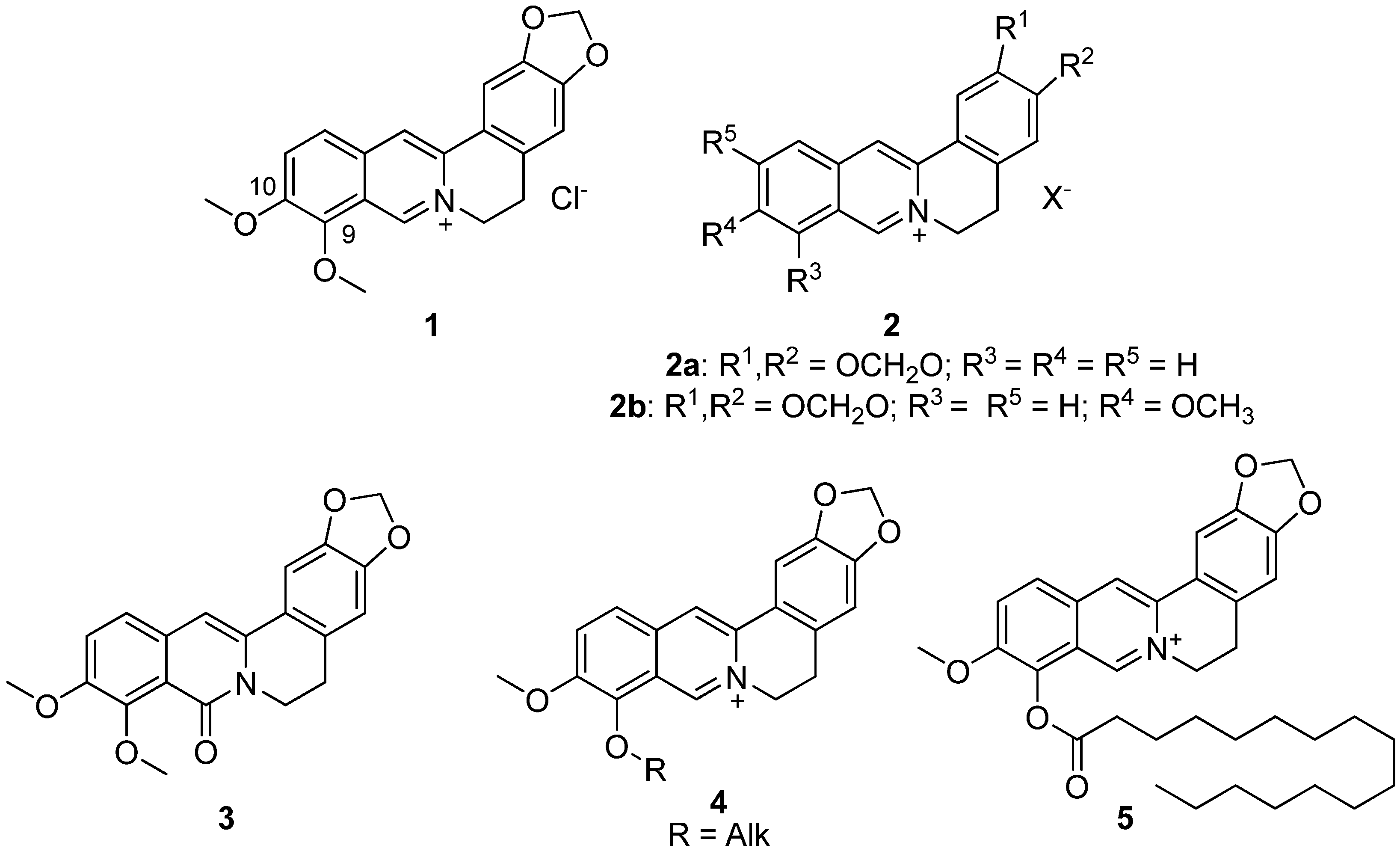
3.1. Synthesis
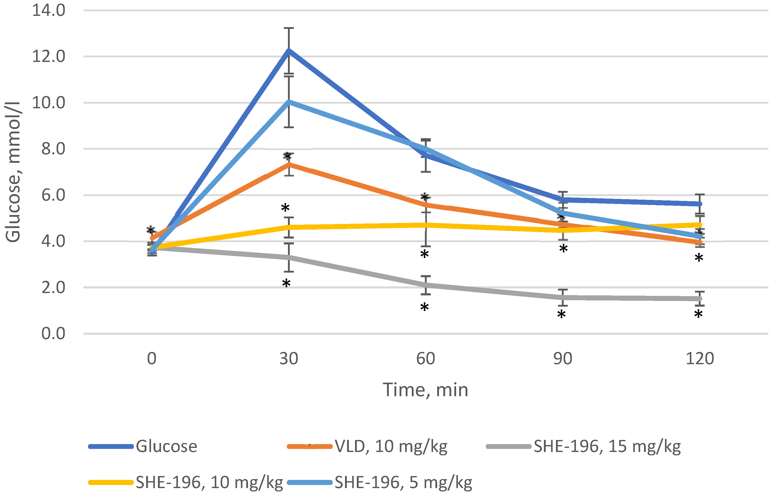
3.2. The OGTT in C57BL/6 Mice
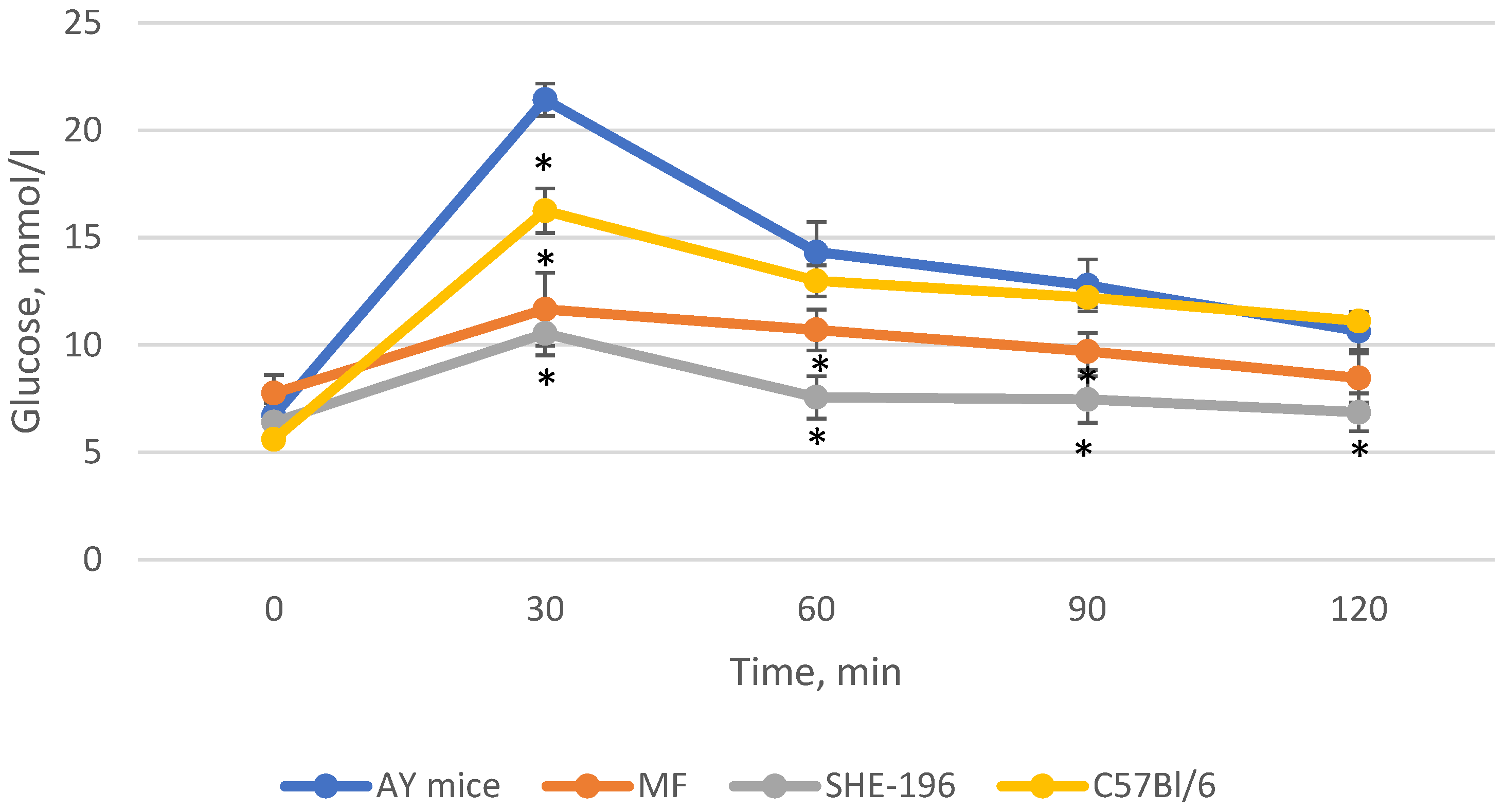
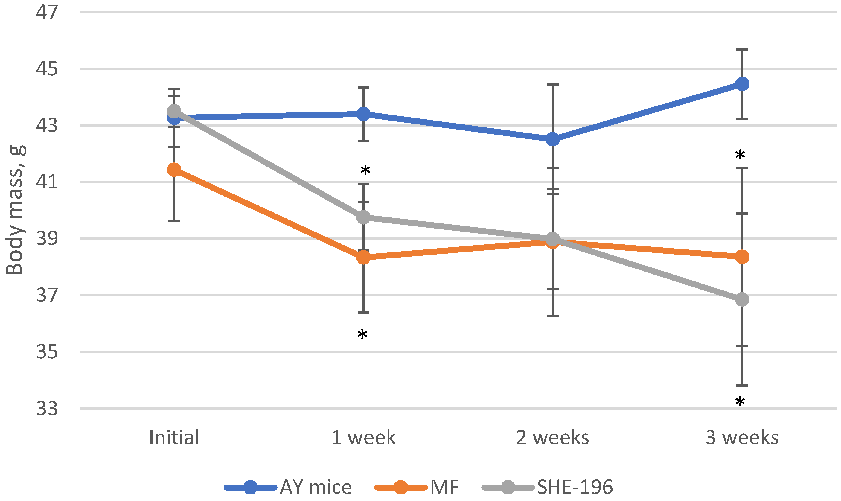
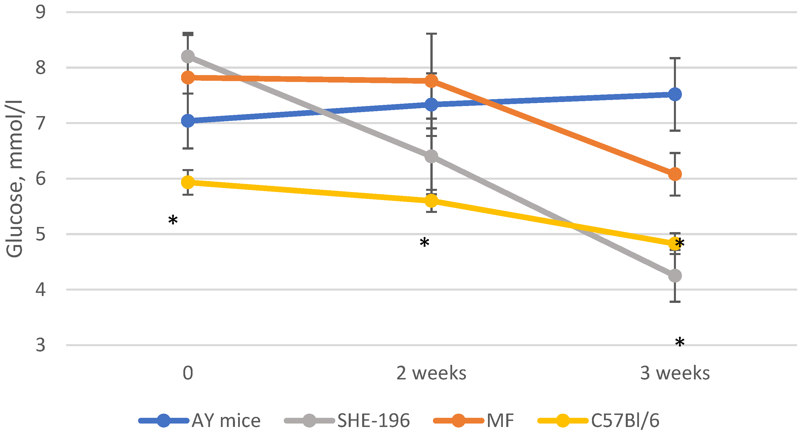
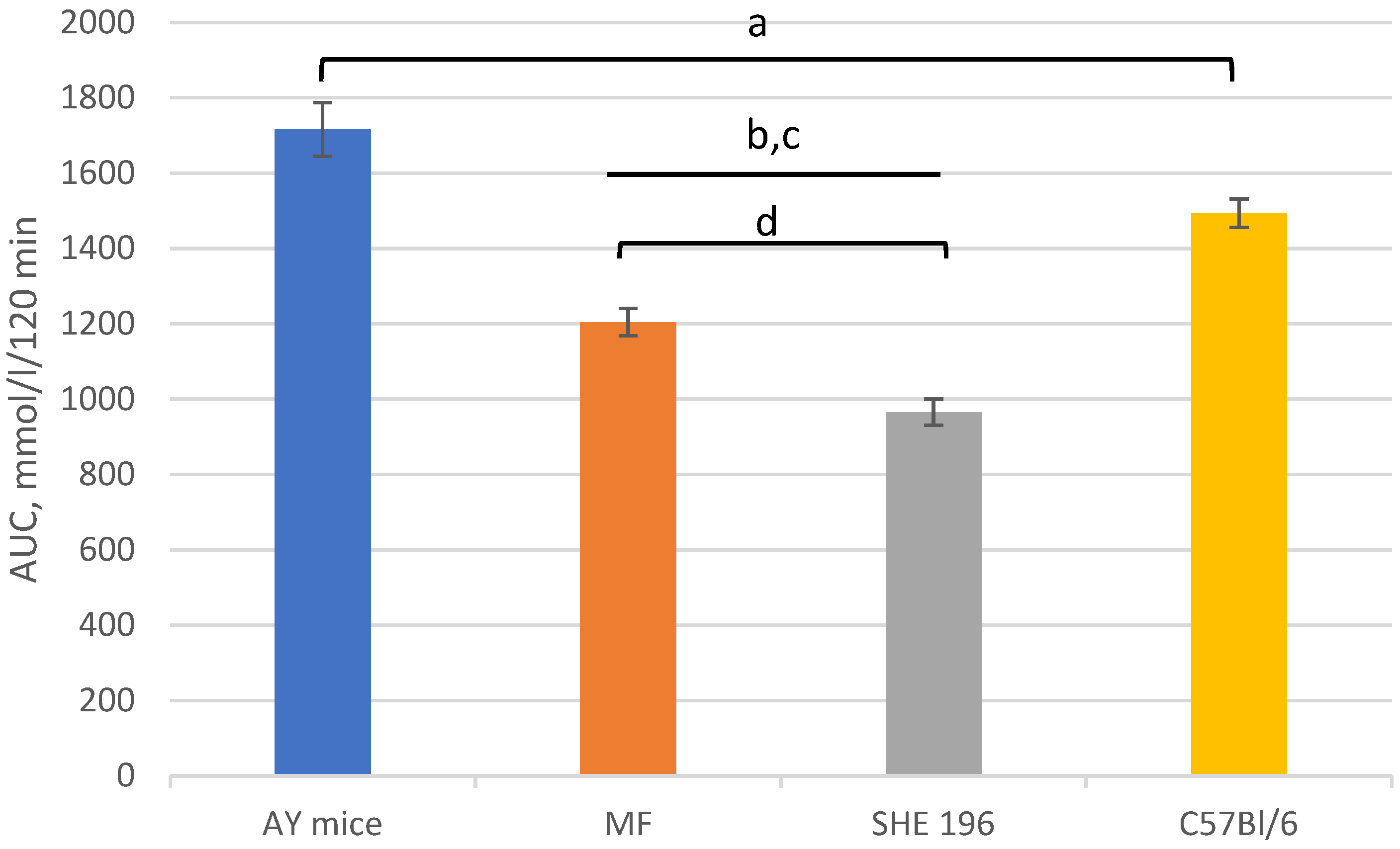
3.3. AY Mice Experiments
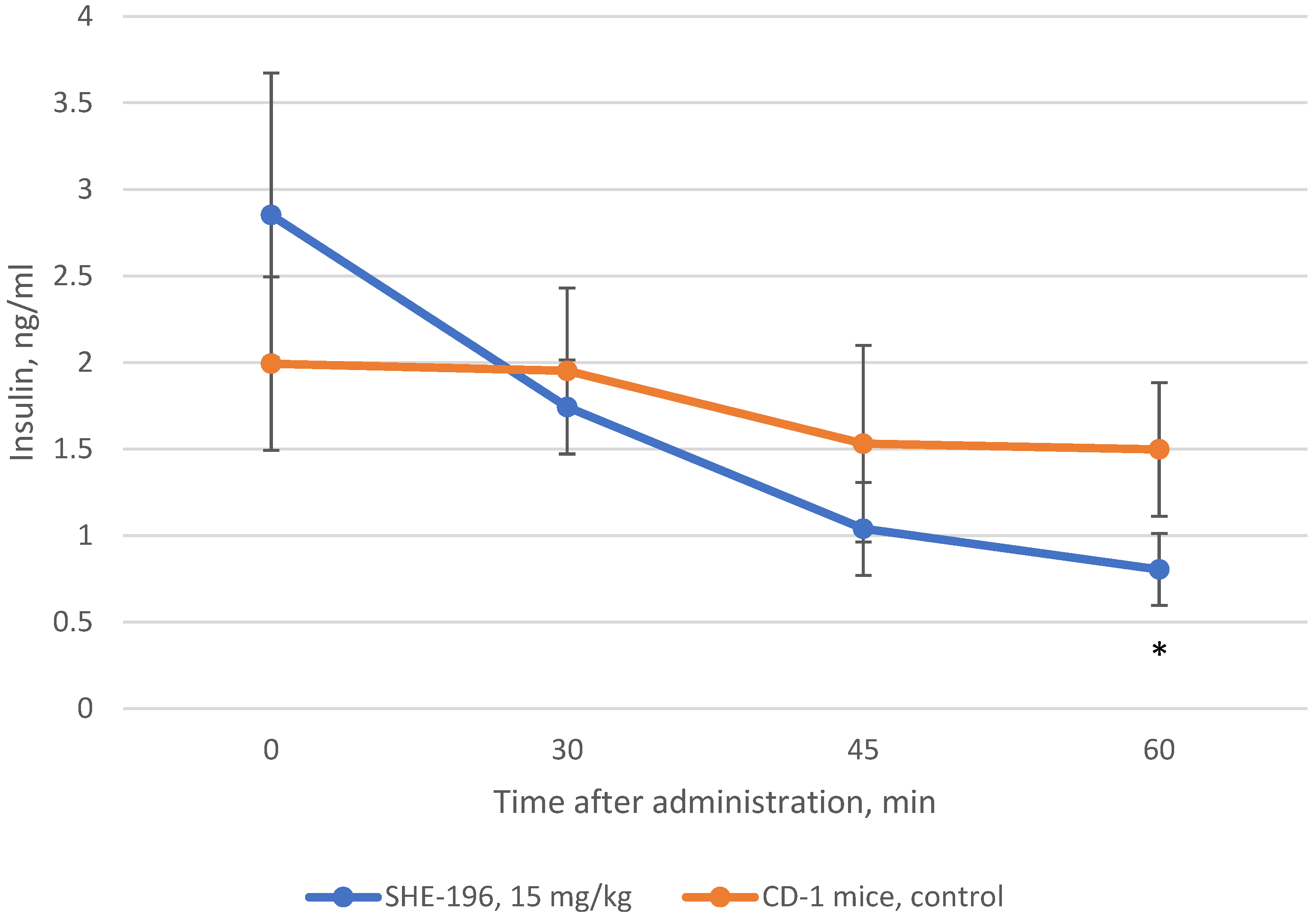
3.4. Insulin Blood Concentration Evaluation after Single Administration
3.5. Toxicology Study
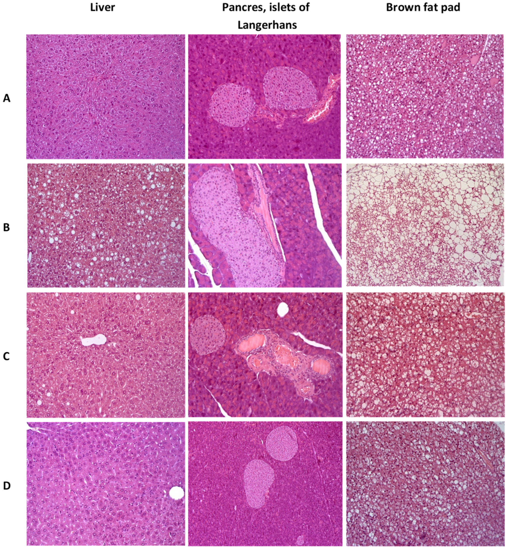
3.6. Hystology
4. Discussion
5. Conclusions
Author Contributions
Funding
Institutional Review Board Statement
Informed Consent Statement
Data Availability Statement
Acknowledgments
Conflicts of Interest
References
- Ma, Y.; Ou, T.-M.; Hou, J.-Q.; Lu, Y.-J.; Tan, J.-H.; Gu, L.-Q.; Huang, Z.-S. 9-N-Substituted berberine derivatives: Stabilization of G-quadruplex DNA and down-regulation of oncogene c-myc. Bioorg. Med. Chem. 2008, 16, 7582–7591. [Google Scholar] [CrossRef]
- Ma, Y.; Ou, T.-M.; Tan, J.-H.; Hou, J.-Q.; Huang, S.-L.; Gu, L.-Q.; Huang, Z.-S. Synthesis and evaluation of 9-O-substituted berberine derivatives containing aza-aromatic terminal group as highly selective telomeric G-quadruplex stabilizing ligands. Bioorg. Med. Chem. Lett. 2009, 19, 3414–3417. [Google Scholar] [CrossRef]
- Long, Y.-H.; Bai, L.-P.; Qin, Y.; Pang, J.-Y.; Chen, W.-H.; Cai, Z.; Xu, Z.-L.; Jiang, Z.-H. Spacer length and attaching position-dependent binding of synthesized protoberberine dimers to double-stranded DNA. Bioorg. Med. Chem. 2006, 14, 4670–4676. [Google Scholar] [CrossRef]
- Zhou, J.; Zhou, S. Berberine regulates peroxisome proliferator-activated receptors and positive transcription elongation factor b expression in diabetic adipocytes. Eur. J. Pharmacol. 2010, 649, 390–397. [Google Scholar] [CrossRef] [PubMed]
- Zhang, Y.; Gu, Y.; Ren, H.; Wang, S.; Zhong, H.; Zhao, X.; Ma, J.; Gu, X.; Xue, Y.; Huang, S.; et al. Gut microbiome-related effects of berberine and probiotics on type 2 diabetes (the PREMOTE study). Nat. Commun. 2020, 11, 5015. [Google Scholar] [CrossRef] [PubMed]
- Sun, R.; Kong, B.; Yang, N.; Cao, B.; Feng, D.; Yu, X.; Ge, C.; Feng, S.; Fei, F.; Huang, J.; et al. The Hypoglycemic Effect of Berberine and Berberrubine Involves Modulation of Intestinal Farnesoid X Receptor Signaling Pathway and Inhibition of Hepatic Gluconeogenesis. Drug Metab. Dispos. 2020, 49, 276–286. [Google Scholar] [CrossRef]
- Liu, D.; Zhang, Y.; Liu, Y.; Hou, L.; Li, S.; Tian, H.; Zhao, T. Berberine Modulates Gut Microbiota and Reduces Insulin Resistance via the TLR4 Signaling Pathway. Exp. Clin. Endocrinol. Diabetes 2018, 126, 513–520. [Google Scholar] [CrossRef]
- Turner, N.; Li, J.-Y.; Gosby, A.; To, S.W.; Cheng, Z.; Miyoshi, H.; Taketo, M.M.; Cooney, G.J.; Kraegen, E.W.; James, D.E.; et al. Berberine and Its More Biologically Available Derivative, Dihydroberberine, Inhibit Mitochondrial Respiratory Complex I: A Mechanism for the Action of Berberine to Activate AMP-Activated Protein Kinase and Improve Insulin Action. Diabetes 2008, 57, 1414–1418. [Google Scholar] [CrossRef] [Green Version]
- Chen, C.; Zhang, Y.; Huang, C. Berberine inhibits PTP1B activity and mimics insulin action. Biochem. Biophys. Res. Commun. 2010, 397, 543–547. [Google Scholar] [CrossRef] [PubMed]
- Wang, S.; Xu, Z.; Cai, B.; Chen, Q. Berberine as a Potential Multi-Target Agent for Metabolic Diseases: A Review of Investigations for Berberine. Endocr. Metab. Immune Disord. Drug Targets 2021, 21, 971–979. [Google Scholar] [CrossRef]
- Ilyas, Z.; Perna, S.; Al-Thawadi, S.; Alalwan, T.; Riva, A.; Petrangolini, G.; Gasparri, C.; Infantino, V.; Peroni, G.; Rondanelli, M. The effect of Berberine on weight loss in order to prevent obesity: A systematic review. Biomed. Pharmacother. 2020, 127. [Google Scholar] [CrossRef] [PubMed]
- Francisco, C.S.; Ma, X.; Zwyssig, M.M.; McDonald, B.A.; Palma-Guerrero, J. Morphological changes in response to environmental stresses in the fungal plant pathogen Zymoseptoria tritici. Sci. Rep. 2019, 9, 9642. [Google Scholar] [CrossRef] [PubMed]
- Zhang, J.; Tang, H.; Deng, R.; Wang, N.; Zhang, Y.; Wang, Y.; Liu, Y.; Li, F.; Wang, X.; Zhou, L. Berberine Suppresses Adipocyte Differentiation via Decreasing CREB Transcriptional Activity. PLoS ONE 2015, 10, e0125667. [Google Scholar] [CrossRef]
- Xia, X.; Yan, J.; Shen, Y.; Tang, K.; Yin, J.; Zhang, Y.; Yang, D.; Liang, H.; Ye, J.; Weng, J. Berberine Improves Glucose Metabolism in Diabetic Rats by Inhibition of Hepatic Gluconeogenesis. PLoS ONE 2011, 6, e16556. [Google Scholar] [CrossRef] [Green Version]
- Chen, W.; Miao, Y.-Q.; Fan, D.-J.; Yang, S.-S.; Lin, X.; Meng, L.-K.; Tang, X. Bioavailability Study of Berberine and the Enhancing Effects of TPGS on Intestinal Absorption in Rats. AAPS Pharmscitech 2011, 12, 705–711. [Google Scholar] [CrossRef] [Green Version]
- Bian, X.; He, L.; Yang, G. Synthesis and antihyperglycemic evaluation of various protoberberine derivatives. Bioorg. Med. Chem. Lett. 2006, 16, 1380–1383. [Google Scholar] [CrossRef] [PubMed]
- Dou, Y.; Huang, R.; Li, Q.; Liu, Y.; Li, Y.; Chen, H.; Ai, G.; Xie, J.; Zeng, H.; Chen, J.; et al. Oxyberberine, an absorbed metabolite of berberine, possess superior hypoglycemic effect via regulating the PI3K/Akt and Nrf2 signaling pathways. Biomed. Pharmacother. 2021, 137, 111312. [Google Scholar] [CrossRef]
- Zhang, S.; Wang, X.; Yin, W.; Liu, Z.; Zhou, M.; Xiao, D.; Liu, Y.; Peng, D. Synthesis and hypoglycemic activity of 9- O -(lipophilic group substituted) berberine derivatives. Bioorg. Med. Chem. Lett. 2016, 26, 4799–4803. [Google Scholar] [CrossRef]
- Wang, Y.-X.; Kong, W.-J.; Li, Y.-H.; Tang, S.; Li, Z.; Li, Y.-B.; Shan, Y.-Q.; Bi, C.-W.; Jiang, J.-D.; Song, D.-Q. Synthesis and structure–activity relationship of berberine analogues in LDLR up-regulation and AMPK activation. Bioorg. Med. Chem. 2012, 20, 6552–6558. [Google Scholar] [CrossRef]
- Li, Y.-H.; Li, Y.; Yang, P.; Kong, W.-J.; You, X.-F.; Ren, G.; Deng, H.-B.; Wang, Y.-M.; Wang, Y.-X.; Jiang, J.-D.; et al. Design, synthesis, and cholesterol-lowering efficacy for prodrugs of berberrubine. Bioorg. Med. Chem. 2010, 18, 6422–6428. [Google Scholar] [CrossRef]
- Cao, S.; Yu, S.; Cheng, L.; Yan, J.; Zhu, Y.; Deng, Y.; Qiu, F.; Kang, N. 9-O-benzoyl-substituted berberine exerts a triglyceride-lowering effect through AMPK signaling pathway in human hepatoma HepG2 cells. Environ. Toxicol. Pharmacol. 2018, 64, 11–17. [Google Scholar] [CrossRef]
- Wang, Y.-X.; Wang, Y.-P.; Zhang, H.; Kong, W.-J.; Li, Y.-H.; Liu, F.; Gao, R.-M.; Liu, T.; Jiang, J.-D.; Song, D.-Q. Synthesis and biological evaluation of berberine analogues as novel up-regulators for both low-density-lipoprotein receptor and insulin receptor. Bioorg. Med. Chem. Lett. 2009, 19, 6004–6008. [Google Scholar] [CrossRef] [PubMed]
- Nechepurenko, I.V.; Boyarskikh, U.A.; Khvostov, M.; Baev, D.; Komarova, N.I.; Filipenko, M.L.; Tolstikova, T.G.; Salakhutdinov, N.F. Hypolipidemic Berberine Derivatives with a Reduced Aromatic Ring C. Chem. Nat. Compd. 2015, 51, 916–922. [Google Scholar] [CrossRef]
- Li, Y.-H.; Yang, P.; Kong, W.-J.; Wang, Y.-X.; Hu, C.-Q.; Zuo, Z.-Y.; Wang, Y.-M.; Gao, H.; Gao, L.-M.; Feng, Y.-C.; et al. Berberine Analogues as a Novel Class of the Low-Density-Lipoprotein Receptor Up-Regulators: Synthesis, Structure−Activity Relationships, and Cholesterol-Lowering Efficacy. J. Med. Chem. 2009, 52, 492–501. [Google Scholar] [CrossRef]
- Ren, G.; Wang, Y.-X.; Li, Y.-H.; Song, D.-Q.; Kong, W.-J.; Jiang, J.-D. Structure-activity relationship of berberine derivatives for their glucose-lowering activities. Int. J. Clin. Exp. Med. 2017, 10, 5054–5060. [Google Scholar]
- Kuranov, S.; Luzina, O.; Khvostov, M.; Baev, D.; Kuznetsova, D.; Zhukova, N.; Vassiliev, P.; Kochetkov, A.; Tolstikova, T.; Salakhutdinov, N. Bornyl Derivatives of p-(Benzyloxy)Phenylpropionic Acid: In Vivo Evaluation of Antidiabetic Activity. Pharmaceuticals 2020, 13, 404. [Google Scholar] [CrossRef]
- Finney, D.J. Probit Analysis. J. Inst. Actuar. 1952, 78, 388–390. [Google Scholar]
- Burov, O.N.; Kletskii, M.E.; Fedik, N.S.; Kurbatov, S.V.; Lisovin, A. Experimental and quantum-chemical study of nucleophilic substitution mechanism in berberine. Chem. Heterocycl. Compd. 2015, 51, 997–1007. [Google Scholar] [CrossRef]
- Wang, J.; Yang, T.; Chen, H.; Xu, Y.-N.; Yu, L.-F.; Liu, T.; Tang, J.; Yi, Z.; Yang, C.-G.; Xue, W.; et al. The synthesis and antistaphylococcal activity of 9, 13-disubstituted berberine derivatives. Eur. J. Med. Chem. 2017, 127, 424–433. [Google Scholar] [CrossRef]
- Thornberry, N.A.; Gallwitz, B. Mechanism of action of inhibitors of dipeptidyl-peptidase-4 (DPP-4). Best Pr. Res. Clin. Endocrinol. Metab. 2009, 23, 479–486. [Google Scholar] [CrossRef]
- Piskunova, Y.V.; Kazantceva, A.Y.; Baklanov, A.V.; Bazhan, N.M. Mutation yellow in agouti loci prevents age-related increase of skeletal muscle genes regulating free fatty acids oxidation. Vavilov J. Genet. Breed. 2018, 22, 265–272. [Google Scholar] [CrossRef]
- Hostalek, U.; Campbell, I. Metformin for diabetes prevention: Update of the evidence base. Curr. Med. Res. Opin. 2021, 37, 1705–1717. [Google Scholar] [CrossRef]
- Guo, W.-R.; Liu, J.; Cheng, L.-D.; Liu, Z.-Y.; Zheng, X.-B.; Liang, H.; Xu, F. Metformin Alleviates Steatohepatitis in Diet-Induced Obese Mice in a SIRT1-Dependent Way. Front. Pharmacol. 2021, 12. [Google Scholar] [CrossRef]
- Miranda, C.S.; Silva-Veiga, F.; Martins, F.F.; Rachid, T.L.; Mandarim-De-Lacerda, C.A.; Souza-Mello, V. PPAR-α activation counters brown adipose tissue whitening: A comparative study between high-fat– and high-fructose–fed mice. Nutrition 2020, 78, 110791. [Google Scholar] [CrossRef] [PubMed]
- Lovejoy, J.; Newby, F.; Gebhart, S.; DiGirolamo, M. Insulin resistance in obesity is associated with elevated basal lactate levels and diminished lactate appearance following intravenous glucose and insulin. Metabolism 1992, 41, 22–27. [Google Scholar] [CrossRef]
- Kong, W.-J.; Zhang, H.; Song, D.-Q.; Xue, R.; Zhao, W.; Wei, J.; Wang, Y.-M.; Shan, N.; Zhou, Z.-X.; Yang, P.; et al. Berberine reduces insulin resistance through protein kinase C–dependent up-regulation of insulin receptor expression. Metabolism 2009, 58, 109–119. [Google Scholar] [CrossRef] [PubMed]
- Och, A.; Podgórski, R.; Nowak, R. Biological Activity of Berberine—A Summary Update. Toxins 2020, 12, 713. [Google Scholar] [CrossRef] [PubMed]
- Gaba, S.; Saini, A.; Singh, G.; Monga, V. An insight into the medicinal attributes of berberine derivatives: A review. Bioorg. Med. Chem. 2021, 38, 116143. [Google Scholar] [CrossRef] [PubMed]
- Pappachan, J.M.; Fernandez, C.J.; Chacko, E.C. Diabesity and antidiabetic drugs. Mol. Asp. Med. 2018, 66, 3–12. [Google Scholar] [CrossRef]


| Group | Gonadal Fat Pad, g | Interscapular Fat Pad, g | Interscapular Brown Fat, g |
|---|---|---|---|
| AY mice | 2.51 ± 0.18 | 1.21 ± 0.02 | 0.21 ± 0.02 |
| MF | 1.91 ± 0.17 | 0.87 ± 0.09 * | 0.13 ± 0.02 * |
| SHE-196 | 2.14 ± 0.27 | 0.92 ± 0.19 * | 0.12 ± 0.03 * |
| C57Bl/6 | 0.29 ± 0.07 | - | 0.03 ± 0.003 * |
| Group | Insulin, ng/mL |
|---|---|
| AY mice | 6.08 ± 1.01 |
| MF | 2.23 ± 0.51 * |
| SHE-196 | 1.75 ± 0.54 * |
| C57Bl/6 | 0.45 ± 0.11 * |
| Group | TC, mmol/L | TG, mmol/L | TP, g/L | Lactate, mmol/L | APH, U/L | ALT, U/L |
|---|---|---|---|---|---|---|
| AY mice | 2.54 ± 0.11 | 0.28 ± 0.03 | 63.49 ± 3.65 | 8.41 ± 0.54 | 98.50 ± 5.94 | 45.38 ± 3.36 |
| MF | 2.52 ± 0.14 | 0.25 ± 0.02 | 59.70 ± 3.64 | 5.88 ± 0.41 * | 94.60 ± 94.68 | 46.20 ± 2.65 |
| SHE-196 | 2.65 ± 0.28 | 0.21 ± 0.01 | 59.93 ± 3.49 | 5.15 ± 0.24 * | 85.50 ± 1.55 | 41.00 ± 3.49 |
| C57Bl/6 | 1.90 ± 0.05 * | 0.18 ± 0.01 * | 59.60 ± 1.72 | 4.40 ± 0.49 * | 87.71 ± 4.92 | 38.43 ± 2.10 |
Publisher’s Note: MDPI stays neutral with regard to jurisdictional claims in published maps and institutional affiliations. |
© 2021 by the authors. Licensee MDPI, Basel, Switzerland. This article is an open access article distributed under the terms and conditions of the Creative Commons Attribution (CC BY) license (https://creativecommons.org/licenses/by/4.0/).
Share and Cite
Khvostov, M.V.; Gladkova, E.D.; Borisov, S.A.; Zhukova, N.A.; Marenina, M.K.; Meshkova, Y.V.; Luzina, O.A.; Tolstikova, T.G.; Salakhutdinov, N.F. Discovery of the First in Class 9-N-Berberine Derivative as Hypoglycemic Agent with Extra-Strong Action. Pharmaceutics 2021, 13, 2138. https://doi.org/10.3390/pharmaceutics13122138
Khvostov MV, Gladkova ED, Borisov SA, Zhukova NA, Marenina MK, Meshkova YV, Luzina OA, Tolstikova TG, Salakhutdinov NF. Discovery of the First in Class 9-N-Berberine Derivative as Hypoglycemic Agent with Extra-Strong Action. Pharmaceutics. 2021; 13(12):2138. https://doi.org/10.3390/pharmaceutics13122138
Chicago/Turabian StyleKhvostov, Mikhail V., Elizaveta D. Gladkova, Sergey A. Borisov, Nataliya A. Zhukova, Mariya K. Marenina, Yuliya V. Meshkova, Olga A. Luzina, Tatijana G. Tolstikova, and Nariman F. Salakhutdinov. 2021. "Discovery of the First in Class 9-N-Berberine Derivative as Hypoglycemic Agent with Extra-Strong Action" Pharmaceutics 13, no. 12: 2138. https://doi.org/10.3390/pharmaceutics13122138
APA StyleKhvostov, M. V., Gladkova, E. D., Borisov, S. A., Zhukova, N. A., Marenina, M. K., Meshkova, Y. V., Luzina, O. A., Tolstikova, T. G., & Salakhutdinov, N. F. (2021). Discovery of the First in Class 9-N-Berberine Derivative as Hypoglycemic Agent with Extra-Strong Action. Pharmaceutics, 13(12), 2138. https://doi.org/10.3390/pharmaceutics13122138