Abstract
Viruses from several families use direct cell-to-cell infection to disseminate between cells. Retroviruses are a relatively recent addition to this list, and appear to spread cell-to-cell by induction of multimolecular complexes termed virological synapses that assemble at the interface between infected and receptor-expressing target cells. Over the past five years, detailed insight into the cellular and molecular basis of virological synapse-mediated retroviral cell-to-cell spread has been obtained, but important questions and controversies have been raised that remain to be resolved. This review will focus on recent advances in the field with emphasis on areas in which work still needs to be done.
1. Introduction
Mammalian viruses have co-evolved with their hosts, and in doing so have developed elegantly adaptive mechanisms for invasion, persistence and onward transmission. Since viruses are obligate intracellular pathogens, they harness the cellular machinery to enter, replicate and exit. The classical paradigm of viral propagation by release of independent infectious virus particles that diffuse freely in extracellular fluids is robust, and for most viruses is probably the best mechanism for long-distance viral dissemination within, and between hosts. However, another mode of viral spread, somewhat neglected in recent years, has relatively recently re-emerged; that of directed movement of viruses between contacting target cells without recourse to long-distance fluid phase diffusion. Such ‘cell-to-cell’ spread was first formally demonstrated for herpesviruses. Varicella Zoster virus was efficiently passed between cells only in a cell-associated form [1], and herpes B virus resisted antibody neutralization when spreading directly between cells, but was neutralization sensitive when spreading in a cell-free mode [2]. Twenty years later, the cell-to-cell movement of rhabdoviruses across neural synapses was captured by electron microscopy [3]. Members of several virus families have adopted cell-to-cell spread by various means including: budding from infected cells and entering target cells across epithelial cell tight junctions (herpesviruses); elaborating actin-rich cellular structures that propel virions from infected cells directly into uninfected cells (poxviruses); breaking down intercellular barriers to spread by inducing limited membrane fusion between infected and uninfected cells (paramyxoviruses) [4,5]. Today the concept of directed cell-to-cell viral spread is well accepted, and for the viruses mentioned above, the cellular and molecular mechanisms are at least partially understood.
2. Retroviruses and Virological Synapses (VS)
Retroviruses have a relatively small genome (~10 kb) and interact in a particularly intimate manner with their cellular hosts. Being enveloped, retroviruses are fragile and do not survive well outside of an infected cell; decay of infectivity is rapid even in tissue culture. Immunodeficiency viruses such as HIV-1 are particularly prone to infectivity decay as their envelope glycoprotein (Env) spike is non-covalently assembled and dissociates into non-functional components over time [6,7]. It is therefore important for their optimal survival and dissemination to find and infect new host cells with the minimum of delay. Many retroviruses infect immune cells, and this is central to the pathogenesis of HIV-1, HIV-2 and simian immunodeficiency virus (SIV)-induced AIDS and human T cell leukemia virus type-I (HTLV-I)-induced disease. Immune cells generally do not have inbuilt polarity, unlike epithelial cells for example [5], and so the spread of retroviruses between immune cells seemed unlikely to be mediated by the same type of mechanism adopted by herpesviruses or rhabdoviruses. Early studies highlighted the ability of HIV-1 to induce syncytium formation between infected and uninfected target cells, suggesting a potential mechanism for intercellular spread of virus similar to that described for paramyxoviruses [5]. However, more recent work revealed that this was in large part an artifact of adaptation of viruses to growth in immortalized cell lines, and restricted principally to CXCR4-using viruses [8]. Interestingly, only limited cell-cell fusion of infected and uninfected cells has been observed during retroviral cell-to-cell transfer, despite estimated infected-target cell conjugate lifespans of several hours or more [9,10,11]. Several studies support the concept of active suppression of HTLV-I [12] and HIV-1 [13,14]–mediated cell-cell fusion by recruitment of the regulatory tetraspans CD9, CD81 or CD82. Other mechanisms, such as exclusion of Env and/or viral receptors from regions of adhesion between infected and target cells, may also operate. What therefore might be the dominant mechanism of cell-to-cell transfer of HIV-1? Clues came from emerging evidence in the field of immunological antigen presentation, which revealed communication between immune cells via immunological synapses [15]). This model implied that both antigen-presenting cells (APC) and lymphocytes were able to co-polarize their cytoskeletons for the purpose of directed exchange of cell-surface (receptor-ligand) and soluble (cytokine) biochemical information leading to modulation of cell function. Similar CD4 [16] and adhesion molecule [17] polarization had already been observed by ourselves and others when HIV-1-infected cells contacted CD4+ target T cells, and this was shown to be gp120- and actin-dependent [18]. Around the same time, pivotal observations were made by the Steinman laboratory that dendritic cells (DC) were able to efficiently transfer HIV-1 to CD4+ T cells via adhesive conjugates [19,20]. These findings led to the concept that retrovirally-infected cells might act in a manner analogous to the immunological synapse by coordinating viral exit on the infected cell and viral receptors on the ‘target’ cell, and the term virological synapse (VS) was proposed by ourselves in 2002 to describe this macromolecular reorganization [21]. Seminal papers describing imaging of synaptic transfer of retroviruses were subsequently published, concerning HIV-1 cell-to-cell spread between DC and CD4+ T cells across an ‘infectious synapse’ [22] and HTLV-I [23] and HIV-1 [9] spread between T cells via VS. All papers alluded to polarization of the cell types involved: McDonald et al. described polarization of HIV-1 particles and viral receptors to the DC-T cell contact zone, Igakura et al. observed clustering of viral Gag in the infected T cell and the actin-associated adaptor molecule talin on the target cell, and Jolly et al. observed co-polarization of HIV-1 Env, Gag and viral receptors CD4 and CXCR4. Several papers rapidly followed that confirmed and extended these observations of VS structure and function for HIV-1 in a variety of cell types including DC, T cells and macrophages [9,10,24,25,26,27,28]. Later studies pointed out that HIV-1 could not only assemble single large-scale VS containing pairs of conjugated cells, but also ‘polysynapses’ containing multiple target cells [28,29].
The structure and function of retroviral VS and their relationship to immunological synapses will be reviewed elsewhere in this issue of Viruses, so here I will only summarize their principal molecular features.
- 1. A central feature of intercellular synaptic structures is the presence of an adhesive junction that is sufficiently stable to allow transfer of signals and soluble mediators between the pre- and post-synaptic cells [5,30]. In the case of the HIV-1 T cell VS, the initial cognate event that holds the interacting T cells together appears to be CD4-gp120 binding [9], and subsequent stable adhesive junctions are probably maintained by integrin-ICAM interactions [17,31,32], although others dispute a functional role for cellular adhesion molecules in this context [33].
- 2. The interaction of Env on the HIV-1-infected cell with CD4 on the target cell recruits filamentous actin into the synaptic zone along with more CD4, HIV-1 coreceptor (CXCR4 or CCR5) and adhesion molecules [9,31]. However, actin may be cleared from the central region of the VS, a strategy proposed to facilitate viral entry into the target cell [34].
- 3. There is evidence that HIV-1 may assemble at the plasma membrane of infected T cells at sites of cell-cell contact, implying polarization of cellular secretory systems towards the VS [35]. This is supported by movement of the microtubule organizing center (MTOC) and other elements of the secretory apparatus proximal to the site of cellular contact in both HTLV-I- and HIV-1-infected T cells [35,36,37,38,39,40,].
The T cell VS consists of relatively large opposing surfaces of polarized infected and target cell plasma membrane containing, respectively, clustered Env and viral receptors [11,28]. However, HIV-1 has also been observed to travel along long intercellular tubular structures termed ‘membrane nanotubes’ joining infected and uninfected T cells [29,41]. Nanotubes emanating from infected and uninfected T cells appear to join at a ‘micro-synapse’ [41], which migrating virions must pass to infect the target cell. HIV-1 infection via membrane nanotubes appears to be less common than via VS [29]. Very recently, the movement of HTLV-I anchored at the infected cell surface in a meshwork of extracellular matrix (termed a ‘viral biofilm’) to uninfected T cells has been proposed as an alternative to VS-mediated transfer [42]. Whether this is a dominant mode of infectious HTLV transfer, and whether it applies to other retroviruses, remains to be seen, although this phenomenon may be related to the ‘antigen caps’ observed in unconjugated T cells infected with HIV-1 for extended periods of time [43,44,45]. A retrovirus that infects predominantly non-immune cells, murine leukemia virus (MLV), spreads between fibroblasts by surfing along projections emanating from the infected cell termed filopodia, anchored to the target cell by interactions between the viral glycoproteins on infected cells and viral receptor on target cells [46,47].
4. VS and evasion of entry inhibition
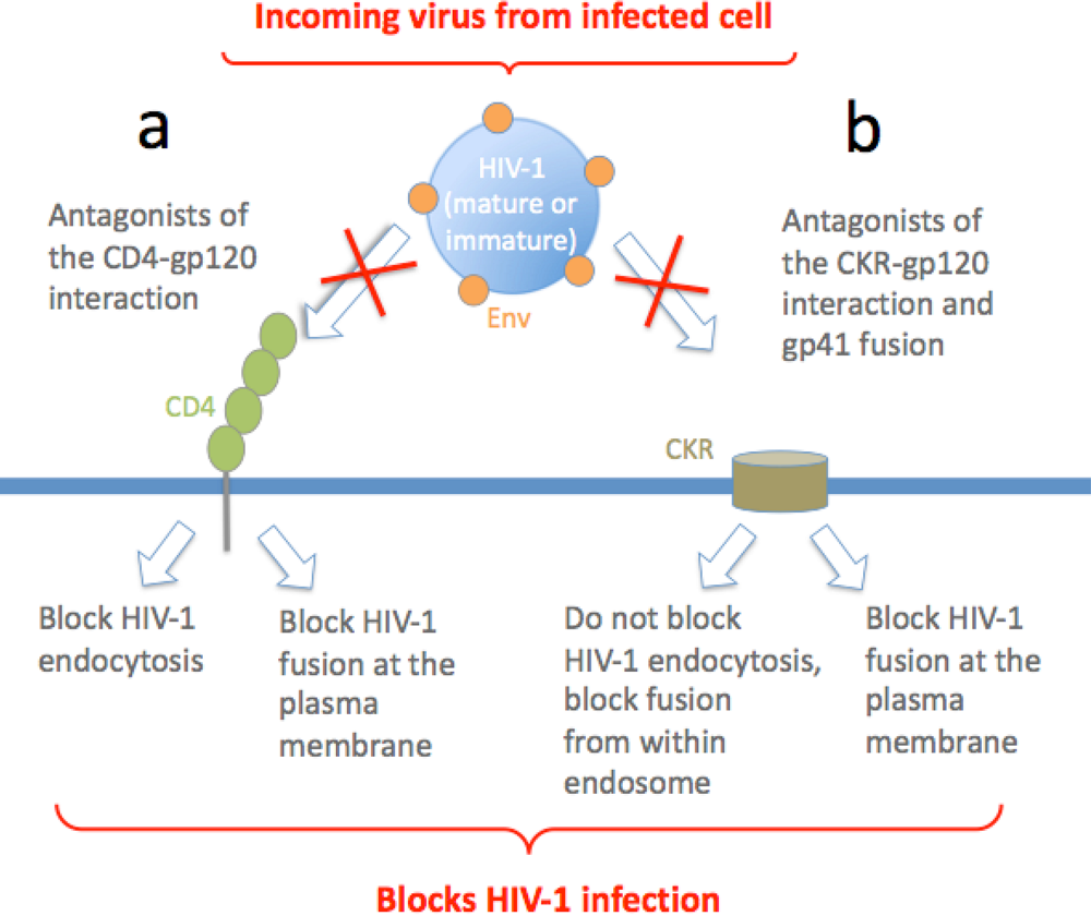
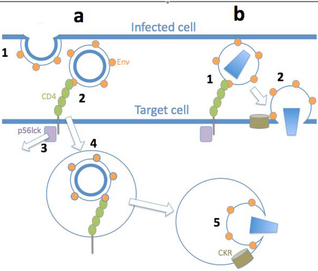
Cell-to-cell spread of herpesviruses was first defined by its resistance to neutralizing antibodies (NAb) of a spreading infection in culture compared to the sensitivity of fluid-phase virion dissemination [2,64]. Neural synapses are though to be sealed from the outside environment by virtue of a tight adhesive junction [30], allowing rhabdoviruses to spread directionally by this route [65] presumably avoiding attack by NAb. Poxviruses [66] and hepatitis C virus [67] have also been proposed to use cell-to-cell spread as a means to escape antibody neutralization. It has therefore been speculated that retroviruses might use VS to evade attack by NAb and other types of entry inhibitor [5,68]. At present, it is unclear whether this is the case, since confusion exists in the field as a result of studies reporting discordant results. There is general agreement that inhibitors of the CD4-gp120 interaction inhibit VS-mediated viral transfer independent of the system used to detect HIV-1 in the target cell [9,10,33,48,60]. By contrast, some of the same groups reporting on Gag transfer between cells using antagonists of events proximal to viral fusion, including NAb to gp41 [10] and HIV-1 coreceptor antagonists [10,69], failed to inhibit Gag transfer but did inhibit infection [70]. At one level these data can be reconciled by the explanation that the CD4-gp120 interaction is required to trigger the stable association of infected-target cell conjugates to form VS, without which no virus transfer could take place [9,48]. In this respect all groups agree that both endocytosis and infection are prevented by inhibitors of the CD4-gp120 interaction [49], and this is consistent with earlier work investigating cell-free viral endocytosis into T cells [62]. With regard to a central role of the Env-CD4 interaction in endocytosis of HIV-1 by the target cell, it is interesting to invoke the endocytic capacity of CD4 conferred by motifs within its cytoplasmic tail [71]. CD4 endocytosis can be triggered by gp120 engagement that dissociates the cytoplasmic tyrosine kinase p56lck [72], liberating CD4 from the endocytosis inhibitory activity of p56lck [73] (Figure 1). However, Blanco and colleagues observed that the cytoplasmic tail of CD4 was not required for HIV-1 uptake by a T cell line, implying that a non-classical mode of viral endocytosis may be operating [48]. Because there is limited time for the virus to mature during VS-mediated transfer to the target cell, this mode of HIV-1 spread would be expected to result in higher levels of immature virus transferring to the target cell than during cell-free infection, and therefore a higher frequency of endocytic uptake of virus [28]. The apparently conflicting data with regard to post-CD4 entry requirements for virus transferred across VS can be reconciled by considering whether the assay used to measure viral transfer detects viral endocytosis or infection as an endpoint (Figure 2). Coreceptor and fusion antagonists fail to interfere with viral transfer across VS mediated by endocytic uptake of virions, since neither the chemokine receptors CCR5 and CXCR4 nor the viral fusion apparatus (gp41) are required for this route of uptake [10,48,70]. By contrast, when viral infectivity is measured using assays of viral reverse transcription, integration or proviral transcription, all inhibitors of viral entry are effective [11,28,49,60,70]. This may be because inhibitor bound to neutralized virus or bound to viral coreceptors will prevent both direct viral fusion at the plasma membrane and membrane fusion within the endosome, and will exert its effect as the virus matures and becomes infectious (Figure 2). Indeed, we observe that when events proximal to HIV-1 infection of target cells are assayed for, inhibitors of all stages of viral entry including NAb interfere with cell-free and cell-to-cell spread with approximate equivalence, regardless of mode of action or molecular weight [11]. An interesting exception to this rule was reported by Hubner et al. [28], who failed to block cell-to-cell spread of HIV-1 with a patient antiserum capable of neutralizing cell-free virus. Whether this represents a qualitatively differential effect of antibody on the two modes of viral transfer, or whether this antiserum contains HIV-1 inhibitory activity of an unusual nature, remains to be seen.

Figure 2.
Inhibition of HIV-1 uptake and entry across VS. (a) HIV-1 (mature or immature) engaging CD4 may trigger either fusion at the plasma membrane (mature virus) or endocytosis (immature virus): both of these events are prevented by blocking the CD4-gp120 interaction, and so HIV-1 cell-to-cell transfer and infection are inhibited. (b) HIV-1 (immature) engaging CD4 and the CKR coreceptor is unable to efficiently activate gp41-mediated fusion, and so virus is internalized by CD4-mediated endocytosis, and antagonists of gp120-CKR interaction or gp41-mediated fusion do not prevent endocytosis. However once in the endosome HIV-1 matures into a fusion-efficient form and gp120-CKR or gp41 antagonists prevent HIV-1 fusion with the endosomal membrane, preventing HIV-1 cell-to-cell infection. HIV-1 (mature) engaging the CKR triggers rapid gp41-induced plasma membrane fusion and both of these events can be blocked by antagonists, preventing cell-to-cell infection.
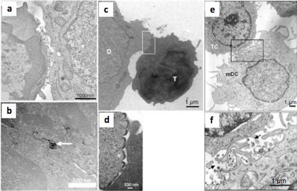
These considerations of entry inhibitor sensitivity apply so far only to transfer of HIV-1 between T cells: relative permissivity to entry inhibition may not be the case for all types of VS, since resistance of DC-T cell viral spread to antibody-mediated inhibition has been reported [74]. Structurally, the macrophage-T cell VS appears to have broader regions of tightly apposed membrane than the T cell-T cell VS [26], although this contrasts with the apparently looser structures visualized for DC-T cell VS [75] (Figure 3). However, the conjugates imaged in [26] were derived from HIV-1-infected macrophages whereas the DCs imaged in [75] were derived from DCs pulsed with inactivated HIV-1, and thus may not be directly comparable. The T cell VS associated with HTLV-I spread [40] appears to be structurally distinct from that observed for HIV-1 [9,11], with a relatively large surface of closely apposed membrane containing occasional ‘pockets’ of virus-like HTLV-I particles [40], which may shield the virus from NAb access (Figure 3). However it is difficult to make general conclusions based upon the currently available electron microscopic data, as they are sparse and derived from different cell types under different conditions.

Figure 3.
Heterogeneity of retroviral VS. (a) Single 2 nm thick digital slice taken from [9]. HIV-1 mature particles are seen at the interface of an HIV-1-infected T cell line (right cell) and a primary CD4+ T cell (left cell). The cells are loosely apposed with few points of adhesion and relatively large (>100 nm) intracellular gaps. (b) Thin section of an HTLV-1 VS taken from [40], showing an HTLV-I particle immunostained for Gag at the interface between a chronically-infected T cell line (top) and an uninfected primary CD4+ T cell (bottom). The two cells have an extensive area of closely-apposed plasma membrane. (c) A VS formed between an acutely-infected T cell line (donor cell, D) and a primary CD4+ T cell target cell (T) from [28]. (d) A higher magnification of the boxed image in (c) showing budding structures at the intracellular interface. (e) A VS formed between a mature DC pulsed with inactivated HIV-1 and a primary CD4+ T cell, showing loose contacts between the cells and multiple filopodia-like structures [75]. (f) A higher magnification of the boxed area in (e) showing filopodia and gaps containing virus-like particles between the cells, some of which are labeled with arrows.
5. Conclusions and perspectives
The concept of retroviral cell-to-cell spread across VS is now well established, and its relevance for in vivo viral dissemination seems certain. Rapid progress in the field has provided a wealth of information about the structure and function of VS, but uncertainties remain concerning the mode of uptake of virus by the target cell and its susceptibility to inhibitors of the fusion cascade. Some of these uncertainties have been raised here, and will be experimentally addressed over the next few years. Probably the most urgent area for further research relates to the question of HIV-1 endocytosis: accumulating data suggest that at least in certain immortalized cell types with some viral isolates, viral fusion from within endosomes may be the principal pathway for cell-free virus [62,76] rather than fusion at the plasma membrane, the dominant paradigm for the past 20 years [77,78]. Resolution of this controversy will help understand discrepancies in data concerning HIV-1 entry and its inhibition from a variety of laboratories. A second area relates to the cell-type dependence of mechanisms of HIV-1 cell-to-cell transfer, and whether there are, or are not, qualitative differences in the type of VS formed. More electron microscopic imaging and tomographic reconstruction of synaptic interfaces would be invaluable. At present, the closest approximation of HIV-1 synaptic spread in vivo is static snapshots of HIV-1 infectious foci in intact lymphoid tissue [79,80,81] associated with mathematical models of cell-to-cell spread [82,83,84]. A third area of research, therefore, that would add enormously to our confidence in the in vivo relevance of this phenomenon would be intravital imaging of HIV-1 cell-to-cell spread in intact secondary lymphoid tissue, as has been carried out for other pathogen-immune cell interactions [85,86].
Acknowledgments
QJS is supported by the Medical Research Council UK, The Bill and Melinda Gates Foundation, The International AIDS Vaccine Initiative Neutralizing Antibody Consortium and the European Union Network of Excellence, EUROPRISE. QJS is a Jenner Institute fellow.
References
- Weller, T.H. Serial propagation in vitro of agents producing inclusion bodies derived from varicella and herpes zoster. Proc. Soc. Exp. Biol. Med. 1953, 83, 340–346. [Google Scholar] [PubMed]
- Black, F.L.; Melnick, J.L. Microepidemiology of poliomyelitis and herpes-B infections: spread of the viruses within tissue cultures. J. Immunol. 1955, 74, 236–242. [Google Scholar] [PubMed]
- Iwasaki, Y.; Clark, H.F. Cell to cell transmission of virus in the central nervous system II. Experimental rabies in mouse. Lab. Invest. 1975, 33, 391–399. [Google Scholar] [PubMed]
- Johnson, D.C.; Huber, M.T. Directed egress of animal viruses promotes cell-to-cell spread. J. Virol. 2002, 76, 1–8. [Google Scholar] [CrossRef] [PubMed]
- Sattentau, Q. Avoiding the void: cell-to-cell spread of human viruses. Nat. Rev. Microbiol. 2008, 6, 815–826. [Google Scholar] [CrossRef]
- McKeating, J.A.; McKnight, A.; Moore, J.P. Differential loss of envelope glycoprotein gp120 from virions of human immunodeficiency virus type 1 isolates: effects on infectivity and neutralization. J. Virol. 1991, 65, 852–860. [Google Scholar] [PubMed]
- Layne, S.P.; Merges, M.J.; Dembo, M.; Spouge, J.L.; Conley, S.R.; Moore, J.P.; Raina, J.L.; Renz, H.; Gelderblom, H.R.; Nara, P.L. Factors underlying spontaneous inactivation and susceptibility to neutralization of human immunodeficiency virus. Virology 1992, 189, 695–714. [Google Scholar] [CrossRef] [PubMed]
- Moore, J.P.; Ho, D.D. HIV-1 neutralization: the consequences of viral adaptation to growth on transformed T cells . AIDS 1995, 9 (Suppl. A), S117–S136. [Google Scholar] [PubMed]
- Jolly, C.; Kashefi, K.; Hollinshead, M.; Sattentau, Q.J. HIV-1 cell to cell transfer across an Env-induced, actin-dependent synapse. J. Exp. Med. 2004, 199, 283–293. [Google Scholar] [CrossRef] [PubMed]
- Chen, P.; Hubner, W.; Spinelli, M.A.; Chen, B.K. Predominant mode of human immunodeficiency virus transfer between T cells is mediated by sustained Env-dependent neutralization-resistant virological synapses. J. Virol. 2007, 81, 12582–12595. [Google Scholar] [CrossRef] [PubMed]
- Martin, N.; Welsch, S.; Jolly, C.; Briggs, J.A.; Vaux, D.; Sattentau, Q.J. Virological Synapse-Mediated Spread of Human Immunodeficiency Virus Type-1 between T cells is Sensitive to Entry Inhibition. J. Virol. 2010, 84, 3516–3527. [Google Scholar] [CrossRef] [PubMed]
- Pique, C.; Lagaudriere-Gesbert, C.; Delamarre, L.; Rosenberg, A.R.; Conjeaud, H.; Dokhelar, M.C. Interaction of CD82 tetraspanin proteins with HTLV-1 envelope glycoproteins inhibits cell-to-cell fusion and virus transmission. Virology 2000, 276, 455–465. [Google Scholar] [CrossRef] [PubMed]
- Gordon-Alonso, M.; Yanez-Mo, M.; Barreiro, O.; Alvarez, S.; Munoz-Fernandez, M.A.; Valenzuela-Fernandez, A.; Sanchez-Madrid, F. Tetraspanins CD9 and CD81 modulate HIV-1-induced membrane fusion. J. Immunol. 2006, 177, 5129–5137. [Google Scholar] [PubMed]
- Weng, J.; Krementsov, D.N.; Khurana, S.; Roy, N.H.; Thali, M. Formation of syncytia is repressed by tetraspanins in human immunodeficiency virus type 1-producing cells. J. Virol. 2009, 83, 7467–7474. [Google Scholar] [CrossRef] [PubMed]
- Monks, C.R.; Freiberg, B.A.; Kupfer, H.; Sciaky, N.; Kupfer, A. Three-dimensional segregation of supramolecular activation clusters in T cells. Nature 1998, 395, 82–86. [Google Scholar] [CrossRef] [PubMed]
- Sattentau, Q.J.; Moore, J.P. The role of CD4 in HIV binding and entry. Philos. Trans. R. Soc. Lond. B Biol. Sci. 1993, 342, 59–66. [Google Scholar] [CrossRef] [PubMed]
- Fais, S.; Capobianchi, M.R.; Abbate, I.; Castilletti, C.; Gentile, M.; Cordiali Fei, P.; Ameglio, F.; Dianzani, F. Unidirectional budding of HIV-1 at the site of cell-to-cell contact is associated with co-polarization of intercellular adhesion molecules and HIV-1 viral matrix protein. AIDS 1995, 9, 329–335. [Google Scholar] [PubMed]
- Iyengar, S.; Hildreth, J.E.; Schwartz, D.H. Actin-dependent receptor colocalization required for human immunodeficiency virus entry into host cells. J. Virol. 1998, 72, 5251–5255. [Google Scholar] [PubMed]
- Cameron, P.U.; Freudenthal, P.S.; Barker, J.M.; Gezelter, S.; Inaba, K.; Steinman, R.M. Dendritic cells exposed to human immunodeficiency virus type-1 transmit a vigorous cytopathic infection to CD4+ T cells. Science 1992, 257, 383–387. [Google Scholar] [PubMed]
- Pope, M.; Betjes, M.G.; Romani, N.; Hirmand, H.; Cameron, P.U.; Hoffman, L.; Gezelter, S.; Schuler, G.; Steinman, R.M. Conjugates of dendritic cells and memory T lymphocytes from skin facilitate productive infection with HIV-1. Cell 1994, 78, 389–398. [Google Scholar] [CrossRef] [PubMed]
- Jolly, C.; Sattentau, Q.J. HIV Env induces the formation of supramolecular activation structures in CD4+ T cells . Mol. Biol. Cell 2002, 13, 401A. [Google Scholar]
- McDonald, D.; Wu, L.; Bohks, S.M.; KewalRamani, V.N.; Unutmaz, D.; Hope, T.J. Recruitment of HIV and its receptors to dendritic cell-T cell junctions. Science 2003, 300, 1295–1297. [Google Scholar] [CrossRef] [PubMed]
- Igakura, T.; Stinchcombe, J.C.; Goon, P.K.; Taylor, G.P.; Weber, J.N.; Griffiths, G.M.; Tanaka, Y.; Osame, M.; Bangham, C.R. Spread of HTLV-I between lymphocytes by virus-induced polarization of the cytoskeleton. Science 2003, 299, 1713–1716. [Google Scholar] [CrossRef] [PubMed]
- Turville, S.G.; Santos, J.J.; Frank, I.; Cameron, P.U.; Wilkinson, J.; Miranda-Saksena, M.; Dable, J.; Stossel, H.; Romani, N.; Piatak Jr., M.; Lifson, J.D.; Pope, M.; Cunningham, A.L. Immunodeficiency virus uptake, turnover, and 2-phase transfer in human dendritic cells. Blood 2004, 103, 2170–2179. [Google Scholar] [CrossRef] [PubMed]
- Arrighi, J.F.; Pion, M.; Garcia, E.; Escola, J.M.; van Kooyk, Y.; Geijtenbeek, T.B.; Piguet, V. DC-SIGN-mediated infectious synapse formation enhances X4 HIV-1 transmission from dendritic cells to T cells. J. Exp. Med. 2004, 200, 1279–1288. [Google Scholar] [CrossRef] [PubMed]
- Groot, F.; Welsch, S.; Sattentau, Q.J. Efficient HIV-1 transmission from macrophages to T cells across transient virological synapses. Blood 2008, 111, 4660–4663. [Google Scholar] [CrossRef] [PubMed]
- Gousset, K.; Ablan, S.D.; Coren, L. V.; Ono, A.; Soheilian, F.; Nagashima, K.; Ott, D.E.; Freed, E.O. Real-time visualization of HIV-1 GAG trafficking in infected macrophages . PLoS Pathog. 2008, 4, e1000015. [Google Scholar] [CrossRef] [PubMed]
- Hubner, W.; McNerney, G.P.; Chen, P.; Dale, B.M.; Gordon, R.E.; Chuang, F.Y.; Li, X.D.; Asmuth, D.M.; Huser, T.; Chen, B.K. Quantitative 3D video microscopy of HIV transfer across T cell virological synapses. Science 2009, 323, 1743–1747. [Google Scholar] [CrossRef] [PubMed]
- Rudnicka, D.; Feldmann, J.; Porrot, F.; Wietgrefe, S.; Guadagnini, S.; Prevost, M.C.; Estaquier, J.; Haase, A.T.; Sol-Foulon, N.; Schwartz, O. Simultaneous cell-to-cell transmission of human immunodeficiency virus to multiple targets through polysynapses. J. Virol. 2009, 83, 6234–6246. [Google Scholar] [CrossRef] [PubMed]
- Dustin, M.L.; Colman, D.R. Neural and immunological synaptic relations. Science 2002, 298, 785–789. [Google Scholar] [CrossRef] [PubMed]
- Jolly, C.; Mitar, I.; Sattentau, Q.J. Adhesion molecule interactions facilitate human immunodeficiency virus type 1-induced virological synapse formation between T cells. J. Virol. 2007, 81, 13916–13921. [Google Scholar] [CrossRef] [PubMed]
- Mothes, W.; Sherer, N.M.; Jin, J.; Zhong, P. Virus cell-to-cell transmission . J. Virol. 2010, April 7 eprint. [Google Scholar]
- Puigdomenech, I.; Massanella, M.; Izquierdo-Useros, N.; Ruiz-Hernandez, R.; Curriu, M.; Bofill, M.; Martinez-Picado, J.; Juan, M.; Clotet, B.; Blanco, J. HIV transfer between CD4 T cells does not require LFA-1 binding to ICAM-1 and is governed by the interaction of HIV envelope glycoprotein with CD4. Retrovirology 2008, 5, 32. [Google Scholar] [CrossRef] [PubMed]
- Vasiliver-Shamis, G.; Cho, M. W.; Hioe, C.E.; Dustin, M.L. Human immunodeficiency virus type 1 envelope gp120-induced partial T-cell receptor signaling creates an F-actin-depleted zone in the virological synapse. J. Virol. 2009, 83, 11341–11355. [Google Scholar] [CrossRef] [PubMed]
- Jolly, C.; Sattentau, Q.J. Regulated secretion from CD4+ T cells. Trends Immunol. 2007, 28, 474–481. [Google Scholar] [CrossRef] [PubMed]
- Barnard, A.L.; Igakura, T.; Tanaka, Y.; Taylor, G.P.; Bangham, C.R. Engagement of specific T-cell surface molecules regulates cytoskeletal polarization in HTLV-1-infected lymphocytes. Blood 2005, 106, 988–995. [Google Scholar] [CrossRef] [PubMed]
- Nejmeddine, M.; Barnard, A.L.; Tanaka, Y.; Taylor, G.P.; Bangham, C.R. Human T-lymphotropic virus, type 1, tax protein triggers microtubule reorientation in the virological synapse. J. Biol. Chem. 2005, 280, 29653–29660. [Google Scholar] [CrossRef] [PubMed]
- Nejmeddine, M.; Negi, V.S.; Mukherjee, S.; Tanaka, Y.; Orth, K.; Taylor, G.P.; Bangham, C.R. HTLV-1-Tax and ICAM-1 act on T-cell signal pathways to polarize the microtubule-organizing center at the virological synapse. Blood 2009, 114, 1016–1025. [Google Scholar] [CrossRef] [PubMed]
- Sol-Foulon, N.; Sourisseau, M.; Porrot, F.; Thoulouze, M.I.; Trouillet, C.; Nobile, C.; Blanchet, F.; di Bartolo, V.; Noraz, N.; Taylor, N.; Alcover, A.; Hivroz, C.; Schwartz, O. ZAP-70 kinase regulates HIV cell-to-cell spread and virological synapse formation. EMBO J. 2007, 26, 516–526. [Google Scholar] [CrossRef] [PubMed]
- Majorovits, E.; Nejmeddine, M.; Tanaka, Y.; Taylor, G.P.; Fuller, S.D.; Bangham, C.R. Human T-lymphotropic virus-1 visualized at the virological synapse by electron tomography . PLoS One 2008, 3, e2251. [Google Scholar] [CrossRef] [PubMed]
- Sowinski, S.; Jolly, C.; Berninghausen, O.; Purbhoo, M.A.; Chauveau, A.; Kohler, K.; Oddos, S.; Eissmann, P.; Brodsky, F. M.; Hopkins, C.; Onfelt, B.; Sattentau, Q.; Davis, D.M. Membrane nanotubes physically connect T cells over long distances presenting a novel route for HIV-1 transmission. Nat. Cell Biol. 2008, 10, 211–219. [Google Scholar] [CrossRef] [PubMed]
- Pais-Correia, A.M.; Sachse, M.; Guadagnini, S.; Robbiati, V.; Lasserre, R.; Gessain, A.; Gout, O.; Alcover, A.; Thoulouze, M.I. Biofilm-like extracellular viral assemblies mediate HTLV-1 cell-to-cell transmission at virological synapses. Nat. Med. 2010, 16, 83–89. [Google Scholar] [CrossRef] [PubMed]
- Jolly, C.; Sattentau, Q.J. Human immunodeficiency virus type 1 virological synapse formation in T cells requires lipid raft integrity. J. Virol. 2005, 79, 12088–12094. [Google Scholar] [CrossRef] [PubMed]
- Jolly, C.; Mitar, I.; Sattentau, Q.J. Requirement for an intact T-cell actin and tubulin cytoskeleton for efficient assembly and spread of human immunodeficiency virus type 1. J. Virol. 2007, 81, 5547–5560. [Google Scholar] [CrossRef] [PubMed]
- Jolly, C.; Sattentau, Q.J. Human immunodeficiency virus type 1 assembly, budding, and cell-cell spread in T cells take place in tetraspanin-enriched plasma membrane domains. J. Virol. 2007, 81, 7873–7884. [Google Scholar] [CrossRef] [PubMed]
- Sherer, N.M.; Lehmann, M.J.; Jimenez-Soto, L.F.; Horensavitz, C.; Pypaert, M.; Mothes, W. Retroviruses can establish filopodial bridges for efficient cell-to-cell transmission. Nat. Cell Biol. 2007, 9, 310–315. [Google Scholar] [CrossRef] [PubMed]
- Sherer, N.M.; Jin, J.; Mothes, W. Directional Spread of Surface Associated Retroviruses Regulated by Differential Virus-Cell Interactions. J. Virol. 2010, 84, 3248–3258. [Google Scholar] [CrossRef] [PubMed]
- Blanco, J.; Bosch, B.; Fernandez-Figueras, M.T.; Barretina, J.; Clotet, B.; Este, J.A. High level of coreceptor-independent HIV transfer induced by contacts between primary CD4 T cells. J. Biol. Chem. 2004, 279, 51305–51314. [Google Scholar] [CrossRef] [PubMed]
- Puigdomenech, I.; Massanella, M.; Cabrera, C.; Clotet, B.; Blanco, J. On the steps of cell-to-cell HIV transmission between CD4 T cells. Retrovirology 2009, 6, 89–95. [Google Scholar] [CrossRef]
- Miyauchi, K.; Kim, Y.; Latinovic, O.; Morozov, V.; Melikyan, G.B. HIV enters cells via endocytosis and dynamin-dependent fusion with endosomes. Cell 2009, 137, 433–444. [Google Scholar] [CrossRef] [PubMed]
- Clotet-Codina, I.; Bosch, B.; Senserrich, J.; Fernandez-Figueras, M.T.; Pena, R.; Ballana, E.; Bofill, M.; Clotet, B.; Este, J.A. HIV endocytosis after dendritic cell to T cell viral transfer leads to productive virus infection. Antiviral Res. 2009, 83, 94–98. [Google Scholar] [CrossRef] [PubMed]
- Fernandez-Larsson, R.; Srivastava, K.K.; Lu, S.; Robinson, H.L. Replication of patient isolates of human immunodeficiency virus type 1 in T cells: a spectrum of rates and efficiencies of entry. Proc. Natl. Acad. Sci. U. S. A. 1992, 89, 2223–2226. [Google Scholar] [CrossRef] [PubMed]
- Sourisseau, M.; Sol-Foulon, N.; Porrot, F.; Blanchet, F.; Schwartz, O. Inefficient human immunodeficiency virus replication in mobile lymphocytes. J. Virol. 2007, 81, 1000–1012. [Google Scholar] [CrossRef] [PubMed]
- Mercer, J.; Schelhaas, M.; Helenius, A. Virus Entry by Endocytosis . Annu. Rev. Biochem. 2010, 79, 1–31. [Google Scholar] [CrossRef] [PubMed]
- Welsch, S.; Keppler, O.T.; Habermann, A.; Allespach, I.; Krijnse-Locker, J.; Krausslich, H.G. HIV-1 buds predominantly at the plasma membrane of primary human macrophages . PLoS Pathog. 2007, 3, e36. [Google Scholar] [CrossRef] [PubMed]
- Deneka, M.; Pelchen-Matthews, A.; Byland, R.; Ruiz-Mateos, E.; Marsh, M. In macrophages, HIV-1 assembles into an intracellular plasma membrane domain containing the tetraspanins CD81, CD9, and CD53. J. Cell Biol. 2007, 177, 329–341. [Google Scholar] [CrossRef] [PubMed]
- Bennett, A.E.; Narayan, K.; Shi, D.; Hartnell, L.M.; Gousset, K.; He, H.; Lowekamp, B.C.; Yoo, T.S.; Bliss, D.; Freed, E.O.; Subramaniam, S. Ion-abrasion scanning electron microscopy reveals surface-connected tubular conduits in HIV-infected macrophages . PLoS Pathog. 2009, 5, e1000591. [Google Scholar] [CrossRef] [PubMed]
- Wyma, D.J.; Jiang, J.; Shi, J.; Zhou, J.; Lineberger, J.E.; Miller, M.D.; Aiken, C. Coupling of human immunodeficiency virus type 1 fusion to virion maturation: a novel role of the gp41 cytoplasmic tail. J. Virol. 2004, 78, 3429–3435. [Google Scholar] [CrossRef] [PubMed]
- Murakami, T.; Ablan, S.; Freed, E.O.; Tanaka, Y. Regulation of human immunodeficiency virus type 1 Env-mediated membrane fusion by viral protease activity. J. Virol. 2004, 78, 1026–1031. [Google Scholar] [CrossRef] [PubMed]
- Ruggiero, E.; Bona, R.; Muratori, C.; Federico, M. Virological consequences of early events following cell-cell contact between human immunodeficiency virus type 1-infected and uninfected CD4+ cells. J. Virol. 2008, 82, 7773–7789. [Google Scholar] [CrossRef] [PubMed]
- Platt, E.J.; Kozak, S.L.; Durnin, J.P.; Hope, T.J.; Kabat, D. Rapid dissociation of HIV-1 from cultured cells severely limits infectivity assays, causes the inactivation ascribed to entry inhibitors, and masks the inherently high level of infectivity of virions. J. Virol. 2010, 84, 3106–3110. [Google Scholar] [CrossRef] [PubMed]
- Schaeffer, E.; Soros, V.B.; Greene, W.C. Compensatory link between fusion and endocytosis of human immunodeficiency virus type 1 in human CD4 T lymphocytes. J. Virol. 2004, 78, 1375–1383. [Google Scholar] [CrossRef] [PubMed]
- Mazurov, D.; Ilinskaya, A.; Heidecker, G.; Lloyd, P.; Derse, D. Quantitative comparison of HTLV-1 and HIV-1 cell-to-cell infection with new replication dependent vectors . PLoS Pathog. 2010, 6, e1000788. [Google Scholar] [CrossRef] [PubMed]
- Andrewes, C.H. Tissue-culture in the study of immunity to herpes. J. Pathol. Bacteriol. 1930, 33, 301–312. [Google Scholar] [CrossRef]
- Wickersham, I.R.; Lyon, D.C.; Barnard, R.J.; Mori, T.; Finke, S.; Conzelmann, K.K.; Young, J.A.; Callaway, E.M. Monosynaptic restriction of transsynaptic tracing from single, genetically targeted neurons. Neuron 2007, 53, 639–647. [Google Scholar] [CrossRef] [PubMed]
- Law, M.; Hollinshead, R.; Smith, G.L. Antibody-sensitive and antibody-resistant cell-to-cell spread by vaccinia virus: role of the A33R protein in antibody-resistant spread. J. Gen. Virol. 2002, 83, 209–222. [Google Scholar] [PubMed]
- Timpe, J.M.; Stamataki, Z.; Jennings, A.; Hu, K.; Farquhar, M.J.; Harris, H.J.; Schwarz, A.; Desombere, I.; Roels, G.L.; Balfe, P.; McKeating, J.A. Hepatitis C virus cell-cell transmission in hepatoma cells in the presence of neutralizing antibodies. Hepatology 2008, 47, 17–24. [Google Scholar] [CrossRef] [PubMed]
- Martin, N.; Sattentau, Q.J. Cell-to-cell HIV-1 spread and its implications for immune evasion. Curr. Opin. HIV AIDS 2009, 4, 143–149. [Google Scholar] [CrossRef]
- Bosch, B.; Grigorov, B.; Senserrich, J.; Clotet, B.; Darlix, J.L.; Muriaux, D.; Este, J.A. A clathrin-dynamin-dependent endocytic pathway for the uptake of HIV-1 by direct T cell-T cell transmission. Antiviral Res. 2008, 80, 185–193. [Google Scholar] [CrossRef] [PubMed]
- Massanella, M.; Puigdomenech, I.; Cabrera, C.; Fernandez-Figueras, M.T.; Aucher, A.; Gaibelet, G.; Hudrisier, D.; Garcia, E.; Bofill, M.; Clotet, B.; Blanco, J. Antigp41 antibodies fail to block early events of virological synapses but inhibit HIV spread between T cells. AIDS 2009, 23, 183–188. [Google Scholar] [CrossRef] [PubMed]
- Pelchen-Matthews, A.; da Silva, R.P.; Bijlmakers, M.J.; Signoret, N.; Gordon, S.; Marsh, M. Lack of p56lck expression correlates with CD4 endocytosis in primary lymphoid and myeloid cells. Eur. J. Immunol. 1998, 28, 3639–3647. [Google Scholar] [CrossRef] [PubMed]
- Juszczak, R.J.; Turchin, H.; Truneh, A.; Culp, J.; Kassis, S. Effect of human immunodeficiency virus gp120 glycoprotein on the association of the protein tyrosine kinase p56lck with CD4 in human T lymphocytes. J. Biol. Chem. 1991, 266, 11176–11183. [Google Scholar] [PubMed]
- Pelchen-Matthews, A.; Parsons, I.J.; Marsh, M. Phorbol ester-induced downregulation of CD4 is a multistep process involving dissociation from p56lck, increased association with clathrin-coated pits, and altered endosomal sorting. J. Exp. Med. 1993, 178, 1209–1222. [Google Scholar] [CrossRef] [PubMed]
- Ganesh, L.; Leung, K.; Lore, K.; Levin, R.; Panet, A.; Schwartz, O.; Koup, R.A.; Nabel, G.J. Infection of specific dendritic cells by CCR5-tropic human immunodeficiency virus type 1 promotes cell-mediated transmission of virus resistant to broadly neutralizing antibodies. J. Virol. 2004, 78, 11980–11987. [Google Scholar] [CrossRef] [PubMed]
- Wang, J.H.; Janas, A.M.; Olson, W.J.; Wu, L. Functionally distinct transmission of human immunodeficiency virus type 1 mediated by immature and mature dendritic cells. J. Virol. 2007, 81, 8933–8943. [Google Scholar] [CrossRef] [PubMed]
- Miyauchi, K.; Kozlov, M.M.; Melikyan, G.B. Early steps of HIV-1 fusion define the sensitivity to inhibitory peptides that block 6-helix bundle formation . PLoS Pathog. 2009, 5, e1000585. [Google Scholar] [CrossRef] [PubMed]
- Stein, B.S.; Gowda, S.D.; Lifson, J.D.; Penhallow, R.C.; Bensch, K.G.; Engleman, E.G. pH-independent HIV entry into CD4-positive T cells via virus envelope fusion to the plasma membrane. Cell 1987, 49, 659–668. [Google Scholar] [CrossRef] [PubMed]
- McClure, M.O.; Marsh, M.; Weiss, R.A. Human immunodeficiency virus infection of CD4-bearing cells occurs by a pH-independent mechanism. EMBO J. 1988, 7, 513–518. [Google Scholar] [PubMed]
- Haase, A.T. Population biology of HIV-1 infection: viral and CD4+ T cell demographics and dynamics in lymphatic tissues. Annu. Rev. Immunol. 1999, 17, 625–656. [Google Scholar] [CrossRef] [PubMed]
- Jung, A.; Maier, R.; Vartanian, J.P.; Bocharov, G.; Jung, V.; Fischer, U.; Meese, E.; Wain-Hobson, S.; Meyerhans, A. Recombination: Multiply infected spleen cells in HIV patients. Nature 2002, 418, 144. [Google Scholar] [CrossRef] [PubMed]
- Hladik, F.; Sakchalathorn, P.; Ballweber, L.; Lentz, G.; Fialkow, M.; Eschenbach, D.; McElrath, M.J. Initial events in establishing vaginal entry and infection by human immunodeficiency virus type-1. Immunity 2007, 26, 257–270. [Google Scholar] [CrossRef] [PubMed]
- Grossman, Z.; Feinberg, M.B.; Paul, W.E. Multiple modes of cellular activation and virus transmission in HIV infection: a role for chronically and latently infected cells in sustaining viral replication. Proc. Natl. Acad. Sci. U. S. A. 1998, 95, 6314–6319. [Google Scholar] [CrossRef] [PubMed]
- Reilly, C.; Schackler, T.; Haase, A.T.; Wietgrefe, S.; Krason, D. The clustering of infected SIV cells in lymphatic tissue. The Journal of the American Statistical Association 2002, 97, 943–954. [Google Scholar] [CrossRef]
- Dixit, N.M.; Perelson, A.S. Multiplicity of human immunodeficiency virus infections in lymphoid tissue. J. Virol. 2004, 78, 8942–8945. [Google Scholar] [CrossRef] [PubMed]
- Mueller, S.N.; Hickman, H.D. In vivo imaging of the T cell response to infection. Curr. Opin. Immunol. 2010, 22, 293–298. [Google Scholar] [CrossRef]
- Chtanova, T.; Han, S.J.; Schaeffer, M.; van Dooren, G.G.; Herzmark, P.; Striepen, B.; Robey, E.A. Dynamics of T cell, antigen-presenting cell, and pathogen interactions during recall responses in the lymph node. Immunity 2009, 31, 342–355. [Google Scholar] [CrossRef] [PubMed]
© 2010 by the authors; licensee Molecular Diversity Preservation International, Basel, Switzerland This is an open-access article distributed under the terms of the Creative Commons Attribution License, which permits unrestricted use, distribution, and reproduction in any medium, provided the original work is properly cited.
