

Analysis of MLKL, RIP1 and RIP3 Immunostaining Markers in Human Liver Tissue from Fatal Yellow Fever Cases: Insights into Necroptosis

 ,
,  , ,
, ,  , ,
, ,  , , and
Abstract
1. Introduction
2. Materials and Methods
2.1. Patients, Samples and Diagnosis of Yellow Fever Infection
2.2. Ethics Statement
2.3. Immunohistochemistry (IHC)
2.4. Quantitative Analysis and Photo-Documentation
2.5. Statistical Analysis
3. Results
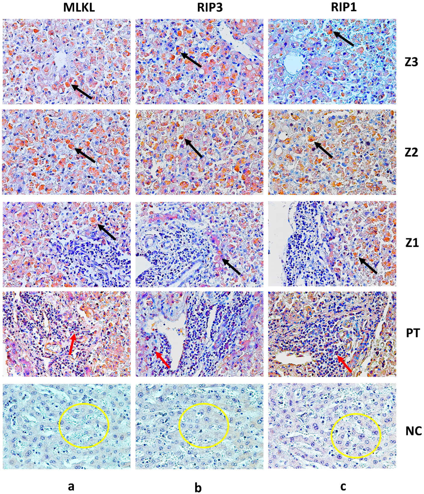
Expression of RIP1, RIP3 and MLKL in the Hepatic Parenchyma in Fatal Yellow Fever Cases
4. Discussion
5. Conclusions
Author Contributions
Funding
Institutional Review Board Statement
Informed Consent Statement
Data Availability Statement
Conflicts of Interest
References
- Gardner, C.L.; Ryman, K.D. Yellow fever: A reemerging threat. Clin. Lab. Med. 2010, 30, 237–260. [Google Scholar] [CrossRef] [PubMed]
- Vasconcelos, P.F.C.; Bryant, J.E.; Travassos Da Rosa, A.P.A.; Tesh, R.B.; Rodrigues, S.G.; Barrett, A.D.T. Genetic Divergence and Dispersal of Yellow Fever Virus, Brazil. Emerg. Infect. Dis. 2004, 10, 1578–1584. [Google Scholar] [CrossRef] [PubMed]
- Staples, J.E.; Monath, T.P. 100 Years of Discovery the Etiology of Yellow Fever: An Additional Note. JAMA Class 2008, 36, 960–962. [Google Scholar] [CrossRef] [PubMed]
- Mutebi, J.-P.; Wang, H.; Li, L.; Bryant, J.E.; Barrett, A.D.T. Phylogenetic and Evolutionary Relationships among Yellow Fever Virus Isolates in Africa. J. Virol. 2001, 75, 6999–7008. [Google Scholar] [CrossRef]
- Couto-Lima, D.; Madec, Y.; Bersot, M.I.; Campos, S.S.; Motta, M.D.A.; Dos Santos, F.B.; Vazeille, M.; Da Costa Vasconcelos, P.F.; Lourenço-De-Oliveira, R.; Failloux, A.B. Potential Risk of Re-Emergence of Urban Transmission of Yellow Fever Virus in Brazil Facilitated by Competent Aedes Populations. Sci. Rep. 2017, 7, 4848. [Google Scholar] [CrossRef]
- Vasconcelos, P.F.C. Febre amarela: A doença e a vacina, uma história inacabada [Yellow fever: The disease and the vaccine, an unfinished history]. Rev. Soc. Bras. Med. Trop. 2003, 36, 275–293. [Google Scholar] [CrossRef]
- Lopes, R.L.; Pinto, J.R.; da Silva Junior, G.B.; Santos, A.K.T.; Souza, M.T.O.; Daher, E.D.F. Kidney Involvement in Yellow Fever: A Review. Rev. Inst. Med. Trop. Sao Paulo 2019, 61, e35. [Google Scholar] [CrossRef]
- Melo, J.M.; Falcão, L.F.M.; da Ponte, L.C.T.; Silva, C.C.; Martins, L.C.; Chiang, J.O.; Martins Filho, A.J.; Franco, E.C.S.; Duarte, M.I.S.; Sousa, J.R.; et al. Emergence of New Immunopathogenic Factors in Human Yellow Fever: Polarisation of the M1/M2 Macrophage Response in the Renal Parenchyma. Viruses 2022, 14, 1725. [Google Scholar] [CrossRef]
- Han, J.; Zhong, C.Q.; Zhang, D.W. Programmed necrosis: Backup to and competitor with apoptosis in the immune system. Nat. Immunol. 2011, 12, 1143–1149. [Google Scholar] [CrossRef]
- Okamoto, T.; Suzuki, T.; Kusakabe, S.; Tokunaga, M.; Hirano, J.; Miyata, Y.; Matsuura, Y. Regulation of apoptosis during flavivirus infection. Viruses 2017, 9, 243. [Google Scholar] [CrossRef]
- Quaresma, J.A.S.; Barros, V.L.R.S.; Pagliari, C.; Fernandes, E.R.; Guedes, F.; Takakura, C.F.H.; Andrade, H.F.; Vasconcelos, P.F.C.; Duarte, M.I.S. Revisiting the liver in human yellow fever: Virus-induced apoptosis in hepatocytes associated with TGF-β, TNF-α and NK cells activity. Virology 2006, 345, 22–30. [Google Scholar] [CrossRef] [PubMed]
- Wallach, D.; Kang, T.-B.; Dillon, C.P.; Green, D.R. Programmed necrosis in inflammation: Toward identification of the effector molecules. Science 2016, 352, aaf2154. [Google Scholar] [CrossRef] [PubMed]
- Wu, X.N.; Yang, Z.H.; Wang, X.K.; Zhang, Y.; Wan, H.; Song, Y.; Chen, X.; Shao, J.; Han, J. Distinct roles of RIP1-RIP3 hetero- and RIP3-RIP3 homo-interaction in mediating necroptosis. Cell Death Differ. 2014, 21, 1709–1720. [Google Scholar] [CrossRef] [PubMed] [PubMed Central]
- Weber, K.; Roelandt, R.; Bruggeman, I.; Estornes, Y.; Vandenabeele, P. Nuclear RIPK3 and MLKL contribute to cytosolic necrosome formation and necroptosis. Commun. Biol. 2018, 1, 6. [Google Scholar] [CrossRef]
- Wu, Y.; Zheng, Z.; Cao, X.; Yang, Q.; Norton, V.; Adini, A.; Maiti, A.K.; Adini, I.; Wu, H. RIP1/RIP3/MLKL Mediates Myocardial Function Through Necroptosis in Experimental Autoimmune Myocarditis. Front. Cardiovasc. Med. 2021, 8, 696362. [Google Scholar] [CrossRef]
- Olímpio, F.A.; Falcão, L.F.M.; Carvalho, M.L.G.; da Costa Lopes, J.; Mendes, C.C.H.; Filho, A.J.M.; da Silva, C.A.M.; Miranda, V.D.S.C.; dos Santos, L.C.; da Silva Vilacoert, F.S.; et al. Endothelium Activation during Severe Yellow Fever Triggers an Intense Cytokine-Mediated Inflammatory Response in the Liver Parenchyma. Pathogens 2022, 11, 101. [Google Scholar] [CrossRef]
- Simões Quaresma, J.A.; Souza Barros, V.L.R.; Rainero Fernandes, E.; Pagliari, C.; Takakura, C.; da Costa Vasconcelos, P.F.; de Andrade, H.F.; Seixas Duarte, M.I. Reconsideration of histopathology and ultrastructural aspects of the human liver in yellow fever. Acta Trop. 2005, 94, 116–127. [Google Scholar] [CrossRef]
- Quaresma, J.A.; Barros, V.L.; Pagliari, C.; Fernandes, E.R.; Andrade HFJr Vasconcelos, P.F.; Duarte, M.I. Hepatocyte lesions and cellular immune response in yellow fever infection. Trans. R. Soc. Trop. Med. Hyg. 2007, 101, 161–168. [Google Scholar] [CrossRef] [PubMed]
- Quaresma, J.A.; Duarte, M.I.; Vasconcelos, P.F. Midzonal lesions in yellow fever: A specific pattern of liver injury caused by direct virus action and in situ inflammatory response. Med. Hypotheses 2006, 67, 618–621. [Google Scholar] [CrossRef] [PubMed]
- Hsu, S.M.; Raine, L.; Fanger, H. Use of Avidin-Biotin-Peroxidase Complex (ABC) in Immunoperoxidase Techniques: A comparison between ABC and unlabeled antibody (PAP) procedures. J. Histochem. Cytochem. 1981, 29, 577–580. [Google Scholar] [CrossRef]
- Vandenabeele, P.; Galluzzi, L.; Vanden Berghe, T.; Kroemer, G. Molecular mechanisms of necroptosis: An ordered cellular explosion. Nat. Rev. Mol. Cell Biol. 2013, 14, 727–736. [Google Scholar] [CrossRef] [PubMed]
- Conos, S.A.; Chen, K.W.; De Nardo, D.; Hara, H.; Whitehead, L.; Nuñez, G.; Masters, S.L.; Murphy, J.M.; Schroder, K.; Vaux, D.L.; et al. Active MLKL triggers the NLRP3 inflammasome in a cell-intrinsic manner. Proc. Natl. Acad. Sci. USA 2017, 114, E961–E969. [Google Scholar] [CrossRef] [PubMed] [PubMed Central]
- Dhuriya, Y.K.; Sharma, D. Necroptosis: A regulated inflammatory mode of cell death. J. Neuroinflammation 2018, 15, 199. [Google Scholar] [CrossRef] [PubMed] [PubMed Central]
- Amin, P.; Florez, M.; Najafov, A.; Pan, H.; Geng, J.; Ofengeim, D.; Dziedzic, S.A.; Wang, H.; Barrett, V.J.; Ito, Y.; et al. Regulation of a distinct activated RIPK1 intermediate bridging complex I and complex II in TNFα-mediated apoptosis. Proc. Natl. Acad. Sci. USA 2018, 115, E5944–E5953. [Google Scholar] [CrossRef] [PubMed] [PubMed Central]
- Scaffidi, P.; Misteli, T.; Bianchi, M.E. Release of chromatin protein HMGB1 by necrotic cells triggers inflammation. Nature 2002, 418, 191–195. [Google Scholar] [CrossRef] [PubMed]
- Iyer, S.S.; Pulskens, W.P.; Sadler, J.J.; Butter, L.M.; Teske, G.J.; Ulland, T.K.; Eisenbarth, S.C.; Florquin, S.; Flavell, R.A.; Leemans, J.C.; et al. Necrotic cells trigger a sterile inflammatory response through the Nlrp3 inflammasome. Proc. Natl. Acad. Sci. USA 2009, 106, 20388–20393. [Google Scholar] [CrossRef] [PubMed] [PubMed Central]
- Rello, S.; Stockert, J.C.; Moreno, V.L.; Gamez, A.; Pacheco, M.; Juarranz, A.; Canete, M.; Villanueva, A. Morphological criteria to distinguish cell death induced by apoptotic and necrotic treatments. Apoptosis 2005, 10, 201–208. [Google Scholar] [CrossRef] [PubMed]
- Verburg, S.G.; Lelievre, R.M.; Westerveld, M.J.; Inkol, J.M.; Sun, Y.L.; Workenhe, S.T. Viral-mediated activation and inhibition of programmed cell death. PLoS Pathog. 2022, 18, e1010718. [Google Scholar] [CrossRef] [PubMed] [PubMed Central]
- Galluzzi, L.; Kepp, O.; Chan, F.K. Necroptosis: Mechanisms and relevance to disease. Annu. Rev. Pathol. Mech. Dis. 2017, 12, 103–130. [Google Scholar] [CrossRef]


| Case | Patient | Gender | Age | State | Year | IT * |
|---|---|---|---|---|---|---|
| 1 | 001/00 | M | 25 | Tocantins | 2000 | 8 |
| 2 | 106/00 | M | 75 | Goiás | 2000 | NR ** |
| 3 | 108/00 | M | 49 | Goiás | 2000 | 7 |
| 4 | 494/00 | M | NR ** | Distrito Federal | 2000 | NR ** |
| 5 | 251/00 | M | 16 | Mato Grosso do Sul | 2000 | 6 |
| 6 | 252/00 | M | 49 | Goiás | 2000 | NR ** |
| 7 | 253/00 | M | 23 | Goiás | 2000 | NR ** |
| 8 | 255/00 | M | NR ** | Goiás | 2000 | NR ** |
| 9 | 291/00 | M | NR ** | Goiás | 2000 | NR ** |
| 10 | 158/00 | M | 33 | Goiás | 2000 | NR ** |
| 11 | 063/03 | M | NR ** | Minas Gerais | 2003 | NR ** |
| 12 | 339/04 | M | 36 | Amazonas | 2004 | 11 |
| 13 | 019/08 | M | 64 | Goiás | 2008 | 7 |
| 14 | 273/08 | M | 57 | Goiás | 2008 | 7 |
| 15 | 068/08 | F | 65 | Goiás | 2008 | 2 |
| 16 | 095/08 | M | 42 | Goiás | 2008 | 3 |
| 17 | 143/08 | M | 37 | Distrito Federal | 2008 | NR ** |
| 18 | 361/15 | F | 53 | Rio Grande do Norte | 2015 | 4 |
| 19 | 062/16 | M | 35 | Goiás | 2016 | NR ** |
| 20 | 346/16 | M | 15 | Goiás | 2016 | 7 |
| 21 | 369/16 | M | 27 | Goiás | 2016 | 1 |
| Markers | Z3 (Cells/mm2) | Z2 (Cells/mm2) | Z1 (Cells/mm2) | PT (Cells/mm2) | ANOVA (p ≤ 0.05) |
|---|---|---|---|---|---|
| RIP1 Control | 143.20 ± 21.75 76.80 ± 13.39 | 350.40 ± 52.40 112.00 ± 25.30 | 125.70 ± 13.83 48.40 ± 9.94 | 102.4 ± 11.19 36.80 ± 4.60 | *** |
| Tukey (p ≤ 0.05) | *** | *** | ** | * | |
| RIP3 Control | 140.2 ± 19.41 59.20 ± 6.57 | 255.10 ± 90.92 92.80 ± 13.39 | 107.3 ± 18.87 30.20 ± 4.38 | 96.33 ± 12.06 22.20 ± 2.68 | *** |
| Tukey (p ≤ 0.05) | *** | *** | * | * | |
| MLKL Control | 147.80 ± 19.53 60.80 ± 17.53 | 318.50 ± 63.38 108.80 ± 28.62 | 109.70 ± 17.73 51.20 ± 17.53 | 89.60 ± 11.79 36.00 ± 8.00 | *** |
| Tukey (p ≤ 0.05) | *** | *** | ** | * |
| Correlation | r | p-Value |
|---|---|---|
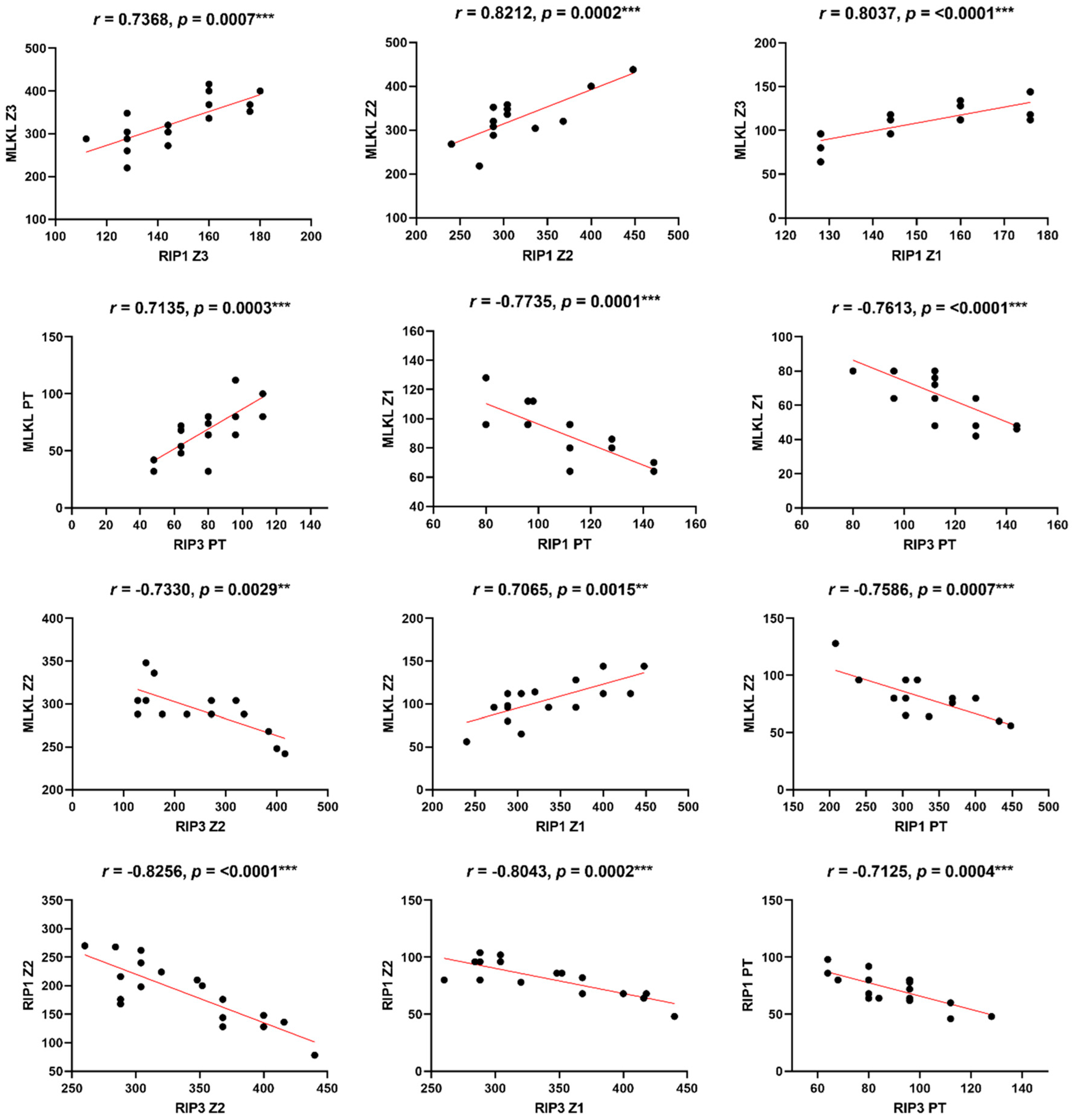
| MLKL Z3 x RIP1 Z3 | 0.5804 | 0.0058 ** |
| MLKL Z3 x RIP1 Z2 | 0.7368 | 0.0007 *** |
| MLKL Z3 x RIP1 Z1 | 0.8037 | <0.0001 *** |
| MLKL Z2 x RIP1 Z2 | 0.8212 | 0.0002 *** |
| MLKL Z2 x RIP1 Z1 | 0.7065 | 0.0015 ** |
| MLKL Z2 x RIP1 PT | −0.7586 | 0.0007 *** |
| MLKL Z1 x RIP1 PT | −0.7735 | 0.0001 *** |
| MLKL Z3 x RIP3 Z3 | 0.4726 | 0.0305 * |
| MLKL Z3 x RIP3 Z2 | 0.6764 | 0.0079 ** |
| MLKL Z3 x RIP Z1 | −0.4788 | 0.0381 * |
| RIP3 Z2 x MLKL Z2 | −0.7330 | 0.0029 * |
| MLKL Z1 x RIP3 PT | −0.7613 | <0.0001 *** |
| MLKL PT x RIP 3 PT | 0.7135 | 0.0003 *** |
| RIP1 Z3 x RIP3 PT | −0.5957 | 0.0044 ** |
| RIP1 Z2 x RIP3 Z2 | −0.8256 | <0.0001 *** |
| RIP1 Z2 x RIP3 Z1 | −0.8043 | 0.0002 *** |
| RIP1 Z2 x RIP3 PT | −0.6483 | 0.0015 ** |
| RIP1 Z1 x RIP3 Z1 | −0.5047 | 0.0327 * |
| RIP1 PT x RIP3 PT | −0.7125 | 0.0004 *** |
| RIP1 Z3 x RIP3 Z2 | 0.4601 | 0.0475 * |
Disclaimer/Publisher’s Note: The statements, opinions and data contained in all publications are solely those of the individual author(s) and contributor(s) and not of MDPI and/or the editor(s). MDPI and/or the editor(s) disclaim responsibility for any injury to people or property resulting from any ideas, methods, instructions or products referred to in the content. |
© 2024 by the authors. Licensee MDPI, Basel, Switzerland. This article is an open access article distributed under the terms and conditions of the Creative Commons Attribution (CC BY) license (https://creativecommons.org/licenses/by/4.0/).
Share and Cite
Miranda, V.d.S.C.; Falcão, L.F.M.; Fuzii, H.T.; Carvalho, M.L.G.; Lopes, J.d.C.; Filho, A.J.M.; Cruz, A.C.R.; Azevedo, R.d.S.d.S.; Sousa, J.R.d.; Wakimoto, M.D.; et al. Analysis of MLKL, RIP1 and RIP3 Immunostaining Markers in Human Liver Tissue from Fatal Yellow Fever Cases: Insights into Necroptosis. Viruses 2025, 17, 3. https://doi.org/10.3390/v17010003
Miranda VdSC, Falcão LFM, Fuzii HT, Carvalho MLG, Lopes JdC, Filho AJM, Cruz ACR, Azevedo RdSdS, Sousa JRd, Wakimoto MD, et al. Analysis of MLKL, RIP1 and RIP3 Immunostaining Markers in Human Liver Tissue from Fatal Yellow Fever Cases: Insights into Necroptosis. Viruses. 2025; 17(1):3. https://doi.org/10.3390/v17010003
Chicago/Turabian StyleMiranda, Vanessa do Socorro Cabral, Luiz Fabio Magno Falcão, Hellen Thais Fuzii, Marcos Luiz Gaia Carvalho, Jeferson da Costa Lopes, Arnaldo Jorge Martins Filho, Ana Cecilia Ribeiro Cruz, Raimunda do Socorro da Silva Azevedo, Jorge Rodrigues de Sousa, Mayumi Duarte Wakimoto, and et al. 2025. "Analysis of MLKL, RIP1 and RIP3 Immunostaining Markers in Human Liver Tissue from Fatal Yellow Fever Cases: Insights into Necroptosis" Viruses 17, no. 1: 3. https://doi.org/10.3390/v17010003
APA StyleMiranda, V. d. S. C., Falcão, L. F. M., Fuzii, H. T., Carvalho, M. L. G., Lopes, J. d. C., Filho, A. J. M., Cruz, A. C. R., Azevedo, R. d. S. d. S., Sousa, J. R. d., Wakimoto, M. D., Vasconcelos, P. F. d. C., & Quaresma, J. A. S. (2025). Analysis of MLKL, RIP1 and RIP3 Immunostaining Markers in Human Liver Tissue from Fatal Yellow Fever Cases: Insights into Necroptosis. Viruses, 17(1), 3. https://doi.org/10.3390/v17010003