Article

Clinical Performance of MAGLUMI Diagnostic Tests for the Automated Detection of Dengue Virus

1
Microbiology Laboratory, Shenzhen Center for Disease Control and Prevention, Shenzhen 518055, China
2
Key-Laboratory of Intelligent Tracking and Forecasting for Infectious Disease, NHC Key Laboratory of Medical Virology and Viral Diseases, National Institute for Viral Disease Control and Prevention, Chinese Center for Disease Control and Prevention, Beijing 102206, China
3
Research & Development Department, Shenzhen New Industries Biomedical Engineering Co., Ltd., No. 23, Jinxiu East Road, Pingshan District, Shenzhen 518122, China
*
Authors to whom correspondence should be addressed.
These authors contributed equally to this work.
Viruses 2025, 17(1), 106; https://doi.org/10.3390/v17010106
Submission received: 1 December 2024 / Revised: 10 January 2025 / Accepted: 12 January 2025 / Published: 14 January 2025
(This article belongs to the Section Human Virology and Viral Diseases)

Abstract

:
Aims: The screening and diagnosis of dengue virus infection play a crucial role in controlling the epidemic of dengue fever, highlighting the urgent need for a highly sensitive, simple, and rapid laboratory testing method. This study aims to assess the clinical performance of MAGLUMI Denv NS1 in detecting dengue virus NS1 antigen. Methods: A retrospective study was conducted to assess the sensitivity and specificity of MAGLUMI Denv NS1 using residual samples. Dengue-confirmed and excluded samples, validated by qPCR, were subjected to testing with MAGLUMI Denv NS1 in accordance with the manufacturer’s instructions. The linear range, endogenous interference, and cross-reactivity of MAGLUMI Denv NS1 were verified, and a consistency analysis with commercial comparator products was carried out. Results: The diagnostic specificity of MAGLUMI Denv NS1 is 98.41% (62/63), and the sensitivity is 98.32% (117/119). It effectively detects various serotypes of dengue virus, with no observed endogenous interference or cross-reactivity. Additionally, the consistency of NS1, IgM, and IgG tests on the MAGLUMI platform compared to commercial comparator reagents reaches 85.71%, 99.25%, and 98.97%, respectively. Conclusions: The MAGLUMI Denv NS1 represents a highly sensitive laboratory testing method capable of enhancing the diagnostic accuracy and efficiency of dengue virus infection detection.

1. Introduction

Dengue virus infection is a prevalent mosquito-borne disease in tropical and subtropical regions. Globally, an estimated 390 million individuals are affected by dengue virus annually, resulting in over 500,000 hospitalizations and 25,000 fatalities [1]. Dengue virus can be categorized into four serotypes based on antigenic disparities known as DENV1-4 [2]. Infection with any of the four serotypes of dengue fever virus may lead to a spectrum of clinical outcomes ranging from mild fever to classic dengue hemorrhagic fever (DHF) and dengue shock syndrome (DSS) [3]. Although the genomes of these four serotypes are similar, they are unable to individually elicit cross-neutralizing antibodies. Severe dengue fever primarily occurs in individuals who have experienced secondary infections with different serotypes of the virus [4]. This is due to pre-existing non-neutralizing or low-affinity antibodies, which, upon encountering a heterologous serotype of dengue virus, are unable to effectively neutralize the new serotype and instead facilitate viral entry into host cells, leading to increased viral replication and inflammation response. Consequently, this increases the risk of developing severe dengue fever upon secondary infection—a phenomenon known as antibody-dependent enhancement (ADE) [5,6]. The intricate cross-reactivity among dengue serotypes poses a significant challenge in the development of effective vaccines for dengue fever. Hence, timely detection, accurate diagnosis, and comprehensive early intervention are pivotal for successful management of dengue fever [7,8].
The WHO guidelines recommended multiple diagnostic methods for dengue fever, including virus isolation, nucleic acid detection, antigen detection, serological testing, and hematological testing [9]. Despite being considered the most sensitive diagnostic approaches, virus isolation through cell culture and nucleic acid detection via real-time quantitative PCR have limitations in terms of rapid diagnosis due to their requirement for specialized knowledge, advanced facilities, expensive laboratory equipment, and a significant amount of time for virus isolation and RNA purification [10,11,12]. The non-structural protein 1 (NS1) is a highly conserved glycoprotein encoded by the dengue virus, which typically reaches high concentrations in the serum of dengue fever patients within 1–5 days after infection and is detected almost simultaneously with viral RNA, long before the production of antibodies. Therefore, NS1 antigen detection serves as an early diagnostic tool for dengue fever [13,14]. Furthermore, the presence of NS1 antigen for up to 9 days or even longer can indicate active viral replication and potential severity of the disease [15,16]. Additionally, a positive NS1 test result can differentiate from other causes of febrile illnesses, such as influenza, Zika virus infection, etc., thus enhancing diagnostic specificity [17]. At present, commercial products for the detection of dengue virus NS1 primarily consist of immunochromatographic assays for rapid on-site screening and enzyme-linked immunosorbent assays (ELISA) commonly utilized for laboratory diagnosis. Several studies evaluating the clinical performance of NS1 detection kits have reported a range of sensitivities from 48.5% to 80.7% and specificities from 92.5% to 99.4% for NS1 antigen rapid diagnostic tests [18,19,20,21], while the sensitivity of ELISA detection methods varies from less than 75% to as high as 100% [22,23]. A recent study on detection sensitivity indicated that combined testing of NS1/IgG/IgM (90.65%) demonstrated higher clinical performance compared to IgG (90.06%) or NS1 (87.50%) alone [24].
In order to enhance the sensitivity, stability, testing speed, and automation level of NS1 detection methods and address clinical rapid diagnostic needs and laboratory efficiency, we have developed a MAGLUMI Denv NS1 chemiluminescent immunoassay (CLIA). This assay employs the sandwich principle of dual antibodies to quantitatively detect dengue fever virus NS1 antigen in human serum and plasma for adjunctive diagnosis of dengue fever infection. In this investigation, we assessed the clinical performance of this assay.

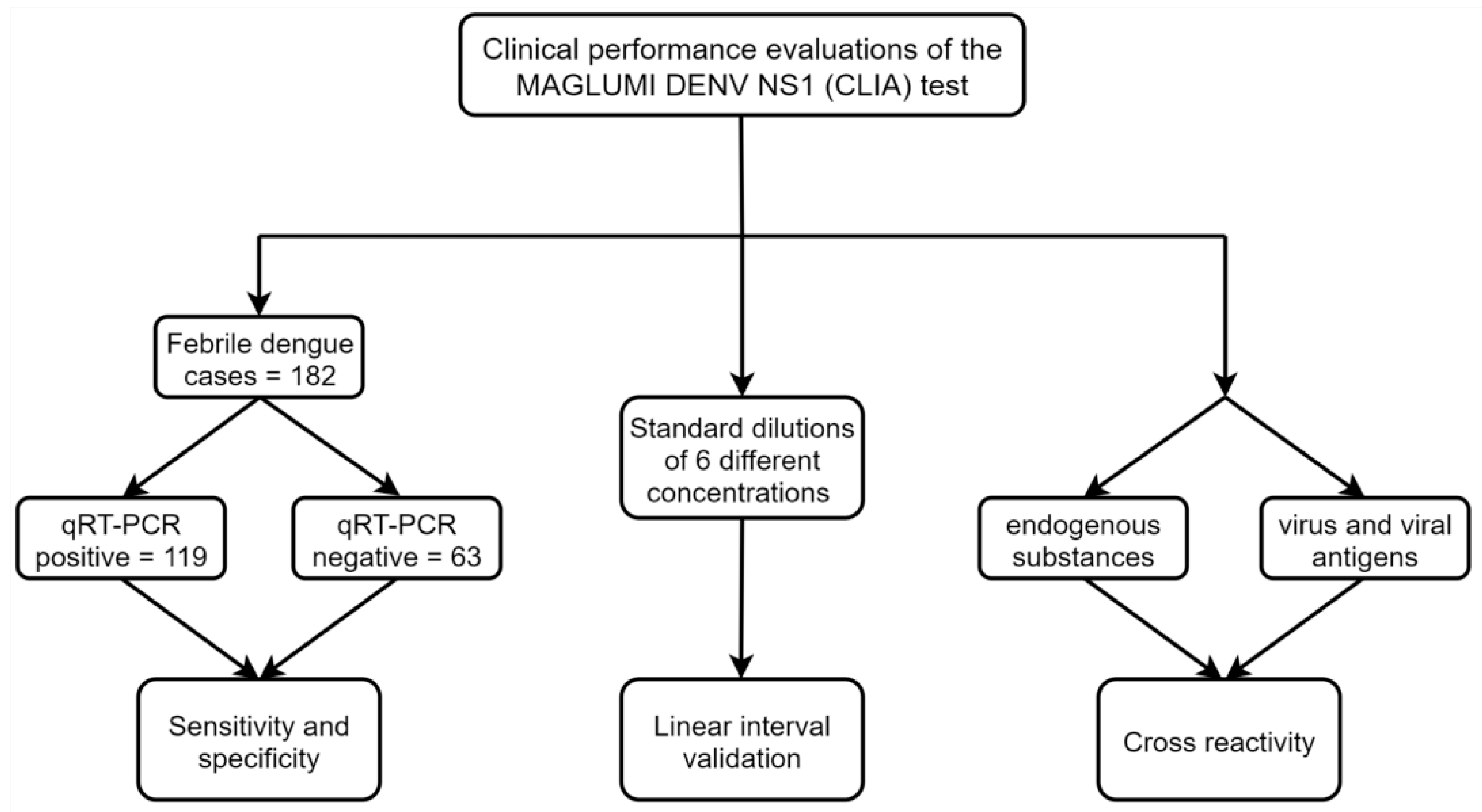
2. Material and Methods

2.1. Clinical Samples

In this study, a total of 182 serum samples were collected from patients that were suspected to have dengue fever. Following real-time qPCR detection, 63 cases were excluded, and 119 were confirmed with a Ct value greater than 40 as the negative criterion. Information about the dengue-positive sample is presented in Supplementary Table S1.
All clinical samples underwent testing at the Center for Disease Control and Prevention of Shenzhen, Guangdong Province, China, between January 2019 and May 2023, with relevant clinical symptoms and demographic data being recorded (Figure 1).

2.2. MAGLUMI Dengue Virus NS1 Antigen Test

The MAGLUMI dengue virus NS1 antigen test utilizes a sandwich chemiluminescence immunoassay, in which the sample is fully mixed with magnetic microbeads coated with NS1 monoclonal antibody, N-(4-Aminobutyl)-n-ethylisoluminol (ABEI) labeled by another NS1 monoclonal antibody, and buffer solution to form a sandwich complex during incubation. The information of the antibodies independently produced by the company is shown in the following table:
Coating AntibodyLabeling Antibody
ImmunogenDengue NS1 antigenDengue NS1 antigen
Sourcemousemouse
Molecular weight (kDa)150150
SDS-PAGE bands (kDa)25, 5525, 55
Purity (SDS-page)≥95%≥95%
Concentration (mg/mL)2.449–3.1782.114–2.869
Following precipitation in the magnetic field, the reaction liquid is introduced to initiate the chemiluminescence reaction. The resulting light signal is measured by photomultiplier tubes as a relative light unit (rlu), providing a proportional indication of the concentration of dengue virus NS1 antigen present in the sample.
In the same instrument system, Maglumi DENV IgG employs the Indirect Chemiluminescence Immunoassay. The prediluted sample, buffer, and magnetic microbeads coated with dengue virus antigen are mixed and precipitated in a magnetic field. Subsequently, ABEI labeled with anti-human IgG monoclonal antibody is added to form immuno-complexes. MAGLUMI DENV IgM utilizes the capture chemiluminescence immunoassay. The sample, buffer, and magnetic microbeads coated with anti-human IgM antibody are mixed and precipitated. Then, ABEI labeled with dengue virus antigen is added and incubated, reacting to form immuno-complexes.

2.3. Analytical Performance Verification

Three distinct batches of reagents were employed to test 4 blank serum samples, 4 low-concentration serum samples, and 6 low-concentration serum samples of known concentrations. LoB, LoD, and LoQ were evaluated, respectively. Each sample was tested over three days, with 5 replicates per day. Linearity verification was conducted in accordance with CLSI standards. The 6 samples provided by the manufacturer, each with a specified concentration, were tested three times within a single run. The mean value of the test results was calculated and then fitted linearly with the theoretical concentration using the least square method. Subsequently, the linear correlation coefficient was determined.

2.4. Endogenous Interference and Cross-Reactivity

A total of 15 samples containing potential cross-reactive substances and 37 samples with interfering substances were assessed. The former consisted of inactivated clinical samples or negative clinical samples supplemented with inactivated pathogen cultures, such as Zika virus, hepatitis A virus, hepatitis B virus, hepatitis C virus, and chikungunya virus. The sample matrix was consistent with the anticipated type of test sample. The latter comprised clinically negative samples supplemented with potential endogenous and exogenous interfering substances, including hemoglobin, HAMA, lipid emulsion, ANA, bilirubin, rheumatoid factor, acetaminophen, lysine acetylsalicylate (LAS), ibuprofen, and dexamethasone.

2.5. Statistical Analysis

Statistical analysis was conducted using MedCalc software version 18.11.3 and Microsoft Office Excel 2013. A four-grid table was established for qualitative data analysis, sensitivity and specificity were calculated as percentages, the Wilson score interval method was employed to calculate the 95% confidence interval of evaluation indicators, and a Kappa consistency test analysis was performed. Between-group comparison utilized the Wilcox rank sum test, with p < 0.05 indicating statistically significant differences. Furthermore, Graphpad Prism software was used to compare various test results and evaluate data correlation generated by two testing methods through simple linear regression analysis.

2.6. Ethics

The performance validation study was carried out by the Shenzhen Center for Disease Control and Prevention in accordance with the guidelines of the Helsinki Declaration and following the principles of good clinical practice. All samples used in this study were residual samples obtained with general ethical approval, and subjects provided broad informed consent through signature.

3. Results

3.1. Diagnostic Specificity and Sensitivity

The results of clinical sample testing using the MAGLUMI Denv NS1 test demonstrated that among 119 dengue-confirmed samples, 117 exhibited positive NS1 antigen reactivity, while only one out of 63 dengue-excluded samples showed NS1 antigen reactivity. Statistical analysis revealed a diagnostic sensitivity of 98.32% (117/119, 95% CI: 98–99%) and a diagnostic specificity of 98.41% (62/63, 95% CI: 98–100%) for the MAGLUMI Denv NS1 test (Table 1 and Table 2).
By categorizing 119 confirmed dengue samples based on serotypes, we can also determine the diagnostic sensitivity of the MAGLUMI Denv NS1 test for four distinct dengue virus serotypes. The aforementioned data illustrates that the MAGLUMI Denv NS1 test exhibits high diagnostic sensitivity and specificity and possesses the capability to detect various serotypes of the dengue virus (Table 3).
Through the assessment of data derived from a MAGLUMI Denv NS1 test and real-time qPCR utilizing simple linear regression analysis, it was determined that there exists no significant correlation between the two detection methodologies, potentially attributed to variances in detection principles (Supplementary Figure S1).

3.2. Analytical Specificity

The analytical specificity of the MAGLUMI Denv NS1 test was determined to be 100% (52/52, 95% CI: 93.12–100%), as evidenced by the lack of reactivity in a total of 52 clinical samples that had been deliberately spiked with cross-interfering substances or a negative serum matrix containing such substances (Table 4).

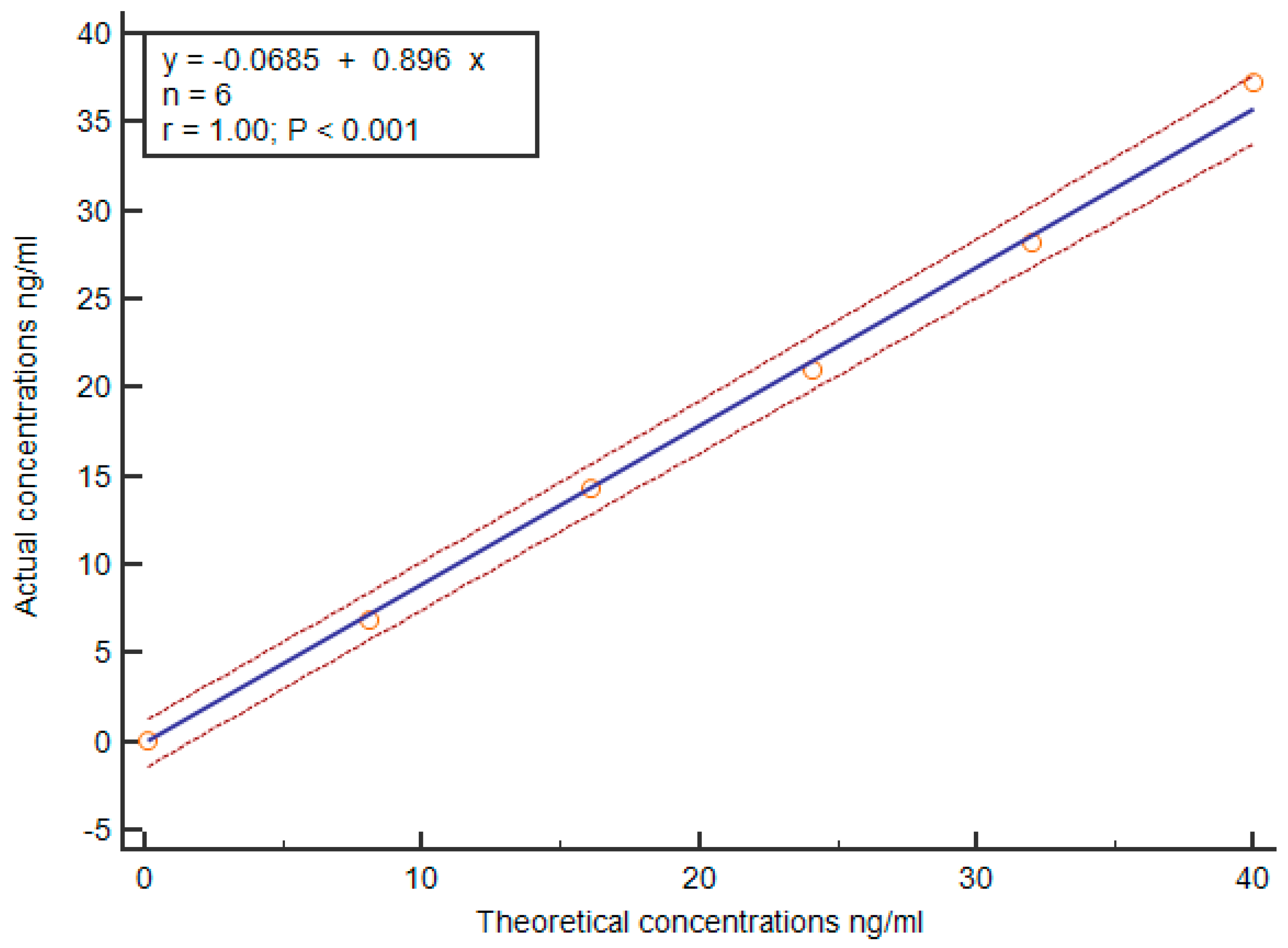
3.3. Analytic Sensitivity and Linearity Verification

LoB, LoD, and LoQ were calculated based on the CLSI Guideline EP17-A2. The LOB for the MAGLUMI Denv NS1 test was 0.500 AU/mL, with the LoD and LoQ at 0.800 AU/mL and 1.000 AU/mL, respectively (Table 5).
The linearity verification results for the MAGLUMI Denv NS1 test are depicted in Figure 2. The best-fit linear regression equation is y = 0.9196 x 0.4553 , with an R-squared value of 0.9978, indicating a significant correlation between the average measured values from three replicate experiments and the expected NS1 concentration within the range of 1~400 AU/mL.

3.4. Serum to Plasma Equivalency

Repeated tests were conducted on eight groups of negative serum or plasma samples treated with various substrates, all yielding negative results. Similarly, 11 groups of positive serum or plasma samples treated with different substrates were retested, and all showed positive results. Despite variations in AU/mL levels among different matrices, the determination of sample positivity or negativity remained unaffected (Table 6).

3.5. Consistency Between Different Dengue Diagnostic Assays

In order to assess the consistency of the MAGLUMI Denv NS1 test in clinical performance with commercial comparator reagents, a total of seven DEN-positive samples and 107 DEN-negative samples were subjected to testing using the MAGLUMI Denv NS1 test and EUROIMMUN Denv NS1, respectively. The results revealed a negative concordance rate of 99.07% between the two products. While the number of positive samples was limited, it was observed that the positive concordance between the two products could reach as high as 85.71% (Table 7).
Simultaneously, we conducted an evaluation of the newly developed dengue virus IgG and IgM reagent kits on the MAGLUMI Diagnostic Platform to assess their clinical performance consistency with comparable products. Our findings revealed a remarkable overall agreement rate of 98.97% for IgG and 99.25% for IgM when compared with EUROIMMUN DENV, indicating a high level of concordance between the dengue diagnostic kits based on the MAGLUMI Diagnostic Platform and recognized testing products (Table 6). These results support the utility of these kits for the simultaneous detection of three biomarkers at different stages of dengue infection.

4. Discussion

Commercial detection products with high specificity and sensitivity are crucial for the prompt diagnosis of dengue fever infection and epidemic prevention. The early presence and abundant levels of NS1 antigen in patient serum have led to its widespread utilization in various immunological analysis products based on monoclonal or polyclonal antibodies, establishing it as a well-researched biomarker for detecting dengue virus. This study presents the clinical performance of MAGLUMI Denv NS1 utilizing the fully automated chemiluminescence immunoassay platform MAGLUMI series. The results demonstrate that MAGLUMI Denv NS1 exhibits a sensitivity of 98.32% and a specificity of 98.41% for detecting dengue virus infection, surpassing the reported sensitivity of similar NS1 antigen detection products at 80.7%. These findings indicate that the exceptional clinical performance of MAGLUMI Denv NS1 meets the diagnostic requirements for dengue virus infection.
Although specific antiviral drugs for the treatment of dengue fever are currently unavailable, understanding the serotype of the virus that patients are infected with may assist clinical doctors’ assessment of disease severity [25,26]. The predominant virus infections in most dengue fever endemic areas globally are DENV-1 and DENV-2 serotypes, with DENV-2 being considered the most common serotype causing severe dengue fever [27,28]. Our study identified 99 samples positive for DENV-1 and 14 samples positive for DENV-2, with a high sensitivity of 112/113. Regrettably, we did not have sufficient positive samples of DENV-3 and DENV-4 serotypes for testing. Given that all four serotypes of dengue virus spread in a similar manner, prior to the application of MAGLUMI Denv NS1 in dengue-endemic areas, verification will be carried out on a larger sample size, especially for samples of DENV 3 and DENV 4.
Cross-reactivity is a significant factor impacting the precision of dengue virus diagnosis. The clinical presentations of dengue fever and other arthropod-borne viral diseases, such as chikungunya fever and Zika fever, can be quite similar at times, manifesting with symptoms like fever, rash, and joint pain [29,30]. This similarity complicates the diagnostic process, necessitating comprehensive laboratory testing and epidemiological history for accurate diagnosis. While our study had a limited sample size, we did not observe cross-reactivity of MAGLUMI Denv NS1 with Zika virus or chikungunya virus. Additionally, we confirmed the excellent detection performance of MAGLUMI Denv NS1 for endogenous interfering substances and its suitability for testing serum or plasma samples from various matrices.
The dengue fever IgM antibody is produced in the early stages of infection, typically beginning to rise 3–7 days after infection and gradually decreasing over time. A positive IgM result indicates a recent or ongoing dengue virus infection, which is crucial for distinguishing between recent and past infections. In contrast, the dengue fever IgG antibody develops during the later phase of infection and throughout the recovery period, persisting for an extended duration [31,32]. It not only signifies a history of prior dengue virus infection but also serves as a tool for assessing potential enhancement effects (ADE) in cases of secondary infections, as a subsequent infection may lead to more severe manifestations of dengue fever. The concurrent detection of dengue virus NS1 antigen, IgG antibodies, and IgM antibodies has the potential to enhance the sensitivity and specificity of diagnosis [33,34]. On the other hand, the antibodies produced by the primary dengue infection might compete with the reagents, thereby diminishing the performance of the detection during the secondary infection. Among the 119 samples of dengue-infected patients employed in this study, seven cases were IgG positive and were within the 4th to 7th days of fever, indicating that they could be patients with secondary infections, and all were detected by the MAGLUMI Denv NS1 test (Supplementary Table S2). To further validate the analytical specificity of the reagent, in future studies, it is necessary to test more acute specimens and compare the performance of NS1 in IgG-positive and IgG-negative samples.
In our study, the overall agreement rate between IgG and EUROIMMUN DENV IgG was 98.97%, while the overall agreement rate between IgM and EUROIMMUN DENV IgM reached 99.25%. These findings demonstrate that the dengue diagnostic kit based on the MAGLUMI Diagnostic Platform exhibits a high level of concordance with recognized similar testing products, thereby enabling its utility for the combined detection of three markers at various stages of dengue infection. Regrettably, limited availability of dengue-positive samples precluded separate investigation into dynamic changes in levels of dengue virus markers during primary and secondary infections. To facilitate improved assessment by healthcare professionals regarding the stage of dengue infection and to guide subsequent treatment plans, further validation is warranted to confirm quantitative detection performance of MAGLUMI Denv NS1 as well as IgG and IgM across different stages of infection.
MAGLUMI Anti-NS1 represents a direct chemiluminescence immunoassay (DCLIA) approach, featuring simplistic technology and a fully automated operational procedure. The time to results merely demands 37 min, which is significantly shorter than the approximately 2 h requisite for the current ELISA reagents [23]. Simultaneously, the diagnostic performance of MAGLUMI Anti-NS1 is also conspicuously superior to the existing rapid diagnostic tests (RDT), with a sensitivity ranging from 48.5% to 80.7% [18,19,20,21]. Additionally, the MAGLUMI platform has developed the ultra-high-speed MAGLUMI X8, suitable for regions with a high prevalence of dengue fever, capable of meeting the demand of 600 tests per hour, and the small-sized MAGLUMI X3, suitable for working environments with constrained resources. The MAGLUMI X3 occupies an area of merely 0.68 m2 and has a speed of 200 tests per hour, jointly contributing to the establishment of a multi-level and three-dimensional dengue fever prevention and control system.

Supplementary Materials

The following supporting information can be downloaded at: https://www.mdpi.com/article/10.3390/v17010106/s1, Figure S1: Correlation between the MAGLUMI Denv NS1 Test and the reference RT-PCR assay; Table S1: Sample size, geographic origin, and clinical characteristics of 119 Dengue positive samples; Table S2: 7 out of 119 cases were IgG positive and all were detected by the NS1 test.

Author Contributions

Conceptualization, B.P., Z.F., K.L., W.R. and X.S.; methodology, C.L., K.L., F.Y., Y.H., C.W., Y.L., D.H., Q.Z., Y.T. and X.L.; validation, C.L., T.W. and K.H.; formal analysis, B.P., Z.F., C.L., T.W., K.H., F.Y., Y.H., C.W., Y.L., D.H., Q.Z., Y.T. and X.L.; investigation, B.P., Z.F., W.R. and X.S.; resources, K.L., W.R. and X.S.; data curation, C.L., K.L., T.W., K.H., F.Y., Y.H., C.W., Y.L., D.H., Q.Z., Y.T. and X.L.; writing—original draft preparation, C.L.; writing—review and editing, B.P., Z.F., C.L., K.L., T.W., K.H., F.Y., Y.H., C.W., Y.L., D.H., Q.Z., Y.T., X.L., W.R. and X.S.; visualization, C.L., F.Y., Y.H., C.W., Y.L., D.H., Q.Z., Y.T. and X.L.; supervision, B.P., Z.F., C.L., W.R. and X.S.; project administration, B.P., Z.F., C.L., K.L., T.W., K.H., F.Y., Y.H., C.W., Y.L., D.H., Q.Z., Y.T., X.L., W.R. and X.S.; funding acquisition, W.R. and X.S. All authors have read and agreed to the published version of the manuscript.

Funding

This work was supported by the Shenzhen Medical Research Fund (B2404002), the Sanming Project of Medicine in Shenzhen (SZSM202211023), and the Opening Foundation of the State Key Laboratory of Pathogen and Biosecurity (SKLPBS2244).

Institutional Review Board Statement

The study was conducted in accordance with the Declaration of Helsinki, and approved by the Ethics Committee of Shenzhen Center for Disease Control and Prevention (SZCDC-IRB2024033, 17 April 2024).

Informed Consent Statement

Informed consent was obtained from all subjects involved in the study.

Data Availability Statement

Data is contained within the article or Supplementary Material. The original contributions presented in the study are included in the article/Supplementary Material, further inquiries can be directed to the corresponding authors.

Conflicts of Interest

The authors have no conflicts of interest. Zhonggang Fang, Cong Li, Kun Liu, Ting Wang and Wei Rao are employees of Shenzhen New Industries Biomedical Engineering Co., Ltd.

References

  1. Bhatt, S.; Gething, P.W.; Brady, O.J.; Messina, J.P.; Farlow, A.W.; Moyes, C.L.; Drake, J.M.; Brownstein, J.S.; Hoen, A.G.; Sankoh, O.; et al. The global distribution and burden of dengue. Nature 2013, 496, 504–507. [Google Scholar] [CrossRef] [PubMed] [PubMed Central]
  2. Guzman, M.G.; Halstead, S.B.; Artsob, H.; Buchy, P.; Farrar, J.; Gubler, D.J.; Hunsperger, E.; Kroeger, A.; Margolis, H.S.; Martínez, E.; et al. Dengue: A continuing global threat. Nat. Rev. Microbiol. 2010, 8 (Suppl. 12), S7–S16. [Google Scholar] [CrossRef] [PubMed] [PubMed Central]
  3. Mairuhu, A.T.A.; Wagenaar, J.; Brandjes, D.P.M.; van Gorp, E.C.M. Dengue: An arthropod-borne disease of global importance. Eur. J. Clin. Microbiol. Infect. Dis. 2004, 23, 425–433. [Google Scholar] [CrossRef]
  4. Halstead, S.B. Etiologies of the experimental dengues of Siler and Simmons. Am. J. Trop. Med. Hyg. 1974, 23, 974–982. [Google Scholar] [CrossRef] [PubMed]
  5. Kok, B.H.; Lim, H.T.; Lim, C.P.; Lai, N.S.; Leow, C.Y.; Leow, C.H. Dengue virus infection—A review of pathogenesis, vaccines, diagnosis and therapy. Virus Res. 2023, 324, 199018. [Google Scholar] [CrossRef] [PubMed] [PubMed Central]
  6. Halstead, S.B.; Mahalingam, S.; Marovich, M.A.; Ubol, S.; Mosser, D.M. Intrinsic antibody-dependent enhancement of microbial infection in mac rophages: Disease regulation by immune complexes. Lancet Infect. Dis. 2010, 10, 712–722. [Google Scholar] [CrossRef] [PubMed Central]
  7. Tatem, A.J.; Hay, S.I.; Rogers, D.J. Global traffic and disease vector dispersal. Proc. Natl. Acad. Sci. USA 2006, 103, 6242–6247. [Google Scholar] [CrossRef] [PubMed Central]
  8. Johansson, M.A.; Hombach, J.; Cummings, D.A.T. Models of the impact of dengue vaccines: A review of current research and potential approaches. Vaccine 2011, 29, 5860–5868. [Google Scholar] [CrossRef] [PubMed Central]
  9. Dengue: Guidelines for Diagnosis, Treatment, Prevention and Control: New Edition; World Health Organization: Geneva, Switzerland, 2009.
  10. Tang, K.F.; Ooi, E.E. Diagnosis of dengue: An update. Expert. Rev. Anti Infect. Ther. 2012, 10, 895–907. [Google Scholar] [CrossRef]
  11. Lanciotti, R.S.; Calisher, C.H.; Gubler, D.J.; Chang, G.J.; Vorndam, A.V. Rapid detection and typing of dengue viruses from clinical samples by using reverse transcriptase-polymerase chain reaction. J. Clin. Microbiol. 1992, 30, 545–551. [Google Scholar] [CrossRef] [PubMed Central]
  12. Najioullah, F.; Viron, F.; Césaire, R. Evaluation of four commercial real-time RT-PCR kits for the detection of dengue viruses in clinical samples. Virol. J. 2014, 11, 164. [Google Scholar] [CrossRef] [PubMed Central]
  13. Young, P.R.; Hilditch, P.A.; Bletchly, C.; Halloran, W. An antigen capture enzyme-linked immunosorbent assay reveals high leve ls of the dengue virus protein NS1 in the sera of infected patients. J. Clin. Microbiol. 2000, 38, 1053–1057. [Google Scholar] [CrossRef] [PubMed] [PubMed Central]
  14. Antunes, P.; Watterson, D.; Parmvi, M.; Burger, R.; Boisen, A.; Young, P.; Cooper, M.A.; Hansen, M.F.; Ranzoni, A.; Donolato, M. Quantification of NS1 dengue biomarker in serum via optomagnetic nanoc luster detection. Sci. Rep. 2015, 5, 16145. [Google Scholar] [CrossRef] [PubMed] [PubMed Central]
  15. Paranavitane, S.A.; Gomes, L.; Kamaladasa, A.; Adikari, T.N.; Wickramasinghe, N.; Jeewandara, C.; Shyamali, N.L.; Ogg, G.S.; Malavige, G.N. Dengue NS1 antigen as a marker of severe clinical disease. BMC Infect. Dis. 2014, 14, 570. [Google Scholar] [CrossRef] [PubMed] [PubMed Central]
  16. Xu, H.; Di, B.; Pan, Y.X.; Qiu, L.W.; Wang, Y.D.; Hao, W.; He, L.J.; Yuen, K.Y.; Che, X.Y. Serotype 1-specific monoclonal antibody-based antigen capture immunoassay for detection of circulating nonstructural protein NS1: Implications for early diagnosis and serotyping of dengue virus infections. J. Clin. Microbiol. 2006, 44, 2872–2878. [Google Scholar] [CrossRef] [PubMed] [PubMed Central]
  17. Erra, E.O.; Korhonen, E.M.; Voutilainen, L.; Huhtamo, E.; Vapalahti, O.; Kantele, A. Dengue in travelers: Kinetics of viremia and NS1 antigenemia and their associations with clinical parameters. PLoS ONE 2013, 8, e65900. [Google Scholar] [CrossRef] [PubMed Central]
  18. Blacksell, S.D.; Jarman, R.G.; Bailey, M.S.; Tanganuchitcharnchai, A.; Jenjaroen, K.; Gibbons, R.V.; Paris, D.H.; Premaratna, R.; de Silva, H.J.; Lalloo, D.G.; et al. Evaluation of six commercial point-of-care tests for diagnosis of acut e dengue infections: The need for combining NS1 antigen and IgM/IgG an tibody detection to achieve acceptable levels of accuracy. Clin. Vaccine Immunol. 2011, 18, 2095–2101. [Google Scholar] [CrossRef] [PubMed Central]
  19. Chong, Z.L.; Sekaran, S.D.; Soe, H.J.; Peramalah, D.; Rampal, S.; Ng, C.-W. Diagnostic accuracy and utility of three dengue diagnostic tests for the diagnosis of acute dengue infection in Malaysia. BMC Infect. Dis. 2020, 20, 210. [Google Scholar] [CrossRef] [PubMed Central]
  20. Haider, M.; Yousaf, S.; Zaib, A.; Sarfraz, A.; Sarfraz, Z.; Cherrez-Ojeda, I. Diagnostic Accuracy of Various Immunochromatographic Tests for NS1 Antigen and IgM Antibodies Detection in Acute Dengue Virus Infection. Int. J. Environ. Res. Public Health 2022, 19, 8756. [Google Scholar] [CrossRef] [PubMed Central]
  21. Ndiaye, O.; Woolston, K.; Gaye, A.; Loucoubar, C.; Cocozza, M.; Fall, C.; Dia, F.; Adams, E.R.; Samb, M.; Camara, D.; et al. Laboratory Evaluation and Field Testing of Dengue NS1 and IgM/IgG Rapi d Diagnostic Tests in an Epidemic Context in Senegal. Viruses 2023, 15, 904. [Google Scholar] [CrossRef] [PubMed Central]
  22. Osorio, L.; Ramirez, M.; Bonelo, A.; Villar, L.A.; Parra, B. Comparison of the diagnostic accuracy of commercial NS1-based diagnost ic tests for early dengue infection. Virol. J. 2010, 7, 361. [Google Scholar] [CrossRef] [PubMed] [PubMed Central]
  23. Lee, H.; Ryu, J.H.; Park, H.S.; Park, K.H.; Bae, H.; Yun, S.; Choi, A.-R.; Cho, S.-Y.; Park, C.; Lee, D.-G.; et al. Comparison of Six Commercial Diagnostic Tests for the Detection of Den gue Virus Non-Structural-1 Antigen and IgM/IgG Antibodies. Ann. Lab. Med. 2019, 39, 566–571. [Google Scholar] [CrossRef] [PubMed] [PubMed Central]
  24. Sánchez-Vargas, L.A.; Sánchez-Marce, E.E.; Vivanco-Cid, H. Evaluation of the SD BIOLINE Dengue Duo rapid test in the course of acute and convalescent dengue infections in a Mexican endemic region. Diagn. Microbiol. Infect. Dis. 2014, 78, 368–372. [Google Scholar] [CrossRef] [PubMed]
  25. Lee, M.F.; Wu, Y.S.; Poh, C.L. Molecular Mechanisms of Antiviral Agents against Dengue Virus. Viruses 2023, 15, 705. [Google Scholar] [CrossRef] [PubMed] [PubMed Central]
  26. Norshidah, H.; Vignesh, R.; Lai, N.S. Updates on Dengue Vaccine and Antiviral: Where Are We Heading? Molecules 2021, 26, 6768. [Google Scholar] [CrossRef] [PubMed Central]
  27. Soo, K.-M.; Khalid, B.; Ching, S.-M.; Chee, H.-Y. Meta-Analysis of Dengue Severity during Infection by Different Dengue Virus Serotypes in Primary and Secondary Infections. PLoS ONE 2016, 11, e0154760. [Google Scholar] [CrossRef] [PubMed Central]
  28. NSirisena, P.D.N.; Mahilkar, S.; Sharma, C.; Jain, J.; Sunil, S. Concurrent dengue infections: Epidemiology & clinical implications. Indian J. Med. Res. 2021, 154, 669–679. [Google Scholar] [CrossRef] [PubMed Central]
  29. Xu, X.; Vaughan, K.; Weiskopf, D.; Grifoni, A.; Diamond, M.S.; Sette, A.; Peters, B. Identifying Candidate Targets of Immune Responses in Zika Virus Based on Homology to Epitopes in Other Flavivirus Species. PLoS Curr. 2016, 8. [Google Scholar] [CrossRef] [PubMed Central]
  30. George, J.; Valiant, W.G.; Mattapallil, M.J.; Walker, M.; Huang, Y.-J.S.; Vanlandingham, D.L.; Misamore, J.; Greenhouse, J.; Weiss, D.E.; Verthelyi, D.; et al. Prior Exposure to Zika Virus Significantly Enhances Peak Dengue-2 Vire mia in Rhesus Macaques. Sci. Rep. 2017, 7, 10498. [Google Scholar] [CrossRef] [PubMed Central]
  31. Nascimento, E.J.M.; Huleatt, J.W.; Cordeiro, M.T.; Castanha, P.M.S.; George, J.K.; Grebe, E.; Welte, A.; Brown, M.; Burke, D.S.; Marques, E.T. Development of antibody biomarkers of long term and recent dengue viru s infections. J. Virol. Methods 2018, 257, 62–68. [Google Scholar] [CrossRef]
  32. Muller, D.A.; Depelsenaire, A.C.I.; Young, P.R. Clinical and Laboratory Diagnosis of Dengue Virus Infection. J. Infect. Dis. 2017, 215 (Suppl. S2), S89–S95. [Google Scholar] [CrossRef] [PubMed]
  33. Gan, V.C.; Tan, L.-K.; Lye, D.C.; Pok, K.-Y.; Mok, S.-Q.; Chua, R.C.-R.; Leo, Y.-S.; Ng, L.-C. Diagnosing dengue at the point-of-care: Utility of a rapid combined di agnostic kit in Singapore. PLoS ONE 2014, 9, e90037. [Google Scholar] [CrossRef] [PubMed] [PubMed Central]
  34. Fry, S.R.; Meyer, M.; Semple, M.G.; Simmons, C.P.; Sekaran, S.D.; Huang, J.X.; McElnea, C.; Huang, C.-Y.; Valks, A.; Young, P.R.; et al. The diagnostic sensitivity of dengue rapid test assays is significantl y enhanced by using a combined antigen and antibody testing approach. PLoS Negl. Trop. Dis. 2011, 5, e1199. [Google Scholar] [CrossRef] [PubMed] [PubMed Central]
Figure 1. Study flow diagram. The MAGLUMI Denv NS1 test was validated for sensitivity and specificity using samples from 119 dengue confirmed cases and 63 dengue excluded cases. Linearity validation involved preparing samples with dilutions of NS1 antigen standard material. Cross-interference validation included samples containing endogenous interferents or potential cross-reacting substances.
Viruses 17 00106 g001
Figure 2. Reference intervals of the MAGLUMI Denv NS1 test. The tested concentrations of 6 samples (orange circles) were linearly fitted to the theoretical concentration through the least square method, generating a scatter diagram with regression line. Two curves on both sides were delineated to represent the 95% confidence interval.
Viruses 17 00106 g002
Table 1. A 2 × 2 table reporting cross-classification of results generated by the MAGLUMI Denv NS1 test and the qPCR reference test.
MAGLUMI Denv NS1 TestqPCR Reference Test
PositiveNegativeTotal
Positive1171118
Negative26264
Total11963182
Table 2. Performance of the MAGLUMI Denv NS1 test on dengue-confirmed and dengue-excluded samples.
MeasureCalculationEstimate95% CI
Specificity62/6398.41%91.54–99.72%
Sensitivity117/11998.32%94.08–99.54%
FPR1/631.59%0.28–8.46%
FNR2/1191.68%0.46–5.92%
PPV117/11899.15%95.36–99.85%
NPV62/6496.88%89.30–99.14%
Accuracy179/18298.35%95.27–99.44%
FPR, false positive rate; FNR, false negative rate; PPV, positive predictive value; NPV, negative predictive value; CI, confidence interval.
Table 3. Sensitivity of the MAGLUMI Denv NS1 test in dengue qRT-PCR-positive samples.
Serotype# SamplesReactiveNon-Reactive
Dengue qPCR
positive samples
Unknown431
Serotype I99981
Serotype II14140
Serotype III220
Serotype IV000
Total 1191172
Sensitivity 98.32%
95% CI 94.08–99.54%
Table 4. Analytical specificity of MAGLUMI Denv NS1 test in samples with potentially cross reacting agents and interfering substances.
ReactiveNon-Reactive
Cross reacting substances/agentsAutoimmune diseases06
HAMA03
Rheumatoid factor positive06
TBE virus04
Yellow fever virus03
Japanese encephalitis virus04
West Nile virus03
Zika virus03
Zika NS1 recombinant antigen02
Chikungunya virus04
Measles virus04
Rubella virus04
Scarlet fever virus04
Leptospirosis03
HAV recombinant antigen03
HBsAg positive05
HCV recombinant antigen03
Interfering substancesAcetaminophen03
Ibuprofen03
Aspirin03
Dexamethasone03
Hemolytic Low02
Hemolytic Medium02
Hemolytic High02
Lipemic Low02
Lipemic Medium01
Lipemic High01
Bilirubin04
Total 090
analytical specificity 100%
95% CI 97.87–100%
HAMA, human anti-mouse antibody; HBsAg, hepatitis B surface antigen; HAV, hepatitis A virus; HCV, hepatitis C virus. Additional nine Denv NS1 recombinant antigen samples tested reactive, serving as reference tests for cross-interference analyzing.
Table 5. Analytic sensitivity for MAGLUMI Denv NS1 test.
Lot IDLot 1Lot 2Lot 3
LoB (AU/mL)0.471 0.480 0.482
LoD (AU/mL)0.7250.7020.743
LoQ (AU/mL)0.8630.9170.914
Table 6. MAGLUMI Denv NS1 test results were obtained on NS1-negative or -positive serum/plasma samples.
Sample TypeNegative SamplesPositive Samples
ReactiveNon-ReactiveReactiveNon-Reactive
Serum/08110
Glass powder08110
Maleic acid and α-olefin08110
PlasmaSodium citrate08110
K2EDTA08110
Lithium heparin08110
Sodium heparin08110
EDTA, ethylenediaminetetraacetic acid; glass powder, including SiO2, Al2O3, and TiO2.
Table 7. Consistency validation of dengue virus NS1, IgG, and IgM diagnostic assays between the MAGLUMI and the EUROIMMUN.
Sample SizeEUROIMMUN Denv NS1 EUROIMMUN Denv IgG EUROIMMUN Denv IgM
114+195+134+
MAGLUMI Denv NS1+61MAGLUMI Denv IgG+772MAGLUMI Denv IgM+211
110621160112
Sensitivity85.71% 100.00% 100.00%
Specificity99.07% 98.31% 99.12%
Accuracy98.25% 98.97% 99.25%
Disclaimer/Publisher’s Note: The statements, opinions and data contained in all publications are solely those of the individual author(s) and contributor(s) and not of MDPI and/or the editor(s). MDPI and/or the editor(s) disclaim responsibility for any injury to people or property resulting from any ideas, methods, instructions or products referred to in the content.

