Article

Potensaphelenchus stammeri (Körner, 1954) Gu, Liu, Abolafia & Pedram, 2021 (Nematoda: Aphelenchoididae) from Pinus pinea Linnaeus, 1753 in Portugal

1
Centre for Functional Ecology, Department of Life Sciences, University of Coimbra, 3000-456 Coimbra, Portugal
2
Molecular Physical-Chemistry R&D Unit, Department of Chemistry, University of Coimbra, 3004-535 Coimbra, Portugal
*
Author to whom correspondence should be addressed.
Forests 2023, 14(5), 962; https://doi.org/10.3390/f14050962
Submission received: 28 February 2023 / Revised: 20 April 2023 / Accepted: 5 May 2023 / Published: 7 May 2023
(This article belongs to the Section Forest Health)

Abstract

:
In a survey for Burspahelenchus species in a declining centennial stone pine, Pinus pinea, in Portugal, few specimens of Potensaphelenchus stammeri were extracted from wood samples, and an isolate was established in fungus cultures. The Portuguese P. stammeri isolate was characterised and identified based on morphological and morphometric diagnostic characters of females and males and by sequencing the D2-D3 expansion region of a large subunit (LSU) ribosomal DNA. Phylogenetic analysis by the multiple sequence alignment of selected relevant D2-D3 sequences including sequences of different isolates of P. stammeri revealed that this Portuguese P. stammeri isolate forms a clade with other P. stammeri isolates. Potensaphelenchus stammeri is reported in Portugal, and it is associated with Pinus pinea; moreover, the morphological and morphometric data of a Portuguese isolate were presented for the first time.

1. Introduction

Potensaphelenchus stammeri (Körner, 1954) Gu, Liu, Abolafia & Pedram, 2021, the type and only species of the genus Potensaphelenchus Gu, Liu, Abolafia & Pedram, 2021, was first described in 1954 as Aphelenchoides stammeri Körner, 1954 [1,2]. Six years later, Goodey considered that this species belongs to the genus Ektaphelenchus Fuchs, 1937, with A. stammeri being a synonym of E. stammeri (Körner, 1954) Goodey, 1960 [1,3,4]. In 2006, the species Seinura lii was described by Huang and Ye and in 2019 was transferred to the genus Aphelenchoides Fisher, 1894, and named A. lii (Huang & Ye, 2006) Kanzaki, Ekino & Masuya, 2019 [5,6]. According to these authors, the taxonomy of A. stammeri needed to be revised, and they suggested that A. lii should be transferred to a new genus or synonymised with A. stammeri. In 2021, Gu et al. revised the taxonomic status of A. stammeri based on detailed morphological studies, such as light microscopy and scanning electronic microscopy, and phylogenetic studies using several genomic regions, and they proposed the species S. lii (=A. lii =A. stammeri) as a synonym of P. stammeri [2]. This mycetophagous nematode species was first isolated from the insect Spondylis buprestoides (Linnaeus, 1758) [1]. In 1988, it was found to be widely distributed in German coniferous forests, in the breeding sites of S. buprestoides, and in the trunks of Pinus spp. and Picea spp. damaged trees [7]. Afterwards, in 2006, it was detected in Pinus massoniana Lambert, 1803; infected with Bursaphelenchus xylophilus (Steiner & Bührer, 1934) Nickle, 1981, in China [5]; isolated from the dead wood of P. sylvestris Linnaeus, 1758, in Slovenia [8]; and later, reported to be associated with P. massoniana, in Turkey [9], observed in packaging wood imported from Spain, and observed in declining P. massoniana, in China [2]. Moreover, some rDNA Potensaphelenchus stammeri sequences (LSUrDNA) from nematodes associated with P. pinaster Aiton, 1789, in Portugal, have been deposited in the NCBI database (accession numbers MG647830, MT786210, and MT786211).
In the present study, in a survey for Bursaphelenchus Fuchs, 1937, species in a centennial Pinus pinea Linnaeus, 1753 displaying severe wilting symptoms at Coimbra, Portugal, few specimens of the family Aphelenchoididae Skarbilovich, 1947, were extracted from wood samples and reared in fungus cultures. The characterisation and identification of this isolate was first determined based on the morphological and morphometric characters of females and males, and then by sequencing the D2-D3 expansion region of the large subunit (LSU) ribosomal DNA, which is identified as P. stammeri.

2. Materials and Methods

2.1. Nematodes Extraction and Culture Establishment

Nematodes were extracted from wood samples by the Whitehead and Hemming tray method [10]. The number of individuals collected was low, and they were immediately transferred to plates colonized by the fungus Botrytis cinerea Persoon, 1794, grown, at 25 °C, on a malt extract agar medium to obtain a nematode isolate culture, as previously described [11]. After one month of culture, the nematodes were washed away from the fungus plates with sterilised distilled water and collected for morphological, morphometric, and molecular studies.

2.2. Morphological and Morphometric Characterisation

Fifteen females and 15 males were gently heat killed in water on a glass slide, mounted in water, immediately photographed, and analysed for the morphological and morphometric characters. Light microscopic photographs were captured in temporary slides with a Leica DM 2500 bright field light microscope (Leica, Heerbrug, Switzerland) using a LeicaDFC 450 digital camera (Leica). Measurements were performed with the Microsystem LAS Interactive Measurement Software Version 4.0.0. (Leica).

2.3. DNA Extraction and Amplification of the D2-D3 LSU rDNA Region

DNA from mix nematodes stages collected from a culture plate was extracted with the DNeasy Blood and Tissue Mini Kit (Qiagen, Hilden, Germany) following the manufacturer’s instructions and quantified using Nanodrop 2000C (ThermoScientific, Waltham, MA, USA). The D2-D3 expansion region of LSU rDNA was amplified in a 50 µL reaction using 50 ng of DNA, 25 U of NZYTaq II Green Master Mix (nzytech, Lisbon, Portugal), and 0.4 µM of primers D2A (5′ACAAGTACCGTGAGGGAAAGTTG-3′) and D3B (5′-TCGGAAGGAACCAGCTACTA-3′) [12]. All reactions were carried out in a Thermal Cycler (Bio-Rad, Madrid, Spain) with a first step of 95 °C for 2.5 min followed by 40 reaction cycles of 95 °C for 30 s, annealing at 56 °C for 30 s, extension at 72 °C for 1 min, and a final extension at 72 °C for 5 min. The amplified PCR product was purified using the NucleoSpin Gel and PCR Clean-up Kit (Macherey-Nagel, Düren, Germany) according to manufacturer instructions.

2.4. Sequencing and Phylogenetic Analysis

The amplified D2-D3 LSU rDNA product was sequenced in both strands in an Automatic Sequencer 3730xl under BigDyeTM terminator cycling conditions at Macrogen Company (Seoul, Republic of Korea), using the same primers as in PCR. Sequence analyses were carried out using BioEdit [13]. Homologous sequences in the databases were searched by BLAST [14], and selected sequences were used for sequence alignment. Phylogenetic analysis was performed in MEGA 11 [15] by the Neighbor-Joining method [16] with 1000 replications of bootstrap and the Jukes-Cantor model used as the substitution model; moreover, all ambiguous positions were removed for each sequence pair (pairwise deletion option).

3. Results

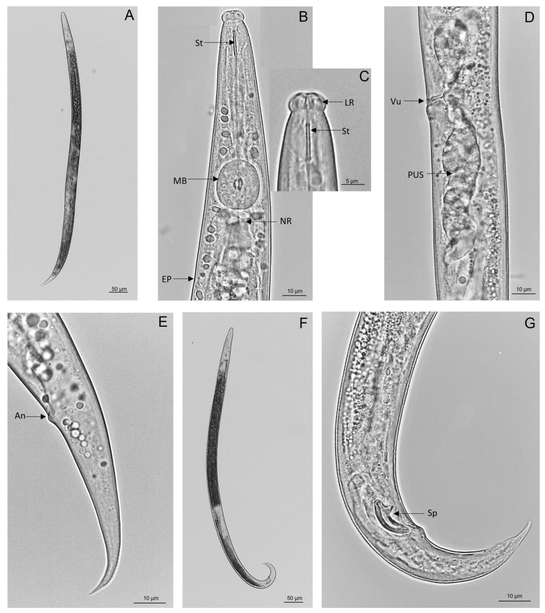
3.1. Morphological and Morphometric Characterisation

Adults (females and males) (Figure 1A,F) presented the main morphological diagnostic characters of P. stammeri [1,2,5,7,8,9]. Large nematodes with a thin body and cuticle; the rounded cephalic region was offset from the body by a noticeable constriction (Figure 1B,C). The stylet has a wide lumen with small basal swellings (Figure 1B,C), and the median bulb was rounded or slightly ellipsoid with large valve plates at the middle or slightly posteriorly (Figure 1B). The pharyngo-intestinal junction was just posterior to the median bulb base with the nerve ring encircling the anterior intestine and pharyngeal glands (Figure 1B). The excretory pore was posterior to the nerve ring (Figure 1B). The females (Figure 1A) had vulva, with simple lips and without flap, located usually at approximately 70% of the total body length, and the post-vulval uterine sac was well developed (Figure 1D). The tail was conoid and dorsally convex, with the distal region ventrally bent (Figure 1E). The males (Figure 1F) had paired spicules that were curved, and had rounded tips. The tail was conoid and strongly curved, with the terminus pointed occasionally, and it exhibited a mucron-like structure. The bursa was not present (Figure 1G).
The morphometrics of females and males were compared with other morphometric data for this species [2,7,8,9] (Table 1 and Table 2). The Portuguese P. stammeri isolate was then designated as PsPt1 and is maintained at the Laboratory of Nematology (NEMATO-lab), Department of Life Sciences, University of Coimbra, Portugal.

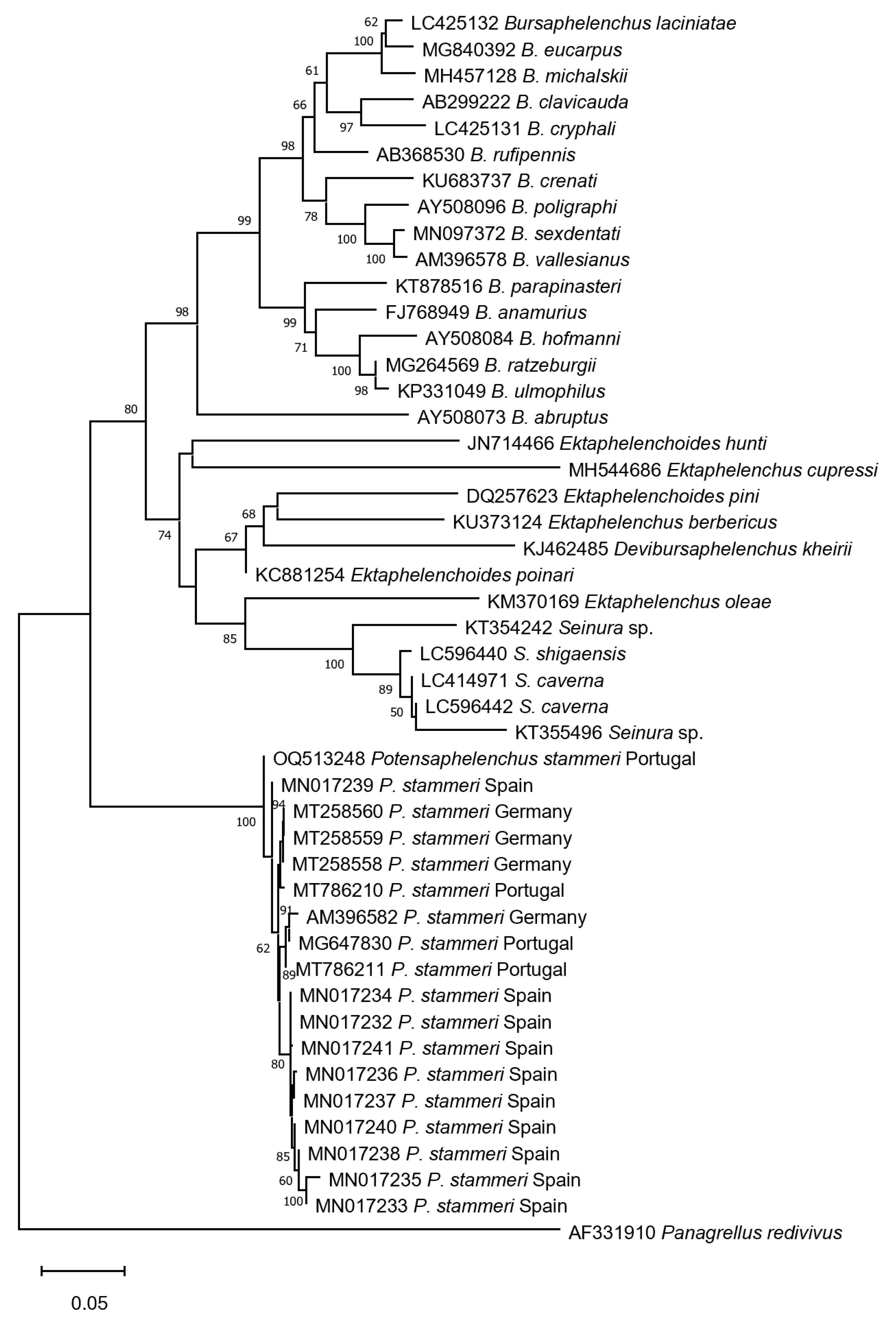
3.2. Molecular Identification

A 744 bp sequence corresponding to the PsPt1 D2-D3 LSU rDNA region was submitted to the NCBI database under accession number OQ513248. The top five BLAST hits using the PsPt1 sequence as the query are presented in Table 3. The phylogenetic analysis of this genomic region revealed that the PsPt1 isolate clusters together with other P. stammeri isolates, forming a separate clade from other closely related nematode species, namely those belonging to the Bursaphelenchus, Devibursaphelenchus Kakuliya, 1967; Ektaphelenchus Fuchs, 1937; Ektaphelenchoides Baujard, 1984; and Seinura Fuchs, 1931, genera (Figure 2).

4. Discussion

In the present study, the species Potensaphelenchus stammeri was found to be associated with Pinus pinea in Portugal, and its morphological and morphometric data were presented for the first time. The morphology of the Portuguese P. stammeri isolate (Figure 1) agrees with the morphological diagnostic characteristics reported by other authors for this species [1,2,5,7,8,9]. Most morphometric data of females and males (Table 1 and Table 2) are within the range of other P. stammeri isolates [2,7,8,9] apart from the total body length (similar to the Slovenian isolate), stylet, and a and c ratios, which are smaller. Some of these variations could be interpreted by intraspecific variations, as already documented for aphelenchoidids [17]. In this study, the D2-D3 rDNA sequence of the newly generated PsPt1 showed the highest best BLAST hit (99.6%) with a Spanish isolate of P. stammeri (Table 3). Phylogenetic analysis using the PsPt1 D2-D3 rDNA sequence showed that the PsPt1 isolate clusters together with other P. stammeri isolates, forming a clade with a clear separation of P. stammeri isolates from other species belonging to Bursaphelenchus, Devibursaphelenchus, Ektaphelenchoides, Ektaphelenchus, and Seinura genera, confirming the morphological and morphometric identification.

5. Conclusions

This study represents the first morphological and morphometric characterisation of a Potensaphelenchus stammeri isolate from a declining centennial Pinus pinea tree, in Portugal. Morphological and morphometric characterisations were compared with other P. stammeri isolates from Germany, China, Slovenia, Turkey, and Spain, and molecular identification was performed by LSU D2-D3 rDNA gene sequencing, revealing that the Portuguese P. stammeri isolate is clearly positioned in a clade with other P. stammeri isolates from different geographical origins.

Author Contributions

Conceptualization, L.F. and I.A.; methodology, H.S., J.M.S.C., R.M.F.d.C. and L.F.; writing—original draft preparation, H.S.; writing—review and editing, all authors; supervision, L.F.; project administration, L.F.; funding acquisition, I.A. and L.F. All authors have read and agreed to the published version of the manuscript.

Funding

This research was supported by the Portuguese Foundation for Science and Technology (FCT) via national funds and the co-funding provided by FEDER, PT2020, and COMPETE 2020 under projects POINTERS- PTDC/ASP-SIL/31999/2017 (POCI-01-145-FEDER-031999), PineWALL, PTDC/ASP-SIL/3142/2020, UIDB/04004/2020; and project ReNATURE—Valorization of the Natural Endogenous Resources of the Centro Region (Centro 2020, Centro-01-0145-FEDER-000007) and Instituto do Ambiente, Tecnologia e Vida.

Data Availability Statement

Genomic data presented in this study can be found in the NCBI database under accession number OQ513248.

Conflicts of Interest

The authors declare no conflict of interest. The funders had no role in the design of the study; in the collection, analyses, or interpretation of data; in the writing of the manuscript; or in the decision to publish the results.

References

  1. Körner, H. Die Nematoden fauna des vergehenden Holzes und ihre Beziehungen zu den Insekten. Zool. Jahrbücher Abt. Für Syst. Ökologie Und Geogr. Der Tiere 1954, 82, 245–353. [Google Scholar]
  2. Gu, J.; Liu, L.; Abolafia, J.; Pedram, M. A revision of the taxonomy of Aphelenchoides stammeri Körner, 1954 (Rhabditida: Aphelenchoididae) and proposal for a new genus. Nematology 2021, 23, 215–228. [Google Scholar] [CrossRef]
  3. Fuchs, G. Neue parasitich und halbparasitische Nematoden bei Borkenkäfern und einige andere Nematoden. Zool. Jb. 1937, 70, 291–442. [Google Scholar]
  4. Goodey, J.B. The classification of the Aphelenchoididae Fuch, 1937. Nematologica 1960, 5, 111–126. [Google Scholar] [CrossRef]
  5. Huang, R.E.; Ye, J.R. Seinura lii n. sp. and S. wuae n. sp. (Nematoda: Seinuridae) from pine wood in China. Nematology 2006, 8, 749–759. [Google Scholar] [CrossRef]
  6. Kanzaki, N.; Ekino, T.; Masuya, H. Seinura caverna n. sp. (Tylenchomorpha: Aphelenchoididae), na androdioecious species isolated from bat guano in a calcareous cave. Nematology 2019, 21, 207–225. [Google Scholar] [CrossRef]
  7. Braasch, H. Aphelenchoides stammeri Körner, 1954–ein in Deutschland weit verbreiteter Holznematode. Nachrichtenbl. Deut. Pflanzenschutzd. 1998, 50, 317–319. [Google Scholar]
  8. Urek, G.; Širca, S.; Geric, B. Morphometrical and molecular characterization of Bursaphelenchus species from Slovenia. Helminthologia 2007, 44, 37–42. [Google Scholar] [CrossRef]
  9. Dayi, M.; Uludamar, E.B.K.; Akbulut, S.; Elekcioğlu, I.H. First record of Aphelenchoides stammeri (Nematoda: Aphelenchoididae) from Turkey. J. Nematol. 2019, 51, e2019-70. [Google Scholar] [CrossRef] [PubMed]
  10. Whitehead, A.G.; Hemming, J.R. A comparison of some quantitative methods of extracting small vermiform nematodes from soil. Ann. Appl. Biol. 1965, 55, 25–38. [Google Scholar] [CrossRef]
  11. Fonseca, L.; Santos, M.C.V.; Santos, M.S.; Curtis, R.H.C.; Abrantes, I.M.O. Morpho-biometrical characterisation of Portuguese Bursaphelenchus xylophilus isolates with mucronate, digitate or round tailed females. Phytopathol. Mediterr. 2008, 47, 223–233. [Google Scholar]
  12. De Ley, P.; Felix, M.; Frisse, L.; Nadler, S.; Sternberg, P.; Thomas, W.K. Molecular and morphological characterisation of two reproductively isolated species with mirror-image anatomy (Nematoda: Cephalobidae). Nematology 1999, 1, 591–612. [Google Scholar] [CrossRef]
  13. Hall, T.A. BioEdit: A user-friendly biological sequence alignment editor and analysis program for Windows 95/98/NT. Nucleic Acids Symp. Ser. 1999, 41, 95–98. [Google Scholar]
  14. Altschul, S.F.; Madden, T.L.; Schaffer, A.A.; Zhang, J.H.; Zhang, Z.; Miller, W.; Lipman, D.J. Gapped BLAST and PSI-BLAST: A new generation of protein database search programs. Nucleic Acids Res. 1997, 25, 3389–3402. [Google Scholar] [CrossRef] [PubMed]
  15. Tamura, K.; Stecher, G.; Kumar, S. MEGA 11: Molecular Evolutionary Genetics Analysis Version 11. Mol. Biol. Evol. 2021, 7, 3022–3027. [Google Scholar] [CrossRef] [PubMed]
  16. Saitou, N.; Nei, M. The Neighbor-Joining method–A new method for reconstructing phylogenetic trees. Mol. Biol. Evol. 1987, 4, 406. [Google Scholar] [PubMed]
  17. Aliramaji, F.; Fouladvand, Z.M.; Pourjam, E.; Mortazavi, P.; Afshar, F.J.; Kanzaki, N.; Giblin-Davis, R.; Pedram, M. A new species of Basilaphelenchus Pedram, Kanzaki, Giblin-Davis & Pourjam, 2018 (Aphelenchoidea: Tylaphelenchinae), from natural forests of Golestan province, Iran. Nematology 2020, 22, 361–371. [Google Scholar]
Figure 1. Light microscopic photographs of nematodes killed with heat and mounted in water from the Portuguese Potensaphelenchus stammeri (Körner, 1954) Gu, Liu, Abolafia & Pedram, 2021 isolate (PsPt1). (A): female (entire body); (B): anterior region; (C): lip region and stylet; (D): part of female reproductive system; (E): female tail; (F): male (entire body); (G): male tail. St: stylet; MB: median bulb; NR: nerve ring; EP: excretory pore; LR: lip region; Vu: vulva; PUS: postuterine sac; An: anus.
Forests 14 00962 g001
Figure 2. Phylogenetic tree generated by the Neighbor-Joining method using the multiple sequence alignment of D2-D3 LSU rDNA sequences. Panagrellus redivivus (Linnaeus, 1767) Goodey, 1945 sequence (AF331910.2) was used as the outgroup. Bootstrap values are shown at branch points, and the scale bar indicates 0.050 substitutions per site.
Forests 14 00962 g002
Table 1. Morphometrics of females of Potensaphelenchus stammeri from Germany, China, Slovenia, Turkey, Spain, and Portugal (PsPt1). Values are mean ± SD. Values in parentheses indicate the minimum and maximum.
CharacterFemales
Braasch, 1998
(n = 6)
Huang and Ye, 2006
(n = 14)
Urek et al., 2007
(n = 5)
Dayi, 2019
(n = 10)
Gu et al., 2021
(n = 15)
This Study
(PsPt1) (n = 15)
Linear (µm)
Body length (L)833.0
(730.0–910.0)
885.0 ± 67.0
(763.0–1038.0)
777.7 ± 98.2
(673.9–905.3)
918.7 ± 46.2
(800.0–972.8)
973.0 ± 45.4
(912.0–1075.0)
768.8 ± 58.0
(690.0–856.4)
Greatest body width (GBW)--------------------28.9 ± 1.9
(25.8–32.4)
27.2 ± 1.6
(24.6–30.0)
Stylet length16.0
(15.0–18.0)
18.0 ± 0.8
(16.8–19.2)
15.2 ± 1.0
(14.1–16.5)
17.4 ± 0.4
(16.0–17.6)
17.1 ± 1.4
(14.0–19.3)
14.6 ± 0.3
(14.0–15.3)
Median bulb length--------------------20.8 ± 1.2
(18.8–23.0)
19.7 ± 1.3
(18.1–22.6)
Median bulb diameter--------------------14.8 ± 1.1
(13.5–16.7)
15.4 ± 0.8
(14.4–16.8)
Excretory pore to anterior end--------------------90.0 ± 4.5
(82.0–96.0)
95.1 ± 4.5
(89.5–103.8)
Anterior end to end of median bulb (AEMB)---------------80.8 ± 2.8
(76.8–84.8)
-----74.6 ± 4.0
(67.8–83.1)
Tail length (TL)-----57.0 ± 5.9
(52.0–69.0)
49.4 ± 5.3
(44.9–59.8)
63.8 ± 3.6
(56.0–68.8)
61.0 ± 4.2
(52.0–69.0)
58.0 ± 4.8
(49.9–65.0)
Body width at anus (BWA)--------------------16.0 ± 0.5
(15.1–16.8)
14.2 ± 0.8
(12.9–15.9)
Vulva to anus---------------230.0 ± 11.1
(214.4–240.0)
-----182.6 ± 15.0
(154.0–200.4)
Ratio
a = L/GBW32.0
(19.0–34.0)
40.0 ± 1.8
(36.4–42.2)
38.3 ± 2.7
(34.9–42.3)
31.9 ± 1.4
(30.4–33.0)
33.6 ± 1.6
(30.2–36.4)
28.2 ± 1.6
(26.1–31.5)
b1 = L/AEMB-------------------------10.3 ± 0.9
(8.7–12.4)
c = L/TL16.0
(13.0–17.0)
15.5 ± 2.3
(12.6–18.5)
15.7 ± 1.1
(14.5–17.8)
14.4 ± 1.2
(13.4–16.7)
16.2 ± 1.3
(14.1–18.9)
13.3 ± 1.4
(11.7–16.6)
c’ = TL/BWA-----4.3 ± 0.4
(3.8–5.1)
4.1 ± 0.4
(3.7–4.6)
4.0 ± 0.3
(3.3–4.5)
3.8 ± 0.3
(3.3–4.3)
4.1 ± 0.3
(3.5–4.5)
Percentage
V = Distance anterior end to vulva × 100/L69.0
(66.0–71.0)
68.7 ± 0.9
(66.7–69.8)
68.1 ± 0.6
(66.9–68.5)
67.8 ± 1.6
(67.0–72.0)
68.1 ± 0.7
(66.9–69.0)
68.8 ± 1.4
(66.9–71.7)
Table 2. Morphometrics of males of Potensaphelenchus stammeri from Germany, China, Slovenia, Turkey, Spain, and Portugal (PsPt1). Values are mean ± SD. Values in parentheses indicate the minimum and maximum.
CharacterMales
Braasch, 1998
(n = 6)
Huang and Ye, 2006
(n = 8)
Urek et al., 2007
(n = 5)
Dayi, 2019
(n = 10)
Gu et al., 2021
(n = 15)
This Study
(PsPt1) (n = 15)
Linear (µm)
Body length (L)853.0
(810.0–920.0)
857.0 ± 26.1
(831.0–908.0)
729.4 ± 53.8
(649.5–790.3)
851.8 ± 61.8
(776.0–976.0)
884.0 ± 44.8
(803.0–983.0)
739.2 ± 34.6
(680.1–788.6)
Greatest body width (GBW)-------------------25.1 ± 1.6
(22.9–27.6)
25.0 ± 1.8
(22.3–29.1)
Stylet length16.0
(15.0–18.0)
17.1 ± 0.6
(16.4–18.0)
14.7 ± 1.4
(13.2–16.6)
17.1 ± 0.6
(16.0–17.6)
16.6 ± 1.6
(14.1–18.8)
14.7 ± 0.4
(14.0–15.3)
Median bulb length--------------------19.7 ± 0.8
(18.3–21.3)
19.4 ± 1.2
(17.6–21.6)
Median bulb diameter--------------------13.5 ± 0.7
(12.5–14.9)
14.6 ± 1.2
(12.6–17.2)
Excretory pore to anterior end--------------------88.0 ± 5.6
(81.0–93.0)
100.5 ± 2.8
(96.3–103.6)
Anterior end to end of median bulb (AEMB)---------------77.6 ± 2.5
(72.0–81.6)
-----78.4 ± 4.7
(72.2–89.8)
Tail length (TL)-----43.0 ± 3.4
(42.0–50.0)
41.0 ± 2.4
(39.0–43.6)
40.0 ± 2.0
(38.4–43.2)
47.0 ± 8.4
(43.0–55.0)
47.1 ± 1.6
(44.7–50.3)
Body width at anus (BWA)--------------------16.9 ± 0.8
(15.5–18.7)
16.3 ± 1.2
(13.8–18.5)
Spicule length (curved median line)19.0
(18.0–20.0)
20.7 ± 0.5
(20.0–21.5)
20.2 ± 1.4
(18.1–21.6)
20.3 ± 3.2
(12.8–24.0)
17.3 ± 1.6
(15.3–21.0)
18.5 ± 0.9
(16.4–19.7)
Ratio
a = L/GBW31.0
(27.0–36.0)
42.0 ± 1.4
(40.4–44.0)
37.4 ± 3.5
(34.4–43.0)
33.5 ± 5.3
(21.0–40.6)
35.3 ± 1.7
(31.4–37.6)
29.7 ± 2.1
(25.5–33.6)
b1 = L/DAEMB-------------------------9.4 ± 0.4
(8.5–10.1)
c = L/TL18.0
(16.0–21.0)
20.0 ± 1.5
(18.4–21.5)
17.8 ± 1.4
(16.3–19.6)
21.1 ± 1.1
(19.4–22.5)
18.5 ± 0.8
(17.5–19.6)
15.7 ± 0.7
(14.5–17.6)
c’ = TL/BWA-----3.2 ± 0.3
(2.8–3.6)
3.2 ± 0.2
(3.1–3.5)
2.4 ± 0.1
(2.2–2.7)
2.9 ± 0.2
(2.6–3.3)
2.9 ± 0.2
(2.4–3.4)
Table 3. Top five BLAST hit sequences obtained by BLASTn analyses using the Potensaphelenchus stammeri (PsPt1) sequence as a query.
Species Query CoverE ValuePercentage IdentityAccession NumberCountry
Potensaphelenchus stammeri100%0.099.60%MN017239.1Spain
P. stammeri100%0.099.19%MN017236.1Spain
P. stammeri96%0.099.72%MT258558.1Germany
P. stammeri96%0.099.72%AM396582.1Germany
P. stammeri95%0.099.72%MG647830.1Portugal
Disclaimer/Publisher’s Note: The statements, opinions and data contained in all publications are solely those of the individual author(s) and contributor(s) and not of MDPI and/or the editor(s). MDPI and/or the editor(s) disclaim responsibility for any injury to people or property resulting from any ideas, methods, instructions or products referred to in the content.

