Article

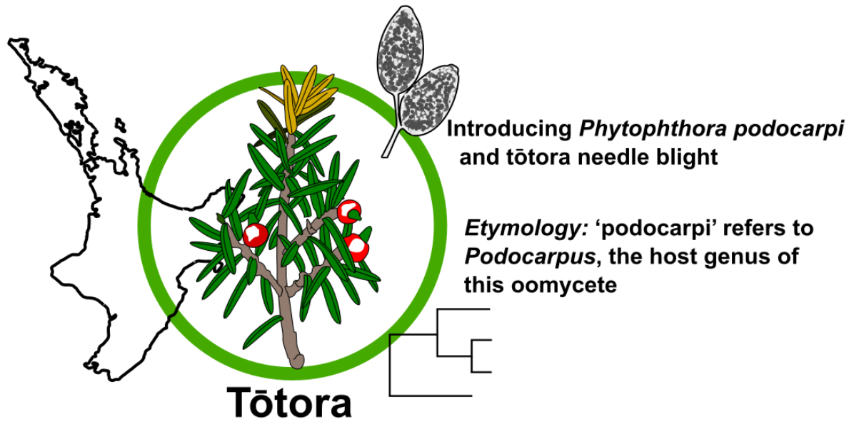
Phytophthora podocarpi sp. nov. from Diseased Needles and Shoots of Podocarpus in New Zealand

1
Scion (New Zealand Forest Research Institute Ltd.), Private Bag 3020, Rotorua 3010, New Zealand
2
Department of Primary Industries and Regional Development, Diagnostics and Laboratory Services, Sustainability and Biosecurity, 3 Baron-Hay Court, Kennsington, South Perth, WA 6151, Australia
3
The New Zealand Institute for Plant and Food Research Ltd., 74 Gerald Street, Lincoln 7608, New Zealand
*
Author to whom correspondence should be addressed.
Forests 2022, 13(2), 214; https://doi.org/10.3390/f13020214
Submission received: 23 December 2021 / Revised: 12 January 2022 / Accepted: 19 January 2022 / Published: 31 January 2022
(This article belongs to the Section Forest Health)

Abstract

:
Foliage samples from Podocarpus totara with severe needle browning and needle loss in the lower part of the crown were observed in 2011 in the Gisborne region of New Zealand. A Phytophthora genus-specific test applied directly to the needles gave a strong positive result, and subsequent isolations yielded colonies of a slow-growing oomycete. Morphological examination in vitro revealed a Phytophthora species. Preliminary comparisons of the rDNA (ITS), and ras-related protein (Ypt) gene regions with international DNA sequence revealed low sequence similarity to species from the downy mildew genus Peronospora, as well as clade 3 Phytophthora species. Other studies have also demonstrated the close relationship with Peronospora. The species was given the interim designation Phytophthora taxon tōtara pending further examination. Here, we formally describe Phytophthora podocarpi sp. Nov. and its associated disease, tōtara needle blight.

Graphical Abstract

1. Introduction

Foliage samples from Podocarpus totara with severe needle browning and needle loss in the lower part of the crown were examined in 2011. Affected tōtara trees were growing in the remnants of the native forest, remaining in steep gullies in a plantation of exotic Pinus radiata in the East Cape back-country of the North Island. Needle symptoms were not consistent with known foliage disorders of Podocarpus totara in New Zealand. A Phytophthora diagnostic test applied directly to symptomatic needles gave a strong positive result and subsequent isolations from needles and shoots yielded colonies of a slow-growing Phytophthora species. Subsequent genome sequencing indicated that it was phylogenetically distinct from other Phytophthora species at that time. It was given the interim designation Phytophthora taxon totara (PTT) [1]. The genome sequence was made publicly available on the NCBI database and was subsequently used in studies by McCarthy and Fitzpatrick [2] and Bourret, Choudhury [3]. Interestingly, these phylogenetic studies showed it had both (i) a close relationship to Phytophthora agathidicida [2], the causal agent of kauri (Agathis australis) dieback, which is another Phytophthora disease of a New Zealand native species, and (ii) strong phylogenetic links to the downy mildew genus Peronospora.
Further locations in Gisborne and Northland areas were recorded in 2011 and 2012. By 2018, PTT had also been recorded from the Bay of Plenty, Hawkes Bay, Taranaki, Taupo, Waikato, and Wairarapa (Figure 1). All records were from Podocarpus totara except for a single record from Podocarpus laetus in 2017.
The disease caused by PTT is now known as tōtara needle blight. The affected trees exhibit needle dieback in the lower crown. Infected needles initially turn khaki in colour then blacken and cast. Shoot infection causes the needles above the point of infection to brown and these needles are retained, giving the trees a fired, scorched appearance (Figure 2). Typically, symptoms are observed during winter and spring.
Podocarpus totara and Podocarpus laetus are endemic species in the Podocarpaceae. They are slow growing and reach up to 25 m. Podocarpus totara grows throughout the North Island and north-eastern South Island at elevations of up to 600 m, whilst Podocaprus laetus grows throughout the North and South Islands of New Zealand, including Stewart Island. The disease has not been extensive to-date; it has only affected a small number of trees in a stand and no mortality of trees has been observed. The disease is therefore not considered to have a high impact at this time. However, tōtara are considered an important tree species in the ngahere/forest and are of cultural importance to the Māori people, having been traditionally used for carvings, waka (traditional Māori canoe), and medicinal purposes. Its berries are also an important food source for native birds. Tōtara is also considered an important species for indigenous forestry initiatives and increasing planting of this species is with reforestation initiatives underway in New Zealand.
Forest biosecurity is critical, especially for protecting the biodiversity of standing native forests and new restoration areas, as well as the sustainability of forest plantations [4]. However, biosecurity relies on the use of species names and clear taxonomic classification [5], therefore species descriptions are important to provide the ability to identify pathogens with certainty, leading to more rapid responses to disease outbreaks. Phytophthora taxon totara is formally described here as a new species, Phytophthora podocarpi sp. nov.

2. Materials and Methods
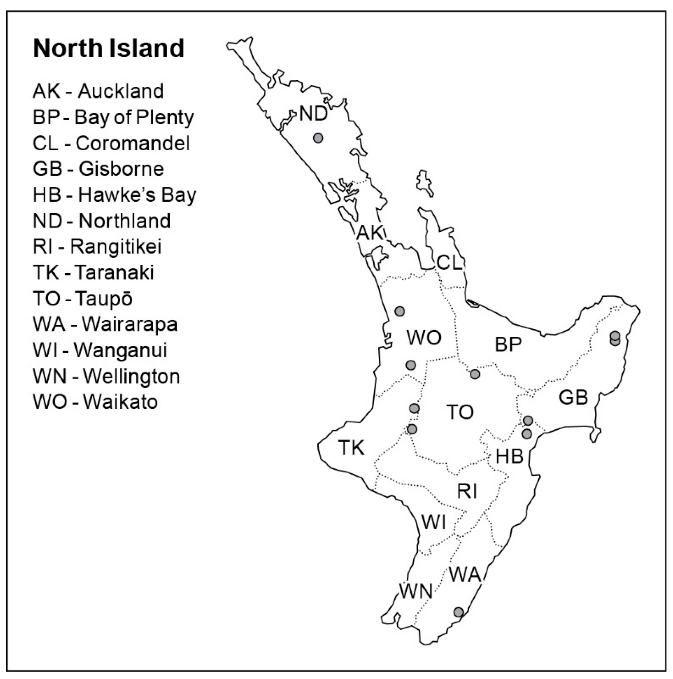
2.1. Sampling

Samples of Podocarpus from around New Zealand were received at the Forest Health Reference Laboratory (FHRL), Scion (Rotorua, New Zealand), as part of routine forest surveillance and from public enquires. Foliage samples were typically sealed in plastic bags and isolations were carried out within 24 to 72 h.

2.2. Detection and Isolation

Upon receipt in the FHRL, samples were assessed to determine if they had typical symptoms of tōtara needle blight, i.e., needle dieback in the lower crown or trees showing a fire scorched appearance and needles or shoots khaki or blackened in colour. Where symptoms were typical, a Phytophthora diagnostic test (Phytophthora ImmunoStrip®, Agdia, Inc., Elkhart, IN, USA, Cat. #92601) was used on symptomatic needle and shoot material. Isolations from symptomatic tissue were carried out by surface sterilising (30 s in 70% v/v ethanol, followed by 30 s in sterile distilled water; twice) 5–6 mm pieces of symptomatic needle and shoot material and plating onto Carrot Rifampicin and Nystatin (CRN) media (carrot agar prepared as described by Erwin and Ribeiro [6] and after autoclaving and cooling, amended with 4 mL of 2.5% ampicillin, 0.05 g nystatin dissolved in 1ml of 90% ethanol, 0.01 g of rifampicin dissolved in 1 mL of acetone and 0.4 mL of pimaricin) and incubated at 17 °C in the dark. Plates were regularly checked for the presence of PTT and any putative isolates were sub-cultured onto carrot agar (CA) [6] for morphological and molecular identification. Phytophthora taxon totara isolates were deposited into the NZFS culture collection (FHRL, Scion). A list of isolates used in this study is given in Table 1.

2.3. DNA Isolation, Amplification, and Sequencing

Cultures were grown on CA with cellophane at 20 °C for 14 days. Mycelium was scraped into lysing matrix A tubes and cell lysis solution CLS-Y was used. DNA was extracted using a FastDNA kit (MP Biomedicals, OH, USA) according to the manufacturers’ instructions and stored at −20 °C. The ITS and Ypt gene regions were amplified by PCR using the following primers: ITS4 and ITS6 primers [7] and Yph1F and Yph2R [8]. PCRs were performed using the KAPA2G Robust HotStart Readymix (2X) (KAPA Biosystems), according to the manufacturer’s instructions. Each 25 μL PCR reaction contained 7.75 μL of PCR grade water, 12.5 μL 2X KAPA2G Robust HotStart Readymix, 1.25 μL of each primer (10 μM), 1.25 μL DMSO, and 1 μL DNA (typically 5–10 ng genomic DNA). The cycling conditions consisted of an initial denaturation step of 95 °C for 1 min, then 35 cycles of 95 °C (15 s), annealing at primer-specific temperatures for (5 s), and 72 °C (15 s), and a final extension step of 72 °C (10 min). Gel electrophoresis was performed with 1.5% (wt/vol) agarose in TAE (120 V, 40 min), and stained with SYBR Safe® (ThermoFisher Scientific Inc., Waltham, MA, USA) for UV transillumination. PCR products were run on 1.5% (wt/vol) agarose in TAE (120 V, 40 min) and stained with SYBR Safe® (Life Technologies) for UV transillumination.
Where the Phytophthora Immunostrip test gave a positive result, from diagnostic plant samples, further molecular analysis on the test strip was performed to confirm the presence of PTT, using the method described by McDougal et al. [9], and the Yph1F and Yph2R primers [8], followed by DNA sequencing.
Before DNA sequencing, PCR products were treated with Exonuclease I (Exo) and Fast Alkaline Phosphatase (FastAP) (Fermentas, Thermo Fischer Scientific, Waltham, MA, USA) according to the manufacturers’ instructions. DNA sequencing was performed in both directions with forward and reverse PCR primers. DNA sequencing was performed on a 3500 Genetic Analyzer machine (Applied Biosystems), using BigDye® Terminator v3.1 Cycle Sequencing Kit (Applied Biosystems), according to the manufacturer’s instructions.

2.4. Phylogenetic Analysis

Multi-locus phylogenetic analysis was performed by concatenating corresponding DNA sequences from 12 genomes [1], using complete or partial DNA sequences from seven gene loci (28S rDNA, 60S ribosomal protein, β-tubulin, elongation factor 1-α, enolase, heat shock protein 90, and tigA gene fusion). The concatenated sequences were then compared to those for 87 related taxa representative of ten clades [10], using Phytopythium vexans P3980 as outgroup. Concatenated nucleotide sequences were aligned using ClustalW [11] in Geneious Pro v9.0 [12] under default settings and adjusted manually as needed. The best-fit evolutionary model (Supplementary Materials) for each of the seven loci (gene fragment) was identified using MrModeltest v2.0 [13] with the Akaike Information Criterion (AIC [14]), after creating partitions corresponding to each individual gene fragment. Because MrBayes cannot implement TIM3+I+G (transitional substitution model with gamma-distributed rate variation and a proportion of invariable sites) model examined by MrModeltest, the GTR+I+G model (general time reversible nucleotide substitution model with gamma-distributed rate variation and a proportion of invariable sites) was selected as the preferred model for each locus. Bayesian analysis was carried out with MrBayes v3.2 [15]; all model parameters were unlinked across partitions, and all partitions were allowed to have different rates. Partition-specific values for each prior were set to the MrModeltest estimates for each locus. A total of two independent Markov chain Monte Carlo (MCMC) runs were performed and each run was composed of four chains, three heated (temperature = 0.2) chains, and one cold chain, starting from random trees for 1,100,000 generations with trees sampled every 100 generations. The first 250 trees corresponding to a burn-in period were discarded and the last trees after convergence were used to construct a majority-rule consensus tree and to summarize posterior probability support for each node. All clades with the exception of 3, 5, and 15 were hidden using the drop.tip function in the ggtree package in R [16].

2.5. Pathogenicity Studies

A total of six isolates were used for pathogenicity testing and are listed in Table 1. Healthy twig material was collected from trees more than 10 years old. A single detached young stem of Podocarpus totara, between 20 and 30 cm long and 3 to 5 mm in diameter, was selected for each pathogen treatment. A total of three needles on each stem were selected for inoculation and a fourth needle was used as a control. A thin layer of tissue was cut with a scalpel (<1 mm deep and 5 mm long) along the adaxial surface of each needle around the midway point of the needle. A scraping of mycelium was placed under the flap on the resulting wound. The flap of tissue was then replaced. Control needles were wounded in the same way but were not inoculated. The stems were then placed into tubes with sterile distilled water and the neck of the tube was sealed with plastic wrap to prevent moisture loss. Tubes were placed in racks within a plastic container with wet paper towels on the bottom, the container was sealed with plastic wrap to further prevent moisture loss. The treated stems were incubated at 22 °C in diurnal light and examined at 7 and 14 days. Where lesions developed, 5 mm square sections of tissue were cut from the dead–live margin, and plated directly on to CRN. Plates were regularly checked for the presence of PTT and any suspect isolates were sub-cultured onto CA for identification.

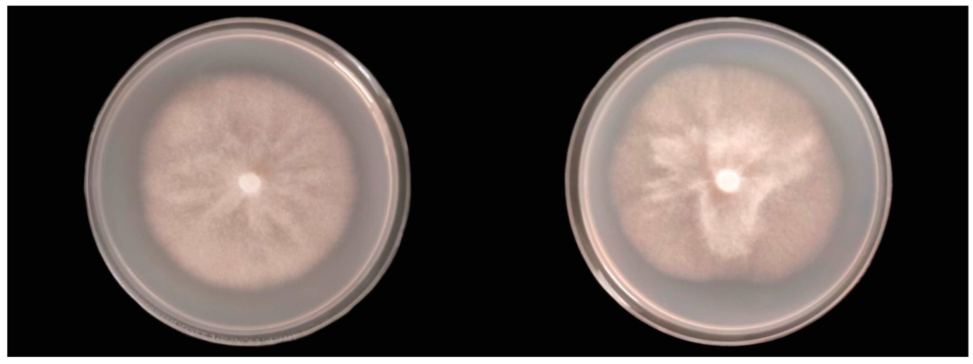
2.6. Colony Morphology, Growth Rates, and Cardinal Temperatures

Colony morphology was assessed on CA after 28 days in the dark and under continuous daylight (Panasonic lamp FL15DF, 15 watt) at 17 °C. Maximum, optimum, and minimum temperatures for growth were assessed on 10% V8 juice agar (as described by Miller [17]), after seven days, in the dark at 2, 8, 10, 15, 17.5, 20, 22.5, 25, and 30 °C. Growth rate was assessed on CA after seven days in the dark at 17 °C. Optimum temperature and growth rate were assessed by placing a 5 mm diam. Inoculum plug from the edge of an actively growing culture in the centre of the media plate, two colony diameters were measured at right angles, of three representative plates per isolate, at seven days.

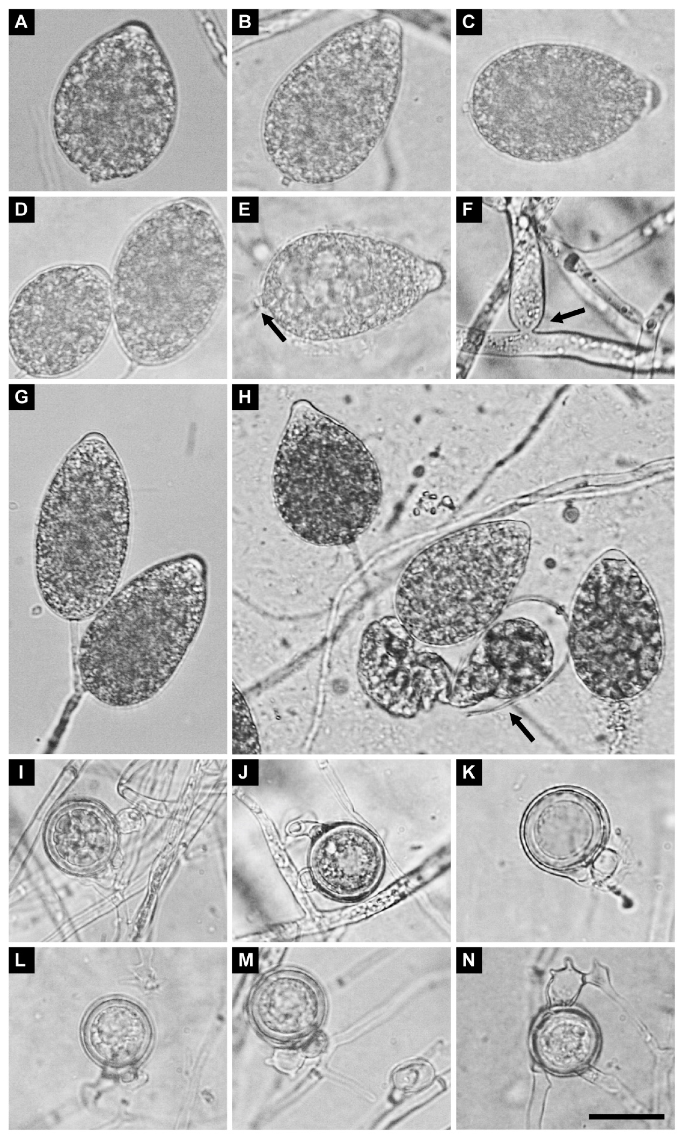
2.7. Morphology of Sexual and Asexual Structures

To produce oogonia, cultures were grown on CA and incubated at 17 °C in the dark for four to eight weeks. Measurements were taken between four and eight weeks; dependent on production in each isolate. Due to the scarcity of oogonia in some isolates only 10 or fewer oogonia could be measured. Oogonia were observed by scanning the under surface of the agar plate at 20× magnification. When oogonia were located, that section of medium was removed, and the under surface scraped with a scalpel to collect the oogonia. Oogonia, oospores, oospore wall, and antheridia were measured. To produce sporangia, 5 mm squares from the edge of an actively growing colony on CA were transferred to unsterile pond water and incubated at 22 °C under diurnal light. After 24 to 36 h, the length, breadth, pore width, and papilla depth of 50 sporangia were measured.

3. Results

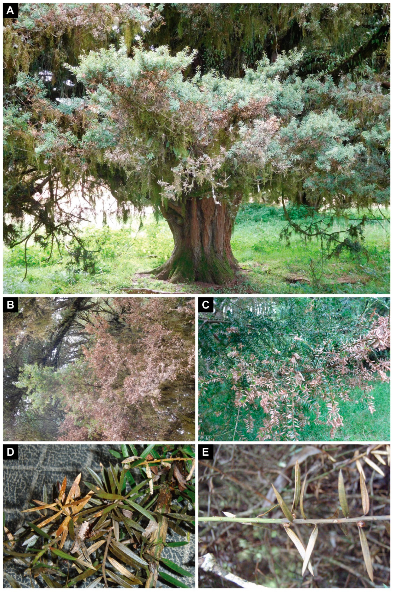
3.1. Sampling and Isolation

The FHRL has received tōtara needle blight samples infrequently, with gaps of two years between 2012 and 2014, and almost three years between 2014 and 2017. Despite concerted efforts to find the disease in the past two years, the last recorded detection was on November 2017. Further to the typical symptoms on needles and shoots, dark bands were occasionally observed on the Podocarpus totara needles, as seen with infection of other conifers with Phytophthora species [18]. Although tōtara blight is scattered around much of the North Island, there are only isolated instances and there is no geographical or climatic link between the records (Figure 1). Blastn analysis of the ITS and Ypt1 gene regions revealed sequence similarity (92.5–92.9% for ITS and 83–86% for Ypt1) to species from the downy mildew genus Peronospora, as well as clade 3 Phytophthora species such as P. pseudosyringae, P. lilics, and P. pluvialis. To date, tōtara blight has not been found in the South Island of New Zealand.

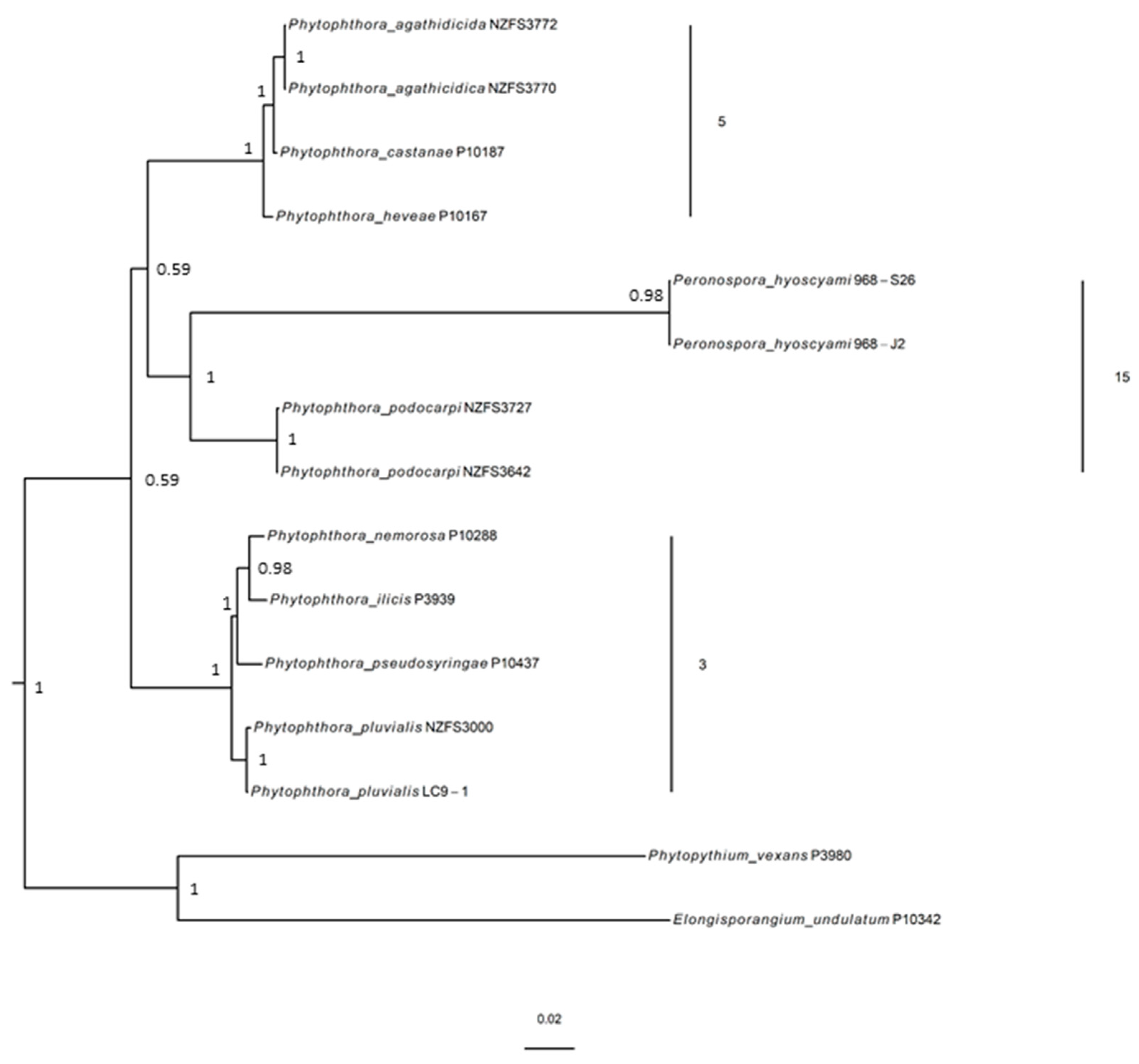
3.2. Molecular Phylogenetic Position

Alignment of seven gene regions with closely related and published Phytophthora Genbank accessions and bayesian phylogenetic analysis confirm that PTT is a new species, supported by strong bayesian support value (Figure 3). The results of the phylogenetic analysis also inferred that PTT is a sister species to Peronospora hyoscyami and differs from Peronospora hyoscyami by over 900 nucleotides in the concatenated DNA sequence alignment of the 28S rDNA, 60S ribosomal protein, β-tubulin, elongation factor 1-α, enolase, heat shock protein 90, and tigA gene regions (Figure 3). The multilocus phylogenetic analysis also showed that PTT is placed adjacent to clade 5 and is closely related but phylogenetically distinct to Phytophthora agathidicida, a Phytophthora species that infects kauri. The results are congruent with a previous study by McCarthy and Fitzpatrick [2], where PTT was considered to be most closely related to P. agathidicida based on species phylogenomic analysis, and not ITS clade 3 as previously suggested from individual gene analyses [1]. More recently, PTT has been placed in a strongly supported, newly assigned clade (clade 15) with downy mildews (graminicolous, brassicolous, and downy mildews with coloured conidia) including Peronospora hyoscyami (previously Peronospora tabacina) [3].

3.3. Pathogenicity

Of the six isolates tested, all produced lesions on some or all of the inoculated leaves after seven days. Lesions were typical of those seen on infected material from field samples, with needles showing khaki colouring and browning. Of the six PTT isolates, three were recovered from lesions on inoculated needles and recovery of the original Phytophthora isolate was confirmed by cultural morphology. No lesions were seen on the control needles.

3.4. Taxonomy

Phytophthora podocarpi K. Dobbie, R.L. McDougal & P.M. Scott sp. nov.
= Phytophthora taxon totara [1,2,3] nom. inval.
Mycobank no. 841407
Etymology:podocarpi’ refers to Podocarpus, the host genus of this oomycete.
Type: NEW ZEALAND: Gisborne: Hampton Forest, from needles of Podocarpus totara, July 2011, Collected by B. J. Rogan, holotype NZFRIM 5991 (dried culture on CA, Scion, Rotorua) ex holotype living culture permanently preserved in a metabolically inactive state as ICMP 24118 = NZFS 3642. Genome sequence GenBank LGSN00000000.
Homothallic, gametangia is rare to absent after 8 weeks and only developed on CA. Oogonia are globose and smooth-walled with a range of mean diam. of 25–30 µm (mean diam. 27.4 µm ± 1.0) with a common range of 20–33 µm. Oogonial stalks are often globose, occasionally with digitate projections. Antheridia areapparently absent, and when present, paragynous and single, mostly globose to club shaped, with a range of means 5–10 × 6–10 µm and a common range of 5–15 × 4–10 µm. Oospores are plerotic with a diameter range of 22–27 µm (mean diam. 24.8 µm ± 1.0) and a common range of 18–30 µm; it has a wall thickness average of 2 µm and a common range of 1–3 µm.
Sporangia borne on simple or compound sympodial sporangiophores are often clustered and form readily within CA, and are less abundant in the aerial mycelium. Sporangia produced from filtered pond water after 72 h are ovoid, obpyriform to limoniform; non-papillate to semi-papillate; and persistent and caducous with short length (1–4 µm) pedicels. Sporangia’s range of means are 47.6–64.5 × 31.3–41.1 µm (mean length 55.7 µm ± 12.2 and breath 37.0 µm ± 8.2) and it has a common range 22–79 × 15–54 µm. Its length to breadth ratio average is 1.5. No internal or external proliferation was observed.
The mycelium on CA has a thickness that is highly variable, from 5–12 µm. It is often prominently constricted at branching, with a resulting swollen wide hypha from the constriction. A few swellings and knot type structures were observed, and no chlamydospores were observed on CA.
Colonies in the dark at 17 °C after 28 days experienced slow growth, and were lightly felted with sparse aerial mycelium, loosely striate, and even radial growth. Under continuous light at 17 °C after 28 days, they experienced slow growth, were lightly felted with an aerial mycelium which was denser in the centre, becoming sparse towards the growing margin, as well as being distinctly striate, and even experiencing radial growth (Figure 5). The optimum growth rate was 17 °C. There was no growth or minimal growth at 2 and 25 °C. At 17 °C in darkness, a radial growth of ca 1.0–1.2 mm d−1 (mean 1.1 mm d−1) occurred.
Phytophthora podocarpi differs morphologically from other species of Phytophthora with non- to semi-papillate sporangia that are persistent and caducous, with homothallic gametangia and paragynous antheridia. The four Phytophthora species that have these same characters are P. brassicae, P. foliorum, P. pseudosyringae, and P. siskiyouensis. Phytophthora brassicae sits in clade 8 [3]. It differs from P. podocarpi in that the antheridia are predominately amphigynous, it has a higher optimum and maximum growth temp at 21 °C and 27 °C, respectively, and has a faster radial growth rate of 6.4 mm d−1 [19]. Phytophthora foliorum is also part of clade 8 [3]. Of the four Phytophthora species discussed here, it is morphologically most similar to P. podocarpi. It differs by the fact that oogonia are produced abundantly on media, it has a higher maximum growth temperature of 28 °C and a slightly faster radial growth rate of 3.0 mm d−1 [20]. Phytophthora pseudosyringae is in clade 3 [3]. It differs from P. podocarpi in having distinctive hyphal swellings in a catenulate arrangement and having a faster radial growth rate of 5 mm d−1 [21]. Phytophthora siskiyouensis sits in clade 2 [3]. This species differs from P. podocarpi in the position of the semi-papillae being apical, subapical, or lateral, the abundant production of oogonia, it has a higher optimum and maximum growth temp at 25 °C and 30 °C, respectively, and a faster radial growth rate of 7.5 mm d−1 [22]. Phylogenetically, P. podocarpi is a sister species to Peronospora hyoscyami. Although these two species are related phylogenetically, morphologically they are distinct. Peronospora hyoscyami has typical downy mildew traits, which include being an obligate biotroph, with determinate, distinct, dichotomously branched sporangiophores, confluent oogonial and oospore walls, and conidia and sporangia which germinate directly to give hyphae [23]. Germination via zoospores has not been confirmed [24]. Some traits that are shared between the two species include sporangia that are non-papillate, a small pedicle that remains after secession, and oogonia with ‘paragynous antheridia’, however the differences far outweigh the similarities from a morphological perspective.

4. Discussion

Symptomatic material was received occasionally at the FHRL, often with two or more years between sightings. This is despite concerted efforts over the past two years to raise awareness of the disease through an online fact sheet and a short note in an industry magazine describing the disease and asking people to record potential sightings and sample. It has now been four years since tōtara needle blight has been reported via FHRL. Despite the infrequent incidence of disease, the importance of describing and naming new species is critical for biosecurity. Phytophthora podocarpi infects a keystone species in New Zealand’s native forests and a species that is utilised for timber and carving.
Laboratory-based inoculations were performed on excised Podocarpus totara twigs to confirm pathogenicity on this host and to demonstrate Koch’s postulates. Although this experiment was limited in replicates, it showed that the lesions typically observed in the field on fully grown trees can be replicated by inoculation with P. podocarpi. Recovery rates of 50% or less (from plant material) are not unusual for Phytophthora species [18]. These results fulfil Koch’s postulates implicating P. podocarpi as the causal agent of tōtara needle blight.
The position of P. podocarpi in some phylogenetic trees has created discussion about the evolutionary relationships it has within the Peronosporaceae. Numerous studies are providing us with a better understanding of the phylogenetics of the Peronosporaceae, and the fact the genus Phytophthora is monophyletic and includes the downy mildews has been well established [2,3,25,26,27,28]. Studholme, McDougal [1] conducted genome sequencing for two isolates of each of six species of Phytophthora selected for their pathogenicity to forest trees, both native and exotic plantation species, in New Zealand. Phytophthora podocarpi appeared to be phylogenetically distinct among the Phytophthora species sequenced. They did however find some sequence similarity in the ITS region with members of clade 3 Phytophthora [27]. McCarthy and Fitzpatrick [2] suggested that downy mildew species, including P. podocarpi, are sister taxa with maximum support in both matrix representation using parsimony and supermatrix analyses. It was concluded that clade 3 is not monophyletic in any of the species’ phylogenies and P. podocarpi is not clade 3 species. From these studies it is apparent that classification of Phytophthora species is difficult when using a single gene region, such as ITS. To understand the evolutionary relationships between the genus Phytophthora and the downy mildews, Bourret, Choudhury [3] investigated phylogenetic relationships between 13 downy mildew and 103 Phytophthora species using two nuclear and four mitochondrial loci. A total of fifteen clades were proposed where P. podocarpi fell into clade 15, along with three of four groups of the downy mildews. They concluded that the close and well supported phylogenetic relationship supported the theory that the downy mildews evolved from a foliar Phytophthora ancestor. Phytophthora podocarpi shares a unique set of morphological and cultural characteristics within the genus Phytophthora; along with unique genomic DNA, it aligns with the downy mildews, within the genus Phytophthora. This leads to us speculate that P. podocarpi could be the foliar Phytophthora ancestor giving rise to the downy mildews.
Downy mildews are obligate biotrophs that occur on aerial plant parts and are usually highly host specific. They share a loose set of morphological characters including determinate, distinct, dichotomously branched sporangiophores (where at the tips of each branch, sporangia enlarge and mature simultaneously); conidia and sporangia that germinate directly, forming hyphae, or indirectly, forming zoospores; confluent oogonial and oospore walls; and oospores germinating via short germ tubes, which occurs infrequently in several species [6,29,30,31,32]. Morphological characters of downy mildews and Phytophthora can be shared by a single species, which confounds taxonomy in some instances. For example, Peronophythora litchi, now accepted as Phytophthora litchi, is an important plant pathogen, causing litchi (Litchi chinensis) downy blight. Discussions as to its placement in the downy mildews have spanned almost four decades. Morphological characters placed it between the downy mildew genus Peronospora and Phytophthora. The fact that it could be cultured, the mode of oospore germination and the presence of both determinate and indeterminate sporangiophores, gave rise to a new downy mildew genus, Peronophythora [31]. Subsequent molecular analysis revealed that it sat squarely within the genus Phytophthora and was renamed by Göker, et al. [32]. Conversely, Sclerophthora macrospora is an obligate parasite with a wide host range, it has determinate sporangiophores, however the sporangiophores cannot be distinguished from mycelium (are micronemous), which is consistent with Phytophthora. It was found that the oogonial and oospore wall was not confluent but could be separated as one would expect for Phytophthora with plerotic oospores [6]. May and Ristaino [33] transferred S. macrospora to Phytophthora, however the name was met with some resistance and has not been accepted.
How does P. podocarpi fit in the midst of the Phytophthora and downy mildew debate? Phylogenetically, it clearly aligns more closely with the downy mildew genera than it does with any species of Phytophthora. The only other traits that align it with the downy mildews are that it has not been associated with other host genera and it is potentially host specific and infects aerial parts of the plant, both of which are traits of some Phytophthora species. It is not an obligate biotroph; it has indeterminate, micronematous sporangiophores, and plerotic oospores. For these reasons, it is retained as a Phytophthora species.

5. Conclusions

Climate change, as seen with changing weather patterns and increasing drought conditions, is impacting not only the growth of trees but susceptibility to disease [34,35]. Combined with current anthropogenic activities, tree diseases are likely to continue to increase [36]. It will be important to understand the genetic diversity, to begin to elucidate the origins of this pathogen. Previous studies have used genomic analyses to establish if Phytophthora species are either introduced [37,38] or endemic to New Zealand [39]. Native pathogens do not typically cause significant disease in native forests, but disturbance of these systems and the ecology within, for example with forestry operations, can result in disease outbreaks [4]. Modification of habitats by destruction of understory plants by introduced browsing mammals is also having a profound effect on forest structure. In addition, Phytophthora species, which have been recognised as significant plant pathogens since the 1800s, are only recently becoming recognised as significant foliar pathogens, having previously been recognised as primarily soil-inhabiting, root and stem infecting pathogens. This means that Phytophthora diseases on foliage may have gone unnoticed for a prolonged period. These factors combined may indicate that this pathogen has been present, but unnoticed for some period, and that land management practices, continued changes in habitat, and changes in weather patterns may have resulted in the emergence of this pathogen in localised pockets in the North Island of New Zealand.

Supplementary Materials

The following are available online at https://www.mdpi.com/article/10.3390/f13020214/s1, Figure S1: A multilocus phylogeny of Phytophthora showing the placement of the two P. podocarpi isolates in clade 15 along with downy mildew, Peronospora hyoscyami (Peronospora tabacina). Complete or partial DNA sequences from seven gene loci (28S rDNA, 60S ribosomal protein, β-tubulin, elongation factor 1-α, enolase, heat shock protein 90, tigA gene fusion) were used to infer phylogeny with MrBayes in Geneious Pro v9.0. Support is displayed as posterior probability values on the nodes. Scale bar shows number of substitutions per site. Table S1: Nucleotide substitution model selection per gene and partitions selected by AIC (MrModeltest).

Author Contributions

Conceptualization, K.D., R.M., P.S. and M.D.; methodology, K.D., R.M., P.S., P.P., M.D. and D.S.; formal analysis, K.D., R.M., P.S., P.T., P.P. and M.D.; investigation, K.D., R.M., P.S., P.T., P.P. and M.D.; resources, R.M. and K.D.; data curation K.D., R.M., P.S., P.T., P.P. and D.S.; writing—original draft preparation, K.D. and R.M.; writing—review and editing, K.D., R.M., P.S., P.T., P.P., D.S. and M.D.; visualization, K.D., P.P. and D.S.; project administration, R.M.; funding acquisition, K.D. and R.M. All authors have read and agreed to the published version of the manuscript.

Funding

The New Zealand Forest Biosecurity Surveillance Programme, The Forest Growers levy Trust, The High-Risk Site Surveillance Program (Ministry for Primary Industries) and Scion (Strategic Science Investment Funding from the New Zealand Ministry of Business, Innovation and Employment; contract C04X1703) are gratefully acknowledged for funding this project.

Acknowledgments

David Studholme and Christine Sambles (University of Exeter) are thanked for early phylogenomic analyses. Anna Caird, Tomoko Pearson, Rita Tetenburg, and Judy Gardner are thanked for technical support. Ian Hood is thanked for reviewing the manuscript and providing advice on nomenclature. Dale Corbett is thanked for assistance with graphics and Darryl Herron is thanked for producing the graphical abstract. Toni Withers, Te Whaeoranga Smallman, and Wharehuia Evans are thanked for project support. The authors would like to thank SPS Biosecurity, Councils, and landowners who have supplied diagnostic samples and access to land for viewing the disease. The authors would like to acknowledge Brent Rogan, (SPS Biosecurity, NZ) for sampling the first known record of this disease in 2011.

Conflicts of Interest

The authors declare no conflict of interest.

References

  1. Studholme, D.; McDougal, R.; Sambles, C.; Hansen, E.; Hardy, G.; Grant, M.; Ganley, R.; Williams, N. Genome sequences of six Phytophthora species associated with forests in New Zealand. Genom. Data 2016, 7, 54–56. [Google Scholar] [CrossRef] [PubMed] [Green Version]
  2. McCarthy, C.G.; Fitzpatrick, D.A. Phylogenomic reconstruction of the oomycete phylogeny derived from 37 genomes. Msphere 2017, 2, e00095-17. [Google Scholar] [CrossRef] [PubMed] [Green Version]
  3. Bourret, T.B.; Choudhury, R.A.; Mehl, H.K.; Blomquist, C.L.; McRoberts, N.; Rizzo, D.M. Multiple origins of downy mildews and mito-nuclear discordance within the paraphyletic genus Phytophthora. PLoS ONE 2018, 13, e0192502. [Google Scholar] [CrossRef] [PubMed] [Green Version]
  4. Burgess, T.I.; Wingfield, M.J. Pathogens on the Move: A 100-Year Global Experiment with Planted Eucalypts. Bioscience 2016, 67, 14–25. [Google Scholar] [CrossRef]
  5. McTaggart, A.R.; van der Nest, M.A.; Steenkamp, E.T.; Roux, J.; Slippers, B.; Shuey, L.S.; Wingfield, M.J.; Drenth, A. Fungal Genomics Challenges the Dogma of Name-Based Biosecurity. PLoS Pathog. 2016, 12, e1005475. [Google Scholar] [CrossRef] [Green Version]
  6. Erwin, D.C.; Ribeiro, O.K. Phytophthora Diseases Worldwide; APS Press: St. Paul, MN, USA, 1996. [Google Scholar]
  7. Cooke, D.E.; Drenth, A.; Duncan, J.M.; Wagels, G.; Brasier, C.M. A molecular phylogeny of Phytophthora and related oomycetes. Fungal Genet. Biol. 2000, 30, 17–32. [Google Scholar] [CrossRef]
  8. Schena, L.; Duncan, J.M.; Cooke, D.E.L. Development and application of a PCR-based ‘molecular tool box’ for the identification of Phytophthora species damaging forests and natural ecosystems. Plant Pathol. 2008, 57, 64–75. [Google Scholar] [CrossRef]
  9. McDougal, R.L.; Cunningham, L.; Hunter, S.; Caird, A.; Flint, H.; Lewis, A.; Ganley, R.J. Molecular detection of Phytophthora pluvialis, the causal agent of red needle cast in Pinus radiata. J. Microbiol. Methods 2021, 189, 106299. [Google Scholar] [CrossRef]
  10. Blair, J.E.; Coffey, M.D.; Park, S.-Y.; Geiser, D.M.; Kang, S. A multi-locus phylogeny for Phytophthora utilizing markers derived from complete genome sequences. Fungal Genet. Biol. 2008, 45, 266–277. [Google Scholar] [CrossRef]
  11. Thompson, J.D.; Higgins, D.G.; Gibson, T.J. CLUSTAL W: Improving the sensitivity of progressive multiple sequence alignment through sequence weighting, position-specific gap penalties and weight matrix choice. Nucleic Acids Res. 1994, 22, 4673–4680. [Google Scholar] [CrossRef] [Green Version]
  12. Kearse, M.; Moir, R.; Wilson, A.; Stones-Havas, S.; Cheung, M.; Sturrock, S.; Buxton, S.; Cooper, A.; Markowitz, S.; Duran, C.; et al. Geneious Basic: An integrated and extendable desktop software platform for the organization and analysis of sequence data. Bioinformatics 2012, 28, 1647–1649. [Google Scholar] [CrossRef] [PubMed]
  13. Nylander, J. MrModeltest v2; Uppsala University, Evolutionary Biology Centre: Uppsala, Sweden, 2004. [Google Scholar]
  14. Akaike, H. A new look at the statistical model identification. IEEE Trans. Autom. Control. 1974, 19, 716–723. [Google Scholar] [CrossRef]
  15. Ronquist, F.; Teslenko, M.; van der Mark, P.; Ayres, D.L.; Darling, A.; Höhna, S.; Larget, B.; Liu, L.; Suchard, M.A.; Huelsenbeck, J.P. MrBayes 3.2: Efficient bayesian phylogenetic inference and model choice across a large model space. Syst. Biol. 2012, 61, 539–542. [Google Scholar] [CrossRef] [PubMed] [Green Version]
  16. Yu, G. Using ggtree to visualize data on tree-like structures. Curr. Protoc. Bioinform. 2020, 69, e96. [Google Scholar] [CrossRef]
  17. Miller, P.M. V-8 juice agar as a general-purpose medium for fungal and bacteria. Phytopathology 1955, 45, 461–462. [Google Scholar]
  18. Dick, M.A.; Williams, N.M.; Bader, M.K.-F.; Gardner, J.F.; Bulman, L.S. Pathogenicity of Phytophthora pluvialis on Pinus radiata and its relation with red needle cast disease in New Zealand. N. Z. J. For. Sci. 2014, 44, 6. [Google Scholar] [CrossRef] [Green Version]
  19. De Cock, A.W.; Ilieva, E.; Lévesque, C.A. Gene flow analysis of Phytophthora porri reveals a new species: Phytophthora brassicae sp. nov. Eur. J. Plant Pathol. 2002, 108, 51–62. [Google Scholar]
  20. Donahoo, R.; Blomquist, C.L.; Thomas, S.L.; Moulton, J.K.; Cooke, D.E.; Lamour, K.H. Phytophthora foliorum sp. nov., a new species causing leaf blight of azalea. Mycol. Res. 2006, 110, 1309–1322. [Google Scholar] [CrossRef]
  21. Thomas, J.; Nechwatal, J.; Cooke, D.E.; Hartmann, G.; Blaschke, M.; OßWALD, W.F.; Duncan, J.M.; Delatour, C. Phytophthora pseudosyringae sp. nov., a new species causing root and collar rot of deciduous tree species in Europe. Mycol. Res. 2003, 107, 772–789. [Google Scholar]
  22. Reeser, P.W.; Hansen, E.M.; Sutton, W. Phytophthora siskiyouensis, a new species from soil, water, myrtlewood (Umbellularia californica) and tanoak (Lithocarpus densiflorus) in southwestern Oregon. Mycologia 2007, 99, 639–643. [Google Scholar] [CrossRef]
  23. Hall, G. Peronospora hyoscyami f. sp. tabacina. Mycopathologia 1989, 106, 191–193. [Google Scholar]
  24. Thines, M.; Choi, Y.-J. Evolution, diversity, and taxonomy of the Peronosporaceae, with focus on the genus Peronospora. Phytopathology 2016, 106, 6–18. [Google Scholar] [CrossRef] [PubMed] [Green Version]
  25. Jung, T.; Scanu, B.; Bakonyi, J.; Seress, D.; Kovács, G.; Durán, A.; Von Stowasser, E.S.; Schena, L.; Mosca, S.; Thu, P. Nothophytophthora gen. nov., a new sister genus of Phytophthora from natural and semi-natural ecosystems. Pers. Mol. Phylogeny Evol. Fungi 2017, 39, 143. [Google Scholar] [CrossRef] [PubMed] [Green Version]
  26. Sun, J.; Gao, Z.; Zhang, X.; Zou, X.; Cao, L.; Wang, J. Transcriptome analysis of Phytophthora litchii reveals pathogenicity arsenals and confirms taxonomic status. PLoS ONE 2017, 12, e0178245. [Google Scholar] [CrossRef] [PubMed] [Green Version]
  27. Ye, W.; Wang, Y.; Shen, D.; Li, D.; Pu, T.; Jiang, Z.; Zhang, Z.; Zheng, X.; Tyler, B.M.; Wang, Y. Sequencing of the litchi downy blight pathogen reveals it is a Phytophthora species with downy mildew-like characteristics. Mol. Plant-Microbe Interact. 2016, 29, 573–583. [Google Scholar] [CrossRef] [Green Version]
  28. Ann, P.J.; Ko, W. Oospore germination of Peronophythora litchii. Mycologia 1980, 72, 611–614. [Google Scholar] [CrossRef]
  29. Ho, H.; Jiayun, L.; Longyin, G. Observations on asexual reproduction by Peronophythora litchii. Mycologia 1984, 76, 745–747. [Google Scholar] [CrossRef]
  30. Ko, W.; Chang, H.; Su, H.; Chen, C.; Leu, L. Peronophythoraceae, a new family of Peronosporales. Mycologia 1978, 70, 380–384. [Google Scholar] [CrossRef]
  31. Michelmore, R.W.; Ilott, T.; Hulbert, S.; Farrara, B. The downy mildews. In Advances in Plant Pathology; Elsevier: Amsterdam, The Netherlands, 1988; Volume 6, pp. 53–79. [Google Scholar]
  32. Göker, M.; Voglmayr, H.; Riethmüller, A.; Oberwinkler, F. How do obligate parasites evolve? A multi-gene phylogenetic analysis of downy mildews. Fungal Genet. Biol. 2007, 44, 105–122. [Google Scholar] [CrossRef]
  33. May, K.J.; Ristaino, J.B. Identity of the mtDNA haplotype(s) of Phytophthora infestans in historical specimens from the Irish Potato Famine. Mycol. Res. 2004, 108, 471–479. [Google Scholar] [CrossRef] [Green Version]
  34. Anderegg, W.R.L.; Trugman, A.T.; Badgley, G.; Anderson, C.M.; Bartuska, A.; Ciais, P.; Cullenward, D.; Field, C.B.; Freeman, J.; Goetz, S.J.; et al. Climate-driven risks to the climate mitigation potential of forests. Science 2020, 368, eaaz7005. [Google Scholar] [CrossRef] [PubMed]
  35. Cavicchioli, R.; Ripple, W.J.; Timmis, K.N.; Azam, F.; Bakken, L.R.; Baylis, M.; Behrenfeld, M.J.; Boetius, A.; Boyd, P.W.; Classen, A.T.; et al. Scientists’ warning to humanity: Microorganisms and climate change. Nat. Rev. Microbiol. 2019, 17, 569–586. [Google Scholar] [CrossRef] [PubMed] [Green Version]
  36. Potter, C.; Urquhart, J. Tree disease and pest epidemics in the Anthropocene: A review of the drivers, impacts and policy responses in the UK. For. Policy Econ. 2017, 79, 61–68. [Google Scholar] [CrossRef]
  37. Brar, S.; Tabima, J.F.; McDougal, R.L.; Dupont, P.-Y.; Feau, N.; Hamelin, R.C.; Panda, P.; LeBoldus, J.M.; Grünwald, N.J.; Hansen, E.M.; et al. Genetic diversity of Phytophthora pluvialis, a pathogen of conifers, in New Zealand and the west coast of the United States of America. Plant Pathol. 2018, 67, 1131–1139. [Google Scholar] [CrossRef]
  38. Tabima, J.F.; Gonen, L.; Gómez-Gallego, M.; Panda, P.; Grünwald, N.J.; Hansen, E.M.; McDougal, R.; LeBoldus, J.M.; Williams, N.M. Molecular phylogenomics and population structure of Phytophthora pluvialis. Phytopathologym 2021, 111, 108–115. [Google Scholar] [CrossRef]
  39. Studholme, D.J.; Panda, P.; Sanfuentes Von Stowasser, E.; González, M.; Hill, R.; Sambles, C.; Grant, M.; Williams, N.M.; McDougal, R.L. Genome sequencing of oomycete isolates from Chile supports the New Zealand origin of Phytophthora kernoviae and makes available the first Nothophytophthora sp. genome. Mol. Plant Pathol. 2019, 20, 423–431. [Google Scholar] [CrossRef] [Green Version]
Figure 1. Distribution of Phytophthora podocarpi across the North Island of New Zealand. Grey dots indicate where the pathogen has been detected.
Forests 13 00214 g001
Figure 2. Symptoms of tōtara needle blight on Podocarpus totara. (AC) branches showing fire scorched appearance from shoot infections, (D,E) khaki and blackened colouration of infected needles and shoots.
Forests 13 00214 g002
Figure 3. Multilocus phylogeny of Phytophthora showing the placement of the two P. podocarpi isolates in clade 15 along with downy mildew, Peronospora hyoscyami. Complete or partial DNA sequences from seven gene loci (28S rDNA, 60S ribosomal protein, β-tubulin, elongation factor 1-α, enolase, heat shock protein 90, and tigA gene fusion) were used to infer phylogeny with MrBayes in Geneious Pro v9.0. Support is displayed as posterior probability values on the nodes. Scale bar shows number of substitutions per site.
Forests 13 00214 g003
Figure 4. Sporangia and oospores of Phytophthora podocarpi. (AH). sporangia and hyphal constriction, (AC). semi papillate, caducous, ovoid sporangia, (D). non-papillate, limoniform sporangia, (E). semi-attached sporangia (indicated by arrow) with zoospores clearly visible, (F). distinct swelling of hyphae after constriction at point of branching (arrow), (G). simple sympodial sporangiophores, (H). caducous sporangia releasing zoospores (arrow), (IN). gametangia, (IL). oogonia with paragynous antheridia, (M,N). oogonial stalk with digitate projections. Scale bar = 25 µm.
Forests 13 00214 g004
Figure 5. Colony morphology of Phytophthora podocarpi, NZFS 3642 (ICMP 24118), grown on carrot agar at 17 °C for 28 days, (Left) In darkness; (Right) Under continuous light.
Forests 13 00214 g005
Table 1. Isolates used in this study.
NZFS No.Host SubstrateLocationYear SampledGenBank
Accession
3603 1,2,3,4Podocarpus totaraHampton Forest, Gisborne2011-
3642 1,2,3,4Podocarpus totaraHampton Forest, Gisborne2011LGSN00000000
3645 1,2,3,4Podocarpus totaraHampton Forest, Gisborne2011-
3702 1,2,3Podocarpus totaraMata Forest, Gisborne2012-
3727 1,2,3,4Podocarpus totaraPipiwai Forest, Northland2012LGSO00000000
3768 1,4Podocarpus totaraHampton Forest, Gisborne2011-
4074 1,4Podocarpus totaraMata Forest, Gisborne2014-
4473 1,2,3Podocarpus totaraOtunui Forest, Taranaki2017-
4525 1Podocarpus totaraCrystals Forest, Hawkes Bay2017-
4526 1,2,3Podocarpus totaraKinleith Forest, Bay of Plenty2017-
1 Isolate used for oogonia and sporangia morphology. 2 Isolates used for colony morphology. 3 Isolates used for growth and temperature analysis. 4 Isolates used for pathogenicity testing.
Publisher’s Note: MDPI stays neutral with regard to jurisdictional claims in published maps and institutional affiliations.

