Article

A Key for the Microhistological Determination of Plant Fragments Consumed by Carpathian Forest Cervids

Department of Applied Zoology and Wildlife Management, Technical University in Zvolen, TG Masaryka 24, 960 01 Zvolen, Slovakia
*
Author to whom correspondence should be addressed.
Forests 2021, 12(9), 1229; https://doi.org/10.3390/f12091229
Submission received: 29 July 2021 / Revised: 3 September 2021 / Accepted: 5 September 2021 / Published: 9 September 2021
(This article belongs to the Section Forest Ecology and Management)

Abstract

:
We present a microhistological key for identification of plant fragments consumed and partially digested by free-roaming, forest cervids based on collection of 92 plant species representing forage availability of the Western Carpathian forests. The key represents a determination tool to facilitate microhistological analyses of faecal and ruminal material. We summarized, integrated, and developed current knowledge on microstructures of plants consumed by Cervidae using specific diagnostic features of plant fragments including type, shape, orientation, and arrangement of cells and stomata, type of venation, presence, and type of trichomes and crystalline inclusions. Since most plant species of the same taxa show common patterns in morphology of the different epidermal traits, we categorized collected material into seven functional botanical groups, i.e., grasses and sedges, herbs and leaves of broadleaved trees, needles, ferns and mosses, seeds and fruits, and genera Rubus, Rosa, Vaccinium. The key is consistent with classifications used in the majority of studies on diet of wild cervids and is supported with photographs of the main diagnostics features. The key has the potential to decrease amount of time needed for processing of the reference material, and to improve consistency between users studying feeding behaviour of forest cervids in central Europe.

1. Introduction

Rapid increase of large herbivores across Europe [1] represents one of the most alarming issues threatening forest ecosystems [2] affecting their ecological stability [3] and producing significant economic losses in forestry management [4]. Thus, understanding the feeding behaviour of wild ungulates is crucial for development of adaptive national management strategies [5,6,7,8]. Microhistological analysis became the gold standard for understanding diet composition of wild herbivores during the past eight decades [9,10,11] worldwide [1,12,13,14]. The main principle is to identify plant fragments obtained from either faecal or stomach content of wild herbivores. Microhistological analysis is widely applicable to a wide range of wild herbivores such as hare (Lepus europeus) [15,16], rabbit (Oryctolagus cuniculus) [17,18,19], roe deer (Capreolus capreolus) [20,21,22], red deer (Cervus elaphus) [23,24,25], elk (Cervus canadensis) [6]) and chamois (Rupricapra rupricapra) [26,27]. Although recent advances in deoxyribonucleic acid (DNA) barcoding have increased options to assess herbivore diets with high precision [28], microhistology remains a valuable technique due to its simplicity, especially in long-term monitoring programs [11].
Despite being a low cost and relatively accurate method, microhistology is limited by processing time, equipment quality and training of a user [29]. However, the most limiting factor for a wider use is the availability of the area-specific reference material [30]. Several region-specific keys have been published worldwide, such as for the region of the Great plains in the USA [31], (south-eastern USA [32], Yellowstone National Park in the USA [33], tropical regions of Northern India [34], rangelands of Argentina [35], arctic parts of Canada [30], and for the Ladakh, India [36]. Although several microhistological studies on wild cervids have been published throughout the Europe [22,23,24,25,37,38,39] a microhistological key is available only for the area of the Gödöllő Hills in Hungary [40], and to our knowledge, there is no key from the area of the Western Carpathians. In this study, we fill this gap by creating an identification key for the determination of a botanical association of partially digested plant fragments by wild cervids from this area. Our goal was to summarize, integrate and extend available data on microscopic characteristics of plant species consumed by wild cervids in the Western Carpathians and to create a tool for faster identification of undigested plant fragments. Our key is based on description of the arrangement, type and size of basic plant structures such as epidermal cells, stomata, inclusions (phytoliths, starch, etc.) and outer structures (trichomes). To minimize concerns about differential digestibility and the number of fragments that cannot be identified, we designed the key at the level of functional botanical groups. We believe that our key has the potential to greatly decrease the amount of time needed for collection and processing of the reference plant material, and that it will improve consistency between users studying feeding behaviour of wild cervids within the Western Carpathians as well as in areas with similar environmental conditions.

2. Materials and Methods

2.1. Study Area

We collected reference plant material within the main habitat types (deciduous, mixed and coniferous forests, pastures and agricultural land) and along an altitudinal gradient of the Western Carpathians (lowland, mountainous and alpine forests). Specifically, we collected samples in the floodplain forests of the Protected Landscape Area Dunajské luhy (<400 m a.s.l.) located in the western Slovakia, which are dominated by the white poplar (Populus alba L.), black poplar (Populus nigra L.), willow (Salix spp. L.), oak (Quercus spp. L.) and narrow-leaved ash (Fraxinus angustifolia Vahl). Dominant herbaceous species were Urtica dioica L., Galium spp. L., Rubus spp. L., Luzula luzuloides Lam., Carex spp. L. The second sampling area was located within mountainous forests of the Štiavnica Mts., Kremnica Mts. and Javorie Mts. (400–1000 m a.s.l.) within central Slovakia. Forests in lower elevations are dominated by the European beech (Fagus sylvatica L.), with admixture of oak (Quercus spp.), European hornbeam (Caprinus betulus L.), maple (Acer spp. L.), ash (Fraxinus excelsior L.) and silver fir (Abies alba Mill.), while Norway spruce (Picea abies L.) becomes dominant at high elevations (>900 m). Dominant herbaceous species were Urtica dioica, Oxalis acetosella L., Filipendula ulmaria L., Athyrium filix-femina L., Dryopteris filix-mas (L.) Schott, Cirsium spp. Mill., Rubus spp., Luzula spp. DC. and Calamagrostis spp. Adans. The third sampling area was located within the Tatra National Park (900–1500 m a.s.l.) in northern Slovakia, highly topographically variable area with mostly deciduous forests dominated by the Norway spruce and admixture of Scots pine (Pinus sylvestris L.), silver fir and larch (Larix decidua Mill.). Alpine meadows with mosaic occurrence of the Mountain pine (Pinus mugo Turra.) dominate elevations above the continuous forest boundary (~1250 m a.s.l Dominant herbaceous species were Rubus spp., Rumex spp. L., Vaccinium spp. L., Avenella flexuosa L., Chamerion angustifolium L., Deschampsia cespitosa (L.) P. Beauv., Calamagrostis spp. and Luzula spp.

2.2. Reference Material

We collected reference material primarily from plant species that are known to be consumed by wild cervids. The preference of plant species by wild cervids was determined either by using published studies from Central Europe [25,26,41,42] or by direct observations during foraging and/or by signs of consumption by cervids. We also included maize (Zea mays L.) and wheat (Triticum spp. L.); plant species commonly used for supplementary feeding in winter (Table S1). We collected above-ground parts of each specimen, including stems and leaves. We excluded flowers because they are usually fully digested. For trees, we collected samples of twigs (<3 mm thick) including wood and bark and buds in winter and leaves during a vegetation season. Fruits of beech, oak, and grains of maize, wheat (Triticum spp.) and barley (Hordeum vulgare L.) were collected in early autumn. In total, we collected 92 plant species as a reference material, of which 19 species were trees (15 deciduous, 4 coniferous), 20 species were monocotyledons, 40 species were forbs, 9 species were shrubs, 3 species were ferns and 1 species of moss (Table S1).
All collected samples were dried at room temperature for 72 h. To soften plant material for the microscopic analysis, we soaked samples in hot water for 6–72 h, depending on the hardness of a structure. Next, we cut approximately 1 cm2 of the material from all parts of each collected specimen, such as stem, petiole, node, internode, leaf tip, leaf blade, bud, needle, flower, seed, fruit and grain [30], or the biggest possible fragment in case of buds and needles. In parts with bilateral surfaces (leaves and needles), we extracted both abaxial (abx) and adaxial (adx) surfaces. To expose the cutinized epidermis, the plant tissue was scraped away from the inside of the examined surface by a scalpel [43]. In cases when fragments were very dark or from a hard material, we used bleach to remove additional pigments and to break down rigid structures inside fragments [44]. Each sample was then mounted in a drop of glycerine upside down on a slide and was observed and photographed under a microscope MOTIC BA 210 (Motic Electric, Linz, Austria) with 4 × 10, 10 × 10 and 40 × 10 magnification.

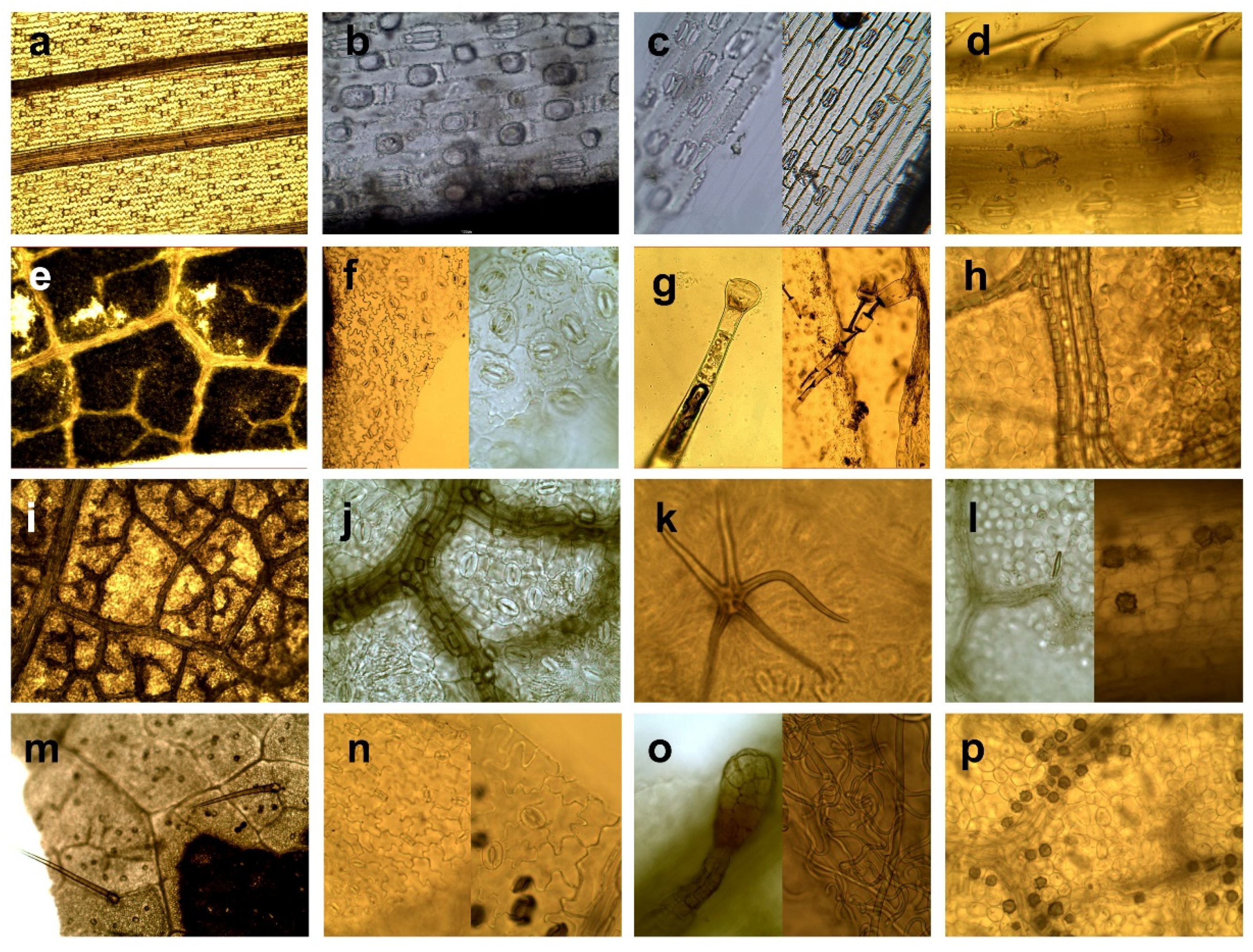
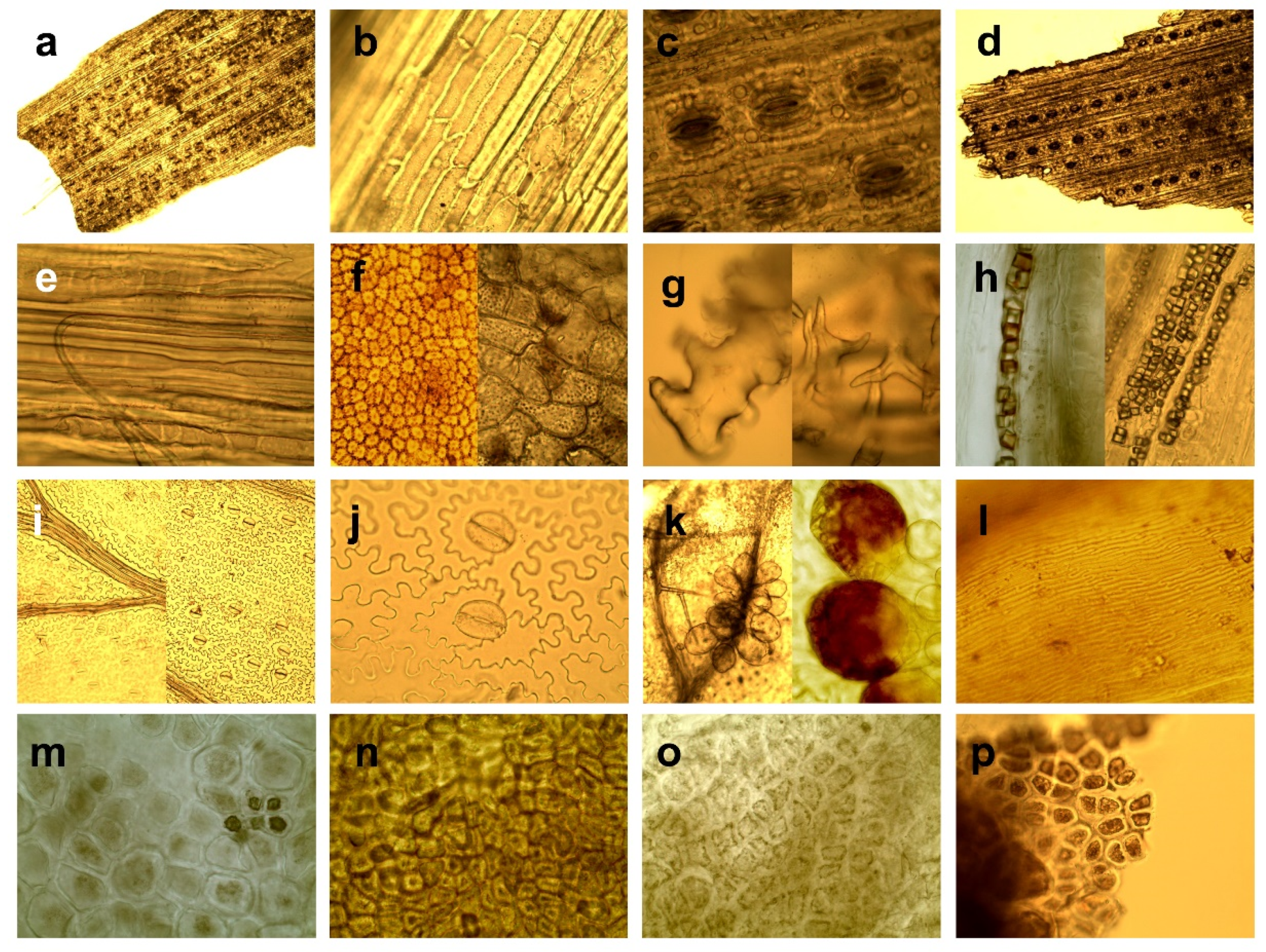
2.3. Morphology Diagnostics and Categorization

Description of the plant microstructures was performed following Metcalfe and Chalk [45], Metcalfe [46], Cutler [47] Esau [48] and Evert [49]. Our identification criteria focused on characteristics of structures that remain preserved after digestion (Figure S1). We considered diagnostic features such as the shape, orientation, and arrangement of cells, the thickness of cell walls, the type of venation, the type, position and orientation of stomata, the shape of guard cells of stomata, the presence, type and arrangement of trichomes and inclusions, the presence and form of sclerenchyma, the presence and type of cork cells, libriform fibres and parts of tracheary elements such as tracheids and vessels. We categorized the reference material based on similar anatomical features into seven functional groups (Table 1), consistent with classifications used in the majority of studies on ungulate diet [25,42,50,51]:
  • Grasses and sedges—monocotyledons of grass-like appearance, such as the Order of Poales, Family Poaceae, Juncaceae, and Cyperaceae.
  • Herbaceous plants and leaves of deciduous trees—dicotyledons, including herbs (except for ferns and genera Rubus, Rosa, Vaccinium) and assimilation organs of deciduous trees and shrubs including buds. We grouped herbs and leaves of trees together due to their similar micromorphology, including shape and arrangement of epidermal cells, type and arrangement of stomata, type of venation and presence of variable types of trichomes.
  • Genera Rubus, Rosa, Vaccinium. We defined this category because of specific morphological structure regarding amount and type of trichomes, and presence of crystalic inclusions, making them well distinguishable from other herbaceous, and because this category has a high nutritional value as a forage source [52], and thus, a specific importance in evaluating forage of wild cervids [1,25,42].
  • Needles—needle-like assimilations organs of coniferous trees.
  • Wood and bark–wood and bark tissue of trees and shrubs.
  • Ferns and mosses—plants from classes Bryopsida and Polypodiopsida.
  • Seeds and fruits—fruits of trees and shrubs, and seeds of agricultural crops.
We excluded anthers and awns from analyses because they spread uncontrollably through vegetation and they often contaminated samples for analysis. We also excluded an internal tissue of stems, due to lack of distinguishable characteristics to classify. Further, we excluded family Liliaceae and Asparagaceae from the key because they can be easily mistaken with grasses or sedges, they have microcharacteristics of monocotyledonous plants although botanists classify these families into herbs.
Table 1. List of seven functional groups based on similar anatomical features as arrangement, type and size of basic plant microstructures consistent with classifications used in the majority of studies on ungulate diet with a description of detailed diagnostic features for each botanical category.
CategoryDiagnostic Features
Grasses and sedgesElongated rectangular shape of epidermal cells arranged parallel to veins [53].
Epidermal cells aligned in longitudinal strips [46].
Parallel venation [49].
Present “short” cells (alone or in pairs) and “long” epidermal cells (except family Juncaceae) [48,49].
Parallel arrangement of stomata [49]
Presence of dumbbell-shaped guard cells of stomata (Gramineous type) exception of the family Juncaceae, which have kidney-shaped of stomata (Amaryllis type) [47].
Presence of macrohairs, microhairs, prickle hairs, and papillae, except family Juncaceae which have the sporadic presence of papillae and trichomes [32].
Herbaceous plants and leaves of deciduous treesEpidermal cells of variable, mostly irregular-shape (hexagonal, round, elongated) [53], cells of elongated organs have elongated shape.
Epidermal cells of leaves arranged diffusely, rarely parallel to the veins [53].
Usually, reticulated venation.
Kidney-shaped guard cells of stomata (Amaryllis type) arranged irregularly or/and randomly [48].
Great morphological variability of trichomes, which occur in different taxa [45].
Presence of different types of crystalline inclusions found in almost all taxa [45].
Genera Rubus, Rosa, VacciniumVenation, shape, and arrangement of epidermal cells identical to category Herbaceous plants and leaves of deciduous trees, except Vaccinium spp. that have also polygonal to rectangular epidermal cells irregularly beaded with straight, thickened, pitted walls [54].
Typical presence of kidney-shaped guard cells of stomata (Amaryllis type) arranged irregularly or/and randomly [45].
Rubus and Rosa: presence of hollow trichomes (simple-unicellular or stellate) sharply pointed, conical with smooth surface. Simple trichomes are twisted or convoluted (length to width ratio min. 10:1), or they are almost straight, narrowed gradually towards to the apex, bases of trichomes have rectangular shape to hexagonal shape with a rosette of epidermal cells; stellate trichomes (with a smooth surface, with pointed apexes, filiform and hollow arms almost equally long) are smaller compared to simple trichomes; sporadic presence of glandular trichomes with multicellular head or presence of multicellular tongue-shaped trichomes Numerous presences of randomly arranged crystals as druses or styloids, especially typical for Rosa [40,45,54,55,56].
Vaccinium: presence of multicellular, glandular tongue-shaped trichomes bent over parallel to the midrib when situated to the apex, also presence of glandular trichomes consist of biseriate or /and multiseriate stalks bearing heads of variable size and small conical with wart-like surface [45].
NeedlesElongated rectangular shape of epidermal cells.
Epidermal cells arranged in rows [57] parallel to vein.
Leaves generally have simple venation and have only one or two long veins running down their centre [58].
Stomata arranged in longitudinal rows forming dense stomatal bands [58].
Stomatal guard cells sunken below the surface of the epidermis [49].
Presence of resin canals in the mesophyll [59].
Wood and barkPresence of parts of tracheary elements and/or vessels elements, fiber tracheids and cells with accompanied libriform fibers or parenchymal cells, crystal-containing cells and sclereids or their combination [49]
Presence of sclereids of various forms such as unbranched (brachysklereids) or sclereids of irregular shapes with a different number of protrusions, as well as large heterogeneous assemblage of sclereids (asterosclerides) [49].
Typical presence of soft cork cells (thin-walled cells of variable shapes) and/or hard cork cells (thick-walled cells in the shape of a square) [60].
Presence of phytoliths; calcium oxalate crystals are common in the secondary phloem of conifers [49].
Ferns and mossesFine structure fragments [61].
Cells of ferns have puzzle-like, and mosses have labyrinth-like pattern.
Mosses single-layered fronds (leaves of mosses) have no developed epidermis [62] and is not differentiated from inner tissue.
A thick layer of photosynthetic cells on a leaf surface of mosses forms lamellae; cells are like furrows or ridges that run parallel to each other.
Ferns have open venation and all cells of the lower and upper epidermis contain chloroplasts [61].
Right (true) vascular tissue contains phloem and xylem absent in mosses [61].
Exclusively presence of Mnium type of stomata [63].
Presence of sporangia on the abaxial side of the fern leaves form clusters, sometimes covered by sharps [62].
Seeds and fruitsVarious type of thick wall cells: sclereids, variable oriented layers or clusters of sclereids, irregularly arranged parenchymal cells with a strongly thickened cell wall, cells with pitted walls, parenchymatic cells and/or irregular polyhedrons with internal filling [48], cells usually arranged diffusely.
Presence of cross cells with thick lignified walls or/and tube cells (lignified cells elongated parallel with the long axis of the grains).
Sporadic presence of stomata randomly arrangement [49].
Presence of starch, starch grains, trichomes.

3. Results

KEY

(1) The fragment consists of regular-shaped cells arranged in rows resp. parallel strips (Figure 1a and Figure 2a); venation (if present) is parallel (Figure 1a) .............................................................2
The fragment consists of irregular-shaped cells (Figure 1f and Figure 2f,j); venation (if present) is reticulated (Figure 1e) ........................................................................................................................... 5
(2) The fragment consists of elongated epidermal cells and/or square and/or rectangular-shaped, mostly angular with arrow-like endings (with sharp or chisel ends); shorter cell wall is not at the right angle to the long axis of the cell ................................................................................... 3
The fragment consists of elongated epidermal cells and/or square and/or rectangular-shaped cells regularly arranged in rows (Figure 1a and Figure 2a); cell walls are smooth or corrugated or sinusoidal or nodular; shorter cell wall approximately at the right angle to the long axis (Figure 1b and Figure 2b), parallel type of venation (Figure 1a and Figure 2a) ............................................ 4
(3) Cells more or less parallel, cells have thickened cell wall and absence of short and long cells; exclusive presence of kidney shape guard cells of stomata (Amaryllis type); stomata randomly arranged (Figure 1n right); exclusive presence of hollow trichomes (simple-unicellular or stellate) sharply pointed, conical with smooth surface, twisted and convoluted (length to width ratio ~10:1; Figure 1o right), or almost straight narrowed gradually towards the apex (Figure 1m), stellate trichomes with equally long arms, sporadic presence of glandular trichomes or presence of multicellular, tongue-shaped trichomes (Figure 1o right) ........................................... Genera Rubus, Rosa, Vaccinium (stem or stalk)
Cells more or less parallel, cells have thickened cell wall and absence of short and long cells; exclusive presence of brachyparacytic stomata randomly arranged (Figure 1n right), with trichomes of two types: stalked, multicellular, glandular, tongue-shaped (Figure 1o right) and small conical with wart-like surface ............................. Genera Rubus, Rosa, Vaccinium (stem or stalk genus Vaccinium)
Cells more or less parallel, thickened or corky cell wall and absence of short and long cells; exclusive presence of Amaryllis type guard cells; stomata randomly arranged (Figure 1f); presence of variable trichomes (Figure 1g) not listed above ................................. Herbaceous plants and leaves of deciduous trees (stem or stalk)
Cells approximately parallel; very fine structure of the fragment (Figure 2l); absence of short and long cells; exclusive Mnium type of stomata randomly arranged (Figure 2j); presence of trichomes except types of trichomes listed above .................... Ferns and mosses (stem or stalk)
(4) Cells of elongated shape, presence of short cells (silica, cork) and long cells, papillae, prickles or trichome bases (Figure 1b); absence of branched trichomes; stomata parallel with long cells (Figure 1c); exclusive dumbbell shape of guard cells of stomata (Gramineous type) levelled with other epidermis cells …….............................................. Grasses and sedges (family Poaceae and Cyperaceae)
Cells have elongated shape; presence of branched trichomes; stomata arranged in rows, parallel with long cells; Amaryllis type of guard cells are in the levelled with epidermal cells ..................................................................... Grasses and sedges (family Juncaceae)
Cells have elongated shape; absence of trichomes; stomata arranged in rows, parallel with long cells (Figure 2d); guard cells of stomata are deeply sunken below the surface of epidermis (Figure 2c); stomata sometimes covered by a wax plug .................................................... Needles
The fragment consists of libriform fibres and/or elements of vessels and/or tracheids and/or sieve elements and/or sclerenchyma cells (sclereids; Figure 2e) and might be accompanied by parenchymatic cells; presence of cells with variable content (crystals, crystalline inclusion, crystalline sand starch, oils, tannins etc.) ...................................................... Wood and bark
(5) Fine structure fragment and cells have puzzle-like (Figure 2j) or labyrinth-like pattern (Figure 2l); open venation; cells of the epidermis may contain chloroplasts; exclusive presence of the Mnium type of stomata (Figure 2j) aligned with leaf veins; long axis of the guard cells aligned with leaf veins (Figure 2i); presence of sporangia (Figure 2k) or presence of elongated, narrow cells forming furrows or backs (Figure 2l); sporadic presence of trichomes .............................................................................................. Ferns and mosses
Predominant part of the fragment consists of various types of thick wall cells or sclereids irregularly arranged (clusters or solitaires; Figure 2n); variable oriented layers or clusters of sclereids, irregularly arranged parenchymal cells with strongly thickened cell wall; cells with pitted walls; presence of parenchymatic cells and/or irregular polyhedrons with internal filling (Figure 2m); presence of starch, starch grains and trichomes; presence of cross cells with thick lignified walls and tube cells (lignified cells elongated parallel with long axis of the grains); absence of cork cells or libriform fibres ............................. Seeds and fruits
Epidermal cells arranged irregularly (Figure 1n); usually reticulated venation (Figure 1m); Amaryllis type of guard cells; stomata randomly arranged (Figure 1n); exclusive presence of hollow trichomes (simple-unicellular or stellate) sharply pointed, conical with smooth surface, twisted and convoluted (length to width ratio ~10:1; Figure 1o right), or almost straight narrowed gradually towards the apex (Figure 1m), base of trichomes have rectangular or hexagonal shape with a rosette of epidermal cells around it; stellate trichomes (smooth surface, pointed apexes, filiform and hollow arms almost equally long) are smaller compared to simple trichomes; sporadic presence of glandular trichomes with multicellular head or presence of multicellular tongue-shaped trichomes (Figure 1o left); presence of druses or prisms in large quantities (Figure 1p) ............................ Genera Rubus, Rosa, Vaccinium (genera Rubus and Rosa)
Epidermal cells irregularly beaded with thickened walls; combinations of randomly arranged, brachyparacytic stomata; glandular trichomes consist of multiseriate stalks bearing heads variable size and multicellular tongue-shaped trichomes bent over parallel to the midrib when situated at the apex of the leaf (Figure 1o left), and small, simple, conical trichomes with wart-like surface ............................................................................ Genera Rubus, Rosa, Vaccinium (genus Vaccinium)
Majority of fragment consists of thick-walled and cork cells (Figure 2f left) and/or partially or completely sclerified cells (Figure 2g) and/or parts of tracheary elements such as tracheids and/or vessels elements; presence of fibre tracheids and cells with accompanied libriform fibres or parenchymal cells (Figure 2e); presence of crystals as solitaires, clusters or crystalline sand in large quantities (Figure 2h); presence of cork cells (Figure 2f left) ......................................................................................................................... Wood and bark
Epidermal cells arranged irregularly (Figure 1f,j); venation usually reticulated (Figure 1e,i); Amaryllis type of guard cells; stomata randomly arranged (Figure 1f,j); various types of trichomes (Figure 1g,k) except for the types listed above; presence of crystals (prisms) in rows over the majority of vascular bundles (Figure 1h) and/or presence of crystalline inclusions of variable forms (Figure 1l) and/or secretory structures.............................. Herbaceous plants and leaves of deciduous trees.
Figure 1. (a) parallel venation on leaf of Agrostis stolonifera L., abx (100×); (b) regular-shaped cells arranged in rows on leaf of Calamagrostis epigejos L. Roth., abx (100×); (c left) stomata parallel with long cells on leaf of Calamagrostis epigejos, adx (400×); (c right) dumbbell shape of guard cells of stomata on leaf of Phleum pratense L., abx (100×); (d) prickles on leaf margin of Deschampsia cespitosa, adx (400×); (e) reticulated venation on leaf of Aegopodium podagraria L., abx, (100×); (f, left), irregular-shaped cells on leaf of Lathyrus niger (L.) Bernh., adx (100×); (f right) kidney shape of guard cells of stomata on leaf of Betonica officinalis L., abx (400×); (g left) glandular trichome on leaf of Saponaria officinalis L., abx (400×); (g right), trichomes on leaf of Impatiens parviflora D. C., abx (100×); (h) prisms in rows over vascular bundles on leaf of Vicia sepium L., adx (400×); (i) reticulated venation on leaf of Populus sp. abx (100×); (j) stomata randomly arranged on leaf of Tillia cordata Mill., abx (400×); (k) stellate trichome on leaf of Quercus cerris L., abx (400×); (l) needle-like crystal on leaf of Robinia pseudoacacia L., adx (400×); (m) almost straight trichomes on leaf of Rubus caesius L., abx (400×); (n left) irregular-shaped cells on leaf of Rubus caesius, abx (400×); (n right) randomly arranged stomata on leaf of Rubus saxatilis L., abx (400×); (o left) tongue-shaped trichome on leaf of Vaccinium myrtillus L., abx (400×); (o right) twisted and convoluted simple trichomes on leaf of Rubus idaeus L., abx (400×); (p) druses on leaf of Rosa canina L., abx (400×).
Forests 12 01229 g001
Figure 2. (a) elongated epidermal cells regularly arranged in rows on fragment of needle of Abies alba (40×); (b) cell wall more less at right angle to the long axis of the cell on fragment of needle of Picea abies (400×); (c) guard cells of stomata are deeply sunken below the surface of other epidermis on needle of Pinus nigra J. F. Arnold, (400×); (d) stomata arranged in rows on needle of Pinus nigra, (100×); (e) fibers of wood from Prunus spinosa L. (400×); (f left) cork cells of bark from Pinus nigra (400×); (f right) cork cells of bark from Picea abies, abx (400×); (g left, right) branched sclereids of bark from Abies alba (400×); (h) crystals in large quantities Abies alba, (400×); (i left) fine structure fragment with open venation on leaf of Athyrium filix-femina, adx (100×); (i right) stomata aligned with leaf veins of Dryopteris filix-mas, abx (400×); (j) Mnium type of stomata on leaf of Dryopteris filix-mas, abx (400×); (k left) sporangia on leaf of Athyrium filix-femina, adx (400×); (k right) sporangia on leaf of Phegopteris connectilis (Michx.) Watt, abx (100×); (l) fine structure fragment of moss sp., with labyrinth-like pattern (400×); (m) irregularly arranged cells with a thickened cell wall and internal filling in acorn (400×); (n) clusters of sclereids in stone of plum (400×); (o) epidermal thick-walled cells on hip (400×); (p) irregularly arranged thick-walled cells with internal filling in beechmast (400×).
Forests 12 01229 g002

4. Discussion

Our key is a simple and effective tool for identification and categorization of plant fragments in faeces of wild cervids of the Western Carpathian region. Roots (except Solanum tuberosum L.), rhizomes, and inflorescence were not included in the key. Each plant part (leaves and stem) of a single species usually includes different diagnostic features. For this reason, microhistological keys cannot be purely dichotomous [30]. Our key allows identification of plant fragments via several steps. First, the key divides possible choices into two groups (coded 2 and 5) based on the arrangement of cells (regular vs. irregular), which narrows possible choices and leads to further categorization based on the shape of cells (for example, 1 → 2 → 3 or 4 → final category) or thickness of the cell walls (1 → 5 → final category). Further identification is based on additional diagnostic features such as trichomes, stomata, sclereids etc., and leads to the final choice. This key helps to identify fragments, not whole plants or plant species; and not all parts of a species are identifiable using epidermal features. For example, some species produced fragments that proved difficult to differentiate using microhistological features, such as Liliaceae and Asparagaceae from monocotyledons. Genera Rubus, Rosa, Vaccinium can also be mistaken with other species of the family Rosaceae which are categorized as herbaceous plants and leaves of deciduous trees. However, based on detailed observation of stomata and type of trichomes can be clearly distinguished. Certain features of some plants that were included in the key are not apparent on the photos, such as the gloss of epidermis on needles. For deeper identification, we recommend using the literature from Metcalfe and Chalk [45,46], resp. the key from the Gödöllő Hills [40] that allow identification at the species level. However, species level identification requires concominant presence of several specific diagnostic features which are often missing and thus should be approached with caution.
On the other hand, certain microscopic features allow to distinguish between some categories with high certainty. For example, regular brick-like arrangement of cells is typical for grasses and needles, while irregular-shaped cells arrangement is typical for herbaceous plants, leaves of deciduous trees, ferns and mosses. Other highly distinctive characteristics is the arrangement of venation, which can be used for distinguishing between monocotyledons from dicotyledons. Further, majority of monocotyledons and dicotyledons, but also ferns and mosses have thin-walled cells, while cells of stems, tree bark, cuticle of seeds and fruits have mostly thickened cell wall. Type and arrangement of stomata, shape of guard cells and subsidiary cells is another important diagnostic feature. For example, the Mnium type of stomata occurs exclusively in the category of mosses and ferns, the dumbbell shape of guard cells of stoma (type Graminae) is typical for grasses and sedges and Amaryllis type of guard cells of stomata occur in herbaceous plants and leaves of deciduous trees, including genera Rubus, Rosa, Vaccinium, and also needles, fruits, and seeds. Moreover, stomata may be at the same level as other epidermal cells in plants but deeply sunken below the level of the surrounding cells in needles, and classifications of stomata according to type of subsidiary cells (anomocytic or brachyparacitic) and amount and type of trichomes may be used to distinguish genera Rubus, Rosa, Vaccinium from other herbaceous plants and leaves of deciduous trees. Finally, “short cells and long cells” occur only in grasses and sedges, sporangia occur exclusively in the category of ferns and mosses, and cross cells and tube cells occur only in the group of seeds and fruits. Cork cells with tracheary elements, vessel elements and libriform fibres are typical for wood and bark and very fine structured fragments are typical for mosses and ferns.

5. Conclusions

The purpose of this study is to provide a tool to identify plant fragments in the faecal remains of forest cervids inhabiting Western Carpathians, in order to improve knowledge on feeding behaviour of free-roaming cervids. Rapid growth of large herbivore populations in Slovakia and across Europe increases intra- and inter-specific competition for food which results in ever increasing damages on forest ecosystems. Since the rough terrain and dense-canopied mixed forests prevent direct observations of food habits, microhistology is a suitable procedure to study feeding behaviour through indirect observations. Using this key will allow researchers to investigate feeding behaviour of cervids with relative ease, and it is expected to save time that can be devoted to other management goals.

Supplementary Materials

The following are available online at https://www.mdpi.com/article/10.3390/f12091229/s1, Table S1: A list of vascular plant species (except for moss) and their parts (leaf, stem, bud, bark and wood, fruit, grain) collected within Western Carpathians, Slovakia and used as a reference material for the microhistological key. Figure S1: Comparison of undigested plant fragments from reference material and digested plant fragments from faecal matter within the same category.

Author Contributions

Conceptualization, A.V. and P.S.; methodology, A.V. and P.S.; investigation, A.V.; writing—original draft preparation, A.V. and P.S.; writing—review and editing, P.S., A.V. and R.K.; visualization, A.V.; supervision, R.K.; project administration, R.K.; funding acquisition, R.K. All authors have read and agreed to the published version of the manuscript.

Funding

This research was funded by the projects VEGA 1/0797/19 and APVV-14-0637 supported by the Ministry of Education, Science, Research and Sport of the Slovak Republic and the projects “Centre of Excellence: Adaptive Forest Ecosystems” (ITMS 26220120006) and “Completing the Centre of Excellence: Adaptive Forest Ecosystems” (ITMS 26220120049) supported by the Operational Programme Research and Development within the European Regional Development Fund.

Institutional Review Board Statement

Not applicable.

Informed Consent Statement

Not applicable.

Acknowledgments

We would like to thank Benčaťová (Technical University in Zvolen, Department of Phytology) for her dedicated assistance with identification of reference plant material, and Homolka (Institute of Vertebrate Biology CAS) for his valuable guidance in the field of microhistology.

Conflicts of Interest

The authors declare no conflict of interest.

References

  1. Spitzer, R.; Felton, A.; Landman, M.; Singh, N.J.; Widemo, F.; Cromsigt, J.P. Fifty years of European ungulate dietary studies: A synthesis. Oikos 2020, 129, 1668–1680. [Google Scholar] [CrossRef]
  2. D’Aprile, D.; Vacchiano, G.; Meloni, F.; Garbarino, M.; Motta, R.; Ducoli, V.; Partel, P. Effects of Twenty Years of Ungulate Browsing on Forest Regeneration at Paneveggio Reserve, Italy. Forests 2020, 11, 612. [Google Scholar] [CrossRef]
  3. Hobbs, N.T. Modification of Ecosystems by Ungulates. J. Wildl. Manag. 1996, 60, 695–713. [Google Scholar] [CrossRef]
  4. Ward, A.I.; White, P.C.; Smith, A.; Critchley, C.H. Modelling the cost of roe deer browsing damage to forestry. For. Ecol. Manag. 2004, 191, 301–310. [Google Scholar] [CrossRef]
  5. Kropil, R.; Smolko, P.; Garaj, P. Home range and migration patterns of male red deer Cervus elaphus in Western Carpathians. Eur. J. Wildl. Res. 2015, 61, 63–72. [Google Scholar] [CrossRef] [Green Version]
  6. Smith, T.N.; Keller, B.J.; Chitwood, M.C.; Hansen, L.P.; Millspaugh, J.J. Diet Composition and Selection of Recently Reintroduced Elk in Missouri. Am. Midl. Nat. 2018, 180, 143–159. [Google Scholar] [CrossRef]
  7. Smolko, P.; Veselovská, A.; Kropil, R. Seasonal dynamics of forage for red deer in temperate forests: Importance of the habitat properties, stand development stage and overstorey dynamics. Wildl. Biol. 2018, 1–10. [Google Scholar] [CrossRef] [Green Version]
  8. Smolko, P.; Kropil, R.; Pataky, T.; Veselovská, A.; Merrill, E. Why do migrants move downhill? The effects of increasing predation and density on red deer altitudinal migration in temperate Carpathian forests. Mammal Res. 2018, 63, 297–305. [Google Scholar] [CrossRef]
  9. Baumgartner, L.L.; Martin, A.C. Plant histology as an aid in squirrel food habits studies. J. Wildl. Manag. 1939, 3, 266–268. [Google Scholar] [CrossRef]
  10. Dusi, J.L. Methods for determination of food habits by plant microtechniques and histology and their application to cottontail rabbit food habits studies. J. Wildl. Manag. 1949, 13, 295–298. [Google Scholar] [CrossRef]
  11. Garnick, S.; Barboza, P.S.; Walker, J.W. Assessment of Animal-Based Methods Used for Estimating and Monitoring Rangeland Herbivore Diet Composition. Rangel. Ecol. Manag. 2018, 71, 449–457. [Google Scholar] [CrossRef]
  12. Christianson, D.; Creel, S. A Review of Environmental Factors Affecting Elk Winter Diets. J. Wildl. Manag. 2010, 71, 164–176. [Google Scholar] [CrossRef]
  13. Baskin, L.; Danell, K. Ecology of Ungulates; Springer: Berlin/Heidelberg, Germany, 2003. [Google Scholar]
  14. Habiba, U.; Anwar, M.; Khatoon, R.; Hussain, M.; Khan, K.A.; Khalil, S.; Hussain, A. Feeding habits and habitat use of barking deer (Muntiacus vaginalis) in Himalayan foothills, Pakistan. PLoS ONE 2021, 16, e0245279. [Google Scholar] [CrossRef]
  15. Green, K.; Davis, N.E.; Robinson, W.A.; McAuliffe, J.; Good, R.B. Diet selection by European hares (Lepus europaeus) in the alpine zone of the Snowy Mountains, Australia. Eur. J. Wildl. Res. 2013, 59, 693–703. [Google Scholar] [CrossRef]
  16. Katona, K.; Altbäcker, V. Diet estimation by faeces analysis: Sampling optimisation for the European hare. Folia. Zool. 2002, 51, 11–15. [Google Scholar]
  17. Chapuis, J.L. Comparison of the diets of two sympatric lagomorphs, Lepus europaeus (Pallas) and Oryctolagus cuniculus (L.) in an agroecosystem of the Ile-de-France. Z. Säugetierkunde 1990, 55, 176–185. [Google Scholar]
  18. Mátrai, K.; Altbäcker, V.; Hahn, I. Seasonal diet of rabbits and their browsing effect on juniper in Bugac Juniper Forest (Hungary). Acta Theriol. 1998, 43, 107–112. [Google Scholar] [CrossRef] [Green Version]
  19. Alves, J.A.B.; Vingada, J.; Rodrigues, P. The wild rabbit (Oryc-tolagus cuniculus L.) diet on a sand dune area in central Portugal: A contribution towards management. Wildl. Biol. Pract. 2006, 2, 63–71. [Google Scholar] [CrossRef]
  20. Gębczynska, Z. Food of the roe deer and red deer in the Białowieża Primeval Forest. Acta Theriol. 1980, 25, 487–500. [Google Scholar] [CrossRef] [Green Version]
  21. Abbas, F.; Picot, D.; Merlet, J.; Cargnelutti, B.; Lourtet, B.; Angibault, J.M.; Verheyden, H. A typical browser, the roe deer, may consume substantial quantities of grasses in open landscapes. Eur. J. Wildl. Res. 2013, 59, 69–75. [Google Scholar] [CrossRef]
  22. Freschi, P.; Fascetti, S.; Riga, F.; Cosentino, C.; Rizzardini, G.; Musto, M. Diet composition of the Italian roe deer (Capreolus capreolus italicus) (Mammalia: Cervidae) from two protected areas. Eur. Zool. J. 2016, 84, 34–42. [Google Scholar] [CrossRef] [Green Version]
  23. Mátrai, K.; Kabai, P. Winter plant selection by red and roe deer in a forest habitat in Hungary. Acta Theriol. 1989, 34, 227–234. [Google Scholar] [CrossRef] [Green Version]
  24. Suter, W.; Suter, U.; Kriisi, B.; Schütz, M. Spatial variation of summer diet of red deer Cervus elaphus in the eastern Swiss Alps. Wildl. Biol. 2004, 10, 43–50. [Google Scholar] [CrossRef]
  25. Krojerová-Prokešová, J.; Barančeková, M.; Šustr, P.; Heurich, M. Feeding patterns of red deer Cervus elaphus along altitudinal gradient in the Bohemian Forest: Effect of habitat and season. Wildl. Biol. 2010, 16, 173–184. [Google Scholar] [CrossRef] [Green Version]
  26. Homolka, M.; Heroldová, M. Native red deer and introduced chamois: Foraging habits and competition in a subalpine meadow-spruce forest area. Folia Zool. 2001, 50, 89–98. [Google Scholar]
  27. Redjadj, C.; Darmon, G.; Maillard, D.; Chevrier, T.; Bastianelli, D.; Verheyden, H.; Saïd, S. Intra-and Interspecific Differences in Diet Quality and Composition in a Large Herbivore Community. PLoS ONE 2014, 9, e84756. [Google Scholar] [CrossRef] [Green Version]
  28. Hartvig, I.; Howe, A.G.; Schmidt, E.N.; Pertoldi, C.; Nielsen, J.L.; Buttenschøn, R.M. Diet of the European bison (Bison bonasus) in a forest habitat estimated by DNA barcoding. Mammal Res. 2020, 66, 123–136. [Google Scholar] [CrossRef]
  29. Holechek, J.L.; Gross, B. Training needed for quantifying simulated diets from fragmented range plants. J. Range Manag. 1982, 35, 644–647. [Google Scholar] [CrossRef] [Green Version]
  30. Carriére, S. Photographic key for the microhistological identification of some arctic vascular plants. Arctic 2002, 247–268. [Google Scholar] [CrossRef] [Green Version]
  31. Howard, G.S.; Samuel, M.J. Atlas of Epidermal Plant Fragments Ingested by Grazing Animals (No. 1582); Department of Agriculture, Science and Education Administration: Seattle, WT, USA, 1979. [Google Scholar]
  32. Johnson, M.K. Microhistological Techniques for Food Habits Analysis; U.S. Department of Agriculture: New Orleans, LA, USA, 1983.
  33. Henderson, J.; Granger, J.; Phillips, G.J. Microhistological Atlas of Greater Yellowstone Moose Browse; Castaway Press: Burbank, CA, USA, 2013. [Google Scholar]
  34. Middleton, B.; Sanchez, E. Microhistological analysis of food habits in the tropics. Rev. Estud. Vida Local 1993, 3, 41–47. [Google Scholar]
  35. Lindström, L.I.; Mújica, M.B.; Bóo, R.M. A key to identify perennial grasses in central Argentina based on microhistological characteristics. Can. J. Bot. 1998, 76, 1467–1475. [Google Scholar] [CrossRef]
  36. Ahmed, T. Photographic Key for the Microhistological Identification of Some Plants of Indian Trans-Himalaya. Not. Sci. Biol. 2015, 7, 171–176. [Google Scholar] [CrossRef] [Green Version]
  37. Cransac, N.; Cibien, C.; Angibault, J.M.; Morellet, N.; Vincent, J.P.; Hewison, A.J.M. Seasonal variation in the diet of roe deer (Capreolus capreolus) according to sex in a very dense forest (Dourdan, France). Mammalia 2001, 65, 1–12. [Google Scholar] [CrossRef]
  38. Azorit, C.; Tellado, S.; Oya, A.; Moro, J. Seasonal and specific diet variations in sympatric red and fallow deer of southern Spain: A preliminary approach to feeding behaviour. Anim. Prod. Sci. 2012, 52, 720–727. [Google Scholar] [CrossRef]
  39. Obidziński, A.; Kiełtyk, P.; Borkowski, J.; Bolibok, L.; Remuszko, K. Autumn-winter diet overlap of fallow, red, and roe deer in forest ecosystems, Southern Poland. Cent. Eur. J. Biol. 2012, 8, 8–17. [Google Scholar] [CrossRef]
  40. Mátrai, K.; Koltay, A.; Tóth, S.; Vízi, G. Key based on leaf epidermal anatomy for food habit studies of herbivores. Acta Bot. Hung. 1986, 23, 255–271. [Google Scholar]
  41. Dzieciolowski, R. Food of the red deer in an annual cycle. Acta Theriol. 1967, 12, 503–520. [Google Scholar] [CrossRef] [Green Version]
  42. Gebert, C.; Verheyden Tixier, H. Variations of diet composition of Red Deer (Cervus elaphus L.) in Europe. Mammal Rev. 2001, 31, 189–201. [Google Scholar] [CrossRef]
  43. Korschgen, L.J. Histological characteristics of plant leaf epidermis and related structures as an aid in food habits studies. Fed. Aid Proj. 1977, 13, R31. [Google Scholar]
  44. Reynolds, H.W.; Hansen, R.M.; Peden, D.G. Diets of the slave river lowland bison herd, northwest territories, Canada. J. Wildl. Manag. 1978, 42, 581–590. [Google Scholar] [CrossRef]
  45. Metcalfe, C.R.; ChaIk, L. Anatomy of the Dicotyledons: Leaves, Stem and Wood in Relation to Taxonomy, with Notes on Economic Uses Volume II; Clarendon Press: Oxford, UK, 1957. [Google Scholar]
  46. Metcalfe, C.R. Anatomy of the Monocotyledons. 1 Gramineae; Clarendon Press: Oxford, UK, 1960. [Google Scholar]
  47. Cutler, D.F. Anatomy of the Monocotyledons, IV. Juncales; Clarendon Press: Oxford, UK, 1969. [Google Scholar]
  48. Esau, K. Anatomy of Seed Plants; John Wiley and Sons: Hoboken, NJ, USA, 1977. [Google Scholar]
  49. Evert, F.R. Esaus Plant Anatomy; Wiley Interscience: Hoboken, NJ, USA, 2006. [Google Scholar]
  50. Latham, J.; Staines, B.W.; Gorman, M.L. Comparative feeding ecology of red (Cervus elaphus) and roe deer (Capreolus capreolus) in Scottish plantation forests. J. Zool. 1999, 247, 409–418. [Google Scholar] [CrossRef]
  51. Lovari, S.; Ferretti, F.; Corazza, M.; Minder, I.; Troiani, N.; Ferrari, C.; Saddi, A. Unexpected consequences of reintroductions: Competition between increasing red deer and threatened Apennine chamois. Anim. Conserv. 2014, 17, 359–370. [Google Scholar] [CrossRef]
  52. Biel, W.; Jaroszewska, A. The nutritional value of leaves of selected berry species. Sci. Agric. 2017, 74, 405–410. [Google Scholar] [CrossRef]
  53. Storr, G.M. Microscopic analysis of faeces, a technique for ascertaining the diet of herbivorous animals. Aust. J. Biol. Sci. 1961, 14, 157–164. [Google Scholar] [CrossRef]
  54. Jackson, B.; Snowdon, D.W. Atlas of Microscopy of Medicinal Plants, Culinary Herbs and Spices; Belhaven Press a Division of Pinter Publishers: London, UK, 1990. [Google Scholar]
  55. Rahfeld, B. Mikroskopischer Farbatlas Pflanzlicher Drogen; Spektrum Akademischer Verlag: Heidelberg, Germany, 2009. [Google Scholar]
  56. Tomaszewski, D.; Zieliński, J.; Gawlak, M. Foliar indumentum in central-European Rubus species (Rosaceae) and its contribution to the systematics of the group. Nord. J. Bot. 2014, 32, 1–10. [Google Scholar] [CrossRef]
  57. Klika, J. Jehličnaté; Nakladatelství Československé Akademie: Praha, Czechoslovakia, 1953. [Google Scholar]
  58. Foster, A.S.; Gifford, E.M. Comparative Morphology of Vascular Plants; W.H. Freeman and Company: New York, NY, USA, 1959. [Google Scholar]
  59. Votrubová, O. Anatomie Rostlin; Karolinum: Praha, Czech Republic, 2010. [Google Scholar]
  60. Požgaj, A. Štruktúra a Vlastnosti Dreva; Príroda a.s.: Bratislava, Slovakia, 1997. [Google Scholar]
  61. Copeland, E.B. The comparative ecology of San Ramon Polypodiaceae. Philipp. J. Sci. 1907, 2, 1–76. [Google Scholar]
  62. Novák, J.; Skalický, M. Botanika; Česká Zemědělská Univerzita V Praze: Praha, Czech Republic, 2009. [Google Scholar]
  63. Scott, T. Concise Encyklopedia Biology; Walter de Gruyter: Berlin, Germany, 1996. [Google Scholar]
Publisher’s Note: MDPI stays neutral with regard to jurisdictional claims in published maps and institutional affiliations.

