Article

Induced Volatile Emissions, Photosynthetic Characteristics, and Pigment Content in Juglans regia Leaves Infected with the Erineum-Forming Mite Aceria erinea

by
Corina Popitanu
1,†,
Andreea Lupitu
2,†,
Lucian Copolovici
2,*,
Simona Bungău
1,
Ülo Niinemets
3,4 and
Dana Maria Copolovici
2
1
Biomedical Sciences Doctoral School, University of Oradea, University St. no. 1, 410087 Oradea, Romania
2
Institute for Research, Development and Innovation in Technical and Natural Sciences, Faculty of Food Engineering, Tourism and Environmental Protection, Aurel Vlaicu University of Arad, Elena Drăgoi St., no. 2, 310330 Arad, Romania
3
Institute of Agricultural and Environmental Sciences, Estonian University of Life Sciences, Kreutzwaldi 1, 51006 Tartu, Estonia
4
Estonian Academy of Sciences, Kohtu 6, 10130 Tallinn, Estonia
*
Author to whom correspondence should be addressed.
These authors contributed equally to this work.
Forests 2021, 12(7), 920; https://doi.org/10.3390/f12070920
Submission received: 8 June 2021 / Revised: 12 July 2021 / Accepted: 13 July 2021 / Published: 15 July 2021
(This article belongs to the Section Forest Health)

Abstract

:
Persian walnut (Juglans regia L., Juglandaceae), one of the essential nut crops, is affected by different diseases, including mite attacks which result in gall and erineum formation. As the proportion of leaf area covered by mite galls or erineum is typically relatively low, the impact on tree photosynthetic productivity is often considered minor, and no pest control management is usually suggested. However, the effect of erineum-forming mites on walnut photosynthesis might be disproportionately larger than can be predicted from the leaf area impacted. In the present study, we studied how the foliage photosynthetic characteristics, pigment contents, and stress-induced volatile organic compounds scaled with the severity of infection varied from 0% (control trees) to 9.9%, by erineum-forming mite Aceria erinea in J. regia. Both leaf net assimilation rate (up to 75% reduction) and stomatal conductance (up to 82%) decreased disproportionately, increasing infection severity. Leaf total chlorophyll and β-carotene contents also decreased with infection severity, although the reduction was less than for photosynthetic characteristics (28% for chlorophyll and 25% for β-carotene). The infection induced significant emissions of green leaves volatiles ((Z)-3-hexenol, (E)-2-hexenal, (Z)-3-hexenyl acetate and 1-hexanol), monoterpenes and the homoterpene 3-(E)-4,8-dimethyl-1,3,7-nonatriene, and these emissions scaled positively with the percentage of leaf area infected. These results collectively indicate that erineum-forming mite infection of walnut leaves results in profound modifications in foliage physiological characteristics that can significantly impact tree photosynthetic productivity.

1. Introduction

Persian walnut (Juglans regia L., Juglandaceae) is a deciduous tree species native to central Asia. It is one of the most important nut crops with a wide area of cultivation, including Europe, North America, Central, and East Asia, North Africa, New Zealand [1]. In Europe, it is primarily grown in central to southern countries, although cultivars selected for superior cold hardiness can survive in the south part of Northern Europe [2]. The most extensive J. regia plantations are in Turkey, France, Romania, Serbia, and Hungary. As a tree species with a sequenced genome, it becomes one of the model species for physiological studies in woody plants [3].
Juglans regia is an early-successional very light-demanding species growing best in deep fertile soils [4]. As a widely planted species, J. regia is confronted by a broad range of pests, including fungi, bacteria, viruses, insect and mite herbivores, and parasites that could disturb roots, fruits, stems, or leaves [1,2,5]. The J. regia leaves can be attacked by a diversity of mites and insects, including leaf miners, chewing and piercing insect herbivores [6,7,8], and gall- and erineum-forming mites [9,10,11] that can lead to extensive defoliation and premature leaf senescence and reduction in leaf physiological activity, thereby potentially having significant economic impacts [5]. However, mite gall and erineum formation infrequent in J. regia plantations [9,10,11], the physiological effects of gall and erineum formation have not been characterized in this species.
Gall and erineum formation result from an active growth reaction of cell plants in reaction to the insect or mite attacks, leading to the creation of tumor-like structures that provide shelter and food for the occupants [12,13]. The gall-inducing insects are found in the orders Hymenoptera, Thysanoptera, Diptera, gall- and erineum-forming mites in Trombidiformes (superfamily Eriophyoidea) [14]. Individual eriophyid species are highly specialized plant feeders affecting only a very narrow range of hosts. Besides high host specificity, a very high diversity of eriophyoid mites (more than 1000 species) can induce galls and erinea on plant species from a wide range of families [15,16].
As gall and erineum formation does not result in defoliation and the surface area covered by galls and erinea is generally a relatively small fraction of the total area, the impact of leaf galling on foliage physiological activity might be considered small. However, available evidence indicates that leaf infection by gall- and erineum-inducing parasites can result in disproportionately greater physiology and biochemistry alterations than predicted based on the percentage of the infected area [17]. In particular, infestation by gall-forming arthropods typically leads to strongly reduced leaf photosynthetic pigment content as observed in Populus nigra var. italica leaves by infestion with petiole gall aphid (Pemphigus spyrothecae) [18], and Alstonia scholaris leaves plagued by Pauropsylla tuberculata [19]. Typically, the gall or erineum-induced leaf physiological alterations include reductions in both light-saturated stomatal conductance and net assimilation rate as confirmed for gall-forming cynipid wasp infections in Quercus robur [17] and eriophyoid erineum-and gall-forming mite infections in Tilia cordata and Alnus glutinosa [20]. Similarly, both stomatal conductance and net assimilation rate were reduced in Carya illinoensis leaves infested by Phylloxera notabilis galls [21]. A lower photosystem II efficiency was found in Carya glabra infested with cecidomyiid midge galls [22]. Net assimilation rate, stomatal conductance, and photosystem II activity were reduced in the leaves of Machilus thunbergii galled by Daphnephila taiwanensis [23]. However, depending on the type of gall inducer, leaf CO2 and H2O exchange can be disturbed to a different extent, potentially leading to modifications in leaf water use efficiency [17,20,24]. For example, in leaves of P. × petrovskiana infested with P. spyrothecae net assimilation rate per area decreased by almost five-fold likely due to restricted nitrogen and carbon availability for leaf lamina assembly. At the same time, increasing the infestation severity determinea the stomatal conductance increase [25]. Furthermore, net assimilation rate and stomatal conductance even increased in Silphium integrifolium leaves infested with the apical meristem galler Antistrophus silphii [26], suggesting profound modifications in sink–source relationships induced by the galler.
Apart from photosynthetic modifications, to respond to herbivore feeding or fungal infection, plants emit various volatiles induced by stress, such as monoterpenes, sesquiterpenes, and green leaf volatiles (GLV) [27,28,29,30,31]. These compounds are implicated in atmospherical chemical reactions with pollutants (as NOx) and determine tropospheric ozone concentration increasing [32]. Much fewer studies have looked at volatile emissions from plants infected with arthropod-formed galls or erinea. Leaves infected with gall- or erineum-forming arthropods typically have enhanced emissions of GLV [17,20], suggesting a sustained damage resulting from the feeding of arthropods. However, induced and constitutive emissions might be differently affected by different gall- or erineum-forming organisms. In [25], both constitutive isoprene and induced monoterpene emissions scaled positively with the infestation severity by petiole gall aphids in leaves of P. × petrovskiana. In [17], in gall wasp-infected leaves of Quercus robur, constitutive isoprene emissions decreased in all cases with increasing infection severity. In contrast, the emission of monoterpenes was increased in leaves infected by Neuroterus albipes, Cynips divisa, and C. quercusfolii wasps and reduced in leaves infected by N. anthracinus [17]. In another study, monoterpene emissions were greater in leaves of Tilia cordata infected by Eriophyes tiliae gall-forming and E. exilis erineum-forming mites, while monoterpene emissions decreased in Alnus glutinosa leaves infected by E. inangulis gall-forming mites [20]. These studies provide encouraging evidence that alterations in constitutive and induced emissions might be used to diagnose the type and severity of infections of gall- and erineum-forming arthropods.
In J. regia, the gall- and erineum-forming mites all belong to Eriophyoidea: Aceria brachytarsus, A. avanensis, A. erinea, and A. tristata [10]. Walnut blister mite (Aceria erinea Nalepa) is a mite that causes erinea resembling yellowish blisters on the upper surface on walnut leaves [11,33]. The blisters correspond to hollows on the leaf underside; the hollows are covered with a dense whitish or pale brown mat of hypertrophic trichomes harboring mites [34] for a description of erineum structure. Although having a spectacular visual appearance, the A. erinea-induced erinea typically covers a low proportion of leaf area, less than 10%. Still, it is likely that leaf physiological characteristics are altered by erinea to a much greater degree than expected based on the degree of infection.
In this study, we hypothesized that A. erinea infections (1) reduce photosynthesis characteristics and photosynthetic pigment concentration, and elicit volatile stress emissions in leaves of J. regia in an infection severity-dependent manner, and (2) that the relative alterations in leaf traits are greater than changes in the proportion of area infected. This study demonstrates that even moderate infections of A. erinea signals strongly reduced leaf photosynthetic activity and enhanced production of stress volatiles that can be employed to predict the reduction in foliage photosynthetic productivity.

2. Materials and Methods

2.1. Study Site and Plant Material

The study takes place in Arad, Romania (46°17′ N, 21°17′ E, elevation ~60 m above sea level) close to Mures river in the middle of July 2020. For July 2020, the average minimum air temperature was 14.6 °C, and the maximum air temperature was 27.8 °C (National Meteorological Administration. Available online: https://www.inmh.ro (accesssed on 28 June 2020)). The Juglans regia trees chosen for performing the measurements were 20–30 years old and 20–25 m tall. South-exposed leaves with varying degrees of visual infection symptoms were selected to measure gas-exchange parameters, and volatile organic compound (VOC) emission rates with the leaves attach to the tree. Control non-infected leaves were measured in nearby healthy trees. After that, all leaves have been collected, used to assess the degree of infection followed by extraction of pigments.

2.2. Foliage Gas-Exchange Measurements

In the field, photosynthetic characteristics of attached leaves with different degrees of infection were measured using a portable gas-exchange system GFS-3000 with 8 cm2 leaf chamber (Waltz, Effeltrich, Germany) as described in [35] under the following environmental conditions: 65% air humidity in the chamber, 400 µmol mol−1 CO2 concentration, 1000 μmol m−2 s−1 (10% blue and 90% red LED light) the incident light intensity, and 25 °C leaf temperature. After the chamber’s enclosure, the leaf was maintained until stomata opened and steady-state CO2 and water vapor exchange rates were reached. The stabilization period was typically 20–30 min. Once in the steady-state, foliage gas exchange rates were logged. For calculation of the values of stomatal conductance to water vapor (gs) and net assimilation (A) the same procedure described in [36] has been used.
In representative leaves with two different degrees of infection, leaf gas exchange’s parameters light-response curves were also recorded with the same system (GFS-3000, Heinz Walz GmbH, Effeltrich, Germany). The light-response curve measurements were conceded in the following sequence (light intensities in μmol m−2 s−1):
0 → 50 → 100 → 200 → 300 → 400 → 500 → 600 → 800 → 1000 → 1200 → 1500 → 1800.
Other environmental conditions were maintained as above. At each light intensity, the gas exchange rates were stabilized until the steady-state values were observed. Light-response curves were measured in triplicate for control and leaves with approximately 3.5% and 5.1% infection. The light response curves were fitted by the rectangular hyperbola of Smith [37] as in [38], and dark respiration rate (Rd), initial quantum yield (α) and maximum net assimilation rate (Amax) were obtained.

2.3. Leaf Pigment Analysis

Pigment extraction was accomplished according to the method defined in [39], followed by the HPLC analysis described in [40].

2.4. Volatile Sampling and GC–MS Analyses

An air sample pump 210-1003 MTX (SKC Inc., Houston, TX, USA) with the constant flow was used to sample volatile organic compounds (VOC) via the outlet of the gas-exchange system using the same procedure and multibed stainless steel cartridge as described in [41]. The flow rate for the volatile collection was 200 mL min−1, and volatiles from the 4 L chamber air was collected. The adsorbent cartridges were analyzed for different volatile organic compounds as green leaf volatiles (GLV) and terpenes using a Shimadzu TD20 automated cartridge desorber integrated with a Shimadzu 2010 Plus GC–MS instrument (Shimadzu Corporation, Kyoto, Japan) following the method of [41,42]. Volatile emission rates were calculated according to Niinemets et al. [43]

2.5. Estimations of the Leaf Area and the Degree of Infection

The leaves were scanned at 200 dpi, and the leaf area was infected. The projected leaf area was estimated using custom-built software “Leaf Area Measurement” (www.plant-image-analysis.org, accessed on 5 May 2021) (Figure 1 for representative examples of control and infected leaflets).

2.6. Statistical Analysis and Data Handling

Altogether 20 leaves with different degrees of infection and five control leaves were measured for foliage gas-exchange characteristics and volatile emissions. Initially, we tested them using the null hypothesis (parametric one-way ANOVA) whether there were any differences between different tree leaves. Because there was no difference and it was demonstrated that the tree effect was not significant, all data were pooled for the regression analyses. Linear and non-linear regressions (Michaelis-Menten type equation: y = a⋅x/(b + x)) were used to explore the relationships of foliage physiological characteristics and pigment contents with the degree of infection. The parameters of the light response curves among control and differently-infected leaves (n = 3 in each case) were compared by parametric ANOVA followed by the Tukey post hoc test for pairwise comparison. All statistical analyses were conducted with GraphPad Prism version 9.1.2 for Windows (GraphPad Software, San Diego, CA, USA), and the statistical tests were considered significant at p < 0.05.

3. Results

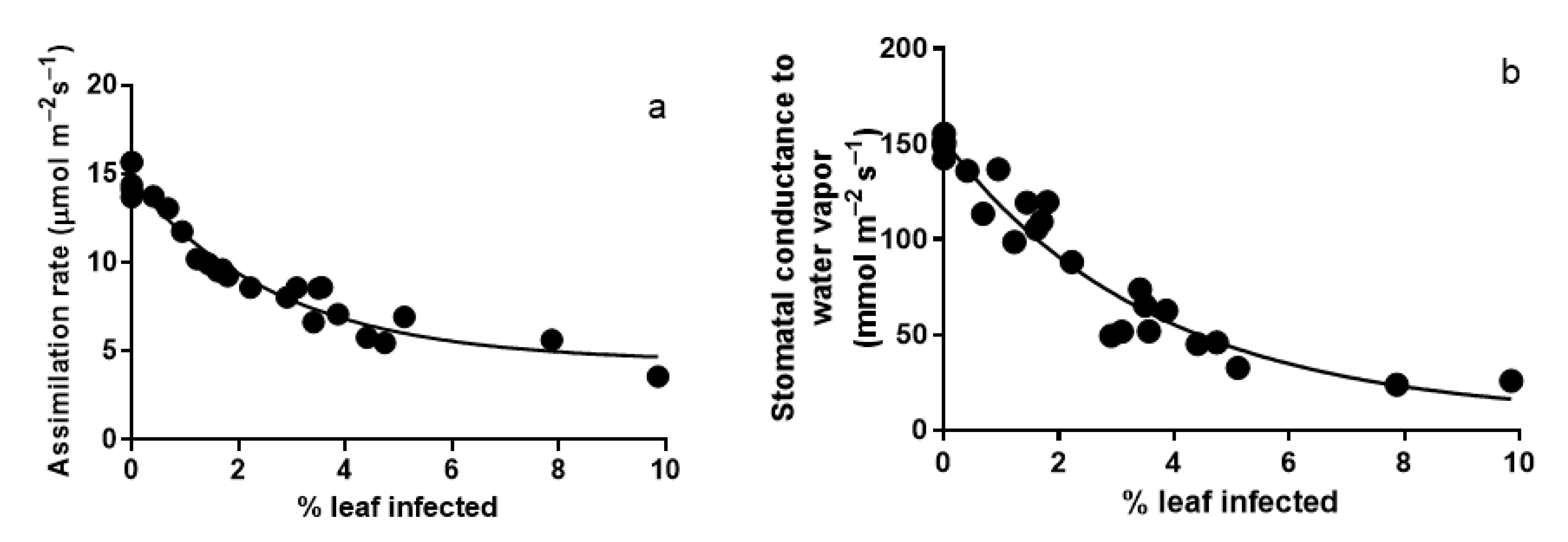
3.1. Photosynthesis Characteristics of Leaves Infected with A. erinea

The infection with A. erinea induced a drastic decrease of average net assimilation rate from 14.8 ± 1.0 µmol m−2 s−1 in control leaves to around three µmol m−2 s−1 in heavily infected leaves (Figure 2a).
Even a minor infection (0.9% degree of infection) reduced leaf assimilation rate by 10–15% (Figure 2a). Stomata conductance to water vapor declined with the infection rate from 151.6 ± 2.3 mmol m−2 s−1 in control leaves to values of 23–25 mmol m−2 s−1 in leaves with more than 7% infection (Figure 2b).

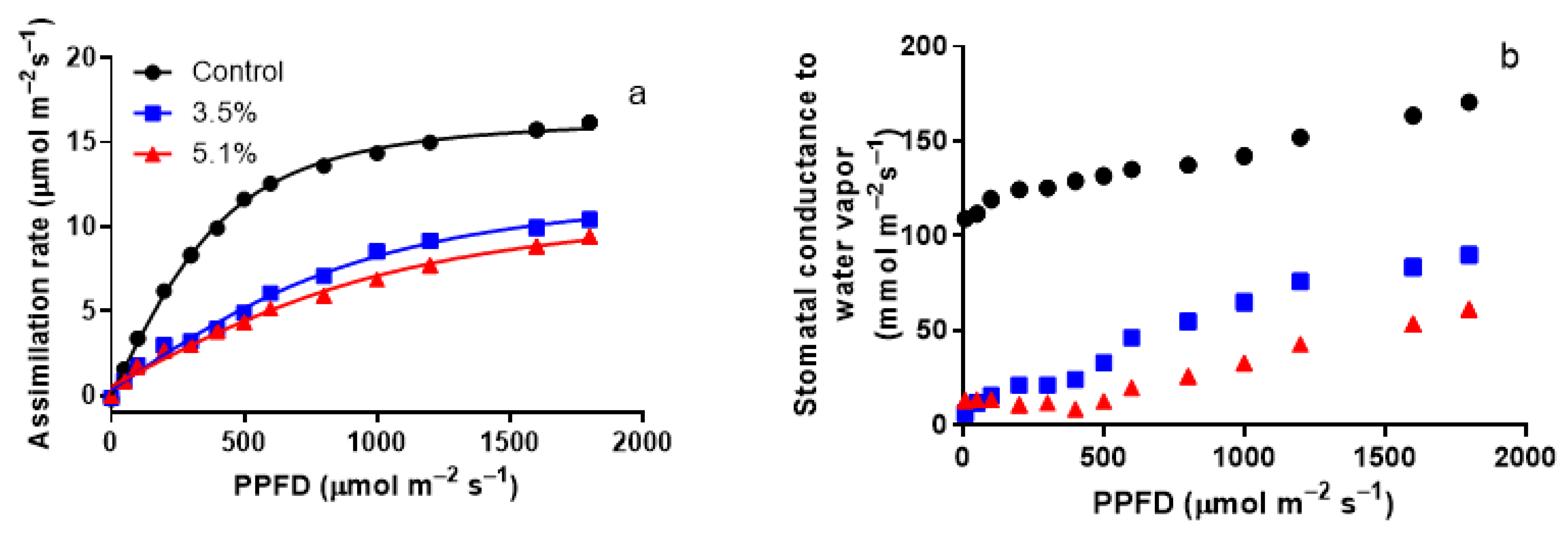
3.2. Modification of Light Responses of Photosynthesis by A. erinea Infection

Analysis of the light response curves further confirmed the major reduction in photosynthesis rate. The maximum net photosynthetic rate was higher for control plants (16.41 ± 0.10 µmol m−2 s−1) than for infected plants (11.94 ± 0.28 µmol m−2 s−1 for average degree of infection of 3.5% and 10.66 ± 0.27 µmol m−2 s−1 for 5.1% infection, p < 0.05 for comparison with the control, Figure 3a). The initial quantum yield decreased from 0.0324 ± 0.0005 μmol μmol−1 in control leaves to 0.0083 ± 0.0003 μmol μmol−1 in leaves with 5.1% infestation (p < 0.05). However, differences in the dark respiration were minor among control and infected plants (Figure 3a).
Similar to the net assimilation rate, stomatal conductance increased with the increasing light intensity (Figure 3b). The difference in stomatal conductance between control and infected leaves was particularly large at a lower light intensity of 0–400 μmol m−2 s−1 than at higher light intensity of 400–1800 μmol m−2 s−1 (Figure 3b). The ratio of stomatal conductance at light intensities of 400 μmol m−2 s−1 and 1800 μmol m−2 s−1 was lower for control (15.13 ± 0.15) than for infected leaves (2.79 ± 0.35 for leaves with 5.1% infection, p < 0.05).

3.3. Responses of Foliage Chlorophylls and β-Carotene Contents to A. erinea Infection

Both chlorophyll a and b contents reduced intensely with the area infected by the A. erinea, e.g., the average chlorophyll content decreased from 742 ± 4 mg m−2 in non-infected leaves to 554 mg m−2 in the leaves with a considerable infestation (Figure 4a). The chlorophyll a/b ratio varied between 2.25 and 2.75 (average ± SE = 2.49 ± 0.17) for all leaves sampled and did not depend on the degree of infestation (p > 0.05).
β-Carotene concentration decreased from 34 mg m−2 in non-infected leaves to 25.69 mg m−2 in highly infected leaves (Figure 4b).

3.4. Effects of A. erinea Infection on Stress-Elicited Volatile Emissions

The infection induced the emission of green leaves volatiles (GLV: (Z)-3-hexenol, (E)-2-hexenal, (Z)-3-hexenyl acetate and 1-hexanol), monoterpenes (α-thujene, (E)-β-ocimene, 3-carene, 3-caren-2-ol) and a nerolidol-derived homoterpene 3-(E)-4,8-dimethyl-1,3,7-nonatriene (DMNT). The emissions of the sum of green leaf volatiles and total monoterpenes increased from nearby zero in control leaves to values as high as ∼8.2 nmol m−2 s−1 and 1.2 nmol m−2 s−1 (Figure 5) in leaves with a high infected percentage.
In addition, the emission of characteristic stress terpenes, the monoterpene (E)-β-ocimene, and DMNT also scaled positively with the infection percentage (Figure 6).

4. Discussion

4.1. The Influence of Aceria erinea Infection on Photosynthetic Characteristics of Juglans regia

The general response of plants to infection by different pathogens includes reducing leaf net assimilation rate [22,25,28,44]. According to previous studies, our data also demonstrated a substantial decrease in the net assimilation rate for infected plants. Notably, the relative reduction in net assimilation rate observed in A. erinea-infected J. regia leaves was much greater (up to 80%) than the degree of infection (up to 10%, Figure 2a). This indicates that erineum-formation led to profound physiological modifications in both erineum-infected and non-infected leaf areas. Furthermore, it suggests that the impact of gall- and erineum-forming mites can be much more significant than generally thought based on a relatively small fraction of the area infected in individual leaves.
The relative reduction in leaf net assimilation rate in erineum-infected leaves can be due to stomatal and non-stomatal (reductions in leaf photosynthetic capacity) factors. In the present study, the relative decrease in net assimilation rate was related to reduced stomatal conductance (Figure 2b) and leaf water loss. An analogous negative influence of gall- and erineum-forming mites on water loss and photosynthesis has been found in Vitis vinifera cultivars infected with erineum-forming mite Colomerus vitis [45], Acer saccharum trees galled by the mite Vasates aceriscrumena [46], in Alnus glutinosa trees infected by Eriophyes inangulis, and Tilia cordata trees infected by E. exilis [20]. The relative reduction in stomatal conductance in pest-infected leaves could be explained by impaired stomatal function in gall- and erineum-covered leaf regions and surrounding leaf tissues due to pathogenic infection or formation of necrotic areas [17,28,47]. However, stomatal conductance is typically reduced less than the net assimilation rate, resulting in reduced leaf water use efficiency [17,48], as observed in our study.
The evidence of a greater reduction in net assimilation rate than in stomatal conductance suggests that the infection also reduced leaf photosynthetic capacity. Typically, leaf photosynthetic capacity is significantly reduced in the infected leaf regions, reflecting altered leaf anatomy and reduced photosynthetic pigment content [46], as observed in our study (Figure 4). A significant reduction in leaf chlorophyll content has been shown in the horn-shaped galls induced by a cecidomyiid on Copaifera langsdorffii leaves [49] and leaves of Cinnamomum tamala with Aceria doctersi galls [50]. The decrease in chlorophyll contents has been connected with a dilution of pigments by cell hypertrophy in leaves with galls [51,52]. However, in our study, the relative decrease in pigment content in J. regia at given infection severity was much less than in the net assimilation rate (cf. Figure 2 and Figure 4).
In fact, there is evidence that the mites might also affect the sink–source relationships (by interaction with others sinks within integrated physiological units) during leaf growth and thereby alter photosynthetic biomass accumulation per unit leaf area and whole leaf photosynthesis rate [53]. Such a sink–source impact has been demonstrated in Populus × petrovskiana infected by petiole gall aphids; the aphid galls are formed on petioles, and thus, the aphids do not directly affect leaf lamina, but nevertheless, leaf photosynthesis rate was strongly reduced in leaves with infested petioles (Ye et al. 2019). It is plausible that the disproportionately more significant reduction in net assimilation rate at the given degree of infection in J. regia is also associated with altered leaf development and decline of photosynthetically active biomass per unit area in infected leaves.

4.2. Emission of Volatile Organic Compounds from Leaves Infected with A. erinea

The green leaf volatiles (GLV, C5 and C6 alcohols, aldehydes, and ketones) are specific elicitor molecules emitted from damaged plant membranes, usually after herbivory, pathogen, or oxidative stresses [28,29,54,55,56]. In the case of A. erinea infection in J. regia, GLV emissions is amplified by increasing the degree of infection (Figure 4a). Similar scaling of GLV emissions with the degree of infection of gall- or erineum- forming arthropods has also been observed in other studies [17,20,25]. However, quantitatively, the rate of GLV emission observed in these studies was less, except in Quercus robur leaves infected by Neuroterus anthracinus wasp galls where the rates of GLV emission were similar to J. regia leaves with A. erinea infection of 1–2% [17] (Figure 5).
A sustained GLV emission could be a direct response to the immediate sites of impact during mite feeding, reflecting the free fatty acids discharge from damaged plant membranes and activation of lipoxygenase at the sites of feeding within the erinea [57,58]. However, once formed, the primary GLV can translocate to other leaf regions with the transpiration stream and can be derivatized before the release [59]. In the case of infection with A. erinea, GLV emissions were dominated by 1-hexanol, suggesting that derivatization of a part of primary aldehydes could have occurred in non-damaged leaf parts. Analogously, a large share of derivatized GLV was found in Quercus robur leaves infected with Neuroterus albipes and Cynips spp. [17] and in Alnus glutinosa infected by Eriophyes inangulis mites and Tilia cordata infected by E. tiliae mites [20].
The leaves of J. regia do not emit monoterpenes in non-stressed conditions, but this species has glandular trichomes on the leaf surface, and leaf essential oil extractions contain different mono- and sesquiterpenes [60,61]. Thus, a certain fraction of monoterpene release in A. erinea-infected mites might be originated from terpene released upon breakage of glandular trichomes. However, given that the erinea harboring the mites are primarily covered by non-glandular hypertrophic trichomes, the contribution of glandular trichomes to terpene release in mite-infected leaves is likely minor. On the other hand, de novo synthesis of monoterpenes is typically elicited in response to pest and parasite attacks as a part of the induced suite of traits involved in direct and indirect defense (for review, see [62,63]). As a part of the direct defense, terpenes can be toxic to infecting organisms, including mites [63,64]. Volatile terpenes also play a compelling role in indirect defense by attracting predators of plant-attacking insects and mites [63,65,66]. In the case of J. regia leaves, there is only limited information on arthropod-induced terpene blends. Infestation of J. regia leaves by Meliboeus ohbayashii ssp. primoriensis larvae induced emissions of monoterpenes limonene, sabinene, β-phellandrene, α-pinene, and β-pinene and sesquiterpenes germacrene D, (E,E)-α-farnesene [67]. In our study, J. regia leaves infected with A. erinea emitted monoterpenes α-thujene, (E)-β-ocimene, and 3-carene and its oxidized derivative 3-caren-2-ol, and total monoterpene emissions scaled with the percentage of the leaf area infected (Figure 4b). In addition, the emissions of two characteristic terpenes de novo synthesized in stressed leaves, (E)-β-ocimene and the homoterpene DMNT, which also scaled positively with the degree of leaf area damage (Figure 6), suggesting that the positive scaling of total terpene emission with the degree of infection in our study (Figure 4b) primarily reflected de novo synthesis of terpenes. The significant differences in the induced terpene emission blends from J. regia infected by the mite A. erinea in our study and the buprestid insect M. ohbayashii ssp. primoriensis [67] suggest that the induced terpene composition provides informative insights for identifying the infecting organism. We suggest that further work with different walnut-infecting organisms is needed to characterize the modification of the emission blend and the degree of elicitation of volatile emissions as determined by various leaf-attacking arthropods.

5. Conclusions

Our study demonstrates the infestation of walnut leaves with Aceria erinea strongly affects the plant’s metabolic and physiological processes. In addition to visual symptoms, the infection led to a decline in photosynthetic characteristics, emission of volatile organic compounds, and a decrease in chlorophylls and carotene contents. All chemical and physiological modifications were quantitatively related to the percentage of the leaf area infected. In contrast, the decline in photosynthetic characteristics was much more significant than could be predicted based directly on the infected leaf area. We conclude that A. erinea infection leads to systemic alterations in J. regia leaf physiological activity.

Author Contributions

Conceptualization, L.C., Ü.N., and D.M.C.; methodology, L.C.; validation, L.C., Ü.N., and D.M.C.; formal analysis, C.P., A.L., L.C., S.B.; investigation, C.P., A.L., S.B., L.C.; resources, L.C.; data curation, C.P., A.L., S.B.; writing—original draft preparation, C.P., A.L., S.B., L.C., Ü.N., and D.M.C.; writing—review and editing, Ü.N., L.C., and D.M.C.; visualization, L.C.; supervision, L.C., D.M.C.; project administration, L.C., D.M.C.; funding acquisition, L.C., D.M.C. All authors have read and agreed to the published version of the manuscript.

Funding

This research was funded by CNFIS-UEFISCDI, project number PN-III-P4-ID-PCE-2020-0410.

Conflicts of Interest

The authors declare no conflict of interest.

References

  1. Bernard, A.; Lheureux, F.; Dirlewanger, E. Walnut: Past and future of genetic improvement. Tree Genet. Genomes 2018, 14, 1. [Google Scholar] [CrossRef] [Green Version]
  2. Durrant, T.H.; Caudullo, G.; Enescu, C.M.; De Rigo, D.; Tinner, W. Juglans regia in Europe: Distribution, habitat, usage and threats. In European Atlas of Forest Tree Species; Schweizerische Zahnärzte-Gesellschaft SSO: Bern, Switzerland, 2016. [Google Scholar]
  3. Martínez-García, P.J.; Crepeau, M.; Puiu, D.; Gonzalez-Ibeas, D.; Whalen, J.; Stevens, K.A.; Paul, R.; Butterfield, T.S.; Britton, M.T.; Reagan, R.L.; et al. The walnut (Juglans regia) genome sequence reveals diversity in genes coding for the biosynthesis of non-structural polyphenols. Plant J. 2016, 87, 507–532. [Google Scholar] [CrossRef] [PubMed] [Green Version]
  4. Clark, J.; Hemery, G.; Savill, P. Early growth and form of common walnut (Juglans regia L.) in mixture with tree and shrub nurse species in southern England. Forestry 2008, 81, 631–644. [Google Scholar] [CrossRef]
  5. Khan, A.; Kundoo, A.A. Pests of Walnut. In Pests and Their Management; Springer: Berlin/Heidelberg, Germany, 2018; pp. 605–647. [Google Scholar]
  6. Poggetti, L.; Raranciuc, S.; Chiabà, C.; Vischi, M.; Zandigiacomo, P. Altitude affects the distribution and abundance of two non-native insect pests of the common walnut. J. Appl. Entomol. 2019, 143, 527–534. [Google Scholar] [CrossRef]
  7. Karczmarz, K. Dynamics of population and bionomics of Panaphis juglandis (Goeze, 1778) (Homoptera, Phyllaphididae) on common walnut (Juglans regia L.) in Lublin’s parks and gardens. Acta Sci. Pol. Hortorum Cultus 2012, 11, 53–70. [Google Scholar]
  8. Cecilio, A.; Ilharco, F. The Control of Walnut Aphid, Chromaphis Juglandicola (Homoptera: Aphidoidea) In Walnut Orchards In Portugal. Acta Hortic. 1997, 442, 399–406. [Google Scholar] [CrossRef]
  9. McGranahan, G.; Leslie, C. Breeding Walnuts (Juglans regia). In Breeding Plantation Tree Crops: Temperate Species; Gradziel, T.M., Ed.; Springer: New York, NY, USA, 2009; pp. 249–273. [Google Scholar]
  10. Ahmad–Hosseini, M.; Khanjani, M.; Karamian, R. Resistance of some commercial walnut cultivars and genotypes to Aceria tristriata (Nalepa) (Acari: Eriophyidae) and its correlation with some plant features. Pest Manag. Sci. 2020, 76, 986–995. [Google Scholar] [CrossRef]
  11. Flechtmann, C.H.W.; Auger, P.; Veraeghe, A.; Cambronne, N.; Kreiter, S. The eriophyoid mites (Acarina) from walnut trees in Grenoble (Isere, France). Acaralogia 2002, 42, 377–388. [Google Scholar]
  12. Shorthouse, J.D.; Wool, D.; Raman, A. Gall-inducing insects—Nature’s most sophisticated herbivores. Basic Appl. Ecol. 2005, 6, 407–411. [Google Scholar] [CrossRef]
  13. Dos Santos, C.S.; Varanda, E. Morphological and histochemical study of leaf galls of Tabebuia ochracea (Cham.) Standl. (Bignoniaceae). Phytomorphol. Int. J. Plant Morphol. 2003, 53, 207–214. [Google Scholar]
  14. Chireceanu, C.; Chiriloaie, A.; Teodoru, A.; Sivu, C. Contribution to knowledge of the gall insects and mites associated with plants in Southern Romania. Sci. Pap. Ser. B Hortic. 2015, 59, 27–36. [Google Scholar]
  15. Amrine, J.W. 4.1.2 Phyllocoptes fructiphilus and biological control of multiflora rose. In World Crop Pests; Lindquist, E.E., Sabelis, M.W., Bruin, J., Eds.; Elsevier: Amsterdam, The Netherlands, 1996; Volume 6, pp. 741–749. [Google Scholar]
  16. Valenzano, D.; Tumminello, M.T.; Gualandri, V.; De Lillo, E. Morphological and molecular characterization of the Colomerus vitis erineum strain (Trombidiformes: Eriophyidae) from grapevine erinea and buds. Exp. Appl. Acarol. 2020, 80, 183–201. [Google Scholar] [CrossRef] [PubMed]
  17. Jiang, Y.; Veromann-Jürgenson, L.-L.; Ye, J.; Niinemets, Ü. Oak gall wasp infections of Quercus roburleaves lead to profound modifications in foliage photosynthetic and volatile emission characteristics. Plant Cell Environ. 2017, 41, 160–175. [Google Scholar] [CrossRef] [PubMed]
  18. Künkler, N.; Brandl, R.; Brändle, M. Changes in Clonal Poplar Leaf Chemistry Caused by Stem Galls Alter Herbivory and Leaf Litter Decomposition. PLoS ONE 2013, 8, e79994. [Google Scholar] [CrossRef] [Green Version]
  19. Biswas, S.M.; Chakraborty, N.; Pal, B. Foliar gall and antioxidant enzyme responses in Alstonia scholaris, r. Br. After Psylloid herbivory—An experimental and statistical analysis. Glob. J. Bot. Sci. 2014, 2, 12–20. [Google Scholar]
  20. Jiang, Y.; Ye, J.; Veromann-Jürgenson, L.-L.; Niinemets, Ü. Gall- and erineum-forming Eriophyes mites alter photosynthesis and volatile emissions in an infection severity-dependent manner in broad-leaved trees Alnus glutinosa and Tilia cordata. Tree Physiol. 2020. [Google Scholar] [CrossRef]
  21. Andersen, P.C.; Mizell, R.F. Physiological Effects of Galls Induced by Phylloxera notabilis (Homoptera: Phylloxeridae) on Pecan Foliage. Environ. Entomol. 1987, 16, 264–268. [Google Scholar] [CrossRef]
  22. Aldea, M.; Hamilton, J.G.; Resti, J.P.; Zangerl, A.R.; Berenbaum, M.R.; Frank, T.D.; DeLucia, E.H. Comparison of photosynthetic damage from arthropod herbivory and pathogen infection in understory hardwood saplings. Oecologia 2006, 149, 221–232. [Google Scholar] [CrossRef]
  23. Huang, M.-Y.; Chou, H.-M.; Chang, Y.-T.; Yang, C.-M. The number of cecidomyiid insect galls affects the photosynthesis of Machilus thunbergii host leaves. J. Asia Pac. Entomol. 2014, 17, 151–154. [Google Scholar] [CrossRef] [Green Version]
  24. Welter, S.C. Arthropod Impact on Plant Gas Exchange. In Insect-Plant Interaction, 1st ed.; CRC Press: Boca Raton, FL, USA, 2019; pp. 135–164. [Google Scholar]
  25. Ye, J.; Jiang, Y.; Veromann-Jürgenson, L.-L.; Niinemets, Ü. Petiole gall aphid (Pemphigus spyrothecae) infestation of Populus × petrovskiana leaves alters foliage photosynthetic characteristics and leads to enhanced emissions of both constitutive and stress-induced volatiles. Trees 2019, 33, 37–51. [Google Scholar] [CrossRef]
  26. Fay, P.A.; Hartnett, D.C.; Knapp, A.K. Plant Tolerance of Gall-Insect Attack and Gall-Insect Performance. Ecology 1996, 77, 521–534. [Google Scholar] [CrossRef]
  27. Copolovici, L.; Kännaste, A.; Remmel, T.; Vislap, V.; Niinemets, Ü. Volatile Emissions from Alnus glutionosa Induced by Herbivory are Quantitatively Related to the Extent of Damage. J. Chem. Ecol. 2011, 37, 18–28. [Google Scholar] [CrossRef] [PubMed]
  28. Copolovici, L.; Vaartnou, F.; Portillo-Estrada, M.; Niinemets, Ü. Oak powdery mildew (Erysiphe alphitoides)-induced volatile emissions scale with the degree of infection in Quercus robur. Tree Physiol. 2014, 34, 1399–1410. [Google Scholar] [CrossRef] [Green Version]
  29. Dicke, M.; Baldwin, I. The evolutionary context for herbivore-induced plant volatiles: Beyond the ‘cry for help’. Trends Plant Sci. 2010, 15, 167–175. [Google Scholar] [CrossRef] [PubMed]
  30. Dicke, M.; Van Loon, J.J.A.; Soler, R. Chemical complexity of volatiles from plants induced by multiple attack. Nat. Chem. Biol. 2009, 5, 317–324. [Google Scholar] [CrossRef]
  31. Holopainen, J.K.; Blande, J.D. Where do herbivore-induced plant volatiles go? Front. Plant Sci. 2013, 4, 185. [Google Scholar] [CrossRef] [PubMed] [Green Version]
  32. Juráň, S.; Grace, J.; Urban, O. Temporal Changes in Ozone Concentrations and Their Impact on Vegetation. Atmosphere 2021, 12, 82. [Google Scholar] [CrossRef]
  33. Dulić-Stojanović, Z.; Stevanović, B.M.; Petanović, R. Morfološke i anatomske promene listova oraha izazvane eriofidama Aceria erinea i Aceria tristriata. (Morphological and anatomical alterations of common walnut leaves caused by eriophyids Aceria erinea and A. tristriata). Zaštita Bilja 2001, 52, 99–114. [Google Scholar]
  34. Karioti, A.; Tooulakou, G.; Bilia, A.R.; Psaras, G.K.; Karabourniotis, G.; Skaltsa, H. Erinea formation on Quercus ilex leaves: Anatomical, physiological and chemical responses of leaf trichomes against mite attack. Phytochemistry 2011, 72, 230–237. [Google Scholar] [CrossRef]
  35. Niinemets, Ü.; Copolovici, L.; Hüve, K. High within-canopy variation in isoprene emission potentials in temperate trees: Implications for predicting canopy-scale isoprene fluxes. J. Geophys. Res. Space Phys. 2010, 115, 115. [Google Scholar] [CrossRef] [Green Version]
  36. Von Caemmerer, S.; Farquhar, G.D. Some relationships between the biochemistry of photosynthesis and the gas exchange of leaves. Planta 1981, 153, 376–387. [Google Scholar] [CrossRef] [PubMed]
  37. Smith, E.L. Photosynthesis in Relation to Light and Carbon Dioxide. Proc. Natl. Acad. Sci. USA 1936, 22, 504–511. [Google Scholar] [CrossRef] [Green Version]
  38. Niinemets, Ü.; Sun, Z.; Talts, E. Controls of the quantum yield and saturation light of isoprene emission in different-aged aspen leaves. Plant Cell Environ. 2015, 38, 2707–2720. [Google Scholar] [CrossRef] [PubMed] [Green Version]
  39. Taschina, M.; Copolovici, D.M.; Bungau, S.; Lupitu, A.I.; Copolovici, L.; Iovan, C. The influence of residual acetaminophen on Phaseolus vulgaris L secondary metabolites. Farmacia 2017, 65, 709–713. [Google Scholar]
  40. Opris, O.; Copaciu, F.; Soran, M.-L.; Ristoiu, D.; Niinemets, Ü.; Copolovici, L. Influence of nine antibiotics on key secondary metabolites and physiological characteristics in Triticum aestivum: Leaf volatiles as a promising new tool to assess toxicity. Ecotoxicol. Environ. Saf. 2013, 87, 70–79. [Google Scholar] [CrossRef]
  41. Kännaste, A.; Copolovici, L.; Niinemets, Ü. Gas Chromatography–Mass Spectrometry Method for Determination of Biogenic Volatile Organic Compounds Emitted by Plants. In Methods in Molecular Biology; Springer: Berlin/Heidelberg, Germany, 2014; Volume 1153, pp. 161–169. [Google Scholar]
  42. Copolovici, L.; Timis, D.; Taschina, M.; Copolovici, D.; Cioca, G.; Bungau, S. Diclofenac influence on photosynthetic parameters and volatile organic compounds emission from Phaseolus vulgaris L. plants. Rev. Chim. 2017, 68, 2076–2078. [Google Scholar] [CrossRef]
  43. Niinemets, Ü.; Kuhn, U.; Harley, P.C.; Staudt, M.; Arneth, A.; Cescatti, A.; Ciccioli, P.; Copolovici, L.; Geron, C.; Guenther, A.; et al. Estimations of isoprenoid emission capacity from enclosure studies: Measurements, data processing, quality and standardized measurement protocols. Biogeosciences 2011, 8, 2209–2246. [Google Scholar] [CrossRef] [Green Version]
  44. Larson, K.C. The impact of two gall-forming arthropods on the photosynthetic rates of their hosts. Oecologia 1998, 115, 161–166. [Google Scholar] [CrossRef]
  45. Khederi, S.J.; Khanjani, M.; Gholami, M.; De Lillo, E. Impact of the erineum strain of Colomerus vitis (Acari: Eriophyidae) on the development of plants of grapevine cultivars of Iran. Exp. Appl. Acarol. 2018, 74, 347–363. [Google Scholar] [CrossRef]
  46. Patankar, R.; Thomas, S.C.; Smith, S.M. A gall-inducing arthropod drives declines in canopy tree photosynthesis. Oecologia 2011, 167, 701–709. [Google Scholar] [CrossRef]
  47. Toome, M.; Randjärv, P.; Copolovici, L.; Niinemets, Ü.; Heinsoo, K.; Luik, A.; Noe, S. Leaf rust induced volatile organic compounds signalling in willow during the infection. Planta 2010, 232, 235–243. [Google Scholar] [CrossRef] [PubMed]
  48. Haiden, S.A.; Hoffmann, J.H.; Cramer, M.D. Benefits of photosynthesis for insects in galls. Oecologia 2012, 170, 987–997. [Google Scholar] [CrossRef]
  49. Castro, A.; Oliveira, D.; Moreira, A.; Lemos-Filho, J.; Isaias, R. Source–sink relationship and photosynthesis in the horn-shaped gall and its host plant Copaifera langsdorffii Desf. (Fabaceae). S. Afr. J. Bot. 2012, 83, 121–126. [Google Scholar] [CrossRef] [Green Version]
  50. Hossain, M.M.; Khalequzzaman, K.M.; Sarkar, M.A.; Sarker, D. Assessment of chlorophyll loss due to infestation of gall mite in bay leaf. Asian J. Appl. Sci. Eng. 2017, 6, 123–128. [Google Scholar]
  51. Oliveira, D.C.; Moreira, A.S.F.P.; Isaias, R.M.S.; Martini, V.; Rezende, U.C. Sink Status and Photosynthetic Rate of the Leaflet Galls Induced by Bystracoccus mataybae (Eriococcidae) on Matayba guianensis (Sapindaceae). Front. Plant Sci. 2017, 8, 1249. [Google Scholar] [CrossRef] [PubMed] [Green Version]
  52. Ferreira, B.G.; Oliveira, D.C.; Moreira, A.S.F.P.; Faria, A.M.; Guedes, L.M.; França, M.G.C.; Álvarez, R.; Isaias, R.M.S. Antioxidant metabolism in galls due to the extended phenotypes of the associated organisms. PLoS ONE 2018, 13, e0205364. [Google Scholar] [CrossRef]
  53. Larson, K.C.; Whitham, T.G. Competition between gall aphids and natural plant sinks: Plant architecture affects resistance to galling. Oecologia 1997, 109, 575–582. [Google Scholar] [CrossRef] [PubMed]
  54. Copolovici, L.; Pag, A.; Kännaste, A.; Bodescu, A.; Tomescu, D.; Copolovici, D.; Soran, M.-L.; Niinemets, Ü. Disproportionate photosynthetic decline and inverse relationship between constitutive and induced volatile emissions upon feeding of Quercus robur leaves by large larvae of gypsy moth (Lymantria dispar). Environ. Exp. Bot. 2017, 138, 184–192. [Google Scholar] [CrossRef] [Green Version]
  55. Copolovici, L.; Kännaste, A.; Remmel, T.; Niinemets, Ü. Volatile organic compound emissions from Alnus glutinosa under interacting drought and herbivory stresses. Environ. Exp. Bot. 2014, 100, 55–63. [Google Scholar] [CrossRef] [Green Version]
  56. Li, S.; Harley, P.C.; Niinemets, Ü. Ozone-induced foliar damage and release of stress volatiles is highly dependent on stomatal openness and priming by low-level ozone exposure in Phaseolus vulgaris. Plant Cell Environ. 2017, 40, 1984–2003. [Google Scholar] [CrossRef] [Green Version]
  57. Andreou, A.; Feussner, I. Lipoxygenases—Structure and reaction mechanism. Phytochemistry 2009, 70, 1504–1510. [Google Scholar] [CrossRef]
  58. Gigot, C.; Ongena, M.; Fauconnier, M.-L.; Wathelet, J.-P.; Du Jardin, P.; Thonart, P. The lipoxygenase metabolic pathway in plants: Potential for industrial production of natural green leaf volatiles. Biotechnol. Agron. Soc. Environ. 2010, 14, 451–460. [Google Scholar]
  59. Matsui, K.; Sugimoto, K.; Mano, J.; Ozawa, R.; Takabayashi, J. Differential Metabolisms of Green Leaf Volatiles in Injured and Intact Parts of a Wounded Leaf Meet Distinct Ecophysiological Requirements. PLoS ONE 2012, 7, e36433. [Google Scholar] [CrossRef]
  60. Rather, M.A.; Dar, B.A.; Dar, M.Y.; Wani, B.A.; Shah, W.A.; Bhat, B.A.; Ganai, B.A.; Bhat, K.A.; Anand, R.; Qurishi, M.A. Chemical composition, antioxidant and antibacterial activities of the leaf essential oil of Juglans regia L. and its constituents. Phytomedicine 2012, 19, 1185–1190. [Google Scholar] [CrossRef]
  61. Verma, R.S.; Padalia, R.C.; Chauhan, A.; Thul, S.T. Phytochemical analysis of the leaf volatile oil of walnut tree (Juglans regia L.) from western Himalaya. Ind. Crop. Prod. 2013, 42, 195–201. [Google Scholar] [CrossRef]
  62. Faiola, C.; Taipale, D. Impact of insect herbivory on plant stress volatile emissions from trees: A synthesis of quantitative measurements and recommendations for future research. Atmos. Environ. X 2020, 5, 100060. [Google Scholar] [CrossRef]
  63. Boncan, D.A.T.; Tsang, S.S.; Li, C.; Lee, I.H.; Lam, H.-M.; Chan, T.-F.; Hui, J.H. Terpenes and Terpenoids in Plants: Interactions with Environment and Insects. Int. J. Mol. Sci. 2020, 21, 7382. [Google Scholar] [CrossRef] [PubMed]
  64. Tak, J.-H.; Isman, M.B. Acaricidal and repellent activity of plant essential oil-derived terpenes and the effect of binary mixtures against Tetranychus urticae Koch (Acari: Tetranychidae). Ind. Crop. Prod. 2017, 108, 786–792. [Google Scholar] [CrossRef]
  65. Gulati, R. Eco-Friendly Management of Phytophagous Mites. In Integrated Pest Management; Abrol, D.P., Ed.; Academic Press: San Diego, CA, USA, 2014; pp. 461–491. [Google Scholar] [CrossRef]
  66. Mumm, R.; Posthumus, M.A.; Dicke, M. Significance of terpenoids in induced indirect plant defence against herbivorous arthropods. Plant Cell Environ. 2008, 31, 575–585. [Google Scholar] [CrossRef]
  67. Cui, Y.; Kong, S.; Liu, X.; Liu, S. Comparison of the volatiles composition between healthy and buprestid infected Juglans regia (Juglandaceae). Rev. Colomb. Entomol. 2020, 46, e8649. [Google Scholar] [CrossRef]
Figure 1. Representative photos of Juglans regia leaflets without (control) and with the erineum-forming mite Aceria erinea infection.
Forests 12 00920 g001
Figure 2. Leaf net CO2 assimilation rate (a) and stomatal conductance to water vapor (b) concerning the percentage of leaf area infected with A. erinea in J. regia. Photosynthetic measurements were conducted under the following conditions: 65% chamber air humidity, 400 µmol mol1 CO2 concentration, 1000 μmol m2 s1 incident light intensity, and leaf temperature of 25 °C (n = 25). The data have been fitted using non-linear regression y = a⋅x/(b + x).
Forests 12 00920 g002
Figure 3. Representative light (photosynthetic photon flux density, PPFD) responses of net CO2 assimilation rate (a) and stomatal conductance to water vapor (b) for J. regia control (non-infected) and A. erinea-infected leaves. The inset in (a) demonstrates the infection percentages. During the measurements, the environmental conditions other than light were as in Figure 2. The rectangular hyperbola of Smith fitted the light response curve (a).
Forests 12 00920 g003
Figure 4. Correlations of chlorophyll a + b (a) and β-carotene (b) with the percentage of A. erinea infection in J. regia leaves (n = 25). The same leaves as used for physiological measurements (Figure 2, Figure 5 and Figure 6). The data have been fitted using linear regression.
Forests 12 00920 g004
Figure 5. Correlations of monoterpene and green leaves volatiles emission rates with the percentage of A. erinea infection in J. regia leaves (n = 25). Measurement conditions as in Figure 2. The data have been fitted using non-linear regression y = a⋅x/(b + x).
Forests 12 00920 g005
Figure 6. Correlations of the emission rates of the homoterpene 3-(E)-4,8-dimethyl-1,3,7-nonatriene (DMNT) and the monoterpene (E)-β-ocimene with the percentage of A. erinea infection in J. regia leaves (n = 25). Environmental conditions during the measurements as in Figure 2. The data have been fitted using non-linear regression y = a⋅x/(b + x).
Forests 12 00920 g006
Publisher’s Note: MDPI stays neutral with regard to jurisdictional claims in published maps and institutional affiliations.

