
On Some Significant Phytoplasma Diseases of Forest Trees: An Update


Abstract
1. Introduction
2. Phytoplasma Diseases of Forest Trees
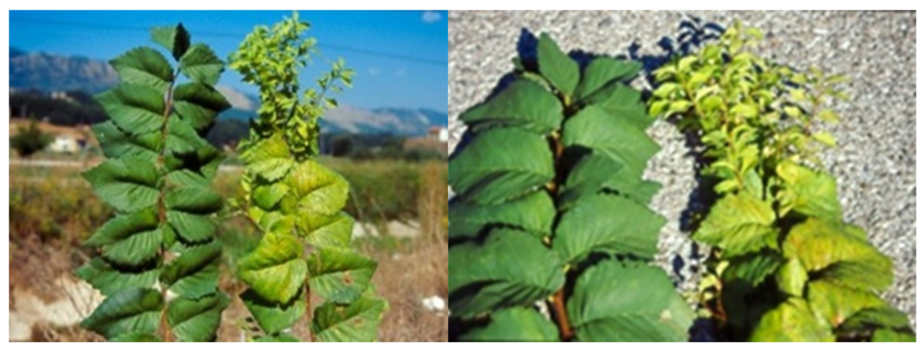
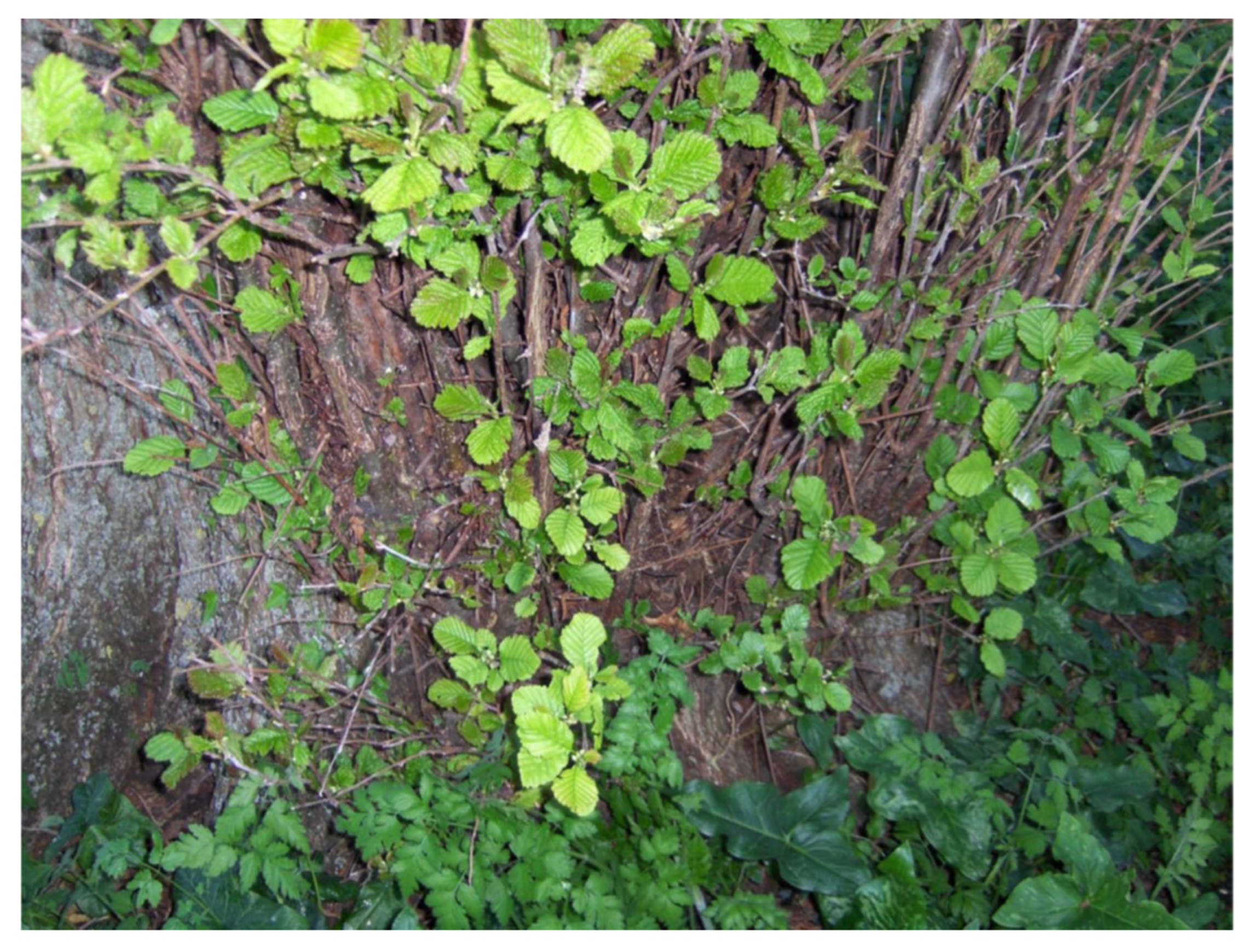
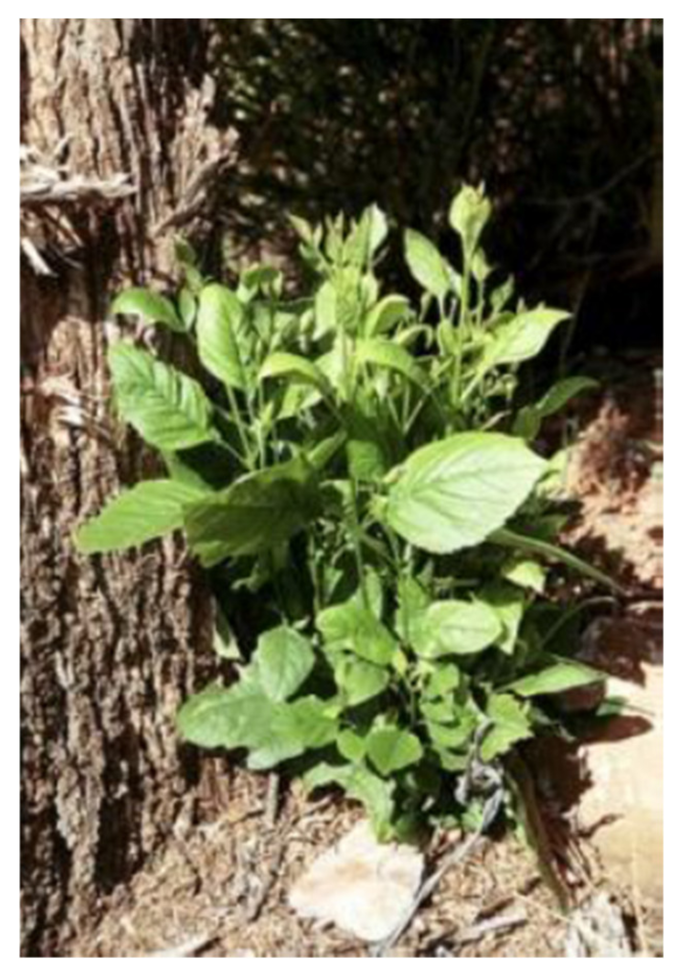
2.1. Elm
2.2. Alder
2.3. Ash
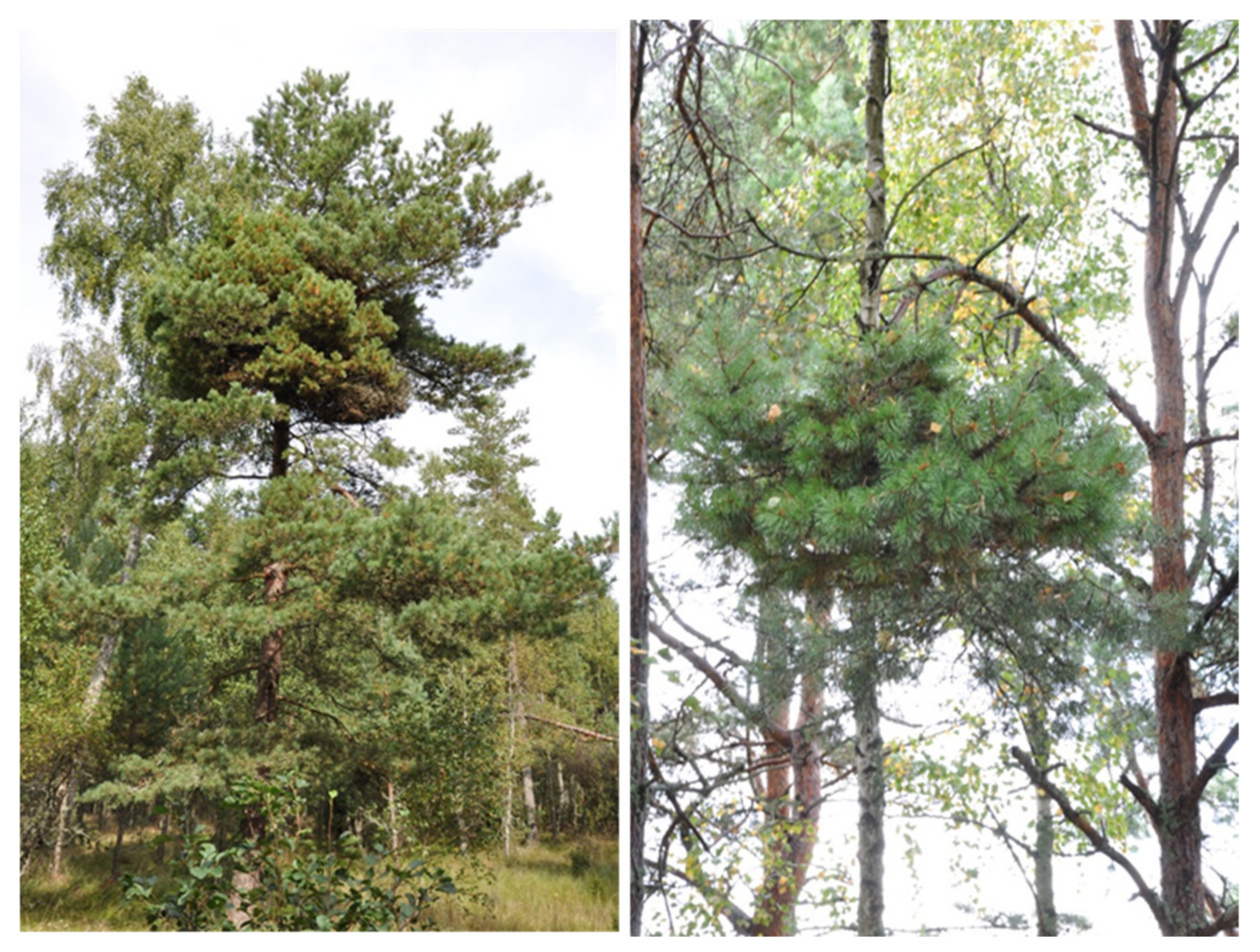
2.4. Conifers
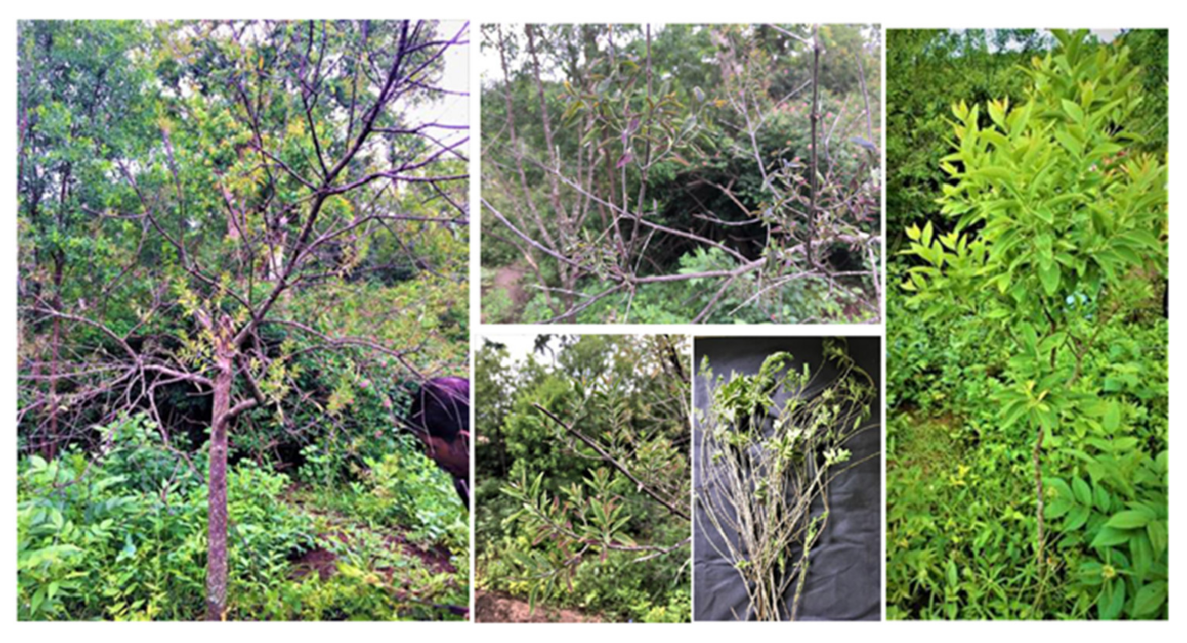
2.5. Sandal
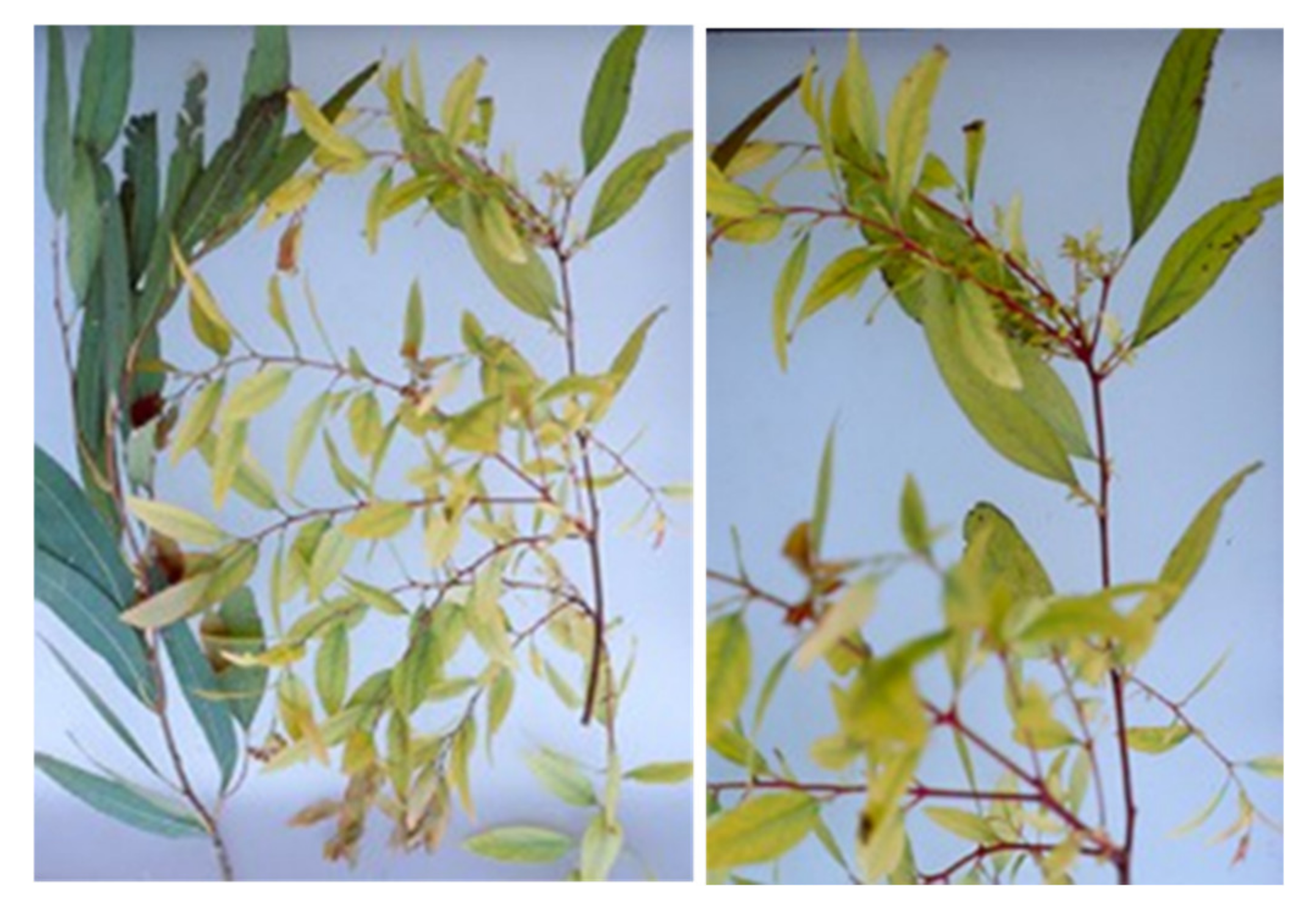
2.6. Eucalyptus
3. Discussion
4. Conclusions
Author Contributions
Funding
Institutional Review Board Statement
Informed Consent Statement
Data Availability Statement
Acknowledgments
Conflicts of Interest
References
- Weintraub, P.G.; Beanland, L. Insect vectors of phytoplasmas. Ann. Rev. Entomol. 2006, 51, 91–111. [Google Scholar] [CrossRef]
- IRPCM Phytoplasma/Spiroplasma Working Team—Phytoplasma Taxonomy Group. ‘Candidatus phytoplasma’, a taxon for the wall-less, non-helical prokaryotes that colonize plant phloem and insects. Int. J. Syst. Evol. Microbiol. 2004, 54, 1243–1255. [Google Scholar] [CrossRef] [PubMed]
- Martini, M.; Marcone, C.; Lee, I.-M.; Firrao, G. The family Acholeplasmataceae (including phytoplasmas). In The Prokaryotes—Firmicutes and Tenericutes, 4th ed.; Rosenberg, E., DeLong, E.F., Lory, S., Stackebrandt, E., Thompson, F., Eds.; Springer-Verlag: Berlin, Germany, 2014; Volume XXII, pp. 469–504. [Google Scholar]
- Bertaccini, A.; Lee, I.-M. Phytoplasmas: An update. In Phytoplasmas: Plant Pathogenic Bacteria-I. Characterization and Epidemiology of Phytoplasma-Associated Diseases; Rao, G.P., Bertaccini, A., Fiore, N., Liefting, L., Eds.; Springer Nature: Singapore, 2018; pp. 1–29. [Google Scholar]
- Zhao, Y.; Davis, R.E. Criteria for phytoplasma 16Sr group/subgroup delineation and the need of a platform for proper registration of new groups and subgroups. Int. J. Syst. Evol. Microbiol. 2016, 66, 2121–2123. [Google Scholar] [CrossRef] [PubMed]
- Marcone, C. Elm yellows: A phytoplasma disease of concern in forest and landscape ecosystems. For. Pathol. 2017, 47, e12324. [Google Scholar] [CrossRef]
- Swingle, R.U. A phloem necrosis of elm. Phytopathology 1938, 28, 757–759. [Google Scholar]
- Forbes, S.A. What is the matter with the elms in Illinois? Ill. Agr. Exp. Sta. Bull. 1912, 154, 1–22. [Google Scholar]
- Garman, H. The elms and their diseases. Kentucky Agr. Exp. Sta. Bull. 1899, 84, 51–75. [Google Scholar]
- Matteoni, J.A.; Sinclair, W.A. Elm yellows and ash yellows. In Tree Mycoplasmas and Mycoplasma Diseases; Hiruki, C., Ed.; University of Alberta Press: Edmonton, AB, Canada, 1988; pp. 19–31. [Google Scholar]
- Conti, M.; D’Agostino, G.; Mittempergher, L. A recent epiphytotic of elm yellows in Italy. In Proceedings of the 7th Congress of the Mediterranean Phytopathological Union, Granada, Spain, 20–27 September 1987; pp. 208–209. [Google Scholar]
- De Jonghe, K.; Deeren, A.-M.; Goedefroit, T.; Ronse, A. First report of ‘Candidatu Phytoplasma ulmi’ on elm in Belgium. Plant Dis. 2019, 103, 1763. [Google Scholar] [CrossRef]
- Schneider, B.; Kätzel, R.; Kube, M. Widespread occurrence of ‘Candidatus Phytoplasma ulmi’ in elm species in Germany. BMC Microbiol. 2020, 20, 74. [Google Scholar] [CrossRef] [PubMed]
- Braun, E.J.; Sinclair, W.A. Phloem necrosis of elms: Symptoms and histopathological observations in tolerant hosts. Phytopathology 1979, 69, 354–358. [Google Scholar] [CrossRef]
- Lee, I.-M.; Davis, R.E.; Sinclair, W.A.; De Witt, N.D.; Conti, M. Genetic relatedness of mycoplasmalike organisms detected in Ulmus spp. in the United States and Italy by means of DNA probes and polymerase chain reactions. Phytopathology 1993, 83, 829–833. [Google Scholar] [CrossRef]
- Mäurer, R.; Seemüller, E.; Sinclair, W.A. Genetic relatedness of mycoplasmalike organisms affecting elm, alder, and ash in Europe and North America. Phytopathology 1993, 83, 971–976. [Google Scholar] [CrossRef]
- Marcone, C.; Ragozzino, A.; Seemüller, E. Identification and characterization of the phytoplasma associated with elm yellows in southern Italy and its relatedness to other phytoplasmas of the elm yellows group. Eur. J. For. Pathol. 1997, 27, 45–54. [Google Scholar] [CrossRef]
- Seemüller, E.; Marcone, C.; Lauer, U.; Ragozzino, A.; Göschl, M. Current status of molecular classification of the phytoplasmas. J. Plant Pathol. 1998, 80, 3–26. [Google Scholar]
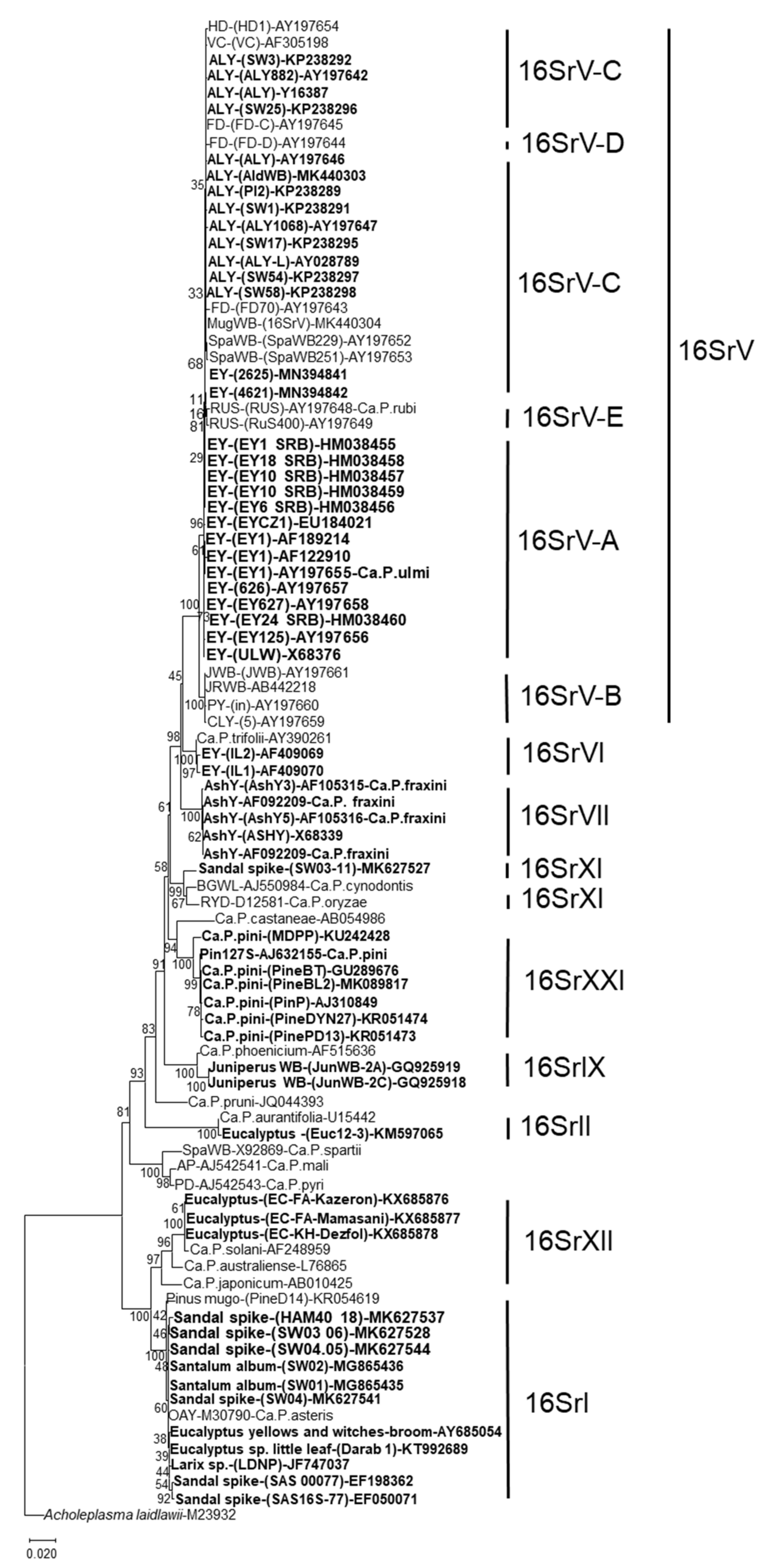
- Lee, I.-M.; Martini, M.; Marcone, C.; Zhu, S.F. Classification of phytoplasma strains in the elm yellows group (16SrV) and proposal of ‘Candidatus Phytoplasma ulmi’ for the phytoplasma associated with elm yellows. Int. J. Syst. Evol. Microbiol. 2004, 54, 337–347. [Google Scholar] [CrossRef] [PubMed]
- Arnaud, G.; Malembic-Maher, S.; Salar, P.; Bonnet, P.; Maixner, M.; Marcone, C.; Boudon-Padieu, E.; Foissac, X. Multilocus sequence typing confirms the close genetic interrelatedness of three distinct flavescence dorée phytoplasma strain clusters and group 16SrV phytoplasmas infecting grapevine and alder in Europe. Appl. Environ. Microbiol. 2007, 73, 4001–4010. [Google Scholar] [CrossRef]
- Jović, J.; Cvrković, T.; Mitrović, M.; Petrović, A.; Krnjajić, S.; Toševski, I. New strain of ‘Candidatus Phytoplasma ulmi’ infecting Ulmus minor and U. laevis in Serbia. Plant Pathol. 2008, 57, 1174. [Google Scholar] [CrossRef]
- Jović, J.; Cvrković, T.; Mitrović, M.; Petrović, A.; Krstić, O.; Krnjajić, S.; Toševski, I. Multigene sequence data and genetic diversity among ‘Candidatus Phytoplasma ulmi’ strains infecting Ulmus spp. in Serbia. Plant Pathol. 2011, 60, 356–368. [Google Scholar] [CrossRef]
- Katanić, Z.; Krstin, L.; Ježić, M.; Zebec, M.; Ćurković-Perica, M. Molecular characterization of elm yellows phytoplasmas in Croatia and their impact on Ulmus spp. Plant Pathol. 2016, 65, 1430–1440. [Google Scholar] [CrossRef]
- Schneider, B.; Hüttel, B.; Zübert, C.; Kube, M. Genetic variation, phylogenetic relationship and spatial distribution of ‘Candidatus Phytoplasma ulmi’ strains in Germany. Sci. Rep. 2020, 10, 21864. [Google Scholar] [CrossRef]
- Kumar, S.; Stecher, G.; Li, M.; Knyaz, C.; Tamura, K. MEGA X: Molecular evolutionary genetics analysis across computing platforms. Mol. Biol. Evol. 2018, 35, 1547–1549. [Google Scholar] [CrossRef] [PubMed]
- Jacobs, K.A.; Lee, I.-M.; Griffiths, H.M.; Miller, F.D., Jr.; Bottner, K.D. A new member of the clover proliferation phytoplasma group (16SrVI) associated with elm yellows in Illinois. Plant Dis. 2003, 87, 241–246. [Google Scholar] [CrossRef]
- Lee, I.-M.; Bertaccini, A.; Vibio, M.; Gundersen, D.E.; Davis, R.E.; Mittempergher, L.; Conti, M.; Gennari, F. Detection and characterization of phytoplasmas associated with disease in Ulmus and Rubus in northern and central Italy. Phytopathol. Mediterr. 1995, 34, 174–183. [Google Scholar]
- Gao, R.; Wang, J.; Shao, Y.-H.; Li, X.-D.; Yang, B.-H.; Chang, W.C.; Zhao, W.-J.; Zhu, S.-F. Molecular identification of a phytoplasma associated with elm witches’-broom in China. For. Pathol. 2011, 41, 355–360. [Google Scholar] [CrossRef]
- De Jonghe, K.; Goedefroit, T.; Deeren, A.-M.; Fauche, F.; Steyer, S. A phytoplasma survey reveals the presence of ‘Candidatus Phytoplasma fragariae’ in Ulmus spp. and Acer pseudoplatanus in Belgium. For. Pathol. 2020, 50, e12635. [Google Scholar]
- Sinclair, W.A. Elm yellows in North America. In The Elms: Breeding, Conservation, and Disease Management; Dunn, C.P., Ed.; Kluwer Academic Publisher: Boston, MA, USA, 2000; pp. 121–136. [Google Scholar]
- Mittempergher, L. Elm yellows in Europe. In The Elms: Breeding, Conservation, and Disease Management; Dunn, C.P., Ed.; Kluwer Academic Publisher: Boston, MA, USA, 2000; pp. 103–119. [Google Scholar]
- Sfalanga, A.; Martini, M.; Surico, G.; Bertaccini, A. Involvement of phytoplasmas in a decline of Ulmus chenmoui in central Italy. For. Pathol. 2002, 32, 265–275. [Google Scholar] [CrossRef]
- Braun, E.J.; Sinclair, W.A. Histopathology of phloem necrosis in Ulmus americana. Phytopathology 1976, 66, 598–607. [Google Scholar] [CrossRef]
- Braun, E.J.; Sinclair, W.A. Translocation in phloem necrosis-diseased American elm seedlings. Phytopathology 1978, 68, 1733–1737. [Google Scholar] [CrossRef]
- Pisi, A.; Marani, F.; Bertaccini, A. Mycoplasma-like organisms associated with Elm witches’ broom symptoms. Phytopathol. Mediterr. 1981, 20, 189–191. [Google Scholar]
- Marcone, C.; Ragozzino, A.; Firrao, G.; Locci, R. Detection of elm witches’-broom agent in Basilicata, Southern Italy. Phytopathol. Mediterr. 1994, 33, 194–199. [Google Scholar]
- Carraro, L.; Ferrini, F.; Ermacora, P.; Loi, N.; Martini, M.; Osler, R. Macropsis mendax as a vector of elm yellows phytoplasma of Ulmus species. Plant Pathol. 2004, 53, 90–95. [Google Scholar] [CrossRef]
- Herath, P.; Hoover, G.A.; Angelini, E.; Moorman, G.W. Detection of elm yellows phytoplasma in elms and insects using real-time PCR. Plant Dis. 2010, 94, 1355–1360. [Google Scholar] [CrossRef]
- Rosa, C.; McCarthy, E.; Duong, K.; Hoover, G.; Moorman, G. First report of the spittlebug Lepyronia quadrangularis and the leafhopper Latalus sp. as vectors of the elm yellows associated phytoplasma, Candidatus Phytoplasma ulmi in North America. Plant Dis. 2014, 98, 154. [Google Scholar] [CrossRef] [PubMed]
- Boudon-Padieu, E.; Larrue, J.; Clair, D.; Hourdel, A.; Jeanneau, A.; Sforza, R.; Collin, E. Detection and prophylaxis of elm yellows phytoplasma in France. Invest. Agrar. Sist. Recu. For. 2004, 13, 71–80. [Google Scholar]
- Jović, J.; Cvrković, T.; Mitrović, M.; Petrović, A.; Krstić, O.; Krnjajić, S.; Toševski, I. Genetic variability among ‘Candidatus Phytoplasma ulmi’ strains infecting elms in Serbia and survey of potential vectors. In COST Action FA0807. Proceedings of the Current Status and Perspectives of Phytoplasma Disease Research and Management, Sitges, Spain, 1–2 February 2010; Bertaccini, A., Laviña, A., Torres, E., Eds.; IRTA: Cabrils, Spain, 2010; p. 18. [Google Scholar]
- Romanazzi, G.; Murolo, S. ‘Candidatus Phytoplasma ulmi’ causing yellows in Zelkova serrata newly reported in Italy. Plant Pathol. 2008, 57, 1174. [Google Scholar] [CrossRef]
- Arismendi, N.; Andrade, N.; Riegel, R.; Zamorano, A.; Fiore, N. Molecular identification of 16SrIII-J and 16SrV-A phytoplasmas in Ugni molinae and Amplicephalus curtulus. In Proceedings of the 20th Chilean Congress of Plant Pathology, Santiago, Chile, 19 November–1 December 2011; pp. 11–12. [Google Scholar]
- Sinclair, W.A.; Townsend, A.M.; Griffiths, H.M.; Whitlow, T.H. Responses of six Eurasian Ulmus cultivars to a North American elm yellows phytoplasma. Plant Dis. 2000, 84, 1266–1270. [Google Scholar] [CrossRef] [PubMed]
- Sinclair, W.A.; Townsend, A.M.; Sherald, J.L. Elm yellows phytoplasma lethal to Dutch elm disease-resistant Ulmus americana cultivars. Plant Dis. 2001, 85, 560. [Google Scholar] [CrossRef]
- Bertaccini, A.; Paltrinieri, S.; Contaldo, N. Standard detection protocol: PCR and RFLP analyses based on 16S rRNA gene. In Phytoplasmas: Methods and Protocols, Methods in Molecular Biology; Musetti, R., Pagliari, L., Eds.; Springer Science+Business Media, LLC: New York, NY, USA, 2019; Volume 1875, pp. 83–95. [Google Scholar]
- Martini, M.; Bottner-Parker, K.D.; Lee, I.-M. PCR-based sequence analysis on multiple genes other than 16S rRNA gene for differentiation of phytoplasmas. In Phytoplasmas: Methods and Protocols, Methods in Molecular Biology; Musetti, R., Pagliari, L., Eds.; Springer Science+Business Media, LLC: New York, NY, USA, 2019; Volume 1875, pp. 97–115. [Google Scholar]
- Marcone, C. Current status of phytoplasma diseases of forest and landscape trees and shrubs. J. Plant Pathol. 2015, 97, 9–36. [Google Scholar]
- Jurga, M.; Zwolińska, A. Artemisia vulgaris, a new host of 16SrV-C phytoplasma related strains infecting black alder in Poland. J. Phytopathol. 2020, 168, 659–667. [Google Scholar] [CrossRef]
- Lederer, W.; Seemüller, E. Occurrence of mycoplasma-like organisms in diseased and non-symptomatic alder trees (Alnus spp.). Eur. J. For. Pathol. 1991, 21, 90–96. [Google Scholar] [CrossRef]
- Foissac, X.; Salar, P.; Olivier, C.; Malembic-Maher, S. Proposal to establish a common taxon for 16SrV-C and 16SrV-D phytoplasmas and three genetic clades based on combined 16S rDNA signatures and variability of housekeeping genes. Phytopathogenic Mollicutes 2019, 9, 85–86. [Google Scholar] [CrossRef]
- Maixner, M.; Reinert, W. Oncopsis alni (Schrank) (Auchenorrhyncha: Cicadellidae) as a vector of the alder yellows phytoplasma of Alnus glutinosa (L.) Gaertn. Eur. J. Plant Pathol. 1999, 105, 87–94. [Google Scholar] [CrossRef]
- Holz, S.; Duduk, B.; Büttner, C.; Kube, M. Genetic variability of alder yellows phytoplasma in Alnus glutinosa in its natural Spreewald habitat. For. Pathol. 2016, 46, 11–21. [Google Scholar] [CrossRef]
- Kube, M.; Furch, A.C.U. Symptomless phytoplasmosis of black alder (Alnus glutinosa): A model for host tolerance. Plant Physiol. Rep. 2019, 24, 550–554. [Google Scholar] [CrossRef]
- Malembic-Maher, S.; Salar, P.; Carle, P.; Foissac, X. Ecology and taxonomy of flavescence doree phytoplasmas: The contribution of genetic diversity studies. In Progrès Agricole et Viticole, Hors Série, Proceedings of the Extended Abstracts of the 16th Meeting of ICVG, Dijon, France, 31 August–4 September 2009; Boudon-Padieu, E., Ed.; Le Progrès Agricole et Viticole: Montpellier, France, 2009; pp. 132–134. [Google Scholar]
- Ember, I.; ACS, Z.; Salar, P.; Danet, J.-L.; Foissac, X.; Kölber, M.; Malembic-Maher, S. Survey and genetic diversity of phytoplasmas from the 16SrV-C and -D subgroups in Hungary. Bull. Insectol. 2011, 64, 33–34. [Google Scholar]
- Marjanovic, M.; Stepanovic, J.; Rekanovic, E.; Kube, M.; Duduk, B. Alder yellows phytoplasmas in Alnus species in Serbia. Phytopathogenic Mollicutes 2019, 9, 57–58. [Google Scholar] [CrossRef]
- Jarausch, B.; Biancu, S.; Lang, F.; Desqué, D.; Salar, P.; Jarausch, W.; Foissac, X.; Malembic-Maher, S.; Maixner, M. Study of the epidemiology of “flavescence dorée” (FD)-related phytoplasmas and potential vectors in a FD-free area. Phytopathogenic Mollicutes 2019, 9, 59–60. [Google Scholar] [CrossRef]
- Filippin, L.; Jović, J.; Cvrković, T.; Forte, V.; Clair, D.; Toševski, I.; Boudon-Padieu, E.; Borgo, M.; Angelini, E. Molecular characteristics of phytoplasmas associated with Flavescence dorée in clematis and grapevine and preliminary results on the role of Dictyophora europaea as a vector. Plant Pathol. 2009, 58, 826–837. [Google Scholar] [CrossRef]
- Malembic-Maher, S.; Salar, P.; Filippin, L.; Carle, P.; Angelini, E.; Foissac, X. Genetic diversity of European phytoplasmas of the 16SrV taxonomic group and proposal of ‘Candidatus Phytoplasma rubi’. Int. J. Syst. Evol. Microbiol. 2011, 61, 2129–2134. [Google Scholar] [CrossRef] [PubMed]
- Malembic-Maher, S.; Salar, P.; Vergnes, D.; Foissac, X. Detection and diversity of “flavescence doree”-related phytoplasmas in alders surrounding infected vineyards in Aquitaine (France). Bull. Insectol. 2007, 60, 329–330. [Google Scholar]
- Mehle, N.; Rupar, M.; Seljak, G.; Ravnikar, M.; Dermastia, M. Molecular diversity of ‘flavescence dorée’ phytoplasma strains in Slovenia. Bull. Insectol. 2011, 64, 29–30. [Google Scholar]
- Desqué, D.; Salar, P.; Danet, J.-L.; Lusseau, T.; Garcion, C.; Moreau, E.; Dubus, C.; Dureuil, J.; Delbac, L.; Binet, D.; et al. Impact of Orientus ishidae on “flavescence dorée” emergence in the vineyards of riparian ecosystems. Phytopathogenic Mollicutes 2019, 9, 69–70. [Google Scholar] [CrossRef]
- Maixner, M.; Rüdel, M.; Daire, X.; Boudon-Padieu, E. Diversity of grapevine yellows in Germany. Vitis 1995, 34, 235–236. [Google Scholar]
- Maixner, M.; Reinert, W.; Darimont, H. Transmission of grapevine yellows by Oncopsis alni (Schrank) (Auchenorrhynca: Macropsinae). Vitis 2000, 39, 83–84. [Google Scholar]
- Angelini, E.; Clair, D.; Borgo, M.; Bertaccini, A.; Boudon-Padieu, E. Flavescence dorée in France and Italy—Occurrence of closely related phytoplasma isolates and their near relationships to Palatinate grapevine yellows and an alder yellows phytoplasma. Vitis 2001, 40, 79–86. [Google Scholar]
- Berges, R.; Seemüller, E. Impact of phytoplasma infection of common alder (Alnus glutinosa) depends on strain virulence. For. Pathol. 2002, 32, 357–363. [Google Scholar] [CrossRef]
- Sinclair, W.A.; Griffiths, H.M. Variation in aggressiveness of ash yellows phytoplasmas. Plant Dis. 2000, 84, 282–288. [Google Scholar] [CrossRef]
- Seemüller, E.; Kiss, E.; Sule, S.; Schneider, B. Multiple infection of apple trees by distinct strains of ‘Candidatus Phytoplasma mali’ and its pathological relevance. Phytopathology 2010, 100, 863–870. [Google Scholar] [CrossRef]
- Rossi, M.; Vallino, M.; Galetto, L.; Marzachì, C. Competitive exclusion of flavescence dorée phytoplasma strains in Catharanthus roseus plants. Plants 2020, 9, 1594. [Google Scholar] [CrossRef]
- Sinclair, W.A.; Griffiths, H.M.; Davis, R.E. Ash yellows and lilac witches’-broom: Phytoplasmal diseases of concern in forestry and horticulture. Plant Dis. 1996, 80, 468–475. [Google Scholar] [CrossRef]
- Griffiths, H.M.; Sinclair, W.A.; Smart, C.D.; Davis, R.E. The phytoplasma associated with ash yellows and lilac witches’-broom: ‘Candidatus Phytoplasma fraxini’. Int. J. Syst. Evol. Microbiol. 1999, 49, 1605–1614. [Google Scholar] [CrossRef]
- Kamińska, M.; Berniak, H. ‘Candidatus Phytoplasma asteris’ in Fraxinus excelsior and its association with ash yellows newly reported in Poland. Plant Pathol. 2009, 58, 788. [Google Scholar] [CrossRef]
- Perilla-Henao, L.M.; Dickinson, M.; Franco-Lara, L. First report of ‘Candidatus Phytoplasma asteris’ affecting woody hosts (Fraxinus udhei, Populus nigra, Pittosporum undulatum, and Croton spp.) in Colombia. Plant Dis. 2012, 96, 1372. [Google Scholar] [CrossRef] [PubMed]
- Filgueira, J.J.; Franco-Lara, L.; Salcedo, J.E.; Gaitan, S.L.; Boa, E.R. Urapan (Fraxinus udhei) dieback, a new disease associated with a phytoplasma in Colombia. Plant Pathol. 2004, 53, 520. [Google Scholar] [CrossRef]
- Filgueira, J.J.; Ávila, K.J.; López, K.R.; Mugno, P.A.; Zambrano, J.C.; Cruz, A.M.; Villamil-Garzón, L.A.; Rodríguez, A.M. Incidence and characterization of the presence of phytoplasmas in Fraxinus uhdei related with Ash Yellow disease in Colombia. J. Plant Dis. Prot. 2018, 125, 63–71. [Google Scholar] [CrossRef]
- Loiseau, M.; Massot, M.; Cousseau-Suhard, P.; Foissac, X.; Piou, D.; Robin, C. Are phytoplasmas associated with dieback symptoms of Fraxinus in France? Phytopathogenic Mollicutes 2019, 9, 53–54. [Google Scholar] [CrossRef]
- Sinclair, W.A.; Griffiths, H.M. Epidemiology of a slow-decline phytoplasmal disease: Ash yellows on old-fields sites in New York State. Phytopathology 1995, 85, 123–128. [Google Scholar] [CrossRef]
- Ioos, R.; Kowalski, T.; Husson, C.; Holdenrieder, O. Rapid in planta detection of Chalara fraxinea by a real-time PCR assay using a dual-labelled probe. Eur. J. Plant Pathol. 2009, 125, 329–335. [Google Scholar] [CrossRef]
- Sinclair, W.A.; Gleason, M.L.; Griffiths, H.M.; Iles, J.K.; Zriba, N.; Charlson, D.V.; Batzer, J.C.; Whitlow, T.H. Responses of 11 Fraxinus cultivars to ash yellows phytoplasma strains of differing aggressiveness. Plant Dis. 2000, 84, 725–730. [Google Scholar] [CrossRef]
- Sinclair, W.A.; Whitlow, T.H.; Griffiths, H.M. Heritable tolerance of ash yellows phytoplasmas in green ash. Can. J. For. Res. 1997, 27, 1928–1935. [Google Scholar] [CrossRef]
- Sinclair, W.A.; Griffiths, H.M.; Whitlow, T.H. Comparisons of tolerance of ash yellows phytoplasmas in Fraxinus species and rootstock-scion combinations. Plant Dis. 1997, 81, 395–398. [Google Scholar] [CrossRef]
- Hill, G.T.; Sinclair, W.A. Taxa of leafhoppers carrying phytoplasmas at sites of ash yellows occurrence in New York State. Plant Dis. 2000, 84, 134–138. [Google Scholar] [CrossRef] [PubMed]
- Schneider, B.; Torres, E.; Martín, M.P.; Schröder, M.; Behnke, H.-D.; Seemüller, E. ‘Candidatus Phytoplasma pini’, a novel taxon from Pinus silvestris and Pinus halepensis. Int. J. Syst. Evol. Microbiol. 2005, 55, 303–307. [Google Scholar] [CrossRef]
- Śliwa, H.; Kaminska, M.; Korszun, S.; Adler, P. Detection of ‘Candidatus Phytoplasma pini’ in Pinus sylvestris trees in Poland. J. Phytopathol. 2008, 156, 88–92. [Google Scholar] [CrossRef]
- Kamińska, M.; Berniak, H. Detection and identification of three ‘Candidatus Phytoplasma’ species in Picea spp. trees in Poland. J. Phytopathol. 2011, 159, 796–798. [Google Scholar] [CrossRef]
- Kamińska, M.; Berniak, H.; Obdrzalek, J. New natural host plants of ‘Candidatus Phytoplasma pini’ in Poland and the Czech Republic. Plant Pathol. 2011, 60, 1023–1029. [Google Scholar] [CrossRef]
- Ježić, M.; Poljak, I.; Šafarić, B.; Idžojtić, M.; Ćurković-Perica, M. ‘Candidatus Phytoplasma pini’ in pine species in Croatia. J. Plant Dis. Prot. 2013, 120, 160–163. [Google Scholar] [CrossRef]
- Valiunas, D.; Jomantiene, R.; Ivanauskas, A.; Urbonaite, I.; Sneideris, D.; Davis, R.E. Molecular identification of phytoplasmas infecting diseased pine trees in the UNESCO-protected Curonian Spit of Lithuania. Forests 2015, 6, 2469–2483. [Google Scholar] [CrossRef]
- Costanzo, S.; Rascoe, J.; Zhao, Y.; Davis, R.E.; Nakhla, M.K. First report of a new ‘Candidatus Phytoplasma pini’-related strain associated with witches’-broom of Pinus spp. in Maryland. Plant Dis. 2016, 100, 1776. [Google Scholar] [CrossRef]
- Huang, S.; Tiwari, A.K.; Rao, G.P. ‘Candidatus Phytoplasma pini’ affecting Taxodium distichum var. imbricarium in China. Phytopathogenic Mollicutes 2011, 1, 91–94. [Google Scholar] [CrossRef]
- Paltrinieri, S.; Martini, M.; Pondrelli, M.; Bertaccini, A. X-disease-related phytoplasmas in ornamental trees and shrubs with witches’-broom and malformation symptoms. J. Plant Pathol. 1998, 80, 261. [Google Scholar]
- Jomantiene, R.; Valiunas, D.; Ivanauskas, A.; Urbanaviciene, L.; Staniulis, J.; Davis, R.E. Larch is a new host for a group 16SrI, subgroup B, phytoplasma in Ukraine. Bull. Insectol. 2011, 64, S101–S102. [Google Scholar]
- Davis, R.E.; Dally, E.L.; Zhao, Y.; Lee, I.-M.; Jomantiene, R.; Detweiler, A.J.; Putnam, M.L. First report of a new subgroup 16SrIX-E (‘Candidatus Phytoplasma phoenicium’-related) phytoplasma associated with juniper witches’ broom disease in Oregon, USA. Plant Pathol. 2010, 59, 1161. [Google Scholar] [CrossRef]
- Gupta, M.K.; Samad, A.; Shasany, A.K.; Ajayakumar, P.V.; Alam, M. First report of a 16SrVI ‘Candidatus Phytoplasma trifolii’ isolate infecting Norfolk Island pine (Auraucaria heterophylla) in India. Plant Pathol. 2010, 59, 399. [Google Scholar] [CrossRef]
- Valiunas, D.; Jomantiene, R.; Ivanauskas, A.; Sneideris, D.; Zizyte-Eidetiene, M.; Shao, J.; Zhao, Y.; Costanzo, S.; Davis, R.E. Rapid detection and identification of ‘Candidatus Phytoplasma pini’-related strains based on genomic markers present in 16S rRNA and tuf genes. For. Pathol. 2019, 49, e12553. [Google Scholar] [CrossRef]
- Cai, W.; Shao, J.; Zhao, Y.; Davis, R.E.; Costanzo, S. Draft genome sequence of ‘Candidatus Phytoplasma pini’-related strain MDPP: A resource for comparative genomics of gymnosperm-infecting phytoplasmas. Plant Dis. 2020, 104, 1009–1010. [Google Scholar] [CrossRef]
- Khan, J.A.; Sing, S.K.; Ahmad, J. Characterisation and phylogeny of a phytoplasma inducing sandal spike disease in sandal (Santalum album). Ann. Appl. Biol. 2008, 153, 365–372. [Google Scholar] [CrossRef]
- Teixeira da Silva, J.A.; Kher, M.M.; Soner, D.; Nataraj, M. Sandalwood spike disease: A brief synthesis. Environ. Exp. Bot. 2016, 14, 199–204. [Google Scholar] [CrossRef]
- Murali, R.; Rangaswamy, K.T.; Nagaraja, H. Occurrence of spike disease in sandal plantations in southern Karnataka and cross transmission studies between sandal spike and Stachytarpheta phyllody through Dodder (Cuscuta subinclusa). Int. J. Curr. Microbiol. App. Sci. 2019, 8, 1041–1046. [Google Scholar] [CrossRef]
- Mondal, S.; Sundararaj, R.; Rao, H.C.Y. A critical appraisal on the recurrence of sandalwood spike disease and its management practices. For. Pathol. 2020, 50, e12648. [Google Scholar] [CrossRef]
- Rao, M.S.; Muniyappa, V. Epidemiology of sandal spike-disease. In Tree Mycoplasmas and Mycoplasma Diseases; Hiruki, C., Ed.; University of Alberta Press: Edmonton, AB, Canada, 1988; pp. 57–58. [Google Scholar]
- Lee, I.-M.; Gundersen-Rindal, D.E.; Davis, R.E.; Bottner, K.D.; Marcone, C.; Seemüller, E. ‘Candidatus Phytoplasma asteris’, a novel phytoplasma taxon associated with aster yellows and related diseases. Int. J. Syst. Evol. Microbiol. 2004, 54, 1037–1048. [Google Scholar] [CrossRef]
- Kirdat, K.; Sundararaj, R.; Mondal, S.; Reddy, M.K.; Thorat, V.; Yadav, A. Novel aster yellows phytoplasma subgroup associated with sandalwood spike disease in Kerala, India. Phytopathogenic Mollicutes 2019, 9, 33–34. [Google Scholar] [CrossRef]
- Ramachandran, S.; Kirdat, K.; Mondal, S.; Reddy, M.K.; Thorat, V.; Rishi, R.; Yadav, A. A threat to sandalwood cultivation in the naturalised Marayoor sandalwood reserve (Kerala, India) through single and mixed phytoplasma infections. Phytopathogenic Mollicutes 2020, 10, 89–95. [Google Scholar] [CrossRef]
- Dijkstra, J.; Lee, P.E. Transmission by dodder of sandal spike disease and the accompanying mycoplasma-like organisms via Vinca rosea. Neth. J. Plant Pathol. 1972, 78, 218–228. [Google Scholar] [CrossRef]
- Sastry, K.S.M.; Thakur, R.N.; Gupta, J.H.; Pandotra, V.R. Three virus disease of Eucalyptus citriodora. Ind. Phytopathol. 1971, 24, 123–126. [Google Scholar]
- Marcone, C.; Ragozzino, A.; Seemüller, E. Detection of an elm yellows-related phytoplasma in eucalyptus trees affected by little leaf disease in Italy. Plant Dis. 1996, 80, 669–673. [Google Scholar] [CrossRef]
- De Souza, A.N.; de Carvalho, S.L.; da Silva, F.N.; Alfenas, A.C.; Valverde Zauza, E.A.; Carvalho, C.M. First report of phytoplasma associated with Eucalyptus urophylla showing witches’ broom in Brazil. Phytopathogenic Mollicutes 2015, 5, S83–S84. [Google Scholar] [CrossRef]
- Salehi, M.; Esmailzadeh Hosseini, S.A.; Salehi, E. First report of a ‘Candidatus Phytoplasma asteris’ related phytoplasma associated with eucalyptus little leaf disease in Iran. J. Plant Pathol. 2016, 98, 175. [Google Scholar]
- Azimi, M.; Farokhi-Nejad, R.; Mehrabi-Koushki, M. First report of a ‘Candidatus Phytoplasma aurantifolia’-related strain associated with leaf roll symptoms on eucalyptus in Iran. New Dis. Rep. 2017, 35, 4. [Google Scholar] [CrossRef]
- Baghaee-Ravari, S.; Jamshidi, E.; Falahati-Rastegar, M. “Candidatus Phytoplasma solani” associated with Eucalyptus witches’ broom in Iran. For. Pathol. 2018, 48, e12394. [Google Scholar] [CrossRef]


Publisher’s Note: MDPI stays neutral with regard to jurisdictional claims in published maps and institutional affiliations. |
© 2021 by the authors. Licensee MDPI, Basel, Switzerland. This article is an open access article distributed under the terms and conditions of the Creative Commons Attribution (CC BY) license (http://creativecommons.org/licenses/by/4.0/).
Share and Cite
Marcone, C.; Valiunas, D.; Mondal, S.; Sundararaj, R. On Some Significant Phytoplasma Diseases of Forest Trees: An Update. Forests 2021, 12, 408. https://doi.org/10.3390/f12040408
Marcone C, Valiunas D, Mondal S, Sundararaj R. On Some Significant Phytoplasma Diseases of Forest Trees: An Update. Forests. 2021; 12(4):408. https://doi.org/10.3390/f12040408
Chicago/Turabian StyleMarcone, Carmine, Deividas Valiunas, Soma Mondal, and Ramachandran Sundararaj. 2021. "On Some Significant Phytoplasma Diseases of Forest Trees: An Update" Forests 12, no. 4: 408. https://doi.org/10.3390/f12040408
APA StyleMarcone, C., Valiunas, D., Mondal, S., & Sundararaj, R. (2021). On Some Significant Phytoplasma Diseases of Forest Trees: An Update. Forests, 12(4), 408. https://doi.org/10.3390/f12040408