Article

Detecting Heterobasidion irregulare in Minnesota and Assessment of Indigenous Fungi on Pines

1
Division of Forestry, Minnesota Department of Natural Resources, Grand Rapids, MN 55744, USA
2
Department of Plant Pathology, University of Minnesota, St. Paul, MN 55108, USA
*
Author to whom correspondence should be addressed.
Currently at Division of Biological Sciences, University of California San Diego, La Jolla, CA 92093, USA.
Forests 2021, 12(1), 57; https://doi.org/10.3390/f12010057
Submission received: 8 December 2020 / Revised: 29 December 2020 / Accepted: 30 December 2020 / Published: 5 January 2021
(This article belongs to the Section Forest Ecophysiology and Biology)

Abstract

:
Heterobasidion irregulare is one of the most problematic forest pathogens in the northern hemisphere, but has only been found relatively recently in the north central United States. Discovered in Wisconsin in 1993, but probably established sometime before that, it quickly spread throughout the state. In November 2014, it was found in southeastern Minnesota. Field surveys were then conducted throughout Minnesota with the focus in the southeast near the initial discovery. To find additional infection sites, surveys were conducted with accompanying aerial imagery of red pine (Pinus resinosa Aiton) stands that were previously thinned. Samples were collected from selected sites with dead and dying trees as well as samples from stumps in recently thinned pine stands. These samples were processed first with a nested polymerase chain reaction (PCR) protocol, which was replaced by a real-time PCR assay after its development. No samples tested positive for H. irregulare using these methods and no cultures from isolations were obtained outside the original infection area. Other indigenous fungi were also identified. The majority were wood decay fungi in the Basidiomycota. A spore collection study was also conducted after field surveys. Automated rotary arm spore collectors were used and assayed with an ITS TaqMan real-time PCR assay. Collectors were placed strategically in different areas of Minnesota. A positive control was used in an infected red pine plantation in Wisconsin and this location had the highest number of spores trapped, with 63,776 over a week period. Spores of H. irregulare were detected at several sites in Minnesota, with the highest spore total observed in traps at 413 over a week period. All other locations sampled also had some spores collected except Itasca State Park located in northwestern Minnesota. The weekly deposition of spores ranged from 0 to 1.26 m−2 h−1. Low spore levels occurring in Minnesota indicate that some spores are present, but they are currently being detected in amounts that may not be sufficient for colonization to be successful.

1. Introduction

The fungal pathogen, Heterobasidion irregulare Garbelotto & Otrosina, is a serious threat to red pine (Pinus resinosa Aiton) and other conifers throughout North America [1]. It is considered one of the most economically damaging forest pathogens in the northern temperate regions of the world. In addition to causing serious economic damage, Heterobasidion root disease (HRD) impacts ecosystem services and can be devastating in urban environments where trees weakened by infection can pose hazards to the general public.
The primary infection is by airborne basidiospores produced by shelf-like basidiocarps [2]. Basidiospores can be disseminated overland to start new infections on recently cut stumps and wounds. The asexual state, Spiniger meineckellus (A.J. Olson) Staplers, produces large numbers of conidiophores and conidia that can form over the surface of exposed infected wood. The role of conidia for infection, however, is not well understood, but they may play a role in local spread within stumps and roots [3]. Managed forests are primarily affected through thinning operations. Cut stumps and to a lesser extent wounds created on residual trees from timber operations can serve as an entry point for airborne spores. After colonization of cut stumps, the pathogen moves into the root system and to adjacent trees by growing through the roots and root grafts to living trees [4]. As the disease progresses on a site, circular infection centers are produced that continually expand, producing circles of death [5].
Currently, the known geographic range of H. irregulare in eastern North America is from eastern Texas through the southeastern United States and north to Wisconsin in the Great Lakes Region, New England, and Eastern Canada [6]. Heterobasidion irregulare is part of the species complex Heterobasidion annosum sensu lato (s.l). Long regarded as a single species, mating studies with H. annosum revealed several intersterile groups (ISGs) [7,8,9]. This species complex is now considered to consist of three species in Eurasia and two in North America [10,11]. In Europe, H. annosum (Fr.) Bref. sensu stricto (s.s.), H. parviporum Niemela & Korhonen, and H. abietinum Niemela & Korhonen are present and in North America, H. irregulare, and H. occidentale Otrosina & Garbelotto are the dominant species.
The disease was first found in Adams County, Wisconsin in the fall of 1993, followed by additional sites in the spring of 1994 in Iowa County, Wisconsin. Both locations had symptomatic trees and basidiocarps present [12]. Periodic surveys for this disease have been done in Minnesota since the initial discovery in Wisconsin. However, prior to that, on two separate occasions in the 1970s, two basidiocarps identified as Heterobasidion annosum were found in Itasca State Park in Minnesota. However, the basidiocarps were not found on species of pine, but on dead white spruce (Picea glauca (Moench) Voss) and balsam fir (Abies balsamea (L.) Mill.) in an old growth protected forest. These were unusual findings since the species commonly infecting these genera in the western United States was Heterobasidion occidentale. We speculate that an isolated population of H. occidentale may have existed on spruce and fir in this remnant old growth forest located in north central Minnesota. The basidiocarps are deposited as herbarium specimens in the U.S.D.A. Forest Service’s Center for Forest Mycology Research in Madison, Wisconsin. Attempts to obtain ribosomal DNA from these basidiocarps through DNA extraction and sequencing was not successful and their species identity remains unknown. No other collections of this fungus have been found in Minnesota.
It was not until late 2014 that this destructive disease was found in Winona County, Minnesota [13]. It was located in a small red pine plantation in southeast Minnesota. At the time of the survey, the age of the stand was 49 years old. The plantation was thinned from December 2003 to January 2004. Even though thinned in the winter, temperatures for these two months were above 0 °C for numerous days according to a weather station located in Thielman, MN, 26 km to the northwest of the site [14]. There was also no snow cover reported for a period of time during these two months. The warm temperatures and lack of snow cover would have made infection from H. irregulare possible. The pathogen was likely introduced via long distance dispersal of basidiospores from Wisconsin (closest infection center is approximately 67–68 km away) or by contaminated logging equipment used to thin the site. The transmission of H. irregulare on logging equipment, however, has not been previously reported.
At the site in Winona County, there was an infection center with adjacent trees having thin crowns. Infected wood was collected from dead and dying trees and pure cultures were isolated. Genomic DNA was extracted from pure cultures and directly from the basidiocarps. Amplification and sequencing of the ITS gene confirmed the identity of H. irregulare. Basidiocarps were later found a small distance away from the main infection center. Genomic rDNA was obtained directly from the basidiocarps through DNA extraction and sequencing [13].
The HRD situation in Wisconsin escalated quickly after it was discovered in 1993. This was due to the lack of a rapid detection method for the disease and a delay in managing the pathogen. The disease is now found in three counties of Wisconsin adjacent to Minnesota. Additionally, as of December 2020, the Wisconsin Department of Natural Resources reports HRD has been confirmed in 28 counties [15]. Therefore, it was important to establish a Minnesota program to detect this pathogen early to prevent its spread. Since Minnesota is at the leading edge of a potential epidemic, this study was conducted to determine the distribution of H. irregulare in Minnesota through field surveys for diseased trees and spore trapping to detect and calculate the density of basidiospores. In addition, culturing studies from dead/dying trees and stumps were conducted to isolate the pathogen as well as identify other indigenous fungi that were present. Indigenous fungi that were isolated were recorded to help determine what native fungi may be present and influencing any outbreaks since some saprophytic fungi, such as Phlebiopsis gigantea, have previously been shown to control the disease [16,17,18,19].

2. Materials and Methods

2.1. Field Surveys

Intensified surveys for H. irregulare started in the spring of 2015 (Figure 1). Surveys focused on red and eastern white pine (Pinus strobus L.) growing areas in southeastern Minnesota, where the first location of H. irregulare had been found and these areas are adjacent to counties in Wisconsin with the disease. Areas in the north and east central regions of Minnesota were also surveyed due to the importance of the pine resource there (Figure 1). The Minnesota Department of Natural Resources provided aerial maps of multiple survey sites and assisted with the surveys. The aerial maps were created to help navigate at sites and also helped detect large pockets of dead and declining trees. Survey sites chosen were focused on red and white pine stands that were thinned recently and up to 20 years ago.
Samples were collected from declining and recently dead trees, stumps, roots, and woody debris. Additionally, basidiocarps resembling H. irregulare were collected. Samples were placed in sterile bags, kept cool during transport, and brought to the University of Minnesota for processing. Genomic DNA was extracted from the majority of wood samples initially with the MO BIO PowerPlant® Pro DNA Isolation Kit (MO BIO Laboratories, Inc., Carlsbad, CA, USA) and also with the Qiagen DNeasy® PowerPlant® Pro Kit (Qiagen, Valencia, CA, USA) following the manufacturer’s protocol. A cetyl trimethylammonium bromide (CTAB) DNA extraction protocol [20] was used for basidiocarp samples and for a set of woody samples. The use of this protocol for woody samples was discontinued, however, due to the DNA kits yielding a more pure DNA extract. Initially, samples were processed with conventional PCR using a nested approach that involved the use of two H. irregulare specific primer sets and two successive PCR reactions. PCR amplification for the first round was targeted for the internal transcribed spacer (ITS) region of rDNA with the primer pair ITS1F/4 [21]. PCR amplification reagents included 12.5 µL of 1X GoTaq Green Master Mix (Promega, Madison, WI, USA), 9.5 µL of sterile water, 1 µL of each primer (0.2 µM), 0.5 µL BSA (0.1 mg/µL), and 1 µL of template DNA in a thermocycler with the following parameters: 94 °C for 5 min, 35 cycles of 94 °C for 1 min, 50 °C for 1 min, 72 °C for 1 min, and a final extension step of 5 min at 72 °C. A 1% agarose gel using SYBR green 1 prestain (Life Technologies, Grand Island, NY, USA) and a Dark Reader DR45 transilluminator (Clare Chemical Research, Denver, CO, USA) was used to visualize PCR amplicons. If PCR amplicons were present, then these samples were used in the second round of PCR. For the second round, specific primers for the genus Heterobasidion were used. These primers were Het_4F (CGAATATCGTGCAAGGTTGAAG) and Het_1R (CCACAAGGGTCTCGCTAATG). The primers were developed in our laboratory at the University of Minnesota and were tested to determine amplification efficiency and accuracy for the genus of Heterobasidion. One µL of the ITS1F/4 PCR product was used and diluted 1/50 in sterile water. The same reagents were used as for the first round. Different thermocycler parameters were used: 95 °C for 4 min, 35 cycles of 95 °C for 1 min, 53 °C for 45 s, 72 °C for 1 min, and a final extension step at 72 °C for 5 min. Additionally, positive and negative controls were used in both rounds of PCR. The positive control was DNA of H. irregulare from a culture that originated from the location positive for H. irregulare in Minnesota. The negative control was sterile water and DNA extracted from a wood sample and negative for H. irregulare via PCR and sequencing. PCR amplicons were visualized using the same methods mentioned previously. Sequencing reactions were performed using the forward primer, ITS1F, for PCR amplicons from the first PCR round using an ABI Prism 3730xl DNA sequencer (Applied Biosystems, Foster City, CA, USA). The majority of DNA sequenced was extracted from fungal cultures isolated from samples. A small amount of DNA was sequenced from sporophore and wood samples via a direct DNA extraction. Assembly of consensus sequences was done using Geneious 9.0 [22] and compared to sequences in the GenBank using BLASTn for identification.

2.2. qPCR Analysis

A real-time PCR protocol was then developed. A LightCycler 96 real-time PCR system (Roche Applied Science, Penzberg, Germany) was used to carry out the reactions. The reagents consisted of 10 µL of 1× FastStart Essential DNA Probes Master (Roche Applied Science), 1 µL of each primer (0.5 µM) (Hirregulare_4A/G_F435 (CATTCTGAAGACATACGAGGGA) and Hirregulare_R500 (GGTCGGGTTCTTTTGAT)), 2 µL of probe (0.1 µM) (Hannosum_ss_irregulare_T464), 0.5 µL of BSA (0.125 mg/µL), and 0.5 µL of sterile water. A total of 5 µL of DNA was used, which was diluted 1/5 with sterile water; this was to minimize pipetting error. Thus, a total reaction volume of 20 µL. The following parameters were used: preincubation at 98 °C for 90 s and a two-step amplification of 45 cycles, 95 °C for 10 s, 58 °C for 25 s. A synthetic standard of the whole ITS region of H. irregulare synthesized using gBlocksTM by Integrated DNA Technologies (IDT) was used to create the standard curves and a correlation coefficient. The stock of the synthetic standard had a concentration of 1.451 × 10¹⁰ H. irregulare ITS copies/µL. A set of four dilutions were made of the synthetic standard with concentrations of 145,100,000, 14,510,000, 145,100, 14,510, and 145.1 H. irregulare ITS copies/µL. After the reaction, the Ct values were plotted against log H. irregulare ITS copy concentration. The lower limit of detection was determined to be 50 H. irregulare ITS copies.

2.3. Culture Methods

Additionally, small segments of wood were cut from samples with aseptic techniques and incubated on culture media. The majority of samples were cultured with a semi-selective media for Basidiomycota (BSA). This media was modified from the original recipe and included 15 g of agar, 15 g of malt extract, 2 g of yeast, and 0.06 g of benlate added to 1 L of water and after autoclaving, 0.1 g of streptomycin sulfate and 2 mL of lactic acid was added [23]. Plates were grown at room temperature (~20°) and checked often for fungal growth that was then sub-cultured onto new BSA. After cultures were grown out, a cetyl trimethylammonium bromide (CTAB) protocol was used to extract DNA from each isolate as well as basidiocarp samples. The primer pair ITS1F/4 was used for PCR amplification. Amplification, sequencing, and sequence assembly was conducted using the previously mentioned methods used for wood samples.

2.4. Spore Surveys

Spore trapping methods followed those used to trap H. irregulare basidiospores in Quebec [24,25]. Two automated rotary arm spore collectors (Aerobiology Research Laboratories, Ottawa, ONT, Canada) were programmed to spin 4 min every 60 min, with rods (1.5 mm × 1.5 mm × 3 cm) placed in the rotary arms that were coated with a 7% silicone-hexane solution. Spore collectors were placed in an open forested setting and moved to different locations after disinfecting with a 10% dilution of bleach, from September to November 2018. The period from late summer to early fall was chosen as the deployment time based on previous research demonstrating that peak deposition is during late summer and fall [5]. The spore collectors were placed at locations previously sampled for H. irregulare and were points of interest due to their proximity to the closest infection site in Wisconsin (Figure 2). Additionally, spore collectors were placed in Itasca State Park due to the finding of possible Heterobasidion basidiocarps on two separate occasions in the 1970s. Spore collectors were deployed for 7 days at each location except for the Wisconsin location, which had deployment for 8 days and the Itasca locations for 9 days. Rods were then collected and placed in separate 1.5 mL microcentrifuge tubes until processed.
Rods were brought to the University of Minnesota and a DNA extraction was performed on each rod based on that of Lamarche et al. (2016) [26]. One mL of Goof Off Pro Strength Remover (W.M. Barr, Memphis, TN, USA) was added to each tube. The rods were vortexed for 30 s or until the rod was totally dissolved. The tubes were centrifuged for 2 min at 20,000× g and the supernatant was discarded. One mL of Goof Off Pro Strength Remover was used to rinse the spore pellet and then discarded. The tube was then left to dry in a bio-safety cabinet for 1 h. A 3 mm stainless steel bead, 180 µL of buffer ATL, and 1 µL of reagent DX (Qiagen) were added to each tube and placed in a MagNA Lyser (Roche Applied Science, Penzberg, Upper Bavaria, Germany) to lyse the samples for 90 s. Next, 20 µL of Proteinase K was added and was mixed by vortexing for 15 s. The samples were then incubated at 56 °C in a water bath overnight. The following day, DNA extraction was performed using the QIAamp® DNA Micro Kit (Qiagen) and following the manufacture’s protocol.
Real-time PCR was performed to obtain basidiospore counts. A ribosomal ITS TaqMan assay was performed based on that of Lamarche et al. (2016) [26]. The reagents consisted of 5 µL of 1× FastStart Essential DNA Probes Master (Roche Applied Science), 1 µL of each primer (0.5 µM), 1 µL of TaqMan probe (0.1 µM), 1 µL of sterile water, and 2 µL of template DNA for a total reaction volume of 11 µL. The amount of template DNA was 2 µL due to the possible risk of pipetting error. Reactions took place on a LightCycler 96 (Roche Applied Science). Calculations of basidiospore equivalents per rod were calculated by translating Ct values using standard curve equations. A mono-basidiosporal isolate of H. irregulare (#98–008) was used to create the standard curves and a correlation coefficient. The isolate was obtained from the Laurentian Forestry Centre culture collection (Canadian Forest Service, Natural Resources Canada) and originated from a basidiocarp sampled in Lac La Blanche, Quebec, Canada. The isolate was confirmed to have 1–2 nuclei/conidia. The protocol to develop the standard curve equations using the mono-basidiosporal isolate of H. irregulare (#98–008) was based on that of Lamarche et al. (2016) [26]. The standard curve produced had a correlation coefficient (r2) of 0.95.
The total amount of basidiospores sampled per m3 was calculated by using the following formula: (number of basidiospores per rod × 1000 L−1 m−3 h−1)/(47.28 L min−1 per rod × 60 min h−1 × 10.73 h). The weekly amount of basidiospores per m3 was divided by 10.73 h to obtain the amount of basidiospores m−2 h−1 for each week. This assumed that the total basidiospores falling on the m2 cross-section was obtained by summing all spores in the m−3 h−1 [27]. Furthermore, the total amount of basidiospores that deposit on a 30-cm stump was calculated by using the following formula: total amount of basidiospores m−3 of a site × (stump radius2 × π). Since the spore collectors were at the Wisconsin site for 8 days, exposure time was a total of 12.27 h and exposure time was 13.8 h at Itasca State Park with spore collectors deployed for 9 days. Calculations were determined for each of the two rods at one location and averaged.

3. Results

3.1. Field Surveys

A total of 949 samples were collected from field survey sites throughout Minnesota (Figure 1). These samples were from dead and dying trees (579), stumps (252), sporophores (87), and downed trees and slash (31). A multitude of different fungi were isolated and subsequently sequenced from collected samples. However, no definitive positive results for H. irregulare were found outside the original H. irregulare infection site. With both conventional PCR and later real-time PCR, H. irregulare was not detected in wood samples. Additionally, isolating from wood did not yield any cultures of H. irregulare except for the original positive location near Minneiska, Minnesota.
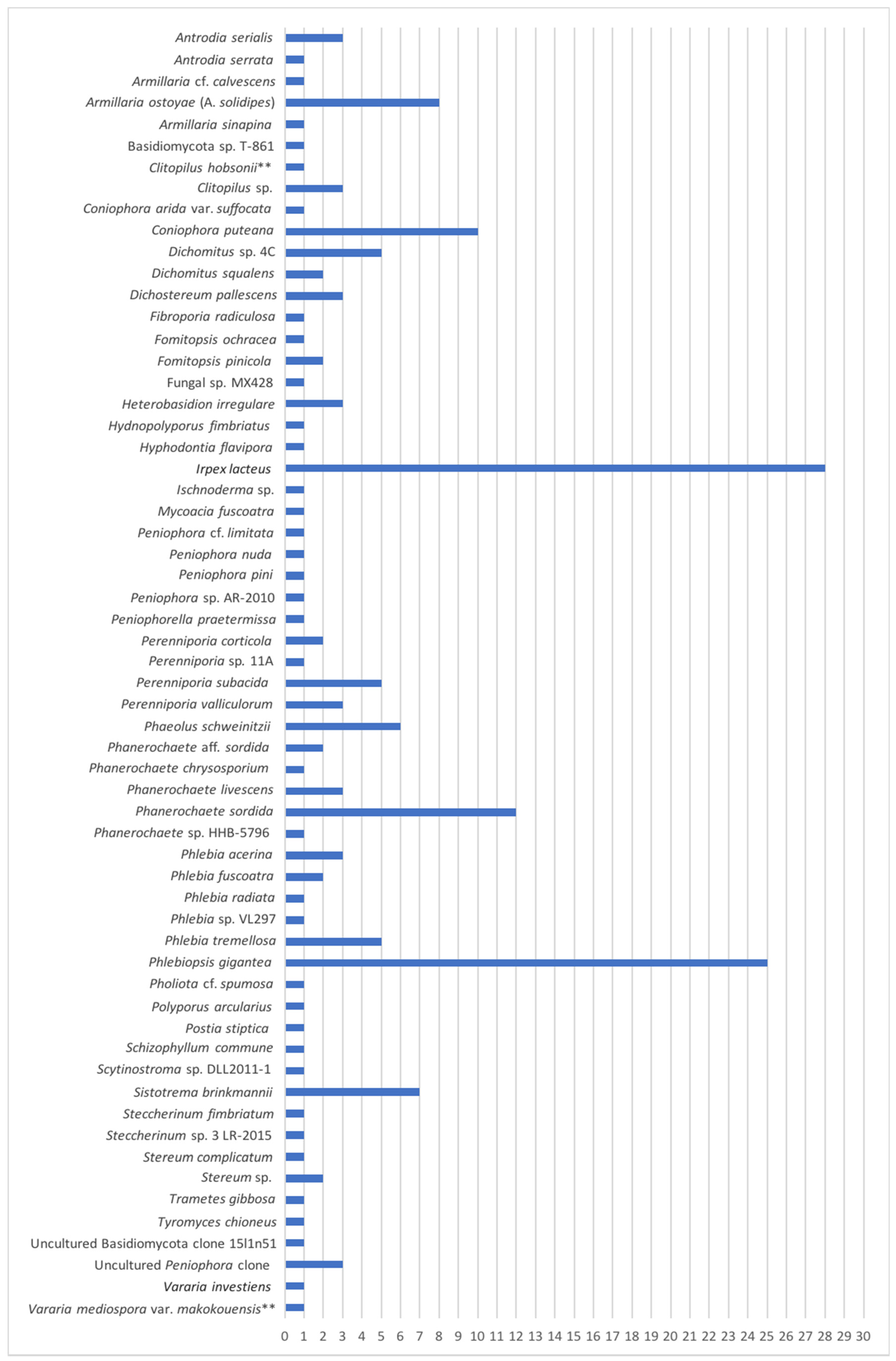
The majority of the fungi isolated and sequenced were fungi in the Basidiomycota. A total of 183 pure cultures were obtained and represented 60 different taxa. Cultures of Ascomycota were isolated, but they are not reported here since the focus of this study was on the Basidiomycota and the media primarily used was a semi-selective media for isolating Basidomycota. If the same taxon was isolated from the same substrate, only one was reported. The most commonly isolated basidiomycete was Irpex lacteus (15%). The early colonizer, Phlebiopsis gigantea was the second most common at 14% and Phanerochaete sordida was the third most common (7%). Other fungi isolated included, Coniophora puteana (5%), Armillaria ostoyae (Armillaria solidipes) (4%), Sistotrema brinkmannii (4%), Phaeolus schweinitzii (3%), and Phlebia tremellosa (3%). Of the total isolates (Figure 3), these eight taxa represented 55% of the total Basidiomycota isolates obtained.
The most commonly isolated substrate was from stumps (76), followed by dead and dying trees (69), sporophores (33), and downed trees and slash (5). Additionally, the majority of samples were collected and isolated from red pine (140) followed by white pine (37), and then white spruce (6). White spruce was selected due to Heterobasidion being reportedly found on these species in Itasca State Park and also being reported as a host to H. irregulare in Wisconsin [15].

3.2. Spore Surveys

The highest counts of H. irregulare spores (Table 1) were collected at the Wisconsin site with a total of 63,776.07 over a week period. This site was infected with H. irregulare for at least 28 years, at the time of the surveys, and had multiple basidiocarps present throughout the stand. The stand was thinned in both 1990 and 2000, but due to high number of basidiocarps present, the initial infection most likely occurred during the earlier thinning. It is also possible that the spores collected at the Wisconsin site inlcuded conidia, but with multiple basidocarps present, the value most likely represented basidiospores. Spores collected at different sites in Minnesota showed the Minneiska site (positive site previously identified) as having the highest concentration at 412.58 for 7 days. The next highest spore count was at William O’Brien State Park with 163.10, Great River Bluffs State Park with 100.75, Wild River State Park with 85.23, Vinegar Ridge Recreation Area with 75.10, Hay Creek with 33.53, Elba with 33.04, and for 7 days. No spores were collected at Itasca State Park for 9 days. The transformation to spore deposition per m−2 h−1 resulted in one location above 1 m−2 h−1 outside of the infested site in Wisconsin at 149.33 m−2 h−1. In Minnesota, the Minneiska site was the highest at 1.26 m−2 h−1 and the lowest amount was 0.10 m−2 h−1 recorded in Hay Creek and Elba. In Minnesota, the range for spores per m3 was 13.55 to 1.09 spores per m3. Furthermore, for cumulative spores deposited on a 30 cm stump the range in Minnesota was from 0.96 to 0.08 spores.

4. Discussion

4.1. Field Surveys

Heterobasidion irregulare was found in Minnesota in November 2014 and other infection sites were expected to be found. However, these studies and field surveys indicated the pathogen is in a very early stage of introduction to Minnesota. No additional basidiocarps were located in the field and no isolates of H. irregulare were obtained from woody samples that were cultured from suspect trees, stumps, and downed conifer wood. Samples collected and processed were of other fungi that colonized the different woody substrates. H. irregulare is known to be a poor competitor and thus needs to colonize stumps early after cutting to become established [4]. H. irregulare can be readily detected from infected trees and stumps via culture methods and qPCR as these methods were successful in detecting it at the original positive location. However, additional sites surveyed were not currently infected and other fungi were present. The discovery of H. irregulare in Minnesota in 2014 leads to the assumption that it was an accidental and isolated introduction or it originated from long distance spore dispersal. As previously noted, the fungus has not been shown to be transported on logging equipment. However, logging equipment could possibly be carrying soil and wood debris from site to site. The asexual stage, Spiniger meineckellus, can survive in the soil for up to 10 months [28], but the exact epidemiological role conidia have is not fully known. However, H. irregulare was notably introduced into Italy, most likely during World War II on transport crates, pallets, and military equipment made of untreated lumber [29]. At this location in Italy it has infected and killed Italian stone pine trees. Thus, it is possible that H. irregulare was introduced on contaminated logging equipment. It could also be possible for infection to occur via long distance dispersal of basidiospores. The closest known infection sites in Wisconsin are approximately 67–68 km away to the first reported site in Minnesota. Previous studies have determined more than 99% of Heterobasidion spores travel within 100 m of a basidiocarp [30]. Studies summarized by Garbelotto and Gonthier (2013) [31] conducted in Fennoscandia, the Alps, and Mediterranean forests found effective spore dispersal that resulted in infection was between 98 and 1255 m. However, in another study spores of Heterobasidion were found to travel very long distances up to 500 km [32]. Therefore, long distance spore dispersal required to reach the Minneiska site from Wisconsin appears possible, but the event may be exceedingly rare.

4.2. Basidiomycota Isolated

In addition to Heterobasidion, the culturing of other Basidiomycota was also of interest since saprophytic fungi such as Phlebiopsis gigantea may serve as a biological control. Since Minnesota has escaped HRD for many years, the possibility exists that native saprophytic Basidiomycota may be involved as a naturally occurring biological control. This study provides new information on the early colonizers of pine wood in Minnesota. The majority of Basidiomycota isolated were saprophytic wood decay fungi and only a few known pathogens. The top three species isolated, Irpex lacteus, Phlebiopsis gigantea, and Phanerochaete sordida are common pioneer colonizing wood decay fungi. Armillaria ostoyae (A. solidipes) and Phaeolus schweinitzii were the most prevalent pathogens isolated. Coniophora puteana, a weak pathogen, was also prevalent. These species could have contributed to the decline and mortality of trees that showed evidence of decline in our surveyed sites. Although important, determining the causative agent of dead and declining trees was not the focus of the study, but evidence is provided (Figure 3) for what could be causing the decline and mortality.
Irpex lacteus, the most commonly isolated fungus, is more typically associated with angiosperms. It is known as a cosmopolitan white rot fungus that inhabits dead hardwood trees [33]. In addition to being a white rot fungus in angiosperms, it can also cause a canker-rot. Although rarely associated with gymnosperms, it is interesting how common it occurred in the coniferous forests that were sampled. This study suggests that it is commonly associated with red and white pine in Minnesota. Although not found fruiting in these forests, its presence was revealed with culturing. Further investigation is warranted to examine the role of this fungus and its degradation potential in coniferous forests ecosystems.
The second most common isolated fungus, Phlebiopsis gigantea, is an effective biological control agent used for Heterobasidion root disease worldwide [16,17,18,19]. Its large presence in coniferous forests of Minnesota could result in it functioning as a native biological control against H. irregulare. After trees are cut, it can quickly colonize stumps and thus prevent other fungi from becoming established.
Phanerochaete sordida was also commonly isolated. This fungus can be found worldwide, especially in the northern hemisphere. It is a saprophyte that causes a white rot decay and is noted to be more common on hardwoods and found occasionally on conifers [34]. This is similar to I. lacteus and interesting to find both commonly isolated from gymnosperms. C. puteana is the only brown rot fungus among the other commonly isolated Basidiomycota and is known to cause a root rot. In one study it was found to have infected 4% of the trees sampled [35].
The root rot fungi, Armillaria ostoyae (A. solidipes) and Phaeolus schweinitzii, were also isolated. Armillaria root disease causes significant ecological and economic damage across North America. Different species of Armillaria that occur in Minnesota include A. ostoyae (A. solidipes), A. mellea, A. sinapina, A. gallica, and A. calvescens. In our study, A. ostoyae (A. solidipes) was the most commonly found and it was also the most commonly isolated Armillaria species in a study conducted previously in Minnesota [36]. Additionally, one isolate of A. calvescens and one isolate of A. sinapina was obtained. Another root rot fungus, P. schweinitzii, causes a brown rot in coniferous hosts, but is mostly considered a weak and opportunistic pathogen [28].

4.3. Spore Surveys

The infected red pine stand in Wisconsin had the highest number of spores collected at 149.33 m−2 h−1. The Minneiska site in Minnesota had a total of 1.26 m−2 h−1 basidiospores. This was the highest concentration of basidiospores collected in Minnesota, and is a moderate number of spores. With the eradication of H. irregulare from the Minneiska site, fruiting bodies should not be present, thus the moderate numbers of spores detected are likely depositing from long distance dispersal and not from a local source. However, due to the moderate numbers of spores, it is possible that there is another source besides the closest known infection site in Wisconsin where these spores originated from. At the other locations, spore numbers were moderate to low. All of these sites most likely had spore deposition from long distance dispersal. A previous slightly similar study by Bérubé et al. (2017) [25] in Quebec showed slightly higher numbers of spores being collected. During their 8-week study, spore totals ranged from 0 to 3.48 m−2 h−1 compared to 0 to 1.26 m−2 h−1 for spores collected in Minnesota. The number of basidiospores collected at an infected plantation we studied in Wisconsin was higher than that of Bérubé et al. (2017) [24] with a highest weekly value of 3.48 m−2 h−1 compared to 149.33 m−2 h−1 in our study. This high number we detected was due to the placement of collectors in a stand with many basidiocarps nearby. Additionally, the automated rotary arm spore collectors used in our study sampled a larger volume of air, 47.8 L/min compared to 21.6 L/min; this could have resulted in a higher number of spores being collected.
Other studies also have reported spore deposition of Heterobasidion species. Edmunds and Driver (1974) [37] used H-shaped Rotorods® in Douglas-fir forests in Washington to examine dispersion and deposition of H. occidentale. The range of natural spore deposition was 0.0003 to 0.007 spores per cm−2 h−1. In four different forests in the western Alps, Gonthier et al. (2005) [38] examined spore deposition for H. annosum s.s., H. parviporum, and H. abietinum. The study used a wood-disk exposure method. Maximum spore totals ranged from 169 to 15,550 m−2 h−1. Spore deposition rates were also examined in three forests of California with means of 298, 61, and 37 m−2 h−1 using the wood-disk exposure method [39]. Also, Kallio (1970) [32] obtained 30,000 spores m−2 h−1 when just 1 m from a basidiocarp. Spore totals in these studies are greater than those we observed. These previous collections were done in infested areas, which would have increased spore deposition. The totals in our study indicate Minnesota is in the very early stages of a potential epidemic.
The majority of previous spore deposition studies used the wood-disk exposure method, which measure differently than those that utilize rotary arm spore collectors. Wood disks measure the amount of infectious basidiospores per m2 compared to rotary arm spore collectors that measure the number of basidiospores per m3 [24]. The aerial basidiospore density would likely be more than the number of basidiospores landing on a stump. The number of basidiospores that colonize a stump would then likely be less than the number of basidiospores that germinate on the stump. The highest number of spores recorded in our study for cumulative spores deposited on a 30 cm stump in Minnesota was 0.96. It is expected that low numbers of spores landing on a stump will not colonize due to factors such as competition with other fungi and environmental factors [38]. Also, aerial basidiospore viability was reported to be between 1% and 5% [40].
The number of monokaryotic basidiospores required to initiate an infection on a stump is not yet known. Stump infection was claimed to be rare if the deposition rate was below 100 colonies m−2 h−1 [41]. Moykkynen and Kontiokari (2001) [42] posed that a deposition rate of 10 colonies m−2 h−1 during a harvest event would result in a higher risk of stump infection, with each colony assumed to originate from one viable basidiospore. The study conducted by Gonthier et al. (2005) [38] suggested that a lower number of 5 spores m−2 h−1, would be needed to cause an infection on a stump. It is important to note that these studies examined the deposition rate of different species of Heterobasidion on different hosts and in different environments, which could affect the deposition rates when applying the findings elsewhere. However, if these deposition rates are compared to the quantity of spores collected in the results reported here, spore numbers in our study are much lower. Yet, it is still of concern that spores are being detected at all. Further investigation examining basidiospore deposition rates of H. irregulare that can initiate infection on red pine in the Great Lakes Region needs to be conducted.
It has been shown that monokaryotic isolates of H. parviporum can cause infection under field conditions when artificially inoculated on a host [43]. However, spore load was found to be low in this study and less than previous thresholds for infection. The number of spores currently being detected may not be sufficient for starting new infections. Additionally, with the spore load being low, single basidiospores are landing and the chance to have two compatible basidiospores grow and fuse is low. Heterokaryons have been suggested as the main colonizers of standing trees as demonstrated from field observations [44]. Thus, monokaryotic basidiospores that can successfully germinate might not be as virulent as heterokaryotic mycelia.
Spore collectors were deployed for a week at a time in our investigations and the number of spores collected might have been higher if deployment was for a longer period of time. Continued monitoring for H. irregulare should be conducted to determine if spore quantities increase with time and thus provide information regarding where to focus more targeted field surveys and management. Spore trapping studies are also important for other forest pathogens, particularly invasive exotic pathogens. Using spore detection networks allows these pathogens to be detected before they can cause environmental and economic damage. Such detection networks are in place for invasive insects, but few are present for invasive pathogens. However, more recent work has used qPCR as well as metagenomics to detect forest invasive pathogens [45,46]. Implementing more spore detection networks in the near future will be paramount to protect our forests. Information gathered from these spore detection networks can provide useful information for forest managers, not just for Heterobasidion, but other invasive forest pathogens as well.

5. Conclusions

Rigorous field surveys for H. irregulare did not result in any positive samples that were studied using nested PCR, qPCR, and traditional culturing. However, a variety of fungi in the Basidiomycota were isolated. Some of these fungi, such as P. gigantea could be acting as a natural biological control agent by quickly colonizing stumps and preventing H. irregulare from colonizing. Additionally, other basidiomycetes isolated could have promise as biological control agents by possessing different antagonistic properties against H. irregualre. Spore trapping efforts did collect moderate to low numbers of H. irregulare spores, thus helping to confirm that Minnesota is at the precipice of a possible epidemic. Continuing to deploy spore traps would be effective for early detection of H. irregulare and other forest pathogens.

Supplementary Materials

The following are available online at https://www.mdpi.com/1999-4907/12/1/57/s1, Table S1: Taxa isolated and GeneBank accession numbers.

Author Contributions

Conceptualization, E.O., R.A.B., and B.H.; field surveys, E.O., R.A.B., B.H., and S.R.; molecular methods, E.O., S.R., and B.H.; formal analysis, E.O. and S.R.; resources, R.A.B.; data curation, E.O.; writing—original draft, E.O.; writing—review and editing, E.O., R.A.B., S.R. and B.H.; supervision, R.A.B. and E.O.; project administration, R.A.B. and E.O.; funding acquisition, R.A.B. All authors have read and agreed to the published version of the manuscript.

Funding

This research was funded by the Minnesota Environment and Natural Resources Trust Fund (ENRTF) and USDA Hatch Project MIN-22081.

Institutional Review Board Statement

Not applicable.

Informed Consent Statement

Not applicable.

Data Availability Statement

All data is presented in the paper and Supplementary Materials.

Acknowledgments

The authors would like to acknowledge the funding for this work that was made available through the Minnesota Environment and Natural Resources Trust Fund. The authors thank Brian Schwingle from the Minnesota Department of Natural Resources for creating maps and assisting with field surveys, and Kyoko Scanlon, Wisconsin Department of Natural Resources, for help locating a study site in Wisconsin. We would like to thank Joshua Weyrens for assisting with field surveys and lab work. Finally, we would like to thank Philippe Tanguay and Don Stewart of Natural Resources Canada for their discussions assisting with the qPCR protocol for detection of H. irregulare spores and data analysis.

Conflicts of Interest

The authors declare no conflict of interest.

References

  1. Filip, G.M.; Morrison, D.J. North America. In Heterobasidion annosum: Biology, Ecology, Impact and Control; CABI: Wallingford, UK, 1998; pp. 405–427. [Google Scholar]
  2. Rishbeth, J. Observations on the biology of Fomes annosus, with particular reference to East Anglian pine plantations. Ann. Bot. 1951, 15, 1–22. [Google Scholar] [CrossRef]
  3. Korhonen, K.; Stenlid, J. Biology of Heterobasidion annosum. In Heterobasidion annosum: Biology, Ecology, Impact and Control; CABI: Wallingford, UK, 1998; pp. 43–70. [Google Scholar]
  4. Stenlid, J.; Redfern, D.B. Spread within the tree and stand. In Heterobasidion annosum: Biology, Ecology, Impact and Control; CABI: Wallingford, UK, 1998; pp. 125–141. [Google Scholar]
  5. Stanosz, G.R.; Smith, D.R.; Juzwik, J. Seasonal availability of the Heterobasidion root disease pathogen in central Wisconsin. Can. J. For. Res. 2016, 46, 1076–1080. [Google Scholar] [CrossRef] [Green Version]
  6. Myers, L.J.; Smith, D.R.; Stanosz, G.R. Survival of Heterobasidion irregulare on red pine discs in cold temperatures. For. Pathol. 2018, 49, e12480. [Google Scholar] [CrossRef] [Green Version]
  7. Capretti, P.; Korhonen, K.; Mugnai, L.; Romagnoli, C. An intersterility group of Heterobasidion annousm specialized to Abies alba. Eur. J. For. Pathol. 1990, 20, 231–240. [Google Scholar] [CrossRef]
  8. Chase, T.E.; Urlich, R.C. Heterobasidion annosum root and butt rot of trees. Adv. Plant. Pathol. 1988, 6, 501–510. [Google Scholar]
  9. Korhonen, K. Intersterility groups of Heterobasidion annosum. In Proceedings of the Communicationes Instituti Forestalis Fenniae, Helsinki, Finland, 1978; Volume 94, pp. 1–25. [Google Scholar]
  10. Niemela, T.; Korhonen, K. Taxonomy of the genus Heterobasidion. In Heterobasidion annosum: Biology, Ecology, Impact and Control; CABI: Wallingford, UK, 1998; pp. 27–33. [Google Scholar]
  11. Otrosina, W.J.; Garbelotto, M. Heterobasidion occidentale sp. nov. and Heterobasidion irregulare nom. nov.: A disposition of North American Heterobasidion biological species. Fungal Biol. 2010, 114, 16–25. [Google Scholar] [CrossRef]
  12. Stanosz, G.R. Root rot of red pine caused by Heterobasidion annosum in Wisconsin. Plant Dis. 1995, 79, 859. [Google Scholar] [CrossRef]
  13. Blanchette, R.A.; Held, B.W.; Mollov, D.; Blake, J.; D’Amato, A.W. First report of Heterobasidion irregulare causing root rot and mortality of red pines in Minnesota. Plant Dis. 2015, 99, 1038. [Google Scholar] [CrossRef]
  14. Minnesota DNR. Past Climate Data. Available online: https://www.dnr.state.mn.us/climate/historical/acis_stn_meta.html (accessed on 8 December 2020).
  15. Wisconsin DNR. Heterobasidion Root Disease (HRD) (Formerly Annosus Root Rot). Available online: https://dnr.wi.gov/topic/foresthealth/annosumrootrot.html (accessed on 8 December 2020).
  16. Dumas, M.T.; Laflamme, G. Efficacy of two Phlebiopsis gigantea formulations in preventing Heterobasidion irregulare colonization of red pine stumps in Eastern Canada. Phytoprotection 2013, 93, 25–31. [Google Scholar] [CrossRef] [Green Version]
  17. Nicolotti, G.; Gonthier, P. Stump treatment against Heterobasidion with Phlebiopsis gigantea and some chemicals in Picea abies stands in the Western Alps. For. Pathol. 2005, 35, 365–374. [Google Scholar] [CrossRef]
  18. Pratt, J.E.; Niemi, M.; Sierota, Z.H. Comparison of three products based on Phlebiopsis gigantea for the control of Heterobasidion annosum in Europe. Biocontrol Sci. Technol. 2000, 10, 467–477. [Google Scholar] [CrossRef]
  19. Terhonen, E.; Sun, H.; Buee, M.; Kasanen, R.; Paulin, L.; Asiegbu, F.O. Effects of the use of biocontrol agent (Phlebiopsis gigantea) on fungal communities on the surface of Picea abies stumps. For. Ecol. Manag. 2013, 310, 428–433. [Google Scholar] [CrossRef]
  20. Blanchette, R.A.; Held, B.W.; Hellmann, L.; Millman, L.; Büntgen, U. Arctic driftwood reveals unexpectedly rich fungal diversity. Fungal Ecol. 2016, 23, 28–65. [Google Scholar] [CrossRef]
  21. Gardes, M.; Bruns, T.D. ITS primers with enhanced specificity for basidiomycetes—Application to the identification of mycorrhizae and rusts. Mol. Ecol. 1993, 2, 113–118. [Google Scholar] [CrossRef]
  22. Kearse, M.; Moir, R.; Wilson, A.; Stones-Havas, S.; Cheung, M.; Sturrock, S.; Buxton, S.; Cooper, A.; Markowitz, S.; Duran, C.; et al. Geneious basic: An integrated and extendable desktop software platform for the organization and analysis of sequence data. Bioinformatics 2012, 28, 1647–1649. [Google Scholar] [CrossRef]
  23. Worrall, J.J. Media for selective isolation of hymenomycetes. Mycologia 1991, 83, 296–302. [Google Scholar] [CrossRef]
  24. Bérubé, J.A.; Potvin, A.; Stewart, D. Importance of local and long-distance Heterobasidion irreugulare aerial basidiospore dispersal for future infection centres in thinned red pine plantation in Quebec. For. Chron. 2017, 93, 241–245. [Google Scholar] [CrossRef] [Green Version]
  25. Bérubé, J.A.; Dubé, J.; Potvin, A. Incidence of Heterobasidion irregulare aerial basidiospores at different locations in southern Quebec. Can. J. Plant. Pathol. 2017, 40, 34–38. [Google Scholar] [CrossRef]
  26. Lamarche, J.; Potvin, A.; Stewart, D.; Blais, M.; Pelletier, G.; Shamoun, S.F.; Hamelin, R.C.; Tanguay, P. Real-time PCR assays for the detection of Heterobasidion irregulare, H. occidentale, H. annosum sensu stricto and the Heterobasidion annosum complex. For. Pathol. 2016, 47, e12321. [Google Scholar] [CrossRef]
  27. Pfender, W.; Graw, R.; Bradley, W.; Carney, M.; Maxwell, L. Emission rates, survival, and modeled dispersal of viable pollen of creeping bentgrass. Crop. Sci. 2007, 47, 2529–2539. [Google Scholar] [CrossRef] [Green Version]
  28. Sinclair, W.A.; Lyon, H.H. Diseases of Trees and Shrubs, 2nd ed.; Cornell University Press: Ithaca, NY, USA, 2006; p. 616. [Google Scholar]
  29. Gonthier, P.; Warner, R.; Nicolotti, G.; Mazzagila, A. Pathogen introduction as a collateral effect of military activity. Mycol. Res. 2004, 108, 468–470. [Google Scholar] [CrossRef] [PubMed] [Green Version]
  30. Stenlid, J. Regional Differentiation in Heterobasidion annosum. In Proceedings of the Eighth International Conference on Root and Butt Roots, Wik, Sweeden/Haikko, Finland, 9–16 August 1993; Johansson, M., Stenlid, J., Eds.; Swedish University of Agricultural Sciences: Uppsala, Sweden, 1994; pp. 243–248. [Google Scholar]
  31. Garbelotto, M.; Gonthier, P. Biology, Epidemiology, and Control of Heterobasidion Species Worldwide. Annu. Rev. Phytopathol. 2013, 51, 39–59. [Google Scholar] [CrossRef] [PubMed] [Green Version]
  32. Kallio, T. Aerial distribution of the root-rot fungus Fomes annosus (Fr.) Cooke in Finland. Acta For. Fenn. 1970, 107, 1–55. [Google Scholar] [CrossRef] [Green Version]
  33. Ryvarden, L.; Gilbertson, R.L. European polypores: Part. 1: Abortiporus—Lindtneria; Fungiflora A/S: Oslo, Norway, 1993; p. 387. [Google Scholar]
  34. Burdsall, H.H. A contribution to the taxonomy of the genus Phanerochaete (Corticiaceae, Aphyllophorales). Mycol. Mem. 1985, 10, 123–128. [Google Scholar]
  35. Whitney, R.D. Root-rotting fungi in white spruce, black spruce, and balsam fir in Northern Ontario. Can. J. For. Res. 1995, 25, 1209–1230. [Google Scholar] [CrossRef]
  36. Kromroy, K.W.; Blanchette, R.A.; Grigal, D.F. Armillaria species on small woody plants, small woody debris, and root fragments in red pine stands. Can. J. For. Res. 2005, 35, 1487–1495. [Google Scholar] [CrossRef] [Green Version]
  37. Edmunds, R.L.; Driver, C.H. Dispersion and deposition of spores of Fomes annosus and fluorescent particles. Phytopathology 1974, 64, 1313–1321. [Google Scholar] [CrossRef]
  38. Gonthier, P.; Garbelotto, M.M.; Nicolotti, G. Seasonal patterns of spore deposition of Heterobasidion species in four forests of the western Alps. Phytopathology 2005, 95, 759–767. [Google Scholar] [CrossRef] [Green Version]
  39. James, R.L.; Cobb, F.W. Spore deposition by Heterobasidion annosum in forests of California. Plant. Dis. 1984, 68, 246–248. [Google Scholar] [CrossRef]
  40. Rishbeth, J. Dispersal of Fomes annosus and Peniophora gigantea. Trans. Br. Mycol. Soc. 1959, 42, 243–260. [Google Scholar] [CrossRef]
  41. Punter, D. Fomes annosus in Eastern Canada. In Root Diseases of Soil-Borne Pathogens; Toussoun, T.A., Bega, R.V., Nelson, P.E., Eds.; University of California Press: Berkeley, CA, USA, 1970; pp. 156–170. [Google Scholar]
  42. Moykkynen, T.; Kontiokari, J. Spore deposition of Heterobasidion annosum coll. in Picea abies stands on North Karelia, eastern Finland. For. Pathol. 2001, 31, 107–114. [Google Scholar] [CrossRef]
  43. Keriö, S.; Niemi, S.M.; Haapanen, M.; Daniel, G.; Asiegbu, F.O. Infection of Picea abies clones with a homokaryotic isolate of Heterobasidion parviporum under field conditions. Can. J. For. Res. 2014, 45, 227–235. [Google Scholar] [CrossRef]
  44. Korhonen, K.; Piri, T. The main hosts and distribution of the S and P groups of Heterobasidion annosum in Finland. In Proceedings of the 8th International Conference on Root and Butt Rots, Wik, Sweden/Haikko, Finland, 9–16 August 1993; Johansson, M., Stenlid, J., Eds.; Swedish University of Agricultural Sciences: Uppsala, Sweden; pp. 260–267. [Google Scholar]
  45. Lamarche, J.; Potvin, A.; Pelletier, G.; Stewart, D.; Feau, N.; Alayon, D.I.O.; Dale, A.L.; Coelho, A.; Uzunovic, A.; Bilodeau, G.J.; et al. Molecular detection of 10 of the most unwanted alien forest pathogens in Canada using real-time PCR. PLoS ONE 2015, 10, e0134265. [Google Scholar] [CrossRef] [PubMed]
  46. Tremblay, E.D.; Duceppe, M.; Bérubé, J.A.; Kimoto, T.; Lemieux, C.; Bilodeau, G.J. Screening for exotic forest pathogens to increase survey capacity using metagenomics. Phytopathology 2018, 108, 1509–1521. [Google Scholar] [CrossRef] [Green Version]
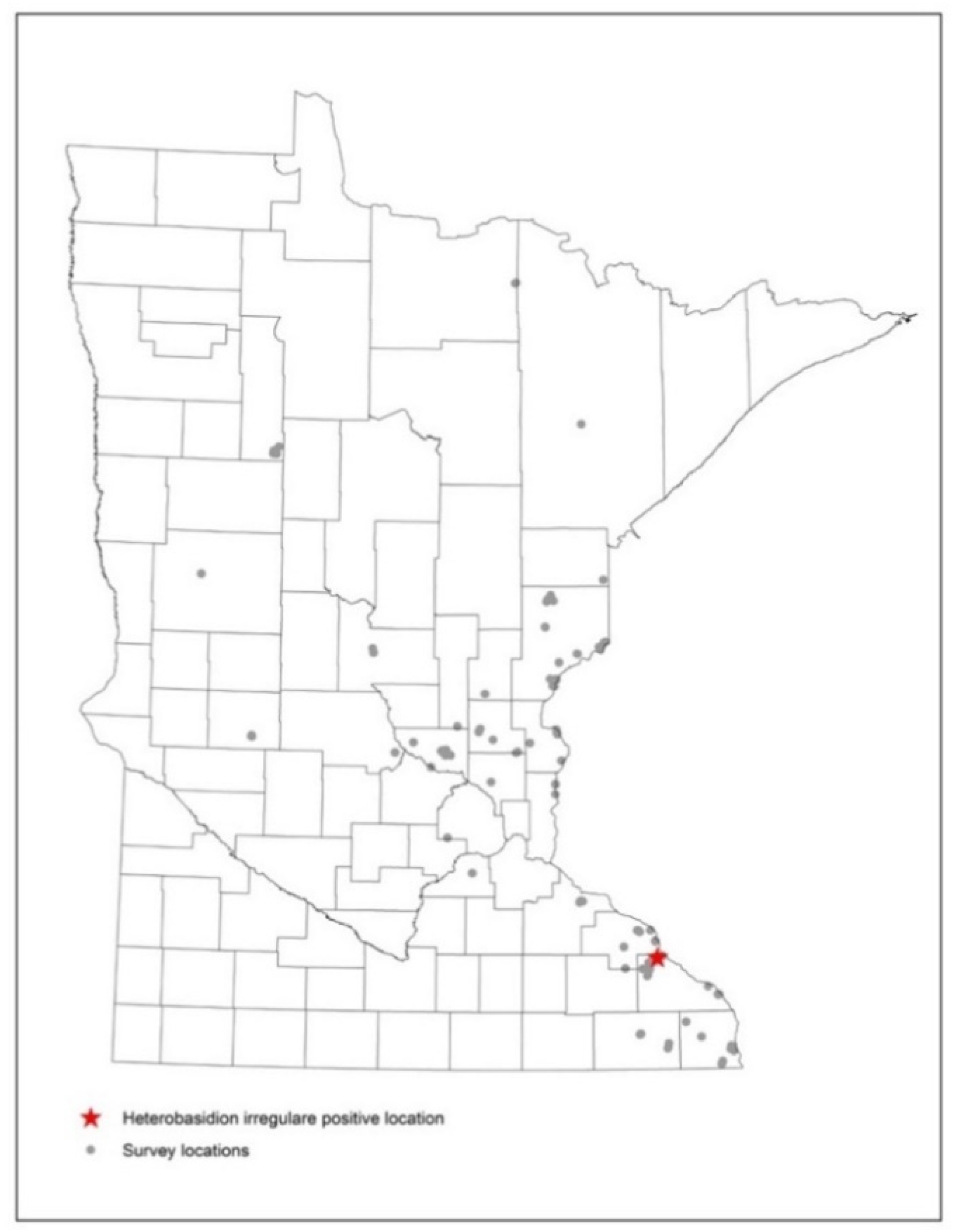
Figure 1. Survey locations for H. irregulare showing results from culturing studies with gray dots as negative locations and the red star as the positive location for the pathogen.
Forests 12 00057 g001
Figure 2. Survey locations for H. irregulare with red dots having spores trapped and detected using qPCR and gray dots showing sampling locations having no spores detected.
Forests 12 00057 g002
Figure 3. Basidiomycota taxa isolated from different woody substrates and sporophores throughout Minnesota during surveys for H. irregulare. ** following the taxon name indicates that the best blast match was below 97% similarity. Bar represents number of isolates. GenBank accession numbers for taxa are presented in Table S1 of Supplementary Materials.
Forests 12 00057 g003
Table 1. Number of H. irregulare mononuclear spores collected at different locations in Minnesota and Wisconsin. The total spore count and other calculations were an average of both rods from each location.
LocationSampling WeekTotal Spore CountSpores Per m3Spores Per m−2 h−1Cumulative Spores Deposited on a 30 cm Stump
Burr Oak, WI18 September 201863,776.071832.25149.33129.51
Minneiska18 September 2018412.5813.551.260.96
Great River Bluffs State Park26 September 2018100.753.310.310.23
Hay Creek26 September 201833.531.100.100.08
Elba03 October 201833.041.090.100.08
Vinegar Ridge Recreation Area03 October 201875.102.470.230.17
William O’Brien State Park10 October 2018163.105.360.500.38
Wild River State Park10 October 201885.232.800.260.10
Itasca State Park (Location 1)23 October 20180000
Itasca State Park (Location 2)23 October 20180000
Publisher’s Note: MDPI stays neutral with regard to jurisdictional claims in published maps and institutional affiliations.