Article

In Vitro Bioactivity of Australian Finger Lime Cultivars as an Initial Evaluation of Their Nutraceutical Potential

1
School of Health, Medical & Applied Science, CQUniversity, Bruce Hwy, North Rockhampton, QLD 4701, Australia
2
Centre for Indigenous Health Equity Research, CQUniversity, Bruce Hwy, North Rockhampton, QLD 4701, Australia
3
Centre for Nutrition and Food Sciences, Queensland Alliance for Agriculture and Food Innovation (QAAFI), The University of Queensland, Coopers Plains, QLD 4108, Australia
*
Author to whom correspondence should be addressed.
Nutraceuticals 2024, 4(4), 596-610; https://doi.org/10.3390/nutraceuticals4040032
Submission received: 26 August 2024 / Revised: 2 October 2024 / Accepted: 21 October 2024 / Published: 24 October 2024

Abstract

:
There is increasing interest in Australian finger lime (Citrus australasica) due to its nutritional and bioactive potential. In this study, polar extracts from five finger lime cultivars were investigated for their potential bioactivity using a range of assays: antioxidant capacity (total phenolic content (TPC), ferric reducing antioxidant power (FRAP), and cupric reducing antioxidant capacity (CUPRAC)), total monomeric anthocyanin content (TMAC), anti-diabetic activity (α-glucosidase and α-amylase inhibition), anti-Alzheimer activity (acetylcholinesterase inhibition), Skin-whitening activity skin-brightening activity (tyrosinase inhibition), and anti-inflammatory activity (COX-2 inhibition). Commercial Tahitian lime was used as a “control” (comparison). The TPC ranged from 328 to 779 mg GAE/100 g dry weight (DW) in the pulp (compared to 1043 mg GAE/100 g for Tahitian lime) and from 755 to 1048 mg GAE/100 g in the peel (1704 mg GAE/100 g for Tahitian lime). A similar range of variation was seen for FRAP, ranging from 114 to 436 mg TE/100 g DW in the pulp (422 mg TE/100 g for Tahitian lime) and 259 to 495 mg TE/100 g DW in the peel (491 mg TE/100 g for Tahitian lime). Similarly, the TFC was generally lower in finger lime pulp (100–392 mg QE/100 g DW) compared to Tahitian lime (312 mg QE/100 g). The polar extracts did not show any significant inhibition of α-glucosidase, α-amylase, tyrosinase, or COX-2. One finger lime variety showed moderate (>50%) inhibition of acetylcholinesterase (AChE) at the highest concentration screened (~1500 mg/L), as did Tahitian lime. Additionally, in silico docking against acetylcholinesterase suggested that some of the polyphenols present, including catechin, quercetin-3-glucoside, and cyanidin-3-glucoside, could potentially dock to AChE and inhibit it. This is the first time the species has been investigated for many of these bioactive properties, and also the first time in silico docking has been performed to explore which potential compounds from this species could provide its bioactivity. Although little bioactivity was generally found across the applied bioassays, these findings nevertheless provide important basic data for future research and any claims about the potential health benefits of Australian finger lime.

1. Introduction

Citrus australasica F.Muell., commonly known as the Australian finger lime or caviar lime, is a shrub or small tree which produces edible lime fruit. It is endemic to the eastern coast of Australia, historically growing in the subtropical rainforest regions of the southeastern Queensland and New South Wales border [1]. It is one of six Citrus species native to Australia, the others being Citrus inodora F.M.Bailey (North Queensland lime) and Citrus garrawayi F.M.Bailey (Mount White lime) from the rainforests of northern Queensland, Citrus australis (A.Cunn. ex Mudie) Planch. (round lime or Australian lime) from southern Queensland, Citrus glauca (Lindl.) Burkill (desert lime) from the drier inland regions of eastern Australia, and Citrus gracilis Mabb. (Humpty Doo lime) from the Northern Territory savannah [2].
Finger lime is probably the best-known of the native Australian Citrus species, as it is commercially used as a gourmet garnish, as a lime replacement in alcoholic drinks, and in preserves [3]. It is also used in some cosmetic/pharmaceutical products for its antioxidant properties and high vitamin C content. In addition to its finger-like shape, one of the major features of finger lime are its small, round vesicles forming the pulp, giving it a caviar-like texture. The species also contains a high level of genetic diversity [3], meaning that there is a wide range of size and colouration found between cultivars. However, the extent of variation in phytochemical composition and bioactive properties is less well-known.
Compared to other plant parts, fruits are a particularly rich source of phytochemical compounds [4], especially polyphenols, which have attracted significant interest due to their near-ubiquitous antioxidant activity, which can lead to other beneficial bioactivities [5]. Common classes of polyphenols include phenolic acids, stilbenes, lignans, and flavonoids [6]; among these, flavonoids are the most abundant in Citrus fruit [7]. Flavonoids are further divided into six subclasses, of which four commonly occur in Citrus: flavones, flavanones, flavonols, and flavans. A fifth subclass of flavonoids—anthocyanins—also occur in red-fleshed Citrus species (e.g., blood oranges). The most abundant flavonoids in Citrus fruit are hesperidin and naringin; polymethoxyflavones are another common flavonoid class which is unique to the Citrus genus [8]. For more information on specific flavonoids found in Citrus, the reader is referred to a recent review [9]. In general, the peel contains higher levels of phenolics compared to the flesh [10].
Much of the analytical work on C. australasica to date has focused on volatile constituents from the peel [11,12,13,14], which include a number of unique terpenyl esters [12,13]. However, the edible pulp is reported to contain several times more ascorbic acid (vitamin C) compared to oranges, as well as moderately high levels of potentially health-benefitting polyphenols [11,15]. One study by Wang et al. [16] found that methanol extracts from finger lime could inhibit inflammation in a mouse microglia BV-2 cell line through preventing nitric oxide (NO) release. Importantly, finger lime extracts have not been found to have any cytotoxic properties at moderate concentrations, as shown by in vitro testing in the aforementioned study and in Cioni et al. [17], along with more comprehensive in vivo testing on zebrafish embryos [18]. Both Cáceres-Vélez et al. [18] and Aznar et al. [19] conducted a comprehensive exploratory analysis of polyphenols and other constituents in finger lime peel using LC-QTof-MS, although the latter work did not include any bioactivity studies. Finally, Cioni et al. [17] used UHPLC-DAD-HR-Orbitrap/ESI-MS to identify a number of anthocyanins and other polyphenols from finger lime peel and pulp extracts, as well as reporting in vitro antioxidant protective effects from both the peel and pulp.
However, many other aspects of finger lime bioactivity have not yet been explored. In this study, we build on our previous work [11] to investigate unexplored aspects of the in vitro and potential in silico bioactivities of five commercial varieties of Australian finger lime. The specific bioactivities tested were chosen based on a combination of studies on other species and historical and contemporary uses of finger lime. Firstly, their anti-diabetic activity (α-glucosidase and α-amylase inhibition) and COX-2 inhibition were tested, as anti-obesity activity and anti-inflammatory activity are commonly reported health benefits of Citrus consumption [20,21]. Finger lime may have historically been used to treat illness [22], which could also suggest the possibility of anti-inflammatory activity in this species. Its anti-Alzheimer activity (acetylcholinesterase inhibition) was tested as hesperidin—a major flavonoid component of other Citrus species—has neuroprotective properties [23]. Finally, its skin whitening activity (tyrosinase inhibition) was also assessed given the increasing interest and commercial uptake of finger lime extracts as cosmetic products.

2. Materials and Methods

2.1. Sample Collection and Preparation

Five commercial cultivars of finger lime (Durhams Emerald, Chartreuse, Rhyne Red, Red Champagne and the P1f2-10 hybrid) were sourced from Rhyne Horticulture (Qunaba, QLD, Australia), as described in Johnson et al. [11]. Species identification was confirmed by the resident CQUniversity botanist (Associate Professor Nanjappa Ashwath) and relevant identification keys in the literature [2]. As the samples were sourced from a commercial plant nursery, herbarium specimens were unable to be collected. A sample of Tahitian lime (Citrus × latifolia) was purchased from a local grocery store (Rockhampton, QLD, Australia) for comparison purposes. Their appearance is shown in Figure 1.
The pulp and peel were manually separated and freeze-dried (−50 °C; 50 mT) before being ground to a fine powder (Breville Coffee & Spice Grinder; Botany, NSW, Australia).

2.2. Extraction and Preparation of Crude Crystals

A polar extraction solvent (90% methanol) was chosen to extract the polar compounds (including the polyphenols and flavonoids mentioned in the Introduction section) from the freeze-dried powder. Briefly, approximately 2.5 g of the freeze-dried powder was combined with 45 mL of 90% methanol and vortexed briefly for 15–20 s before being shaken end-over-end at 50 rpm for 60 min [24]. After centrifuging (1000 rcf for 10 min), the supernatant was decanted and the pellet re-extracted with another 45 mL of 90% methanol. After end-over-end shaking (50 rpm for 20 min) and centrifuging (1000 rcf for 10 min), the supernatant was combined with that previously collected and made up to a total volume of 100 mL with 90% methanol.
The 90% methanol extract was used to determine the ferric reducing antioxidant power (FRAP), cupric reducing antioxidant capacity (CUPRAC), total phenolic content (TPC), and total monomeric anthocyanin content (TMAC). For further bioactivity testing, a crude crystal product was prepared from the methanol extracts according to the following procedure:
Approximately 80 mL of the 90% methanol extract was concentrated to dryness in a rotary evaporator (Büchi R-114 Rotavapor; Flawil, Switzerland), with the waterbath temperature limited to 27 °C. The polar constituents were then re-dissolved in approximately 20 mL of Milli-Q water, which was then freeze-dried (−50 °C; 50 mT) until a constant mass was reached. This process was performed to semi-purify the extracts and concentrate the polar components to allow for bioactivity testing. The resultant freeze-dried crude crystals were kept in a refrigerator (4 °C) until use in subsequent bioactivity testing.

2.3. Determination of Antioxidant Capacity, Total Flavonoid Content, and TMAC

The antioxidant capacity of the methanolic extracts was measured using three spectrophotometric methods as described by Johnson, et al. [25]. For the FRAP assay, 100 µL of the extract was incubated for 4 min at 37 °C with 3 mL of the FRAP reagent before the absorbance was measured at 593 nm. In the CUPRAC assay, 100 µL of extract was combined with 1 mL of 10 mM CuCl2, 1 mL of 1 M ammonium acetate, 1 mL of water, and 1 mL of 7.5 mM neocuproine. After incubating at 50 °C for 30 min, absorbance at 450 nm was measured. The TPC assay used the Folin–Ciocalteu method, where 400 µL of extract was incubated with 1:10 diluted Folin–Ciocalteu reagent before being incubated with 7.5% w/v Na2CO3 for 10 min. The absorbance was measured at 760 nm.
Results for FRAP and CUPRAC were expressed in terms of Trolox equivalents (TEs) per 100 g of sample (freeze-dried weight (DW) basis), while TPC results were expressed in gallic acid equivalents (GAEs) per 100 g of sample (DW). All of these are electron-transfer assays which assess the reducing capacity of the antioxidants in the sample [26,27].
The TMAC was determined using the pH-differential method, as described by Johnson, et al. [28]. Briefly, 200 µL of extract was combined with 1.8 mL of buffer at pH 1 or pH 4.5, and the absorbance measured at 510 and 700 nm after 15 min. The equivalent amount of cyanidin-3-glucoside (cyd-3-glu) per 100 g of sample (DW) was calculated from the corrected absorbance using the Beer–Lambert law after accounting for extract haziness (at 700 nm): A = (pH1: ABS510 nm − ABS700 nm) − (pH4.5: ABS510 nm − ABS700 nm).
To determine the total flavonoid content (TFC), 800 µL of methanolic extract was combined with 800 µL of methanolic aluminium chloride solution (10%) and 1200 µL of aqueous sodium acetate solution (1 M) followed by a 30 min incubation at room temperature. The absorbance of the resulting mixture was measured at 415 nm. The results were expressed in quercetin equivalents (QEs) per 100 g of sample (DW).

2.4. Determination of α-Glucosidase Inhibition

The ability of the extracts to inhibit α-glucosidase was tested following the method of Ušjak et al. [29]. Firstly, 50 µL of the crude crystal solution (at a concentration in the range of 1450–1580 mg/L) was placed in a 96-well plate along with 50 µL of 0.4 U/mL α-glucosidase from Saccharomyces cerevisiae (made up in 0.1 M phosphate buffer adjusted to pH 6.7). After incubating the plate at 37 °C for 15 min, 50 µL of 1.5 mg/mL PNP-G (p-nitrophenyl glucopyranoside) was added to initiate the reaction. The absorbance of each well was measured at 405 nm every 15 s for 5 min using a microplate reader (Bio-Rad iMark microplate reader, Bio-Rad, Hercules, CA, USA), with the slope of the change in absorbance being used in subsequent calculations. The % inhibition of α-glucosidase was calculated using the following formula:
% i n h i b i t i o n = 100 ( Δ e x t r a c t Δ n o   i n h i b Δ n o   e n z y m e × 100 )
where Δextract, Δno inhib, and Δno enzyme indicate the average slope of the test extract well, no inhibitor well, and no enzyme well, respectively.

2.5. Determination of α-Amylase Inhibition

The procedure for testing for α-amylase inhibition also followed Ušjak et al. [29]. Firstly, 25 µL of the crude crystal solution (at a concentration in the range of 1450–1580 mg/L) was combined with 50 µL of 0.8 U/mL α-amylase from Aspergillus oryzae (made up in 0.1 M phosphate buffer at pH 6.9) in a 96-well plate and incubated at 37 °C for 10 min. Following this, 25 µL of 1 M HCl and 100 µL of I2/KI reagent (comprising 2 mM I2 and 6 mM KI) was added and the absorbance measured at 590 nm. The same Bio-Rad microplate reader was used for all assays. The percentage inhibition was calculated using the same formula as for α-glucosidase inhibition, but with the absorbance values instead of the change in absorbance (slope).

2.6. Determination of Acetylcholinesterase Inhibition

Acetylcholinesterase inhibition was assessed following the method reported by Zheng et al. [30] and previously used by our laboratory [31]. Methanol extracts were used, rather than crude crystal dilutions, as per our previously developed methods [31]. Briefly, 40 µL of the methanol extract (diluted in phosphate buffer if necessary) was placed in a 96-well plate with 160 µL of 0.2 M phosphate buffer, adjusted to pH 7.7. Then, 80 µL of 1 mM DTNB (5,5′-dithiobis-(2-nitrobenzoic acid)) and 10 µL of 2 U/mL acetylcholinesterase solution (from Electrophorus electricus) were added to each well, followed by incubation at room temperature for 5 min. After this, 15 µL of 8 mM acetylthiocholine iodide was added, before incubation for a further 5 min. The change in absorbance at 405 nm was then recorded at 15 s intervals for 5 min. The blank-corrected absorbance values were used to calculate the inhibitory activity of the extracts using a similar formula as that presented for α-glucosidase inhibition. The 90% methanol extract was also tested to ensure it did not inhibit AChE activity. Samples showing >50% inhibition were subjected to further testing at different dilutions (3×, 9×, 27×, 81×, 243×, 729×, 2187×) to calculate their IC50 values.

2.7. Determination of Tyrosinase Inhibition

Tyrosinase inhibition was assessed using a method developed by our laboratory and adapted from Momtaz et al. [32]. Firstly, 50 µL of the crystal solution (at a concentration in the range of 1450–1580 mg/L) was combined with 50 µL of 200 U/mL tyrosinase solution (from mushroom; made up in 0.05 M phosphate buffer, adjusted to pH 6.5) in a 96-well plate. After incubating at room temperature (25 °C) for 5 min, 100 µL of 12 mM L-Dopa was added. The absorbance of the wells was measured at 475 nm every minute for 30 min. The slope of the change in absorbance was used to quantify the % inhibition of tyrosinase using the following equation:
% i n h i b i t i o n = 100 ( Δ e x t r a c t Δ n o   i n h i b Δ n o   e n z y m e × 100 )
where Δextract, Δno inhib, and Δno enzyme indicate the average slope of the test extract well, no inhibitor well, and no enzyme well, respectively.

2.8. Determination of Anti-Inflammatory Activity (COX-2 Inhibition)

COX-2 inhibition screening was conducted using a commercial inhibitor screening assay kit (Cayman Chemical, Ann Arbor, MI, USA; item No. 701080) following the manufacturer’s instructions.
Briefly, 10 µL of the test solution (at a concentration ranging from 1450 to 1510 mg/L) was mixed with 10 µL of working concentration COX-2 (human recombinant), 10 µL of heme solution, and 160 µL of reaction buffer in a 96-well plate and incubated for 10 min at 37 °C. Then, 10 µL of arachidonic acid substrate was added, followed by gentle mixing, incubation for another 2 min at 37 °C, and addition of 30 µL of saturated stannous chloride solution. After a final incubation for 15 min at 37 °C, the reaction substrate was diluted 2000× using ELISA buffer, and 50 µL of the diluted sample was placed in a mouse anti-rabbit IgG-coated 96-well plate, along with 50 µL of PGF AChE tracer solution and 50 µL of ELISA antiserum.
After 18 hrs of incubation at 4 °C, the wells were emptied and rinsed five times with ELISA wash buffer, and 200 µL of Ellman’s Reagent was added to each well. After allowing for colour development for 40 min, the absorbance of the wells was measured at 415 nm, with results quantified against a series of 8 PGF standards (3.9–500 pg/mL). After subtracting the PGF concentration of the background control wells, the percent COX inhibition was calculated using the following equation:
% i n h i b i t i o n = A v   c o n c .   o f   i n i t i a l   a c t i v i t y   w e l l c o n c .   o f   s a m p l e   w e l l A v   c o n c .   o f   i n i t i a l   a c t i v i t y   w e l l × 100
where conc. refers to the concentration of the PGF in the sample/control.
Corresponding controls were prepared using 10 µL of inhibitor vehicle (either water or DMSO) in place of the inhibitor. The negative control (corresponding to no COX-2 inhibition) was prepared containing 10 µL of COX-2 with no inhibitor. Background activity samples were prepared by placing a small amount of COX-2 enzyme in boiling water for 3 min; 10 µL of this was taken for the corresponding COX reactions. These samples correspond to complete COX-2 inhibition.

2.9. In Silico Docking of Selected Polyphenols

Although no characterisation of individual compounds was conducted in this work, Johnson et al. [11] previously reported tentative identifications of 25 compounds from the aqueous extracts of these finger lime varieties. Of these, 8 compounds (gallic acid, catechin, gentisic acid, catechol, cyanidin-3-glucoside, rutin, quercetin-3-glucoside, and apigenin) were identified from matching both their UV spectra and retention time with authentic standards. Consequently, in silico docking of these 8 polyphenols was conducted against the binding site centre of acetylcholinesterase from Tetronarce californica (PDB entry 1E3Q) using the MCule application (https://mcule.com/; accessed on 23 September 2024). This software provides docking scores, where more negative docking scores are indicative of stronger docking strength. Scores are also calculated for several different possible ‘poses’ (orientations) of the ligand. Consequently, compounds with more negative scores are more likely to be stronger inhibitors of AChE—particularly those with very negative scores across several different poses.

2.10. Data Analysis and Statistics

Statistical analysis was performed in R Studio, running R 4.2.3 [33]. This was typically a one-way ANOVA if the data were approximately normally distributed. If a significant result (p < 0.05) was obtained from the ANOVA, then post hoc Tukey testing was subsequently performed at α = 0.05. Where applicable, data are shown as mean ± standard deviation (SD) and are presented on a dry-weight basis (DW).

3. Results

3.1. Antioxidant Capacity, TFC, and TMAC

The fruit showed moderately high TPC, FRAP, and CUPRAC (Table 1), particularly in the peel. The antioxidant capacity was consistently higher in the peel, compared to the pulp of the corresponding variety.
The varieties also had differing trends between pulp and peel antioxidant capacity. For example, the Durhams Emerald variety consistently showed low antioxidant capacity in both the pulp and peel. On the other hand, the P1f2-10 hybrid showed relatively lower antioxidant capacity in the peel but quite high values in the pulp. Conversely, Red Champagne showed the opposite trend, with relatively high values in the peel but low values in the pulp. Additionally, the TPC, FRAP, and CUPRAC values of all finger lime varieties were all lower than those found in commercial Tahitian lime.
TFC was generally significantly higher in the peel extracts compared to the pulp extracts, again with considerable variation between different varieties. In the pulp, the highest TFCs were seen for Rhyne Red and Tahitian lime (392 and 312 mg QE/100 g, respectively), while the highest TFCs in the peel were Tahitian lime and Chartreuse (1935 and 1786 mg QE/100 g, respectively).
All of the electron transfer antioxidant assays showed a strong to moderate positive correlation with one another, between TPC-FRAP (r8 = 0.829, p < 0.001), TPC-CUPRAC (r8 = 0.872, p < 0.001), and FRAP-CUPRAC (r8 = 0.633, p < 0.001). TFC also showed moderately strong positive correlations with TPC (r8 = 0.786, p < 0.001), FRAP (r8 = 0.707, p < 0.001), and CUPRAC (r8 = 0.722, p < 0.001). However, none of the other assays were significantly correlated with TMAC (p > 0.05).
The volatile profiles, vitamin C content, and aqueous extract antioxidant capacities of the samples have been previously reported [11] and thus are not discussed here.

3.2. Anti-Diabetic Activity

To test for potential anti-diabetic activity, two assays were used: α-amylase and α-glucosidase inhibition. These enzymes are involved in the hydrolysis of carbohydrates into monomeric sugars and thus play a significant role in leading to postprandial hyperglycaemia. Consequently, inhibition of α-amylase and/or α-glucosidase is a common target for the treatment of type 2 diabetes [34,35].
Finger lime crude crystals (tested at a concentration of approximately 1500 mg/L) showed no to low inhibition of α-amylase for the pulp samples (9–20% inhibition) and slightly higher α-amylase inhibition for the peel samples (14–27% inhibition) (Table 2). Furthermore, no pulp or peel samples demonstrated any significant (>10%) inhibition of α-glucosidase. This suggests that the polar constituents of C. australasica are unlikely to exert any significant anti-diabetic effects through acting on these two enzymes, although it does not rule out other pathways for exerting anti-obesity effects.
Limonene—the dominant terpenoid found in finger lime [11]—has been found to display anti-diabetic activity [36] and act as a non-competitive inhibitor of α-glucosidase [37]. The reason for the lack of activity observed here may be two-fold. Firstly, the inhibition found by Basak and Candan [37] was moderately weak. Additionally, it is unlikely that the polar solvent used here (methanol) extracted much of the limonene from the peel samples, as it is a non-polar compound. In the future, we hope to further explore the activity of non-polar extracts from finger lime and clarify this issue.
Other studies on Citrus species have suggested that naringenin may play a role in α-glucosidase inhibition [38]. Nevertheless, Lim and Loh [39] only found moderate inhibition of α-glucosidase (16–44%) and α-amylase (38–62%) by four commercial Citrus varieties at a concentration of 200 mg/L.

3.3. Anti-Alzheimer Activity

The AChE inhibition assay was used to screen for potential anti-Alzheimer activity of the samples, as overexpression of AChE has been identified as one of the symptoms of Alzheimer’s disease [40,41]. It should be noted that as per our previously developed in-house AChE protocol, methanol extracts were used to screen for AChE inhibition rather than crude crystal dilutions.
Overall, the AChE inhibition achieved by the pulp extracts (mean of 27 ± 14% inhibition) was lower than the average inhibition achieved by the peel extracts (40 ± 22% inhibition; Table 3), but this difference was not statistically significant according to Student’s t test (p > 0.05). Furthermore, there was no clear correlation between the level of AChE inhibition found in the pulp and peel samples from the same lime variety (r4 = 0.08, p > 0.05).
Only two of the peel samples (Durhams Emerald and Tahitian lime) were found to display >50% inhibition at the highest concentration tested, so these were tested at seven further dilutions (3×, 9×, 27×, 81×, 243×, 729×, 2187×) to calculate their IC50 values. The IC50 values for Durhams Emerald peel and Tahitian lime peel were calculated to be 58.4 mg/mL and 12.8 mg/mL, respectively. It should be noted that these values are calculated with respect to the original freeze-dried powders, not the crude crystal extracts.
The compound(s) responsible for AChE inhibition in finger lime peel have not yet been identified, but due to their relatively weak activity, they may not be of great commercial interest. Limonene has been identified an inhibitor of AChE [42] and is found in high concentrations in finger lime peel [13], but AChE inhibition did not correlate with the limonene concentrations of the peel samples tested here [11]. It also did not correlate with the total flavonoid content of the samples (Table 1). Consequently, it is more likely that the observed AChE inhibition was due to the action of specific flavonoids or other polar phytochemicals.
The major flavonoids previously reported from finger lime peel include neohesperidin, α-glucosyl hesperidin, and methylnaringenin 7-O-rutinoside [16]. Notably, neohesperidin, hesperidin, and naringin have previously been identified as moderately potent AChE inhibitors from C. limon (L.) Burm.f. [43], while naringenin also acts as an AChE inhibitor [44].

3.4. Anti-Tyrosinase Activity

The pulp samples showed no to negligible tyrosinase inhibition (0–0.9%; Table 4), while a few peel samples had low to moderate inhibitory activity (4–25%). The highest inhibition was seen for Chartreuse peel, which provided 25.4% inhibition of tyrosinase activity at a concentration of 1510 mg/L. This was considerably higher than the inhibition seen for Tahitian lime (11.2% inhibition), in contrast to the results found for AChE inhibition (Section 2.3). Notably, the two samples showing the highest tyrosinase inhibition also had the highest total flavonoid contents (see Table 1). Nevertheless, these findings indicate a general lack of tyrosinase inhibition for the polar constituents of Australian finger lime pulp and peel.
Studies on other Citrus species have reported various components (primarily from Citrus peel) as tyrosinase inhibitors, including the terpenoids citral and myrcene [45] and the flavonoids nobiletin and hesperidin [46]. This concurs somewhat with the present study, where low to moderate tyrosinase inhibition was seen in the peel extracts of most lime varieties, but not in the pulp extracts (Table 4). The reason for the low tyrosinase inhibition in this study is unclear; it may be due to the choice of the polar extraction solvent, or it is possible that finger limes and Tahitian limes contain lower levels of these tyrosinase inhibitory compounds. It should be noted that Abirami et al. [47] reported strong tyrosinase inhibitory activity from the juice of Citrus hystrix DC. and C. maxima Burm.) Merr. fruits, demonstrating that in these species, the tyrosinase inhibitors were polar compounds.

3.5. Anti-Inflammatory Activity

Due to the high cost and limited availability of reagents associated with the COX-2 inhibition assay, only the pulp and peel extracts from one finger lime sample (Durhams Emerald) were initially screened for their activity (Table 5). This variety was chosen on the basis of its relatively high α-amylase and AChE inhibition, as well as its wide use as an ornamental/commercial finger lime cultivar [13].
Neither the pulp nor peel crystals from the Durhams Emerald finger lime variety showed any inhibition of COX-2 at the concentration tested (~1500 mg/L). As this concentration was considered the maximum concentration of biological relevance, further COX-2 inhibition testing was not conducted. It should be noted that this does not exclude the possibility of finger lime possessing anti-inflammatory activity but does suggest that the polar constituents do not impart significant anti-inflammatory activity through the COX-2 pathway.
Indeed, a recent study by Wang et al. [16] reported that methanol extracts from finger lime inhibited nitric oxide (NO) release in a mouse microglia BV-2 cell line, indicative of potential anti-inflammatory activity. Further investigation of changes in gene expression suggested that NO inhibition was mediated by preventing the upregulation of the iNOS, IL-6, JAK2, TNFα, IL-1β, NF-κB, TLR2 and TLR4 genes and preventing the downregulation of the IκBα gene. This was tentatively ascribed to the action of flavonoids such as naringenin, which has previously been found to display this type of activity [48]. Because anti-inflammatory activity can be mediated through a range of different biological pathways, further investigation into the true extent of anti-inflammatory activity in Australian finger lime is required.

3.6. Docking of Selected Polyphenols Against Acetylcholinesterase

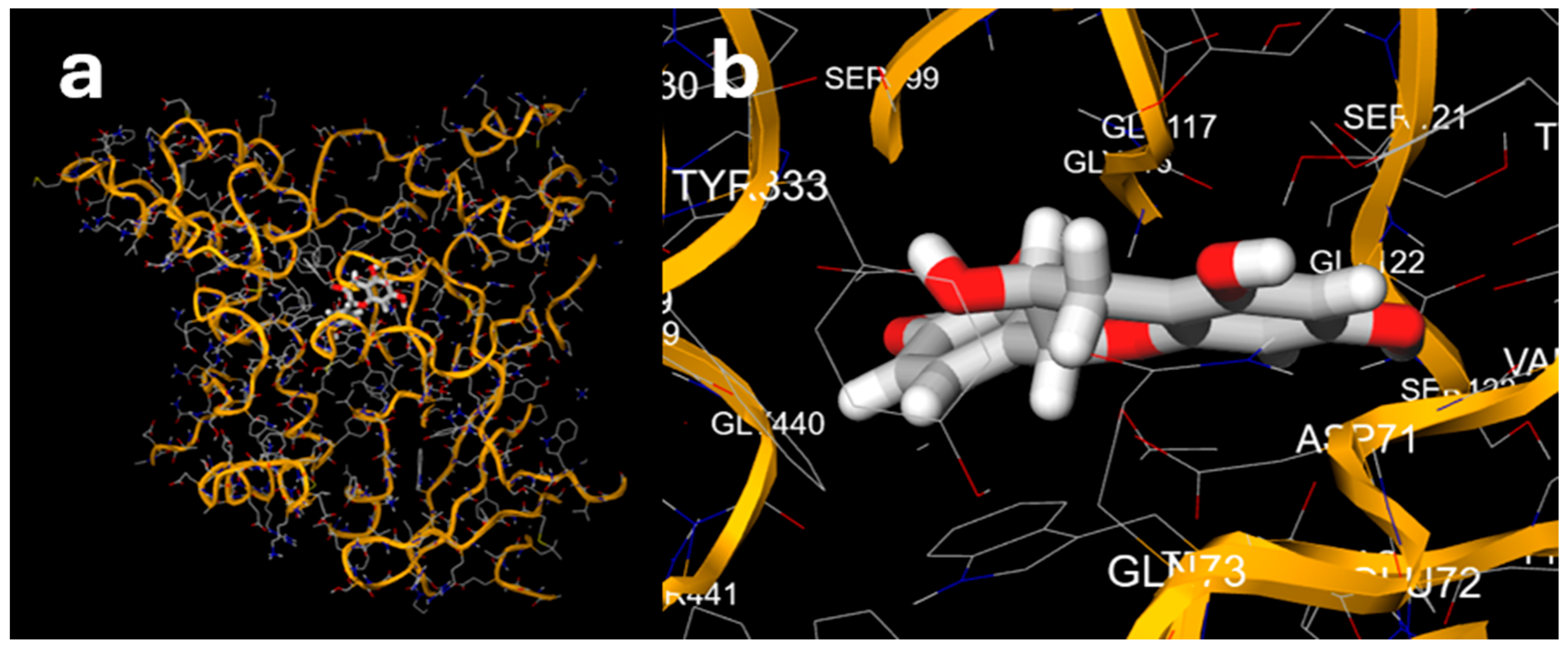
Further in silico testing was conducted to confirm the results of the strongest bioactivity observed out of those tested—acetylcholinesterase inhibition. Table 6 shows the in silico docking results from the eight selected polyphenol compounds against the acetylcholinesterase enzyme (PDB code 1E3Q). Several of the compounds, including catechin (−10.0 kcal/mol), quercetin-3-glucoside (−9.3 kcal/mol), cyanidin-3-glucoside (−9.1 kcal/mol), and apigenin (−9.0 kcal/mol), showed quite negative docking energy scores. Visualisation of the docking results for the most potent compound (catechin; Figure 2) showed that it bound to the active site of the enzyme, confirming the validity of the docking scores. This suggests that these compounds may bind strongly to acetylcholinesterase and could potentially inhibit its activity, supporting the observations of anti-AChE activity seen in Section 3.3. It should be noted that further in silico and in vitro screening would be required to validate this.

3.7. General Discussion

Compared to previous reports on the hydrophilic antioxidant capacity of these same varieties [11], the methanol-soluble TPC and FRAP found here were considerably higher. This suggests that the major antioxidant-active compounds were more soluble in methanol than water—i.e., they had moderate polarity. There is also a moderately high level of vitamin C found in these finger lime varieties [11], which be extracted by both the aqueous and methanol extraction protocols. However, the TMAC was lower here than the previously reported hydrophilic TMAC. This may be attributed to the high polarity of anthocyanins, particularly glycosylated anthocyanins. Although no previous studies appear to have compared the antioxidant capacity of water and methanol extracts from C. australasica, it is noted that most of the antioxidant capacity of desert lime (C. glauca) could be attributed to the hydrophilic fraction [49].
No significant α-glucosidase inhibition was observed for any sample (<10% for all) and only a few samples showed low levels of α-amylase inhibition (up to 20% for pulp and 27% for the peel samples). This was somewhat unexpected, as flavonoids are well-known inhibitors of α-glucosidase and α-amylase [50,51], and previous studies on other species have reported the inhibition of these enzymes [52]. However, this does not mean that finger lime necessarily has no anti-diabetic or anti-obesity activity, as this could be imparted through other biological pathways.
Similarly, there was no inhibition of COX-2 observed from the sample extracts, despite previous reports of anti-inflammatory activity from Citrus flavonoids—such as polymethoxyflavones [53,54]—and despite the results of Wang et al. [16], who noted the strong anti-inflammatory effect of finger lime. This suggests that the anti-inflammatory activity of finger lime is not mediated through COX-2 inhibition. For example, Wang et al. [16] highlighted that the inhibition of NO release was mediated through the upregulation and downregulation of several different signalling pathways.
Finally, only one of the finger lime varieties (Chartreuse) showed a moderate but significant inhibition of tyrosinase activity. Other studies have reported strong tyrosinase inhibition by non-native Australian Citrus species [47] and by nobiletin and hesperidin, two flavonoids commonly found in Citrus [46]. However, it is worth noting that at least some of this tyrosinase inhibitory activity may be attributable to volatile compounds from essential oils [45], which would not have been captured by the methanolic extraction protocol of this study. There are also anecdotal reports that α-hydroxy acids present in fruit can activate the Transient Receptor Potential Vanilloid-3 (TRPV3) in keratinocyte cells, triggering the skin renewal process [55]. This may make fruit extracts a useful part of cosmetic formulations.
While low bioactivity was generally observed in the assays selected in this study, finger lime is yet to be explored for a range of bioactivities reported for other Citrus species, such as anti-obesity activity [20], reducing cancer risk [56], and lowering plasma triglyceride levels [57].

4. Conclusions

The finger lime varieties investigated in this study showed moderately high antioxidant capacities, with higher values in the peel. The relative amounts of antioxidants, phenolics, and flavonoids in the peel and pulp varied widely with the specific variety. The strongest bioactivity observed was anti-acetylcholinesterase inhibition, where the most potent finger lime extracts showed 54% inhibition, still lower than Tahitian lime (72% inhibition), which was used as a “control”. Although little bioactivity was generally found across most of the other bioassays, this nevertheless provides important basic data for future research and any claims about the potential health benefits of Australian finger lime. Additionally, in silico screening of selected polyphenols suggested that some of these could bind strongly to the enzyme acetylcholinesterase. It would also be interesting to explore the bioactivity of finger limes in other areas where other Citrus species are known to show bioactivity, such as anti-obesity effects. Additionally, further work could explore the bioactive properties of the non-polar constituents of C. australasica or explore different extraction protocols to maximise the bioactive content of the extracts. Furthermore, in vivo bioavailability and bioactivity studies with human subjects are also warranted to substantiate the obtained in vitro results in an in vivo setting.

Author Contributions

Conceptualisation, J.B.J. and M.N.; methodology, J.B.J. and M.N.; software, J.B.J.; validation, J.B.J.; formal analysis, J.B.J., S.-Y.C. and P.R.T.; investigation, J.B.J., S.-Y.C. and P.R.T.; resources, M.N.; data curation, J.B.J.; writing—original draft preparation, J.B.J.; writing—review and editing, J.B.J., P.R.T., S.-Y.C., J.S.M., M.E.N. and M.N.; visualisation, J.B.J.; supervision, M.N.; project administration, J.B.J. and M.N.; funding acquisition, M.N. All authors have read and agreed to the published version of the manuscript.

Funding

This work was supported by a New Staff Grant (RSH/5343) awarded by CQUniversity to one of the authors (M.N.).

Institutional Review Board Statement

Not applicable.

Informed Consent Statement

Not applicable.

Data Availability Statement

The datasets supporting the findings of this research are available from the corresponding author upon request.

Acknowledgments

The authors wish to thank Daniel Manson from Rhyne Horticulture for supplying the finger lime samples used in this study.

Conflicts of Interest

The authors declare no conflicts of interest.

References

  1. Agrifutures Australia. Finger Lime. Available online: https://www.agrifutures.com.au/farm-diversity/finger-lime/ (accessed on 8 March 2024).
  2. Mabberley, D.J. Australian Citreae with notes on other Aurantioideae (Rutaceae). Telopea 1998, 7, 333–344. [Google Scholar] [CrossRef]
  3. Delort, E.; Yuan, Y.-M. Finger lime/The Australian Caviar—Citrus australasica. In Exotic Fruits; Rodrigues, S., de Oliveira Silva, E., de Brito, E.S., Eds.; Academic Press: Cambridge, MA, USA, 2018; pp. 203–210. [Google Scholar]
  4. Johnson, J.; Mani, J.; Batley, R.; Hoyos, B.; Novello, N.; Thani, P.; Arachchige, C.; Neupane, P.; Naiker, M. Functional Foods or Over-Hyped? Observations on the Antioxidant and Phenolic Content of Australian Foodstuffs. Biol. Life Sci. Forum 2023, 26, 17. [Google Scholar] [CrossRef]
  5. Bors, W.; Michel, C. Chemistry of the Antioxidant Effect of Polyphenols. Ann. N. Y. Acad. Sci. 2002, 957, 57–69. [Google Scholar] [CrossRef]
  6. Manach, C.; Scalbert, A.; Morand, C.; Rémésy, C.; Jiménez, L. Polyphenols: Food sources and bioavailability. Am. J. Clin. Nutr. 2004, 79, 727–747. [Google Scholar] [CrossRef]
  7. Dadwal, V.; Joshi, R.; Gupta, M. A comparative metabolomic investigation in fruit sections of Citrus medica L. and Citrus maxima L. detecting potential bioactive metabolites using UHPLC-QTOF-IMS. Food Res. Int. 2022, 157, 111486. [Google Scholar] [CrossRef]
  8. Saini, R.K.; Ranjit, A.; Sharma, K.; Prasad, P.; Shang, X.; Gowda, K.G.M.; Keum, Y.-S. Bioactive Compounds of Citrus Fruits: A Review of Composition and Health Benefits of Carotenoids, Flavonoids, Limonoids, and Terpenes. Antioxidants 2022, 11, 239. [Google Scholar] [CrossRef] [PubMed]
  9. Tripoli, E.; Guardia, M.L.; Giammanco, S.; Majo, D.D.; Giammanco, M. Citrus flavonoids: Molecular structure, biological activity and nutritional properties: A review. Food Chem. 2007, 104, 466–479. [Google Scholar] [CrossRef]
  10. Peleg, H.; Naim, M.; Rouseff, R.L.; Zehavi, U. Distribution of bound and free phenolic acids in oranges (Citrus sinensis) and Grapefruits (Citrus paradisi). J. Sci. Food Agric. 1991, 57, 417–426. [Google Scholar] [CrossRef]
  11. Johnson, J.B.; Batley, R.; Manson, D.; White, S.; Naiker, M. Volatile compounds, phenolic acid profiles and phytochemical content of five Australian finger lime (Citrus australasica) cultivars. LWT 2022, 154, 112640. [Google Scholar] [CrossRef]
  12. Delort, E.; Jaquier, A. Novel terpenyl esters from Australian finger lime (Citrus australasica) peel extract. Flavour Fragr. J. 2009, 24, 123–132. [Google Scholar] [CrossRef]
  13. Delort, E.; Jaquier, A.; Decorzant, E.; Chapuis, C.; Casilli, A.; Frérot, E. Comparative analysis of three Australian finger lime (Citrus australasica) cultivars: Identification of unique citrus chemotypes and new volatile molecules. Phytochemistry 2015, 109, 111–124. [Google Scholar] [CrossRef] [PubMed]
  14. Cozzolino, R.; Câmara, J.S.; Malorni, L.; Amato, G.; Cannavacciuolo, C.; Masullo, M.; Piacente, S. Comparative Volatilomic Profile of Three Finger Lime (Citrus australasica) Cultivars Based on Chemometrics Analysis of HS-SPME/GC-MS Data. Molecules 2022, 27, 7846. [Google Scholar] [CrossRef] [PubMed]
  15. Adhikari, B.; Dutt, M.; Vashisth, T. Comparative phytochemical analysis of the fruits of four Florida-grown finger lime (Citrus australasica) selections. LWT 2021, 135, 110003. [Google Scholar] [CrossRef]
  16. Wang, Y.; Ji, S.; Zang, W.; Wang, N.; Cao, J.; Li, X.; Sun, C. Identification of phenolic compounds from a unique citrus species, finger lime (Citrus australasica) and their inhibition of LPS-induced NO-releasing in BV-2 cell line. Food Chem. Toxicol. 2019, 129, 54–63. [Google Scholar] [CrossRef]
  17. Cioni, E.; Migone, C.; Ascrizzi, R.; Muscatello, B.; De Leo, M.; Piras, A.M.; Zambito, Y.; Flamini, G.; Pistelli, L. Comparing Metabolomic and Essential Oil Fingerprints of Citrus australasica F. Muell (Finger Lime) Varieties and Their In Vitro Antioxidant Activity. Antioxidants 2022, 11, 2047. [Google Scholar] [CrossRef]
  18. Cáceres-Vélez, P.R.; Ali, A.; Fournier-Level, A.; Dunshea, F.R.; Jusuf, P.R. Phytochemical and Safety Evaluations of Finger Lime, Mountain Pepper, and Tamarind in Zebrafish Embryos. Antioxidants 2022, 11, 1280. [Google Scholar] [CrossRef]
  19. Aznar, R.; Rodríguez-Pérez, C.; Rai, D.K. Comprehensive Characterization and Quantification of Antioxidant Compounds in Finger Lime (Citrus australasica L.) by HPLC-QTof-MS and UPLC-MS/MS. Appl. Sci. 2022, 12, 1712. [Google Scholar] [CrossRef]
  20. Wang, X.; Li, D.; Liu, F.; Cui, Y.; Li, X. Dietary citrus and/or its extracts intake contributed to weight control: Evidence from a systematic review and meta-analysis of 13 randomized clinical trials. Phytother. Res. 2020, 34, 2006–2022. [Google Scholar] [CrossRef]
  21. Miles, E.A.; Calder, P.C. Effects of Citrus Fruit Juices and Their Bioactive Components on Inflammation and Immunity: A Narrative Review. Front. Immunol. 2021, 12, 712608. [Google Scholar] [CrossRef]
  22. Richmond, R.; Bowyer, M.; Vuong, Q. Australian native fruits: Potential uses as functional food ingredients. J. Funct. Foods 2019, 62, 103547. [Google Scholar] [CrossRef]
  23. Hajialyani, M.; Hosein Farzaei, M.; Echeverría, J.; Nabavi, S.M.; Uriarte, E.; Sobarzo-Sánchez, E. Hesperidin as a Neuroprotective Agent: A Review of Animal and Clinical Evidence. Molecules 2019, 24, 648. [Google Scholar] [CrossRef] [PubMed]
  24. Johnson, J.B.; Skylas, D.J.; Mani, J.S.; Xiang, J.; Walsh, K.B.; Naiker, M. Phenolic Profiles of Ten Australian Faba Bean Varieties. Molecules 2021, 26, 4642. [Google Scholar] [CrossRef] [PubMed]
  25. Johnson, J.; Collins, T.; Power, A.; Chandra, S.; Portman, D.; Blanchard, C.; Naiker, M. Antioxidative properties and macrochemical composition of five commercial mungbean varieties in Australia. Legume Sci. 2020, 2, e27. [Google Scholar] [CrossRef]
  26. Huang, D.; Ou, B.; Prior, R.L. The Chemistry behind Antioxidant Capacity Assays. J. Agric. Food Chem. 2005, 53, 1841–1856. [Google Scholar] [CrossRef] [PubMed]
  27. Prior, R.L.; Wu, X.; Schaich, K. Standardized Methods for the Determination of Antioxidant Capacity and Phenolics in Foods and Dietary Supplements. J. Agric. Food Chem. 2005, 53, 4290–4302. [Google Scholar] [CrossRef]
  28. Johnson, J.; Collins, T.; Walsh, K.; Naiker, M. Solvent extractions and spectrophotometric protocols for measuring the total anthocyanin, phenols and antioxidant content in plums. Chem. Pap. 2020, 74, 4481–4492. [Google Scholar] [CrossRef]
  29. Ušjak, L.J.; Milutinović, V.M.; Đorđić Crnogorac, M.J.; Stanojković, T.P.; Niketić, M.S.; Kukić-Marković, J.M.; Petrović, S.D. Barks of Three Wild Pyrus Taxa: Phenolic Constituents, Antioxidant Activity, and in Vitro and in Silico Investigations of α-Amylase and α-Glucosidase Inhibition. Chem. Biodivers. 2021, 18, e2100446. [Google Scholar] [CrossRef]
  30. Zheng, B.; Yuan, Y.; Xiang, J.; Jin, W.; Johnson, J.B.; Li, Z.; Wang, C.; Luo, D. Green extraction of phenolic compounds from foxtail millet bran by ultrasonic-assisted deep eutectic solvent extraction: Optimization, comparison and bioactivities. LWT 2021, 154, 112740. [Google Scholar] [CrossRef]
  31. Johnson, J.B.; Neupane, P.; Bhattarai, S.P.; Trotter, T.; Naiker, M. Phenolic profiles and potential anti-Alzheimer activity of Australian adzuki bean. In Proceedings of the 72nd Australasian Grain Science Conference, Canberra, Australia, 24–26 August 2022; p. 48. [Google Scholar]
  32. Momtaz, S.; Mapunya, B.M.; Houghton, P.J.; Edgerly, C.; Hussein, A.; Naidoo, S.; Lall, N. Tyrosinase inhibition by extracts and constituents of Sideroxylon inerme L. stem bark, used in South Africa for skin lightening. J. Ethnopharmacol. 2008, 119, 507–512. [Google Scholar] [CrossRef]
  33. R Core Team. R: A Language and Environment for Statistical Computing, version 4.2.3; R Foundation for Statistical Computing: Vienna, Austria, 2023.
  34. Kajaria, D.; Ranjana; Tripathi, J.; Tripathi, Y.B.; Tiwari, S. In-vitro α amylase and glycosidase inhibitory effect of ethanolic extract of antiasthmatic drug—Shirishadi. J. Adv. Pharm. Technol. Res. 2013, 4, 206–209. [Google Scholar] [CrossRef]
  35. Tundis, R.; Loizzo, M.R.; Menichini, F. Natural Products as α-Amylase and α-Glucosidase Inhibitors and their Hypoglycaemic Potential in the Treatment of Diabetes: An Update. Mini Rev. Med. Chem. 2010, 10, 315–331. [Google Scholar] [CrossRef] [PubMed]
  36. Murali, R.; Saravanan, R. Antidiabetic effect of d-limonene, a monoterpene in streptozotocin-induced diabetic rats. Biomed. Prev. Nutr. 2012, 2, 269–275. [Google Scholar] [CrossRef]
  37. Basak, S.S.; Candan, F. Effect of Laurus nobilis L. essential oil and its main components on α-glucosidase and reactive oxygen species scavenging activity. Iran. J. Pharm. Res. 2013, 12, 367. [Google Scholar]
  38. Benayad, O.; Bouhrim, M.; Tiji, S.; Kharchoufa, L.; Addi, M.; Drouet, S.; Hano, C.; Lorenzo, J.M.; Bendaha, H.; Bnouham, M.; et al. Phytochemical Profile, α-Glucosidase, and α-Amylase Inhibition Potential and Toxicity Evaluation of Extracts from Citrus aurantium (L) Peel, a Valuable By-Product from Northeastern Morocco. Biomolecules 2021, 11, 1555. [Google Scholar] [CrossRef] [PubMed]
  39. Lim, S.; Loh, S. In vitro antioxidant capacities and antidiabetic properties of phenolic extracts from selected citrus peels. Int. Food Res. J. 2016, 23, 211. [Google Scholar]
  40. Konrath, E.L.; Passos, C.d.S.; Klein-Júnior, L.C.; Henriques, A.T. Alkaloids as a source of potential anticholinesterase inhibitors for the treatment of Alzheimer’s disease. J. Pharm. Pharmacol. 2013, 65, 1701–1725. [Google Scholar] [CrossRef]
  41. Mehta, M.; Adem, A.; Sabbagh, M. New Acetylcholinesterase Inhibitors for Alzheimer’s Disease. Int. J. Alzheimer’s Dis. 2012, 2012, 728983. [Google Scholar] [CrossRef]
  42. Saito, T.; Nishida, Y.; Tabata, M.; Isobayashi, A.; Tomizawa, H.; Miyahara, Y.; Sugizaki, Y. Molecular Interactions between an Enzyme and Its Inhibitor for Selective Detection of Limonene. Anal. Chem. 2022, 94, 7692–7702. [Google Scholar] [CrossRef]
  43. Liu, C.; Hou, W.; Li, S.; Tsao, R. Extraction and isolation of acetylcholinesterase inhibitors from Citrus limon peel using an in vitro method. J. Sep. Sci. 2020, 43, 1531–1543. [Google Scholar] [CrossRef]
  44. Haider, S.; Liaquat, L.; Ahmad, S.; Batool, Z.; Siddiqui, R.A.; Tabassum, S.; Shahzad, S.; Rafiq, S.; Naz, N. Naringenin protects AlCl3/D-galactose induced neurotoxicity in rat model of AD via attenuation of acetylcholinesterase levels and inhibition of oxidative stress. PLoS ONE 2020, 15, e0227631. [Google Scholar] [CrossRef]
  45. Matsuura, R.; Ukeda, H.; Sawamura, M. Tyrosinase Inhibitory Activity of Citrus Essential Oils. J. Agric. Food Chem. 2006, 54, 2309–2313. [Google Scholar] [CrossRef] [PubMed]
  46. Zhang, C.; Lu, Y.; Tao, L.; Tao, X.; Su, X.; Wei, D. Tyrosinase inhibitory effects and inhibition mechanisms of nobiletin and hesperidin from citrus peel crude extracts. J. Enzym. Inhib. Med. Chem. 2007, 22, 83–90. [Google Scholar] [CrossRef] [PubMed]
  47. Abirami, A.; Nagarani, G.; Siddhuraju, P. In vitro antioxidant, anti-diabetic, cholinesterase and tyrosinase inhibitory potential of fresh juice from Citrus hystrix and C. maxima fruits. Food Sci. Hum. Wellness 2014, 3, 16–25. [Google Scholar] [CrossRef]
  48. Park, H.Y.; Kim, G.-Y.; Choi, Y.H. Naringenin attenuates the release of pro-inflammatory mediators from lipopolysaccharide-stimulated BV2 microglia by inactivating nuclear factor-κB and inhibiting mitogen-activated protein kinases. Int. J. Mol. Med. 2012, 30, 204–210. [Google Scholar] [CrossRef] [PubMed]
  49. Konczak, I.; Zabaras, D.; Dunstan, M.; Aguas, P.; Roulfe, P.; Pavan, A. Health Benefits of Australian Native Foods: An Evaluation of Health-Enhancing Compounds; Rural Industries Research and Development Corporation: Barton, ACT, Australia, 2009. [Google Scholar]
  50. Xiao, J.; Ni, X.; Kai, G.; Chen, X. A Review on Structure–Activity Relationship of Dietary Polyphenols Inhibiting α-Amylase. Crit. Rev. Food Sci. Nutr. 2013, 53, 497–506. [Google Scholar] [CrossRef]
  51. Hua, F.; Zhou, P.; Wu, H.-Y.; Chu, G.-X.; Xie, Z.-W.; Bao, G.-H. Inhibition of α-glucosidase and α-amylase by flavonoid glycosides from Lu’an GuaPian tea: Molecular docking and interaction mechanism. Food Funct. 2018, 9, 4173–4183. [Google Scholar] [CrossRef]
  52. Sahnoun, M.; Trabelsi, S.; Bejar, S. Citrus flavonoids collectively dominate the α-amylase and α-glucosidase inhibitions. Biologia 2017, 72, 764–773. [Google Scholar] [CrossRef]
  53. Wang, Y.; Jin, R.; Chen, J.; Cao, J.; Xiao, J.; Li, X.; Sun, C. Tangeretin maintains antioxidant activity by reducing CUL3 mediated NRF2 ubiquitination. Food Chem. 2021, 365, 130470. [Google Scholar] [CrossRef]
  54. Wang, X.; Li, S.; Wei, C.-C.; Huang, J.; Pan, M.-H.; Shahidi, F.; Ho, C.-T. Anti-inflammatory effects of polymethoxyflavones from citrus peels: A review. J. Food Bioact. 2018, 3, 76–86. [Google Scholar] [CrossRef]
  55. Cornara, L.; Xiao, J.; Smeriglio, A.; Trombetta, D.; Burlando, B. Emerging Exotic Fruits: New Functional Foods in the European Market. eFood 2020, 1, 126–139. [Google Scholar] [CrossRef]
  56. Li, W.-Q.; Kuriyama, S.; Li, Q.; Nagai, M.; Hozawa, A.; Nishino, Y.; Tsuji, I. Citrus consumption and cancer incidence: The Ohsaki cohort study. Int. J. Cancer 2010, 127, 1913–1922. [Google Scholar] [CrossRef] [PubMed]
  57. Santos, K.G.d.; Yoshinaga, M.Y.; Glezer, I.; Chaves-Filho, A.d.B.; Santana, A.A.d.; Kovacs, C.; Magnoni, C.D.; Lajolo, F.M.; Miyamoto, S.; Aymoto Hassimotto, N.M. Orange juice intake by obese and insulin-resistant subjects lowers specific plasma triglycerides: A randomized clinical trial. Clin. Nutr. ESPEN 2022, 51, 336–344. [Google Scholar] [CrossRef] [PubMed]
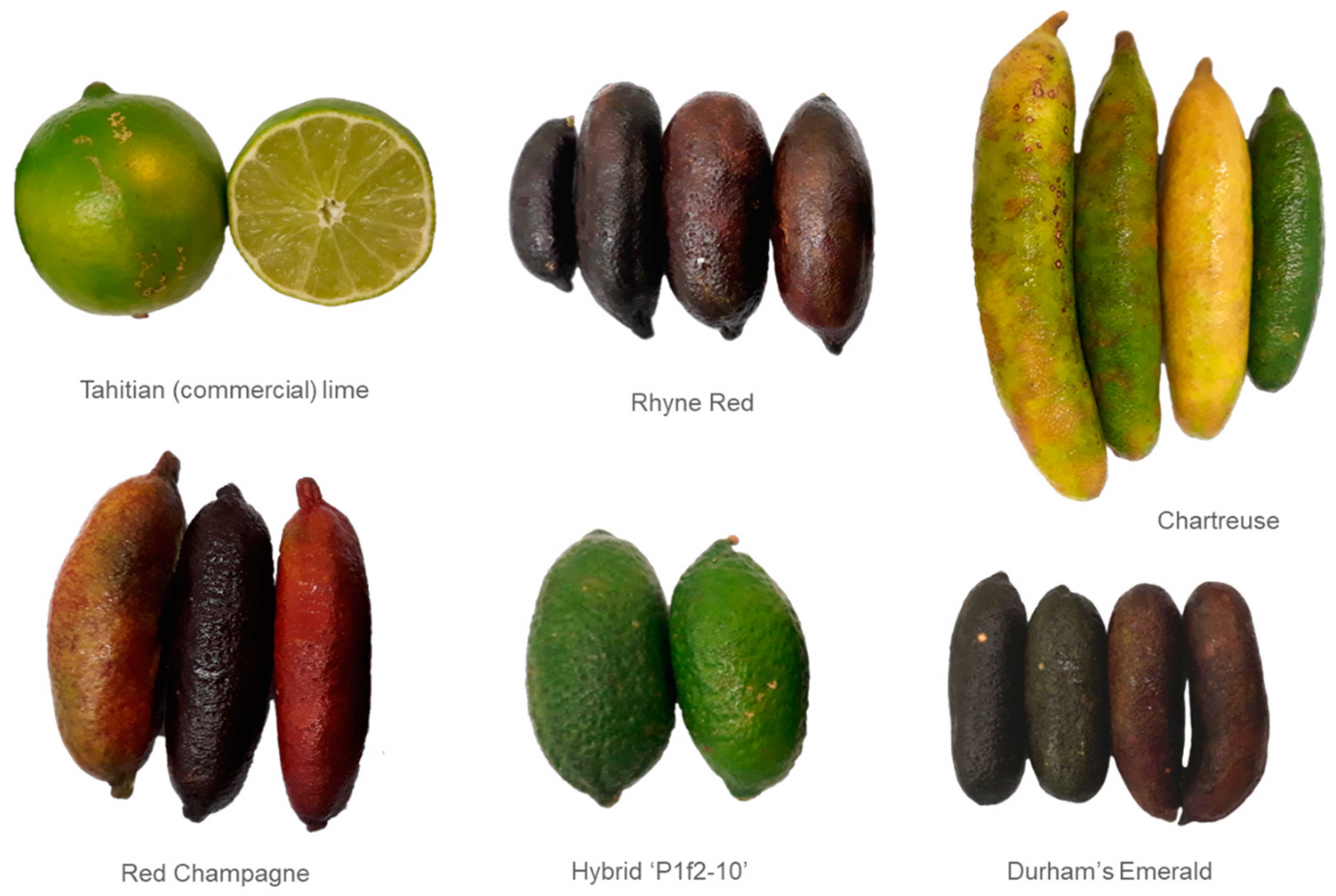
Figure 1. The five finger lime cultivars investigated in this study, along with the commercial Tahitian lime used for comparison. Not to scale.
Nutraceuticals 04 00032 g001
Figure 2. (a) Docking results of catechin against the enzyme acetylcholinesterase. (b) Close-up of the docking pose catechin (with residues labelled).
Nutraceuticals 04 00032 g002
Table 1. Antioxidant capacity and TMAC of the five finger lime cultivars and commercial Tahitian lime, as measured in methanol extracts.
VarietyTPC (mg GAE/100 g DW)FRAP (mg TE/100 g DW)CUPRAC (mg TE/100 g DW)TMAC (mg cyd-3-glu/100 g DW)TFC (mg QE/100 g DW)
Pulp
Durhams Emerald328 ± 6 f114 ± 15 g727 ± 0 g1 ± 2 ef100 ± 1 h
Chartreuse341 ± 2 f204 ± 4 f886 ± 5 g1 ± 1 ef112 ± 1 h
P1f2-10 hybrid344 ± 0 f234 ± 3 ef1235 ± 20 f4 ± 0 bcdef145 ± 1 h
Rhyne Red779 ± 22 e436 ± 5 bc1987 ± 55 e6 ± 1 bcde392 ± 5 fg
Red Champagne385 ± 3 f225 ± 5 f839 ± 18 g2 ± 0 def108 ± 0 h
Tahitian lime1043 ± 1 b422 ± 10 c7849 ± 160 c9 ± 1 b312 ± 10 g
Peel
Durhams Emerald851 ± 32 d349 ± 9 d2063 ± 32 e7 ± 0 bcd526 ± 10
Chartreuse1048 ± 8 b449 ± 8 bc8207 ± 27 b0 ± 0 f1786 ± 20 b
P1f2-10 hybrid755 ± 9 e259 ± 1 e1898 ± 12 e4 ± 1 bcdef511 ± 6 ef
Rhyne Red850 ± 24 d465 ± 14 ab2397 ± 74 d8 ± 4 bc1560 ± 63 c
Red Champagne966 ± 15 c495 ± 4 a2415 ± 5 d25 ± 0 a640 ± 16 d
Tahitian lime1704 ± 3 a491 ± 8 a11104 ± 140 a3 ± 0 cdef1935 ± 82 a
Data are mean ± SD, n = 2 extractions; rows with different superscript letters are significantly different to one another according to a one-way ANOVA followed by post hoc Tukey testing at α = 0.05.
Table 2. Inhibition of α-glucosidase and α-amylase activity found for the finger lime and commercial lime crude crystals.
VarietyCrystal Concentration (mg/L)α-Glucosidase Inhibition (%)α-Amylase Inhibition (%)
Pulp
Durhams Emerald14509.520.2
Chartreuse1490019.3
P1f2-10 hybrid1490015.2
Rhyne Red1520012.0
Red Champagne152008.9
Tahitian lime158009.5
Peel
Durhams Emerald1510023.7
Chartreuse1510017.3
P1f2-10 hybrid1450026.9
Rhyne Red1480018.7
Red Champagne1460013.5
Tahitian lime1490014.2
One replicate analysis was performed per sample. The analytical variability associated with these assays is typically <10%.
Table 3. Inhibition of acetylcholinesterase activity found for finger lime and commercial lime samples. The equivalent sample concentration is calculated with respect to the freeze-dried powder, not the crude crystals.
VarietyEquivalent Sample Concentration (mg/mL)Acetylcholinesterase Inhibition (%)
Pulp
Durhams Emerald81.321.5
Chartreuse75.745.3
P1f2-10 hybrid69.27.5
Rhyne Red76.824.1
Red Champagne91.623.5
Tahitian lime93.139.4
Peel
Durhams Emerald71.954.6
Chartreuse77.320.2
P1f2-10 hybrid71.536.2
Rhyne Red77.443.8
Red Champagne71.511.4
Tahitian lime82.871.9
One replicate analysis was performed per sample. The analytical variability associated with this assay is typically <10%.
Table 4. Inhibition of tyrosinase activity found for finger lime and commercial lime crude crystals.
VarietyCrystal Concentration (mg/L)Tyrosinase Inhibition (%)
Pulp
Durhams Emerald14500
Chartreuse14900.4
P1f2-10 hybrid14900
Rhyne Red15200.9
Red Champagne15200
Tahitian lime15800
Peel
Durhams Emerald15104.0
Chartreuse151025.4
P1f2-10 hybrid14504.5
Rhyne Red14809.1
Red Champagne14605.1
Tahitian lime149011.2
One replicate analysis was performed per sample.
Table 5. Inhibition of COX-2 activity found for Durhams Emerald finger lime pulp and peel crystals.
VarietyCrystal Concentration (mg/L)COX-2 Inhibition (%)
Durhams Emerald pulp1450Not detected
Durhams Emerald peel1510Not detected
One replicate analysis was performed per sample. The analytical variability associated with this assay is typically <10%.
Table 6. Docking scores for selected polyphenols previously identified from finger lime extracts [11] against the enzyme acetylcholinesterase (from Torpedo californica; PDB code 1E3Q). Docking was performed in MCule; more negative scores indicate stronger binding activity.
CompoundDocking Scores
Pose #1Pose #2Pose #3Pose #4
Gallic acid−6.4−6.4−6.4−6.4
Catechin−10.0−9.0−8.9−8.7
Gentisic acid−6.2−6.2−6.1−6.0
Catechol−5.4−5.4−5.1−5.0
Cyanidin-3-glucoside−9.1−7.7−7.6−7.6
Rutin−8.8−8.8−8.6−8.5
Quercetin-3-glucoside−9.3−9.2−8.7−8.3
Apigenin−9.0−8.7−8.5−8.3
Disclaimer/Publisher’s Note: The statements, opinions and data contained in all publications are solely those of the individual author(s) and contributor(s) and not of MDPI and/or the editor(s). MDPI and/or the editor(s) disclaim responsibility for any injury to people or property resulting from any ideas, methods, instructions or products referred to in the content.