
Changes in Foot Biomechanics during Pregnancy and Postpartum: Scoping Review

 ,
,  ,
,  and
and 
Abstract
1. Introduction
2. Materials and Methods
2.1. Criteria for Eligibility
2.2. Research Strategy and Databases
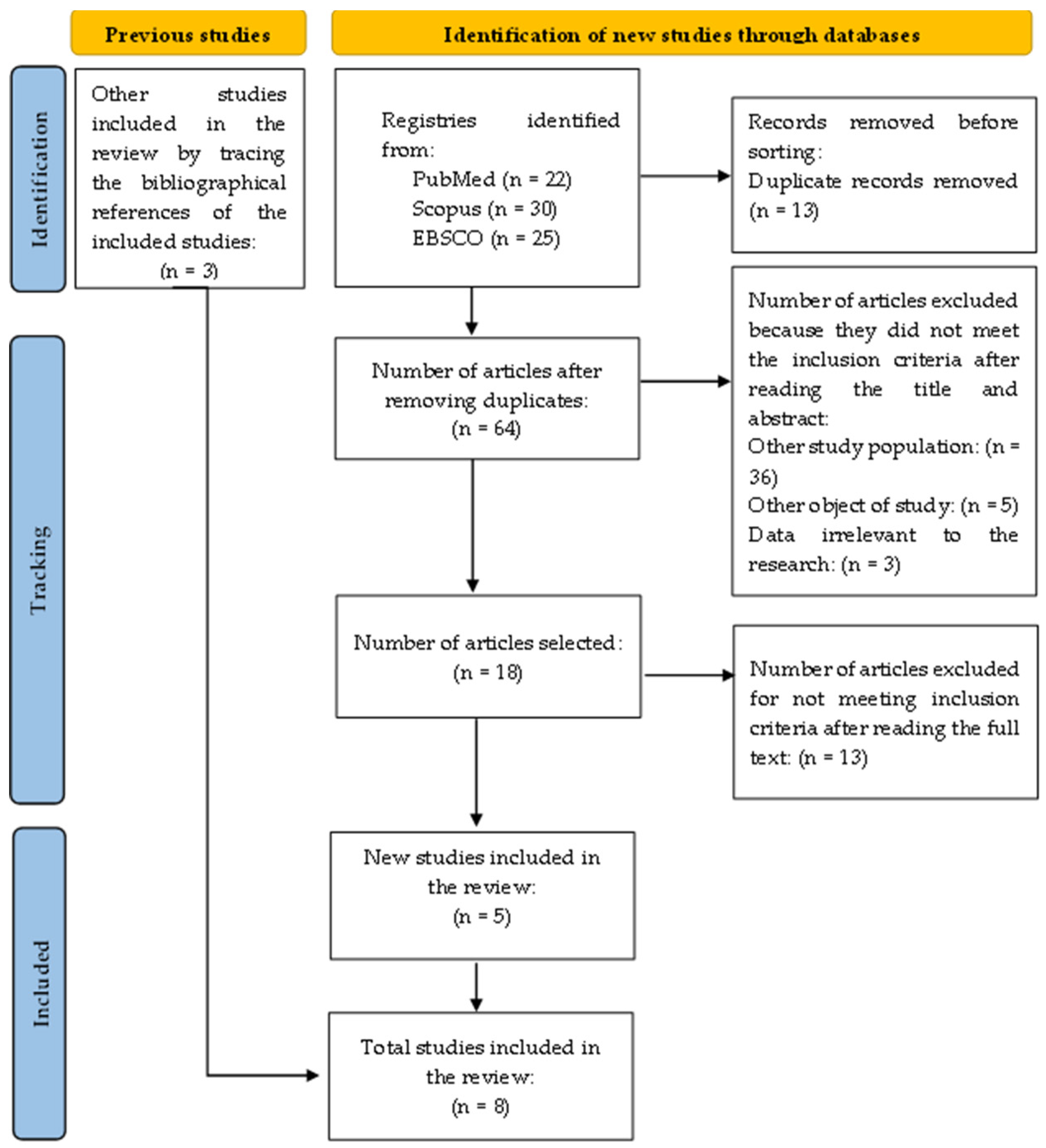
2.3. Study Selection
2.4. Data Extraction
3. Results
4. Discussion
4.1. Limitations
4.2. Implications for Future Research
5. Conclusions
Author Contributions
Funding
Conflicts of Interest
References
- Lowdermilk, D.L.; Perry, S.E.; Cashion, K.; Alden, K.R.; Olshansky, E.F. Maternity and Women’s Health Care, 11th ed.; Elsevier: St. Louis, MO, USA, 2016; ISBN 978-0-323-16918-9. [Google Scholar]
- Nyska, M.; Sofer, D.; Porat, A.; Howard, C.B.; Levi, A.; Meizner, I. Planter foot pressures in pregnant women. Isr. J. Med. Sci. 1997, 33, 139–146. [Google Scholar] [PubMed]
- Masłoń, A.; Suder, A.; Curyło, M.; Frączek, B.; Salamaga, M.; Ivanenko, Y.; Forczek-Karkosz, W. Influence of pregnancy related anthropometric changes on plantar pressure distribution during gait-A follow-up study. PLoS ONE 2022, 17, e0264939. [Google Scholar] [CrossRef] [PubMed]
- Ribeiro, A.P.; Trombini-Souza, F.; de Camargo Neves Sacco, I.; Ruano, R.; Zugaib, M.; João, S.M. Changes in the plantar pressure distribution during gait throughout gestation. J. Am. Podiatr. Med. Assoc. 2011, 101, 415–423. [Google Scholar] [CrossRef] [PubMed]
- Mei, Q.; Gu, Y.; Fernandez, J. Alterations of Pregnant Gait during Pregnancy and Postpartum. Sci. Rep. 2018, 8, 2217. [Google Scholar] [CrossRef] [PubMed]
- Vico Pardo, F.J.; López Del Amo, A.; Pardo Rios, M.; Gijon-Nogueron, G.; Yuste, C.C. Changes in foot posture during pregnancy and their relation with musculoskeletal pain: A longitudinal cohort study. Women Birth 2018, 31, e84–e88. [Google Scholar] [CrossRef]
- Yoo, H.; Shin, D.; Song, C. Changes in the spinal curvature, degree of pain, balance ability, and gait ability according to pregnancy period in pregnant and nonpregnant women. J. Phys. Ther. Sci. 2015, 27, 279–284. [Google Scholar] [CrossRef] [PubMed]
- Segal, N.A.; Boyer, E.R.; Teran-Yengle, P.; Glass, N.A.; Hillstrom, H.J.; Yack, H.J. Pregnancy leads to lasting changes in foot structure. Am. J. Phys. Med. Rehabil. 2013, 92, 232–240. [Google Scholar] [CrossRef]
- Bertuit, J.; Leyh, C.; Rooze, M.; Feipel, V. Plantar Pressure During Gait in Pregnant Women. J. Am. Podiatr. Med. Assoc. 2016, 106, 398–405. [Google Scholar] [CrossRef] [PubMed]
- Mitternacht, J.; Klement, A.; Lampe, R. Plantar pressure distribution during and after pregnancy. Eur. Orthop. Traumatol. 2013, 4, 229–236. [Google Scholar] [CrossRef]
- Karadag-Saygi, E.; Unlu-Ozkan, F.; Basgul, A. Plantar pressure and foot pain in the last trimester of pregnancy. Foot Ankle Int. 2010, 31, 153–157. [Google Scholar] [CrossRef]
- Forczek, W.; Ivanenko, Y.P.; Bielatowicz, J.; Wacławik, K. Gait assessment of the expectant mothers—Systematic review. Gait Posture 2018, 62, 7–19. [Google Scholar] [CrossRef] [PubMed]
- Gimunová, M.; Kasović, M.; Zvonar, M.; Turčínek, P.; Matković, B.; Ventruba, P.; Vaváček, M.; Knjaz, D. Analysis of ground reaction force in gait during different phases of pregnancy. Kinesiology 2015, 47, 236–241. [Google Scholar]
- Segal, N.A.; Neuman, L.N.; Hochstedler, M.C.; Hillstrom, H.L. Static and dynamic effects of customized insoles on attenuating arch collapse with pregnancy: A randomized controlled trial. Foot 2018, 37, 16–22. [Google Scholar] [CrossRef] [PubMed]
- Aromataris, E.; Lockwood, C.; Porritt, K.; Pilla, B.; Jordan, Z. (Eds.) JBI Manual for Evidence Synthesis. JBI. 2024. Available online: https://synthesismanual.jbi.global (accessed on 1 February 2024). [CrossRef]
- Gijon-Nogueron, G.A.; Gavilan-Diaz, M.; Valle-Funes, V.; Jimenez-Cebrian, A.M.; Cervera-Marin, J.A.; Morales-Asencio, J.M. Anthropometric foot changes during pregnancy: A pilot study. J. Am. Podiatr. Med. Assoc. 2013, 103, 314–321. [Google Scholar] [PubMed]
- Ramachandra, P.; Kumar, P.; Kamath, A.; Maiya, A.G. Do Structural Changes of the Foot Influence Plantar Pressure Patterns During Various Stages of Pregnancy and Postpartum? Foot Ankle Spec. 2017, 10, 513–519. [Google Scholar] [CrossRef] [PubMed]
- Mikeska, O.; Gimunová, M.; Zvonař, M. Assessment of distribution of plantar pressures and foot characteristics during walking in pregnant women. Acta Bioeng. Biomech. 2019, 21, 49–56. [Google Scholar] [PubMed]
- Wetz, H.H.; Hentschel, J.; Drerup, B.; Kiesel, L.; Osada, N.; Veltmann, U. Changes in shape and size of the foot during pregnancy. Orthopade 2006, 35, 1124–1130. [Google Scholar] [CrossRef] [PubMed]
- Albino, M.A.; Moccellin, A.S.; Firmento Bda, S.; Driusso, P. Gait force propulsion modifications during pregnancy: Effects of changes in feet’s dimensions. Rev. Bras. Ginecol. Obstet. 2011, 33, 164–169. [Google Scholar] [CrossRef] [PubMed]
- Butler, E.E.; Colón, I.; Druzin, M.L.; Rose, J. Postural equilibrium during pregnancy: Decreased stability with an increased reliance on visual cues. Am. J. Obstet. Gynecol. 2006, 195, 1104–1108. [Google Scholar] [CrossRef]
- Bird, A.R.; Menz, H.B.; Hyde, C.C. The effect of pregnancy on footprint parameters. A prospective investigation. J. Am. Podiatr. Med. Assoc. 1999, 89, 405–409. [Google Scholar] [CrossRef]
- Alvarez, R.; Stokes, I.A.; Asprinio, D.E.; Trevino, S.; Braun, T. Dimensional changes of the feet in pregnancy. J. Bone Joint Surg. Am. 1988, 70, 271–274. [Google Scholar] [CrossRef]
- Widen, E.M.; Gallagher, D. Body composition changes in pregnancy: Measurement, predictors and outcomes. Eur. J. Clin. Nutr. 2014, 68, 643–652. [Google Scholar] [CrossRef]
- Alcahuz-Griñan, M.; Nieto-Gil, P.; Perez-Soriano, P.; Gijon-Nogueron, G. Alterações morfológicas e posturais no pé durante a gravidez e puerpério: Um estudo longitudinal. J. Int. Pesqui. Ambient. E Saúde Pública 2021, 18, 2423. [Google Scholar]
- Foti, T.; Davids, J.R.; Bagley, A. A biomechanical analysis of gait during pregnancy. J. Bone Joint Surg. Am. 2000, 82, 625–632. [Google Scholar] [CrossRef] [PubMed]
- Machado, Á.S.; Bombach, G.D.; Duysens, J.; Carpes, F.P. Differences in foot sensitivity and plantar pressure between young adults and elderly. Arch. Gerontol. Geriatr. 2016, 63, 67–71. [Google Scholar] [CrossRef] [PubMed]
- Stolwijk, N.M.; Duysens, J.; Louwerens, J.W.; Keijsers, N.L. Plantar pressure changes after long-distance walking. Med. Sci. Sports Exerc. 2010, 42, 2264–2272. [Google Scholar] [CrossRef]
- Chiou, W.K.; Chiu, H.T.; Chao, A.S.; Wang, M.H.; Chen, Y.L. The influence of body mass on foot dimensions during pregnancy. Appl. Ergon. 2015, 46 Pt A, 212–217. [Google Scholar] [CrossRef]
- Santos-Rocha, R. Guia da Gravidez Ativa—Atividade Física, Exercício, Desporto e Saúde na Gravidez e Pós-Parto; Escola Superior de Desporto de Rio Maior: Rio Maior, Portugal, 2020; ISBN 978-989-8768-26-1 (print); 978-989-8768-27-4 (e-book). [Google Scholar]
- Forczek, W.; Ivanenko, Y.; Curyło, M.; Frączek, B.; Masłoń, A.; Salamaga, M.; Suder, A. Progressive changes in walking kinematics throughout pregnancy-A follow up study. Gait Posture 2019, 68, 518–524. [Google Scholar] [CrossRef]

| Database | Limiters | Search Strategy |
|---|---|---|
| Scopus | Health Professions Multidisciplinary Studies Nursing Article Full Text Biomechanical Phenomena Pregnancy Foot Postpartum Period | (foot AND (pregnancy OR postpartum)) AND (biomechanical phenomena) |
| PubMed | Full Text | S1: ((Foot) AND (Pregnancy OR postpartum)) AND (Biomechanical Phenomena) S2: ((Foot) AND (Pregnancy OR postpartum)) AND (Biomechanics) |
| Via EBSCO: MEDLINE with Full Text BioOne Complete CINAHL Plus with Full Text Academic Search Complete SPORTDiscus with Full Text | Full Text Types of Resources: Academic Journals Subject: pregnancy; foot Expanders: Search also in the full text of articles; apply equivalent subjects | foot AND (pregnancy OR postpartum) AND biomechanical phenomena |
| Title, Authors, Country, and Year | Study Method | Population | Concept | Key Findings * |
|---|---|---|---|---|
| ‘Plantar Pressure and Foot Pain in the Last Trimester of Pregnancy’ Karadag-Sayg et al., Turkey, 2010 [11] | Randomized controlled trial | Thirty-five pregnant women in the third trimester with a body mass index (BMI) greater than 25 kg/m2 (overweight). Thirty-five overweight women in the control group who were matched according to BMI and age. | Understanding that the plantar pressure difference of the foot caused weight gain during pregnancy by assessing changes in blood pressure and the differences in the postural balance of pregnant women. | Pregnant women presented more significant pressures in the forefoot areas while standing. The length of the anteroposterior oscillation was also longer in the intervention group. Results demonstrated a dominant load on the foot on the right side in the static condition for pregnant and overweight women. There was no difference between pregnant women and the overweight group according to the pressure load of the midfoot. In contrast to the midfoot, the forefoot pressures of pregnant women are greater than those of overweight women. A significantly higher forefoot pressure peak was identified on the right side in pregnancy compared to the control group. Time of contact has also been established as longer during forefoot loading, correlating with visual analog scale (VAS) scores. |
| ‘Anthropometric Foot Changes During Pregnancy’ Gijon-Nogueron et al., Spain, 2013 [16] | Prospective cohort | Ten pregnant women evaluated at the 12th, 24th and 34th weeks of gestation. | Learning the anthropometric changes in the feet of pregnant women, the implications of these changes in the rest of the musculoskeletal system, and their relationship with other complications that usually occur during pregnancy. | Minimal increase in foot length. The authors verified the volume increase. A decrease in the longitudinal length and height of the internal arch is noticeable in the present study. There was a decrease in arch height, leading to pronation. |
| ‘Do Structural Changes of the Foot Influence Plantar Pressure Patterns During Various Stages of Pregnancy And Postpartum?’ Ramachandra et al., India, 2016 [17] | Cohort | A total of 84 primiparous women with singleton pregnancy, with gestational age equal to or less than 12 weeks, aged between 18 and 35 years. | Prospectively studying the structural changes of in the feet and patterns of static plantar pressure in women in various trimesters of pregnancy and postpartum. | Except for foot length and the length of the truncated foot, all other parameters showed significant differences between the various pregnancy and postpartum periods. The authors found that the variations in static pressures showed substantial changes. Maximal pressure was recorded in the hindfoot of the dominant foot, and peak maximal pressure was observed at 32 weeks gestation compared to other periods. The drop in arch height, which indicates foot pronation during pregnancy, cannot be neglected. Therefore, proper modifications in footwear need to be suggested during pregnancy. We may also consider implementing footwear measures that can prevent the effect of increased plantar pressures, especially on the hindfoot, with the advancement of pregnancy. |
| ‘Assessment of Distribution of Plantar Pressures and Foot Characteristics During Walking in Pregnant Women’ Mikeska et al., Czech Republic, 2019 [18] | Randomized controlled trial | Thirty-five pregnant women in the experimental group who wore specific orthopedic shoes developed in cooperation between Masaryk University and J Hanák R. Thirty-eight women made up the control group and were matched according to age, height, and weight. | Analyzing the distribution of plantar pressure and foot characteristics during walking between the 27th and 36th weeks of pregnancy, and verifying the influence of specific orthopedics, i.e., shoes delivered to the experimental group. | There were changes in heel width, foot length, and forefoot width in both feet of both groups. There was a significant increase in peak pressure only in the hindfoot and midfoot areas for both groups. As both groups recorded similar values in certain areas in comparing pre/post-measurement characteristics, the influence of specific orthopedic shoes in this study cannot be conclusively demonstrated in the experimental group. |
| ‘Influence of Pregnancy-related Anthropometric Changes on Plantar Pressure Distribution During Gait—A Follow-up Study’ Masłoń et al. Poland, 2022 [3] | Prospective cohort | Thirty pregnant women who were evaluated at three different time points (first, second, and third trimester of pregnancy). | Understanding the pregnancy-related gradual changes in the pattern of distribution of plantar pressure to each foot separately. It is expected that for pregnancy, it will be the progressive flattening of the longitudinal arch of both feet. In addition, the authors expected to observe the adaptations of individual feet (some changes in the angle of the foot and the relative load of the foot) to achieve a more stable gait, dependent on the gestational period. | The flattening of the foot arch during gait correlated with the mass, consistent with the influence of individual biomechanical factors (e.g., internal loads related to the anatomical structure of the body) on the foot load. The results showed a trend of longitudinal flattening of the arch of the foot for both feet, but the observed changes were statistically significant only for the right foot when compared to the second and third trimesters. While the flattening of the arch of the foot correlated with body mass in all trimesters, the mediolateral load index correlated only in the first and second trimesters. The forefoot–hindfoot load index was not influenced by body mass. In the third trimester, there was a small but significant increase in the angle of the right foot. |
| ‘Changes in shape and size of the foot during pregnancy’ Wetz et al. Germany, 2006 [19] | Prospective cohort | Forty pregnant women with no age restrictions, height, weight, or number of previous pregnancies. | Evaluating the length, width, height, and volume of the pregnant women’s feet over three consultations, one in each trimester. | The height of the foot has decreased only slightly. This decrease is not significant. There was an increase in the length and width of the foot. |
| ‘Gait force propulsion modifications during pregnancy: effects of changes in feet’s dimensions’ Albino et al. Brazil, 2011 [20] | Randomized controlled trial | Two groups: one control with 20 non-pregnant women volunteers; and another with 13 pregnant volunteers who were evaluated in the three gestational trimesters. | Analyzing the propulsion force in the women’s’ gait and relating it to changes in the dimensions of the feet and the influence on the quality of life of the pregnant woman. | There was a gain in body mass during pregnancy and an increase in perimetry in both ankles, suggesting a strong correlation between these two characteristics. Increased body mass can interfere with the characteristics of the gait of pregnant women, expanding the heel support time and decreasing the propulsion force of gait. In the third trimester of pregnancy, in the dominant limb, there was an increase in the maximum pressure point in the heel about the whole pressure point on the metatarsals. The difference between the maximum and minimum values of the horizontal component of the propulsion force in the mediolateral direction (max–min Fx) increased with the gestational trimesters. |
| ‘Pregnancy Leads to Lasting Changes in Foot Structure’ Segal et al. USA, 2013 [8] | Prospective cohort | Forty-nine women who were evaluated in the first trimester and 19 weeks postpartum. | Determining whether loss of arch height persists after delivery. | The results suggest that pregnancy appears to be associated with a permanent loss of arch height and stiffness that could potentially lead to an abnormal arthrokinematics in the lower limb, ultimately placing atypical stress on the musculoskeletal system in postpartum women. |
Disclaimer/Publisher’s Note: The statements, opinions and data contained in all publications are solely those of the individual author(s) and contributor(s) and not of MDPI and/or the editor(s). MDPI and/or the editor(s) disclaim responsibility for any injury to people or property resulting from any ideas, methods, instructions or products referred to in the content. |
© 2024 by the authors. Licensee MDPI, Basel, Switzerland. This article is an open access article distributed under the terms and conditions of the Creative Commons Attribution (CC BY) license (https://creativecommons.org/licenses/by/4.0/).
Share and Cite
Zangão, M.O.B.; Poeira, A.F.; Branco, M.; Santos-Rocha, R. Changes in Foot Biomechanics during Pregnancy and Postpartum: Scoping Review. Int. J. Environ. Res. Public Health 2024, 21, 638. https://doi.org/10.3390/ijerph21050638
Zangão MOB, Poeira AF, Branco M, Santos-Rocha R. Changes in Foot Biomechanics during Pregnancy and Postpartum: Scoping Review. International Journal of Environmental Research and Public Health. 2024; 21(5):638. https://doi.org/10.3390/ijerph21050638
Chicago/Turabian StyleZangão, Maria Otília Brites, Ana Filipa Poeira, Marco Branco, and Rita Santos-Rocha. 2024. "Changes in Foot Biomechanics during Pregnancy and Postpartum: Scoping Review" International Journal of Environmental Research and Public Health 21, no. 5: 638. https://doi.org/10.3390/ijerph21050638
APA StyleZangão, M. O. B., Poeira, A. F., Branco, M., & Santos-Rocha, R. (2024). Changes in Foot Biomechanics during Pregnancy and Postpartum: Scoping Review. International Journal of Environmental Research and Public Health, 21(5), 638. https://doi.org/10.3390/ijerph21050638