Systematic Review

Effectiveness and Safety of Different Treatment Modalities for Patients Older Than 60 Years with Distal Radius Fracture: A Network Meta-Analysis of Clinical Trials

by
Héctor Gutiérrez-Espinoza
1,
Felipe Araya-Quintanilla
2,*,
Iván Cuyul-Vásquez
3,
Rodrigo Gutiérrez-Monclus
4,
Sara Reina-Gutiérrez
5,
Iván Cavero-Redondo
5,6 and
Sergio Núñez de Arenas-Arroyo
5
1
Escuela de Fisioterapia, Universidad de las Americas, Quito 170504, Ecuador
2
Escuela de Kinesiología, Facultad de Odontología y Ciencias de la Rehabilitación, Universidad San Sebastián, Santiago 7510157, Chile
3
Faculty of Health, Therapeutic Process Department, Temuco Catholic University, Temuco 4780000, Chile
4
Traumatology Institute of Santiago, Santiago 7501241, Chile
5
Health and Social Research Center, Universidad de Castilla-La Mancha, 16071 Cuenca, Spain
6
Facultad de Ciencias de la Salud, Universidad Autónoma de Chile, Talca 7500912, Chile
*
Author to whom correspondence should be addressed.
Int. J. Environ. Res. Public Health 2023, 20(4), 3697; https://doi.org/10.3390/ijerph20043697
Submission received: 16 January 2023 / Revised: 8 February 2023 / Accepted: 13 February 2023 / Published: 19 February 2023
(This article belongs to the Special Issue Research of Orthopaedic and Traumatic Injuries and Surgical Outcomes)

Abstract

:
The aim of this study was to compare the clinical effectiveness and complications of different treatment modalities for elderly patients with distal radius fracture (DRF). Methods: We performed a network meta-analysis (NMA) of randomized clinical trials (RCTs). Eight databases were searched. The eligibility criteria for selecting studies were RCTs that compared different treatment modalities (surgical or nonoperative) in patients older than 60 years with displaced or unstable intra-articular and/or extra-articular DRFs. Results: Twenty-three RCTs met the eligibility criteria (2020 patients). For indirect comparisons, the main findings of the NMA were in volar locking plate (VLP) versus cast immobilization, with the mean differences for the patient-rated wrist evaluation (PRWE) questionnaire at −4.45 points (p < 0.05) and grip strength at 6.11% (p < 0.05). Additionally, VLP showed a lower risk ratio (RR) of minor complications than dorsal plate fixation (RR: 0.02) and bridging external fixation (RR: 0.25). Conversely, VLP and dorsal plate fixation showed higher rates of major complications. Conclusions: Compared with other treatment modalities, VLP showed statistically significant differences for some functional outcomes; however, most differences were not clinically relevant. For complications, although most differences were not statistically significant, VLP was the treatment modality that reported the lowest rate of minor and overall complications but also showed one of the highest rates of major complications in these patients. PROSPERO Registration: CRD42022315562.

1. Introduction

Distal radius fracture (DRF) is one of the most common types of fractures [1]. DRFs have a bimodal age distribution in the population, with the peak incidence in patients younger than 18 years and the second peak in patients older than 60 years [2]. In elderly patients, DRFs constitute 18% of all fractures [2]. Currently, there are different treatment modalities, such as open reduction and internal fixation with volar or dorsal plates, external fixation (bridging or nonbridging), closed reduction with percutaneous K-wire fixation, closed reduction and cast immobilization, or a combination of these techniques [3].
Generally, treatment depends on the type of DRF, age, activity level, and patient and surgeon preferences [4]. Although surgeon preference and fracture characteristics are the most influential factors in therapeutic decision making, in most cases, surgical treatment is guided by the radiological reduction criteria [5,6]. In the treatment of displaced or unstable DRFs, open reduction and internal fixation with volar locking plates are increasingly being performed because of the general belief that anatomic reduction is positively related to functional outcomes [5,7]. However, the evidence for an association between radiological reduction criteria and functional outcomes is still inconclusive, especially in patients older than 60 years [8,9].
Despite the high incidence of displaced or unstable DRFs, no consensus has been reached on the optimal treatment method in patients older than 60 years [10]. Several systematic reviews with or without meta-analyses have been published on the comparison between surgical and nonoperative treatment in this age group [11,12,13,14,15,16,17,18,19]. These meta-analyses found no statistically significant or clinically relevant differences (the observed differences did not exceed published estimates of the minimally clinically important difference) in functional outcomes between surgical and nonoperative treatment in these patients [13,14,15,16,17,18,19]. Regarding the safety of different treatment modalities in patients with DRF, a recent network meta-analysis (NMA) showed no difference in the risk of complications between nonoperative treatment and any surgical intervention in patients older than 60 years [20].
Although there is a relative increase in studies exploring different treatment modalities for DRFs, all traditional meta-analyses can only compare two treatment options at a time, thereby excluding a large number of studies [20]. To our knowledge, only two NMAs have been published that compared different treatment modalities in patients with DRF; however, these NMAs included a wide age range in patients [3,20]. Accordingly, the aim of this study was to perform an NMA of randomized clinical trials (RCTs) comparing the clinical effectiveness and safety, in terms of functional outcomes and complications, respectively, of different treatment modalities for patients older than 60 years with displaced or unstable DRF. Therefore, the study questions for this NMA were as follows: Which treatment modality showed better functional outcomes? Which treatment modality is associated with the lowest risk of complications in these patients?

2. Materials and Methods

2.1. Protocol and Registration

This study was conducted and reported according to the PRISMA extension statement for reporting systematic reviews incorporating NMA (PRISMA-NMA) guidelines [21] (PRISMA-NMA Checklist is available in Supplemental Table S1) and followed the recommendations of the Cochrane Collaboration Handbook [22]. The registration number in the International Prospective Register of Systematic Reviews (PROSPERO) is CRD42022315562.

2.2. Eligibility Criteria

Studies on the effectiveness of surgical interventions and nonoperative treatment for functional outcomes and complications in patients older than 60 years with DRF were considered eligible for inclusion if the following criteria were fulfilled: (1) Population: patients older than 60 years with displaced or unstable intra-articular and/or extra-articular DRFs; (2) Type of intervention: patients treated with any type of surgical intervention for the reduction and/or fixation of a DRF, such as internal fixation with volar or dorsal plates (locking or nonlocking), external fixation (bridging or nonbridging), closed reduction with percutaneous K-wire fixation, or intramedullary fixation; (3) Type of comparison: patients treated with nonoperative treatment (closed reduction and cast immobilization); (4) Types of outcomes: studies that assess clinical effectiveness of wrist and upper limb function with the patient-rated wrist evaluation (PRWE) and the disabilities of the arm, shoulder, and hand (DASH) questionnaires. Secondary functional outcomes included grip strength, wrist range of motion (flexion, extension, pronation, supination), and pain intensity. Studies in which the safety of different treatment modalities was assessed with clinical complications were included. Although there is no standard criteria for classifying complications as major or minor [15,20], our criteria was based on the need for surgery after the initial planned treatment, for surgical treatment interventions, this constitutes reoperation, and for all patients, an unplanned surgery. In this sense, we categorized as minor complications that did not require surgery (i.e., flexor or extensor tendon tenosynovitis, carpal tunnel syndrome, complex regional pain syndrome, delayed fracture, infection, or wrist pain), major complications that required surgery (i.e., implant removal, tendon rupture repair, wrist deformity requiring corrective osteotomy or plate fixation, surgical release for carpal tunnel syndrome, or implant malposition requiring surgical revision), or overall complications; (5) Types of studies: RCTs. Conversely, we excluded studies that included patients with associated fractures of the distal ulna or carpal bones, patients with dislocations of the distal radioulnar joint, patients with vascular injury that required surgical repair, or patients with open fractures.

2.3. Electronic Search

We searched the MEDLINE, Central Register of Controlled Trials (CENTRAL), EMBASE, Physiotherapy Evidence Database (PEDro), Latin American and the Caribbean Literature in Health Sciences (LILACS), Cumulative Index to Nursing and Allied Health Literature (CINAHL), SPORTDiscus, and Web of Science (WoS) databases from inception until November 2022. The search strategy for each database is available in Supplemental Table S2.

2.4. Study Selection

Two reviewers (HG-E and FA-Q) independently reviewed the titles and abstracts of the potentially eligible studies. Subsequently, we obtained the full text. Finally, we added a third reviewer (IC-R) to check for any discrepancy in the selection of a study.

2.5. Data Extraction Process

Two authors (IC-V and RG-M) used an ad hoc form to independently extract data on the outcomes for each trial. When data were missing, the researchers contacted the study authors to see the feasibility of obtaining them. If this was not possible, it was decided by consensus to exclude it from the network meta-analysis.

2.6. Risk of Bias in Individual Studies

Two researchers (SR-G and SN-A) independently conducted a risk of bias assessment on the RCTs that included using the Cochrane Collaboration’s tool for assessing risk of bias (RoB2) [23]. This tool evaluates the risk of bias according to five domains: randomization process, deviations from intended interventions, missing outcome data, measurement of the outcome, and selection of the reported result. A third reviewer (IC-R) was involved if a consensus could not be reached.

2.7. Statistical Methods

Our analysis was performed by the following steps. First, we used a network geometry graph in which the size of the nodes was relative to the number of trials included, and the width of the continuous line connecting nodes was proportional to the number of trials that directly compared two treatment modalities [24]. Network plot graphs were performed with the Confidence In Network Meta-Analysis (CINeMA) [25].
Second, consistency was assessed by checking whether the intervention effects estimated from direct comparisons were consistent with those estimated by indirect comparisons. Confidence was assessed with the CINeMA web application [25].
Third, a standard pairwise meta-analysis was conducted for each direct comparison by using the DerSimonian and Laird random effect method [26]. Pooled estimates of the mean difference (MD) for functional outcomes and risk ratio (RR) with the respective 95% confidence intervals (CIs) for complication rates were calculated. We examined the statistical heterogeneity using the I2 statistic; ranging was classified as unimportant (0 to 40%), moderate (30 to 60%), substantial (50 to 90%), or considerable (75 to 100%) [22]. In addition, we considered the corresponding p values. Finally, a league table was generated to illustrate these results.
Fourth, we assessed transitivity by checking whether direct comparisons of different treatment modalities included had similar clinical characteristics. Therefore, we evaluated that the patients included in these studies were similar in the baseline distribution of factors such as age, gender, dominant side involved, time of surgery, and time of immobilization post treatment.
Fifth, once we estimated the effectiveness and safety of the different treatment modalities, the surface under the cumulative ranking (SUCRA) was calculated to rank the probability of each type of treatment being the most effective or safe. SUCRA involves assigning a numerical value from 0 to 100 to simplify the classification in the rankogram, with values close to 100 being the best intervention and 0 being the worst [27].
Additionally, subgroup analyses were used to assess the clinical effectiveness and complications of different treatment modalities by the time of follow-up (3, 6, 12, and 24 months) and type of DRF (extra-articular, intra-articular, or both). Finally, Egger’s regression asymmetry test was used to assess publication bias, considering p < 0.10 as statistically significant [28]. All analyses were performed using Stata 16.0 (Stata, College Station, TX, USA).

3. Results

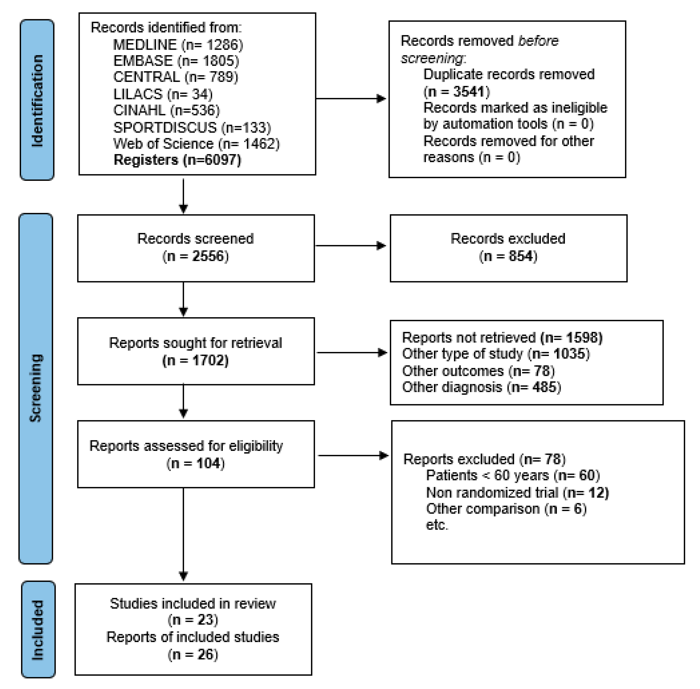
3.1. Study Selection

Electronic searches identified a total of 6097 articles (Figure 1). Finally, 26 studies were included in this NMA [29,30,31,32,33,34,35,36,37,38,39,40,41,42,43,44,45,46,47,48,49,50,51,52,53,54], which included 23 RCTs [29,30,31,32,33,34,36,37,38,39,40,41,42,44,45,46,47,48,49,50,51,53,54] and three articles publishing the long-term follow-up of some of these trials [35,43,52]. The kappa agreement rate between the reviewers was 0.91. Some of these RCTs included patients younger than 60 years; however, the authors of those trials provided their raw data to allow inclusion of their patients older than 60 years in this NMA [30,33,40,42,43,44,47,49,52]. The excluded studies and their reasons are available in Supplemental Table S3. Additionally, a summary of the results of direct and indirect comparisons is available in the network forest figures (Supplemental Figures S1–S3).

3.2. Study Characteristics

A summary of the included studies is available in Supplemental Table S4. The overall population included 2020 patients (1328 treated with different surgical interventions and 692 with nonoperative treatment). The mean age was 71.1 years (±5.2), and the mean follow-up period was 13.6 months (range, 3 to 36 months). Additionally, the included trials were classified into the following comparisons: (i) volar locking plate versus cast immobilization [29,32,34,35,38,46,48,51,53]; (ii) volar locking plate versus percutaneous K-wire fixation [34,35,37,40,47]; (iii) volar locking plate versus bridging external fixation [34,35,44,49,52]; (iv) percutaneous K-wire fixation versus cast immobilization [31,34,35,54]; (v) volar locking plate versus dorsal plate fixation [42,43]; (vi) bridging external fixation versus percutaneous K-wire fixation [34,35]; (vii) bridging external fixation versus cast immobilization [34,35,36,39,41,50]; (viii) volar locking plate versus intramedullary fixation [33]; and (ix) nonbridging versus bridging external fixation [30].

3.3. Risk of Bias Assessment in the Individual Studies

The RoB2 assessment for the included studies is presented in Figure 2 and Figure 3. For the overall risk of bias, 53.8% of the clinical trials were rated as having a low risk of bias [29,30,34,35,38,45,46,47,48,49,51,52,53,54], 34.7% were rated as having some concerns [31,32,33,37,39,42,43,44,50], and 11.5% were rated as having a high risk of bias [36,40,41]. Regarding the randomization process, 53.8% of the clinical trials were rated as having a low risk of bias [29,30,33,36,37,45,46,47,48,49,51,52,53,54]. For deviations from intended interventions, 30.8% of the clinical trials were rated as having a high risk of bias [32,36,37,39,40,41,42,43]. For missing outcome data, 100% of the clinical trials were rated as having a low risk of bias [29,30,31,32,33,34,35,36,37,38,39,40,41,42,43,44,45,46,47,48,49,50,51,52,53,54]. Finally, for the selection of the reported result, 34.7% of the clinical trials were rated as having a low risk of bias [32,34,35,38,44,46,49,51,52].

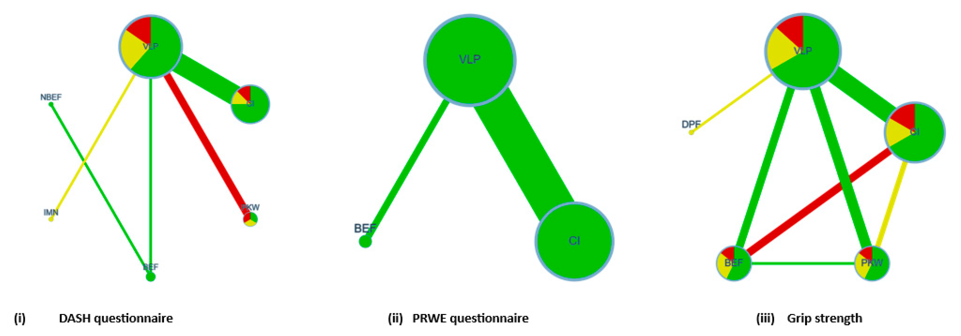
3.4. Network Analyses

The network geometry graphs show the relative amount of evidence available for the different modalities of treatment for functional outcomes such as DASH, PRWE, and grip strength, involving five, two, and seven pairwise direct comparisons, respectively (Figure 4), and on minor, major, and overall clinical complications, involving eight, six, and eight pairwise direct comparisons, respectively (Figure 5). The colors on the graph correspond to the risk of bias in studies included in these comparisons. Additionally, the transitivity assumption was met for all the comparisons by including participants with similar baseline characteristics (age, sex, dominant side involved, time of surgery, and time of immobilization posttreatment). For each comparison, the p value was <0.05.

3.4.1. Different Modalities of Treatment on Functional Outcomes

For indirect comparisons, the main summary measure for the analysis was MD. Table 1 shows the MD estimates for the DASH questionnaire. Although no MD was significant, all estimates were in favor of treatment with volar locking plates. Table 2 shows the MD estimates for the PRWE questionnaire. In the NMA, volar locking plate versus cast immobilization (−4.45 points; 95% CI, −8.62 to −0.28) showed a statistically significant difference. Table 3 shows the MD estimates for grip strength. In the NMA, volar locking plates showed statistically significant differences compared to dorsal plate fixation (30%; 95% CI, 16.16 to 43.84), bridging external fixation (6.16%; 95% CI, 1.12 to 11.21), and cast immobilization (6.11%; 95% CI, 2 to 10.21).

3.4.2. Probabilities

For the DASH and PRWE questionnaires, the highest SUCRA values were observed for volar locking plates, as 78.8% and 79%, respectively, and for bridging external fixation, as 57% and 59%, respectively (Table 4). For grip strength, the highest SUCRA values were observed for volar locking plate (97.6%) and percutaneous K-wire fixation (73.9%) (Table 4).

3.4.3. Different Modalities of Treatment on Clinical Complications

For indirect comparisons, the main summary measure for the analysis was the RR. Table 5 shows the RR estimates for minor complications. In the NMA, volar locking plates showed a lower rate of complications than dorsal plate fixation (RR: 0.02; 95% CI, 0.0009 to 0.6) and bridging external fixation (RR: 0.25; 95% CI, 0.07 to 0.93). Table 6 shows the RR estimates for major complications. In the NMA, although no RRs were significant, volar locking plate and dorsal plate fixation showed a tendency toward a higher rate of major complications. Table 7 shows the RR estimates for overall complications. In the NMA, volar locking plate fixation showed a lower rate of complications than dorsal plate fixation (RR: 0.006; 95% CI, 0.0001 to 0.28). Conversely, dorsal plate fixation showed the highest rate of overall complications.

3.4.4. Probabilities

For minor complications, the highest SUCRA values were observed in volar locking plates (84.6%) and intramedullary nails (80.7%) (Table 4). For overall complications, the highest SUCRA values were observed in volar locking plate (84.3%) and cast immobilization (67.5). Conversely, for major complications, the lowest SUCRA values were observed in intramedullary nails (17.8%), dorsal plate fixation (17.9%), and volar locking plates (41.1%) (Table 4).

3.5. Subgroup and Publication Bias Analyses

Subgroup analyses were performed to assess the clinical effectiveness and safety of the different treatment modalities according to follow-up time and type of DRF. For direct comparisons in the meta-analysis, compared to cast immobilization, volar locking plates showed statistically significant differences at the 3-month follow-up in grip strength (13.9%; 95% CI, 4.2 to 23.5; p = 0.005), DASH (−5.7 points; 95% CI, −8.2 to −3.1; p = 0.000), and PRWE questionnaire (−9.2 points; 95% CI, −18.1 to −0.3; p = 0.044). At 1 year of follow-up, only grip strength maintained this difference (7.1%; 95% CI, 1.1 to 13.1; p = 0.019). Compared to percutaneous K-wire fixation, volar locking plate fixation only showed a statistically significant difference in grip strength at the 3-month follow-up (9.9%; 95% CI, 3.9 to 15.8; p = 0.001). A summary of the main findings is available in Supplemental Table S5. Additionally, at the 6-month follow-up, the volar locking plate showed a lower risk of minor and overall complications than percutaneous K-wire fixation (RR: 0.12; 95% CI, 0.02 to 0.88; p = 0.037) and bridging external fixation (RR: 0.5; 95% CI, 0.29 to 0.86; p = 0.025). At 24 months of follow-up, volar locking plate showed a lower risk of minor and overall complications than cast immobilization (RR: 0.69; 95% CI, 0.34 to 1.39; p = 0.012). A summary of the main findings is available in Supplemental Table S6.
Regarding the type of DRF, for direct comparisons in the meta-analysis, compared to cast immobilization, the volar locking plate showed statistically significant differences in both intra-articular and extra-articular DRFs for grip strength (9.8%; 95% CI, 6 to 13.6; p = 0.000), DASH (−2.3 points; 95% CI, −4.2 to −0.4; p = 0.016), and PRWE questionnaire (−4.3 points; 95% CI, −7.1 to −1.5; p = 0.003). A summary of the main findings is available in Supplemental Table S7. Only for intra-articular DRFs did volar locking plates show a lower risk of minor and overall complications than cast immobilization (RR: 0.17; 95% CI, 0.09 to 0.32; p = 0.000). Compared to percutaneous K-wire fixation, volar locking plates showed, in both intra-articular and extra-articular fixation, a lower risk of minor complications (RR: 0.3; 95% CI, 0.13 to 0.71; p = 0.006) and a higher risk of major complications (RR: 18.38; 95% CI, 3.07 to 110.04; p = 0.001). Additionally, for intra-articular DRFs, volar locking plates showed a lower risk of minor and overall complications than bridging external fixation (RR: 0.5; 95% CI, 0.29 to 0.86; p = 0.025). A summary of the main findings is available in Supplemental Table S8.
Finally, evidence of publication bias was observed through Egger’s test for the following comparisons: volar locking plate versus cast immobilization (p = 0.79), volar locking plate versus percutaneous K-wire fixation (p = 0.82), volar locking plate versus bridging external fixation (p = 0.68), and percutaneous K-wire fixation versus cast immobilization (p = 0.85). Additionally, the funnel plot figures are available in Supplemental Figures S4–S7.

4. Discussion

This NMA of RCTs aimed to compare the clinical effectiveness and complications of different treatment modalities for patients older than 60 years with DRF. Despite the increasing number of published RCTs and meta-analyses, no consensus has been reached on the best treatment in elderly patients with displaced or unstable DRF. The main findings of our NMA were that, compared with cast immobilization, volar locking plates showed statistically significant differences in wrist function and grip strength. For DASH, PRWE, and grip strength, the highest SUCRA values were observed in volar locking plates. Conversely, volar locking plate was the treatment modality with the lowest risk of minor and overall complications but it also showed a trend toward a highest rate of major complications. Additionally, subgroup meta-analysis showed that at the 3-month follow-up, volar locking plates were more effective than cast immobilization for upper limb and wrist function and grip strength; at the 1-year follow-up, only grip strength maintained this difference between both treatments. However, these statistically significant differences in the functional outcomes should be interpreted with caution because most differences do not reach the threshold to be considered minimally clinically important [55,56].
Despite the high incidence of displaced or unstable intra-articular and/or extra-articular DRFs and the potential implications of suboptimal management, high-level scientific evidence on the best method of treatment in patients older than 60 years is not available [10,19]. Accordingly, the findings of two NMAs of RCTs performed in all patients with DRF showed controversial results [3,20]. An NMA concluded that open reduction and internal fixation with volar locking plates showed the best results for adults and patients older than 60 years with DRF in terms of short- and long-term functional recovery and the lowest rate of fracture healing complications [3]. Conversely, another NMA showed no clinically important differences in functional outcomes between the different surgical treatments at the 1-year follow-up. For patients older than 60 years, nonoperative treatment may still be the preferred option because there is no reliable evidence showing a decrease in minor or major complications between the different treatment modalities in these patients [20].
Regarding patient-reported outcome measures, most of the studies included in this NMA compared volar locking plate versus cast immobilization [29,32,34,35,38,46,48,51,53]. Consistent with our findings, previous meta-analyses reported no statistically significant or clinically relevant differences in functional outcomes between surgical and nonoperative treatments in patients older than 60 years with DRF [13,14,15,16,17,18,19]. Despite the worse radiographic outcomes associated with closed reduction and cast immobilization, these findings suggest that redisplacement does not affect functional outcomes in these patients [8,19]. Possible explanations could be related to a lower functional demand on the upper limbs, which is thought to be associated with aging, and on the other hand, patient-perceived function or disability is only partially represented by radiological measurements. Accordingly, patient self-evaluation scores will always be subjective, and factors other than those related to the fracture might influence the degree of perceived function or disability [19]. Furthermore, none of the included RCTs reported between-group MD that exceeded the predefined minimally clinically important differences between volar locking plate and nonoperative treatment in functional outcomes [55,56]. However, some of the trials showed small, statistically but not clinically significant differences in favor of patients treated with volar locking plates, which could explain this trend in the magnitude of the effect size in favor of this intervention.
Some surgical modality treatments, such as intramedullary nails, have been used for the stabilization of unstable DFRs, with the aim of reducing soft tissue complications due to a smaller surgical incision while maintaining the benefits of rigid fracture fixation [57,58]. On the other hand, a dorsal plate was used with the aim of allowing the most direct exposure and reconstruction of the joint by a capsular incision but required dissection of the extensor retinaculum and subsequent plate positioning beneath this tendon, which often led to tendonitis or tendon rupture [59]. Despite this, only two trials have compared the effectiveness of these surgical interventions with volar locking plates in patients older than 60 years [33,42]. Further high-quality trials are required to establish the role of these surgical interventions in distal radius fixation.
With the paradigm shift toward surgical treatment of displaced or unstable DRFs with volar locking plates, the overall incidence of residual deformity has decreased [60]. However, this surgical technique has reported variable rates of nerve, tendon, and hardware-related complications [61]. Our findings show that in the short to medium term, volar locking plates showed a lower risk of minor complications than dorsal plate fixation and bridging external fixation. Conversely, compared with other treatment modalities, volar locking plate and dorsal plate fixation showed a trend toward a higher rate of major complications. Despite these findings, there are many discrepancies in the literature on complication rates after a DRF, and we hypothesized that most of the discrepancies are related to the different definitions of a ‘complication’, the stringency with which authors reported complications, and how a major or minor complication is defined in the included studies.
To our knowledge, this NMA of RCTs specifically compared the clinical effectiveness and safety of different treatment modalities in patients older than 60 years with displaced or unstable DRFs. Based on the PRISMA guidelines, the recommendations of the Cochrane Collaboration Handbook, and protocol registration in PROSPERO, this study used a transparent method of assessing and reporting the evidence.

Limitations

The limitations of our NMA are as follows: (1) there is a moderate to considerable degree of statistical heterogeneity among the included studies, and potential sources of heterogeneity could be variations in the fracture types, different treatment modalities occupied, and cointerventions; (2) methodological limitations, such as a lack of adequate sample size, unclear randomization, inadequately concealed allocation, and lack of blinding of patients and assessors, could overestimate the effect size of the interventions studied; and (3) this study focused only on patients older than 60 years without immediate complications; therefore, the findings do not apply to the younger population, whose functional demands on their hands are greater and might have reported different outcome scores or rates of complications. Finally, our results should be interpreted with caution because high-quality RCTs are needed to establish the clinical effectiveness and safety of different treatment modalities in these patients. Future trials should include methodological strategies to minimize the potential risk of bias, including adequate sample size, randomization process, allocation concealment, and blinding of patients and outcome assessors.

5. Conclusions

This NMA showed that compared with other treatment modalities, volar locking plates showed statistically significant differences for some functional outcomes; however, most differences were not clinically relevant. In terms of safety, although most differences were not statistically significant, volar locking plates were the treatment modality that reported the lowest rates of minor and overall complications but also showed one of the highest rates of major complications. This should not be interpreted as a recommendation for surgical management in all patients. The indications for different treatment modalities should be judged based on the balance of risk and benefit in patients older than 60 years with DRF.

Supplementary Materials

The following supporting information can be downloaded at: https://www.mdpi.com/article/10.3390/ijerph20043697/s1, Table S1: PRISMA NMA checklist of items to include when reporting a systematic review involving a network meta-analysis; Table S2: Search strategy; Table S3: Excluded studies; Table S4: Characteristics of the randomized controlled trials included in this network meta-analyses; Table S5: Subgroup analyses of different treatment modalities on functional outcomes by time of follow-up; Table S6: Subgroup analyses of different treatment modalities on clinical complications by time of follow-up; Table S7: Subgroup analyses of different treatment modalities on functional outcomes by type of DRF; Table S8: Subgroup analyses of different treatment modalities on clinical complications by type of DRF. Figure S1: Contribution plot for the DASH questionnaire; Figure S2: Contribution plot for the PRWE questionnaire; Figure S3: Contribution plot for grip strength; Figure S4: Funnel plot for grip strength (volar locking plate versus cast mobilization); Figure S5: Funnel plot for DASH (volar locking plate versus k-wire fixation); Figure S6: Funnel plot for PRWE (volar locking plate versus bridging external fixation); Figure S7: Funnel plot complications (percutaneous K-wire fixation versus cast immobilization).

Author Contributions

Conceptualization, H.G.-E., I.C.-R. and F.A.-Q.; methodology, F.A.-Q., S.R.-G. and I.C.-R.; software, H.G.-E. and S.N.d.A.-A.; validation, H.G.-E., I.C.-V. and R.G.-M.; formal analysis, H.G.-E. and I.C.-R.; investigation, H.G.-E. and I.C.-V.; resources, H.G.-E. and F.A.-Q.; data curation, H.G.-E. and R.G.-M.; writing—original draft preparation, H.G.-E., I.C.-V., F.A.-Q., R.G.-M. and I.C.-R.; writing—review and editing, H.G.-E. and F.A.-Q.; visualization, H.G.-E., S.R.-G., I.C.-V., S.N.d.A.-A., R.G.-M. and F.A.-Q.; supervision, H.G.-E., I.C.-V., I.C.-R. and F.A.-Q.; project administration, H.G.-E.; funding acquisition, H.G.-E. and F.A.-Q. All authors have read and agreed to the published version of the manuscript.

Funding

This research received no external funding.

Data Availability Statement

Al relevant data are within the manuscript and its supporting information files.

Conflicts of Interest

The authors declare no conflict of interest.

References

  1. Nellans, K.W.; Kowalski, E.; Chung, K.C. The epidemiology of distal radius fractures. Hand Clin. 2012, 28, 113–125. [Google Scholar] [CrossRef] [PubMed] [Green Version]
  2. MacIntire, N.J.; Dewan, N. Epidemiology of distal radius fracture and factors predicting risk and prognosis. J. Hand Ther. 2016, 29, 136–145. [Google Scholar] [CrossRef] [PubMed]
  3. Vannabouathong, C.; Hussain, N.; Guerra-Farfan, E.; Bhandari, M. Interventions for Distal Radius Fractures: A Network Meta-analysis of Randomized Trials. J. Am. Acad. Orthop. Surg. 2019, 27, e596–e605. [Google Scholar] [CrossRef]
  4. Chung, K.C.; Sasor, S.E.; Speth, K.A.; Wang, L.; Shauver, M.J.; WRIST Group. Patient satisfaction after treatment of distal radial fractures in older adults. J. Hand Surg. Eur. Vol. 2020, 45, 77–84. [Google Scholar] [CrossRef]
  5. Walenkamp, M.M.J.; Mulders, M.A.M.; Goslings, J.C.; Westert, G.P.; Schep, N.W.L. Analysis of variation in the surgical treatment of patients with distal radial fractures in the Netherlands. J. Hand Surg. Eur. Vol. 2017, 42, 39–44. [Google Scholar] [CrossRef]
  6. Waljee, J.F.; Zhong, L.; Shauver, M.J.; Chung, K.C. The influence of surgeon age on distal radius fracture treatment in the United States: A population-based study. J. Hand Surg. Am. 2014, 39, 844–851. [Google Scholar] [CrossRef] [Green Version]
  7. Chung, K.C.; Shauver, M.J.; Birkmeyer, J.D. Trends in the United States in the treatment of distal radial fractures in the elderly. J. Bone Joint Surg. Am. 2009, 91, 1868–1873. [Google Scholar] [CrossRef] [Green Version]
  8. Mulders, M.A.M.; Detering, R.; Rikli, D.A.; Rosenwasser, M.P.; Goslings, J.C.; Schep, N.W.L. Association between Radiological and Patient-Reported Outcome in Adults with a Displaced Distal Radius Fracture: A Systematic Review and Meta-Analysis. J. Hand Surg. Am. 2018, 43, 710–719. [Google Scholar] [CrossRef]
  9. Gutiérrez-Monclus, R.; Gutiérrez-Espinoza, H.; Zavala-González, J.; Olguín-Huerta, C.; Rubio-Oyarzún, D.; Araya-Quintanilla, F. Correlation between Radiological Parameters and Functional Outcomes in Patients Older than 60 Years of Age with Distal Radius Fracture. Hand 2019, 14, 770–775. [Google Scholar] [CrossRef]
  10. Luokkala, T.; Laitinen, M.K.; Hevonkorpi, T.P.; Raittio, L.; Mattila, V.; Launonen, A.P. Distal radius fractures in the elderly population. EFORT Open Rev. 2020, 5, 361–370. [Google Scholar] [CrossRef] [PubMed]
  11. Gutiérrez, H.; Gutiérrez, R.; Aguilera, R.; Ortiz, L. Manejo terapéutico de pacientes con fractura del extremo distal de radio mayores de 60 años: Revisión Sistemática. Rev. Chil. Ortop. Y Traum. 2010, 51, 79–90. [Google Scholar]
  12. Diaz-Garcia, R.J.; Oda, T.; Shauver, M.J.; Chung, K.C. A systematic review of outcomes and complications of treating unstable distal radius fractures in the elderly. J. Hand Surg. Am. 2011, 36, 824–835. [Google Scholar] [CrossRef] [Green Version]
  13. Ju, J.H.; Jin, G.Z.; Li, G.X.; Hu, H.Y.; Hou, R.X. Comparison of treatment outcomes between nonsurgical and surgical treatment of distal radius fracture in elderly: A systematic review and meta-analysis. Langenbecks Arch. Surg. 2015, 400, 767–779. [Google Scholar] [CrossRef]
  14. Song, J.; Yu, A.X.; Li, Z.H. Comparison of conservative and operative treatment for distal radius fracture: A meta-analysis of randomized controlled trials. Int. J. Clin. Exp. Med. 2015, 8, 17023–17035. [Google Scholar]
  15. Chen, Y.; Chen, X.; Li, Z.; Yan, H.; Zhou, F.; Gao, W. Safety and efficacy of operative versus nonsurgical management of distal radius fractures in elderly patients: A systematic review and meta-analysis. J. Hand Surg. Am. 2016, 41, 404–413. [Google Scholar] [CrossRef]
  16. Li, Q.; Ke, C.; Han, S.; Xu, X.; Cong, Y.X.; Shang, K.; Liang, J.-D.; Zhang, B.-F. Nonoperative treatment versus volar locking plate fixation for elderly patients with distal radial fracture: A systematic review and meta-analysis. J. Orthop. Surg. Res. 2020, 15, 263. [Google Scholar] [CrossRef]
  17. Stephens, A.R.; Presson, A.P.; McFarland, M.M.; Zhang, C.; Sirniö, K.; Mulders, M.A.M.; Schep, N.W.L.; Tyser, A.R.; Kazmers, N.H. Volar Locked Plating Versus Closed Reduction and Casting for Acute, Displaced Distal Radial Fractures in the Elderly: A Systematic Review and Meta-Analysis of Randomized Controlled Trials. J. Bone Joint Surg. Am. 2020, 102, 1280–1288. [Google Scholar] [CrossRef]
  18. Zhang, Y.X.; Li, C.; Wang, S.W.; Zhang, M.L.; Zhang, H.W. Volar plate fixation vs. non-operative management for distal radius fractures in older adults: A meta-analysis. Eur. Rev. Med. Pharmacol. Sci. 2021, 25, 3955–3966. [Google Scholar]
  19. Gutiérrez-Espinoza, H.; Araya-Quintanilla, F.; Olguín-Huerta, C.; Gutiérrez-Monclus, R.; Valenzuela-Fuenzalida, J.; Román-Veas, J.; Campos-Jara, C. Effectiveness of surgical versus conservative treatment of distal radius fractures in elderly patients: A systematic review and meta-analysis. Orthop. Traumatol. Surg. Res. 2022, 108, 103323. [Google Scholar] [CrossRef]
  20. Woolnough, T.; Axelrod, D.; Bozzo, A.; Koziarz, A.; Koziarz, F.; Oitment, C.; Gyemi, L.; Gormley, J.; Gouveia, K.; Johal, H. What Is the Relative Effectiveness of the Various Surgical Treatment Options for Distal Radius Fractures? A Systematic Review and Network Meta-analysis of Randomized Controlled Trials. Clin. Orthop. Relat. Res. 2021, 479, 348–362. [Google Scholar] [CrossRef]
  21. Hutton, B.; Salanti, G.; Caldwell, D.M.; Chaimani, A.; Schmid, C.H.; Cameron, C.; Ioannidis, J.P.A.; Straus, S.; Thorlund, K.; Jansen, J.P.; et al. The PRISMA extension statement for reporting of systematic reviews incorporating network meta-analyses of health care interventions: Checklist and explanations. Ann. Intern. Med. 2015, 162, 777–784. [Google Scholar] [CrossRef] [PubMed] [Green Version]
  22. Higgins, J.; Thomas, J. Cochrane Handbook for Systematic Reviews of Interventions Version 6.3 [Updated 2022]. The Cochrane Collaboration. 2022. Available online: http:/handbook.cochrane.org (accessed on 20 November 2022).
  23. Sterne, J.A.C.; Savović, J.; Page, M.J.; Elbers, R.G.; Blencowe, N.S.; Boutron, I.; Cates, C.J.; Cheng, H.Y.; Corbett, M.S.; Eldridge, S.M.; et al. RoB 2: A revised tool for assessing risk of bias in randomised trials. BMJ 2019, 366, l4898. [Google Scholar] [CrossRef] [Green Version]
  24. Salanti, G.; Ades, A.E.; Ioannidis, J.P. Graphical methods and numerical summaries for presenting results from multiple-treatment meta-analysis: An overview and tutorial. J. Clin. Epidemiol. 2011, 64, 163–171. [Google Scholar] [CrossRef] [PubMed]
  25. Nikolakopoulou, A.; Higgins, J.P.T.; Papakonstantinou, T.; Chaimani, A.; Giovane, C.D.; Egger, M.; Salanti, G. CINeMA: An approach for assessing confidence in the results of a network meta-analysis. PLoS Med. 2020, 17, e1003082. [Google Scholar] [CrossRef] [PubMed] [Green Version]
  26. DerSimonian, R.; Laird, N. Meta-analysis in clinical trials. Control Clin. Trials. 1986, 7, 177–188. [Google Scholar] [CrossRef] [PubMed]
  27. Chaimani, A.; Higgins, J.P.; Mavridis, D.; Spyridonos, P.; Salanti, G. Graphical tools for network meta-analysis in STATA. PLoS One 2013, 8, e76654. [Google Scholar] [CrossRef] [Green Version]
  28. Sterne, J.A.; Egger, M.; Smith, G.D. Systematic reviews in health care: Investigating and dealing with publication and other biases in meta-analysis. BMJ 2001, 323, 101–105. [Google Scholar] [CrossRef]
  29. Arora, R.; Lutz, M.; Deml, C.; Krappinger, D.; Haug, L.; Gabl, M. A prospective randomized trial comparing nonoperative treatment with volar locking plate fixation for displaced and unstable distal radial fractures in patients sixty-five years of age and older. J. Bone Joint Surg. Am. 2011, 93, 2146–2153. [Google Scholar] [CrossRef] [Green Version]
  30. Atroshi, I.; Brogren, E.; Larsson, G.U.; Kloow, J.; Hofer, M.; Berggren, A.M. Wrist-bridging versus non-bridging external fixation for displaced distal radius fractures: A randomized assessor-blind clinical trial of 38 patients followed for 1 year. Acta Orthop. 2006, 77, 445–453. [Google Scholar] [CrossRef]
  31. Azzopardi, T.; Ehrendorfer, S.; Coulton, T.; Abela, M. Unstable extra-articular fractures of the distal radius: A prospective, randomised study of immobilisation in a cast versus supplementary percutaneous pinning. J. Bone Joint Surg. Br. 2005, 87, 837–840. [Google Scholar] [CrossRef] [Green Version]
  32. Bartl, C.; Stengel, D.; Bruckner, T.; Gebhard, F.; ORCHID Study Group. The treatment of displaced intra-articular distal radius fractures in elderly patients. Dtsch. Arztebl. Int. 2014, 111, 779–787. [Google Scholar] [CrossRef] [Green Version]
  33. Chappuis, J.; Bouté, P.; Putz, P. Dorsally displaced extra-articular distal radius fractures fixation: Dorsal IM nailing versus volar plating. A randomized controlled trial. Orthop. Traumatol. Surg. Res. 2011, 97, 471–478. [Google Scholar] [CrossRef]
  34. Chung, K.C.; Kim, H.M.; Malay, S.; Shauver, M.J.; Wrist and Radius Injury Surgical Trial Group. The Wrist and Radius Injury Surgical Trial: 12-Month Outcomes from a Multicenter International Randomized Clinical Trial. Plast. Reconstr. Surg. 2020, 145, 1054e–1066e. [Google Scholar] [CrossRef]
  35. Chung, K.C.; Kim, H.M.; Malay, S.; Shauver, M.J.; WRIST Group. Comparison of 24-Month Outcomes after Treatment for Distal Radius Fracture: The WRIST Randomized Clinical Trial. JAMA Netw. Open. 2021, 4, e2112710. [Google Scholar] [CrossRef]
  36. Földhazy, Z.; Ahrengart, L. External fixation versus closed treatment of displaced distal radial fractures in elderly patients: A randomized controlled trial. Curr. Orthop. Pract. 2010, 21, 288–295. [Google Scholar] [CrossRef]
  37. Goehre, F.; Otto, W.; Schwan, S.; Mendel, T.; Vergroesen, P.P.; Lindermann-Sperfeld, L. Comparison of palmar fixed-angle plate fixation with K-wire fixation of distal radius fractures (AO A2, A3, C1) in elderly patients. J. Hand Surg. Eur. Vol. 2014, 39, 249–257. [Google Scholar] [CrossRef]
  38. Hassellund, S.S.; Williksen, J.H.; Laane, M.M.; Pripp, A.; Rosales, C.P.; Karlsen, O.; Madsen, J.E.; Frihagen, F. Cast immobilization is non-inferior to volar locking plates in relation to QuickDASH after one year in patients aged 65 years and older: A randomized controlled trial of displaced distal radius fractures. Bone Joint J. 2021, 103-B(2), 247–255. [Google Scholar] [CrossRef]
  39. Hegeman, J.H.; Oskam, J.; van der Palen, J.; ten Duis, H.J.; Vierhout, P.A.M. Primary external fixation versus plaster immobilization of the intra-articular unstable distal radial fracture in the elderly. Aktuelle Traumatol. 2004, 34, 64–70. [Google Scholar] [CrossRef]
  40. Hollevoet, N.; Vanhoutie, T.; Vanhove, W.; Verdonk, R. Percutaneous K-wire fixation versus palmar plating with locking screws for Colles’ fractures. Acta Orthop. Belg. 2011, 77, 180–187. [Google Scholar]
  41. Horne, J.G.; Devane, P.; Purdie, G. A prospective randomized trial of external fixation and plaster cast immobilization in the treatment of distal radial fractures. J. Orthop. Trauma. 1990, 4, 30–34. [Google Scholar]
  42. Jakubietz, R.G.; Gruenert, J.G.; Kloss, D.F.; Schindele, S.; Jakubietz, M.G. A randomised clinical study comparing palmar and dorsal fixed-angle plates for the internal fixation of AO C-type fractures of the distal radius in the elderly. J. Hand Surg. Eur. Vol. 2008, 33, 600–604. [Google Scholar] [CrossRef] [PubMed]
  43. Jakubietz, M.G.; Gruenert, J.G.; Jakubietz, R.G. Palmar and dorsal fixed-angle plates in AO C-type fractures of the distal radius: Is there an advantage of palmar plates in the long term? J. Orthop. Surg. Res. 2012, 7, 8. [Google Scholar] [CrossRef] [PubMed] [Green Version]
  44. Jeudy, J.; Steiger, V.; Boyer, P.; Cronier, P.; Bizot, P.; Massin, P. Treatment of complex fractures of the distal radius: A prospective randomised comparison of external fixation ‘versus’ locked volar plating. Injury 2012, 43, 174–179. [Google Scholar] [CrossRef] [PubMed] [Green Version]
  45. Koshimune, M.; Kamano, M.; Takamatsu, K.; Ohashi, H. A randomized comparison of locking and non-locking palmar plating for unstable Colles’ fractures in the elderly. J. Hand Surg. Br. 2005, 30, 499–503. [Google Scholar] [CrossRef]
  46. Lawson, A.; Naylor, J.M.; Buchbinder, R.; Ivers, R.; Balogh, Z.J.; Smith, P.; Xuan, W.; Howard, K.; Vafa, A.; Perriman, D.; et al. Surgical Plating vs. Closed Reduction for Fractures in the Distal Radius in Older Patients: A Randomized Clinical Trial. JAMA Surg. 2021, 156, 229–237. [Google Scholar]
  47. Marcheix, P.S.; Dotzis, A.; Benkö, P.E.; Siegler, J.; Arnaud, J.P.; Charissoux, J.L. Extension fractures of the distal radius in patients older than 50: A prospective randomized study comparing fixation using mixed pins or a palmar fixed-angle plate. J. Hand Surg. Eur. Vol. 2010, 35, 646–651. [Google Scholar] [CrossRef]
  48. Martinez-Mendez, D.; Lizaur-Utrilla, A.; de-Juan-Herrero, J. Intra-articular distal radius fractures in elderly patients: A randomized prospective study of casting versus volar plating. J. Hand Surg. Eur. Vol. 2018, 43, 142–147. [Google Scholar] [CrossRef]
  49. Mellstrand Navarro, C.; Ahrengart, L.; Törnqvist, H.; Ponzer, S. Volar Locking Plate or External Fixation with Optional Addition of K-Wires for Dorsally Displaced Distal Radius Fractures: A Randomized Controlled Study. J. Orthop. Trauma. 2016, 30, 217–224. [Google Scholar] [CrossRef]
  50. Moroni, A.; Vannini, F.; Faldini, C.; Pegreffi, F.; Giannini, S. Cast vs external fixation: A comparative study in elderly osteoporotic distal radial fracture patients. Scand. J. Surg. 2004, 93, 64–67. [Google Scholar] [CrossRef] [Green Version]
  51. Saving, J.; Severin Wahlgren, S.; Olsson, K.; Enocson, A.; Ponzer, S.; Sköldenberg, O.; Wilcke, M.; Mellstrand Navarro, C. Nonoperative Treatment Compared with Volar Locking Plate Fixation for Dorsally Displaced Distal Radial Fractures in the Elderly: A Randomized Controlled Trial. J. Bone Joint Surg. Am. 2019, 101, 961–969. [Google Scholar] [CrossRef]
  52. Saving, J.; Enocson, A.; Ponzer, S.; Mellstrand-Navarro, C. External Fixation Versus Volar Locking Plate for Unstable Dorsally Displaced Distal Radius Fractures-A 3-Year Follow-Up of a Randomized Controlled Study. J. Hand Surg. Am. 2019, 44, 18–26. [Google Scholar] [CrossRef] [Green Version]
  53. Tahir, M.; Khan Zimri, F.; Ahmed, N.; Jamali, A.R.; Mehboob, G.; Watson, K.R.; Faraz, A. Plaster immobilization versus anterior plating for dorsally displaced distal radial fractures in elderly patients in Pakistan. J. Hand Surg. Eur. Vol. 2021, 46, 647–653. [Google Scholar] [CrossRef]
  54. Wong, T.C.; Chiu, Y.; Tsang, W.L.; Leung, W.Y.; Yam, S.K.; Yeung, S.H. Casting versus percutaneous pinning for extra-articular fractures of the distal radius in an elderly Chinese population: A prospective randomised controlled trial. J. Hand Surg. Eur. Vol. 2010, 35, 202–208. [Google Scholar] [CrossRef]
  55. Walenkamp, M.M.; de Muinck Keizer, R.J.; Goslings, J.C.; Vos, L.M.; Rosenwasser, M.P.; Schep, N.W. The Minimum Clinically Important Difference of the Patient-rated Wrist Evaluation Score for Patients with Distal Radius Fractures. Clin. Orthop. Relat. Res. 2015, 473, 3235–3241. [Google Scholar] [CrossRef] [Green Version]
  56. Sorensen, A.A.; Howard, D.; Tan, W.H.; Ketchersid, J.; Calfee, R.P. Minimal clinically important differences of 3 patient-rated outcomes instruments. J. Hand Surg. Am. 2013, 38, 641–649. [Google Scholar] [CrossRef] [Green Version]
  57. Hardman, J.; Al-Hadithy, N.; Hester, T.; Anakwe, R. Systematic review of outcomes following fixed angle intramedullary fixation of distal radius fractures. Int. Orthop. 2015, 39, 2381–2387. [Google Scholar] [CrossRef]
  58. Chen, Z.; Zhu, Y.; Zhang, W.; Eltagy, H.; Elerian, S. Comparison of Intramedullary Nail and Volar Locking Plate for Distal Radius Fractures: A Systematic Review and Meta-Analysis of Randomised Controlled Trials. Cureus 2021, 13, e17972. [Google Scholar] [CrossRef]
  59. Wei, J.; Yang, T.B.; Luo, W.; Qin, J.B.; Kong, F.J. Complications following dorsal versus volar plate fixation of distal radius fracture: A meta-analysis. J. Int. Med. Res. 2013, 41, 265–275. [Google Scholar] [CrossRef] [Green Version]
  60. Del Piñal, F.; Jupiter, J.B.; Rozental, T.D.; Arora, R.; Nakamura, T.; Bain, G.I. Distal radius fractures. J. Hand Surg. Eur. Vol. 2022, 47, 12–23. [Google Scholar] [CrossRef]
  61. Alter, T.H.; Sandrowski, K.; Gallant, G.; Kwok, M.; IIas, A.M. Complications of Volar Plating of Distal Radius Fractures: A Systematic Review. J. Wrist Surg. 2019, 8, 255–262. [Google Scholar] [CrossRef]
Figure 1. Flow chart diagram.
Ijerph 20 03697 g001
Figure 2. Risk of bias summary: review authors’ judgements about each risk of bias item for each included study [29,30,31,32,33,34,35,36,37,38,39,40,41,42,43,44,45,46,47,48,49,50,51,52,53,54]. Note: “low,” (green ball or “+”) “unclear”, (yellow ball or “!”) and “high” (red ball or “-“) Risk of bias.
Ijerph 20 03697 g002
Figure 3. Risk of bias graph: review authors’ judgments about each risk of bias item presented as percentages across all included studies.
Ijerph 20 03697 g003
Figure 4. Network geometry for functional outcomes.
Ijerph 20 03697 g004
Figure 5. Network geometry for clinical complications.
Ijerph 20 03697 g005
Table 1. Absolute and relative mean difference estimates (95% CI) on DASH questionnaire. Upper right triangle gives the mean difference from pairwise comparisons (column intervention relative to row) and lower left triangle gives the mean difference from the network meta-analysis (row intervention relative to column).
Cast ImmobilizationNANANANA−1.84 (−3.78, 0.10)
−0.7 (−17.12, 15.72)Intramedullary NailNANANA−2.3 (−16.82, 12.22)
−1 (−7.1, 5.1)−0.3 (−17.23, 16.63)Bridging External FixationNA4 (−2.48, 10.48)−2 (−3.58, −0.42)
0.64 (−5.78, 7.07)1.34 (−15.73, 18.42)1.64 (−6.04, 9.33)Percutaneous Kirshner WireNA−3.8 (−12.19, 4.59)
3 (−8.33, 14.33)3.7 (−15.74, 23.14)4 (−5.55, 13.55)2.36 (−9.9, 14.61) Nonbridging External FixationNA
−3 (−6.17, 0.17)−2.3 (−18.41, 13.81)−2 (−7.21, 3.21)−3.64 (−9.29, 2.01)−6 (−16.88, 4.88)Volar Locking Plate
CI: confidence interval; DASH: Disabilities of the Arm, Shoulder, and Hand questionnaire (scores range from 0 to 100 points, lower scores indicate a better upper limb function); NA: not available. Mean difference in bold: statistically significant. Negative mean differences: the first intervention of the comparison improves upper limb function compared to the second one.
Table 2. Absolute and relative mean difference estimates (95% CI) on PRWE questionnaire. Upper right triangle gives the mean difference from pairwise comparisons (column intervention relative to row) and lower left triangle gives the mean difference from the network meta-analysis (row intervention relative to column).
Cast ImmobilizationNA−4.48 (−8.61, −0.34)
−3.45 (−12.46, 5.57)Bridging External Fixation−1 (−3.68, 1.68)
−4.45 (−8.62, −0.28)−1 (−9, 6.99)Volar Locking Plate
CI: confidence interval; PRWE: Patient-Rated Wrist Evaluation questionnaire (scores range from 0 to 100 points, lower scores indicate a better wrist function); NA: not available. Mean difference in bold: statistically significant. Negative mean differences: the first intervention of the comparison improves wrist function compared to the second one.
Table 3. Absolute and relative mean difference estimates (95% CI) on grip strength (*). Upper right triangle gives the mean difference from pairwise comparisons (column intervention relative to row) and lower left triangle gives the mean difference from the network meta-analysis (row intervention relative to column).
Cast Immobilization−2.04 (−8.88, 4.8)2 (1.82, 2.19)NA7.25 (1.75, 12.76)
−0.06 (−5.43, 5.32)Bridging External Fixation9 (0.36, 17.64)NA4.36 (−4.36, 13.09)
3.69 (−2.45, 9.84)3.75 (−3.14, 10.64)Percutaneous Kirshner WireNA4 (−4.97, 12.97)
−23.89 (−38.33, −9.45)−23.83 (−9.1, −38.56)−27.58 (−12.51, −42.65)Dorsal Plate Fixation30 (20.59, 39.41)
6.11 (2, 10.21)6.16 (1.12, 11.21)2.42 (−3.54, 8.37)30 (16.16, 43.84)Volar Locking Plate
CI: confidence interval; NA: not available; (*): grip strength was measured as the percentage of the injured hand compared with the uninjured hand. Mean difference in bold: statistically significant. Positive mean differences: the first intervention of the comparison improves grip strength compared to the second one.
Table 4. Relative rankings of SUCRA values for functional outcomes and complications of different treatment modalities.
Volar Locking PlateNonbridging External FixationDorsal Plate FixationPercutaneous Kirshner WireBridging External FixationIntramedullary NailCast Immobilization
DASHRank (PrBest)1st (33.1)6th (1.3)-5th (5.9)2nd (32.7)3rd (17.1)4th (9.9)
SUCRA78.830.5-39.657.052.142.0
Mean rank2.14.5-4.03.13.43.9
PRWERank (PrBest)1st (58.7)---2nd (40.2)-3rd (1.1)
SUCRA79.0---59.0-12.0
Mean rank1.4---1.8-2.8
Grip strengthRank (PrBest)1st (90.2)-5th (0)2nd (9.8)4th (0)-3rd (0)
SUCRA97.6-0.073.930.4-48.1
Mean rank1.1-5.02.03.8-3.1
Minor complicationsRank (PrBest)1st (57.3)5th (1.5)7th (0.5)4th (3)6th (0.5)2nd (28.4)3rd (8.9)
SUCRA84.638.57.544.436.880.757.5
Mean rank1.94.76.54.34.82.23.6
Major complicationsRank (PrBest)5th (3.3)4th (7.3)6th (3.2)2nd (23)1st (42.5)7th (0.1)3rd (20.6)
SUCRA41.163.517.968.375.917.865.6
Mean rank4.53.25.92.92.45.93.1
Overall complicationsRank (PrBest)1st (35.3)5th (5.8)7th (0.3)4th (9.3)6th (1.2)3rd (13.4)2nd (34.7)
SUCRA84.342.42.548.041.264.067.5
Mean rank1.94.56.94.14.53.22.9
Table 5. Absolute and relative RR estimates (95% CI) on minor complications (*). Upper right triangle gives the mean difference from pairwise comparisons (column intervention relative to row) and lower left triangle gives the mean difference from the network meta-analysis (row intervention relative to column).
Cast ImmobilizationNA2.49 (0.77, 8.1)0.52 (0.04, 6.02)NANA0.48 (0.2, 1.13)
0.28 (0.01, 7.49)Intramedullary NailNANANANA1.5 (0.29, 7.73)
1.87 (0.51, 6.77)6.7 (0.22, 100)Bridging External FixationNANA1.08 (0.68, 1.72)0.73 (0.37, 1.45)
1.5 (0.28, 8.08)5.26 (0.16, 100)0.8 (0.11, 5.88)Percutaneous Kirshner WireNANA0.3 (0.13, 0.71)
19.48 (0.67, 563.7)100 (0.75, 1000)10 (0.31, 1000)12.5 (0.36, 1000)Dorsal Plate FixationNA0.15 (0.04, 0.57)
2.36 (0.11, 51.77)8.3 (0.09, 1000)1.27 (0.08, 20)1.56 (0.05, 50)0.12 (0.001, 11.1)Nonbridging External FixationNA
0.46 (0.19, 1.14)1.61 (0.07, 33.3)0.25 (0.07, 0.93)0.3 (0.65, 1.45)0.02 (0.0009, 0.6)0.2 (0.009, 4.35)Volar Locking Plate
RR: relative risk; CI: confidence interval; NA: not available; (*): number of patients with minor complications. Mean difference in bold: statistically significant. Values less than 1 indicated a protector effect (minor risk) of first intervention compared to the second one.
Table 6. Absolute and relative RR estimates (95% CI) on major complications (*). Upper right triangle gives the mean difference from pairwise comparisons (column intervention relative to row) and lower left triangle gives the mean difference from the network meta-analysis (row intervention relative to column).
Cast ImmobilizationNANA1.6 (0.2, 12.54)NANA1.35 (0.78, 2.32)
8.59 (0.36, 204.7)Intramedullary NailNANANANA0.2 (0.01, 3.92)
0.78 (0.31, 2)0.09 (0.004, 2.33)Bridging External FixationNANANA1.88 (0.87, 4.1)
0.87 (0.22, 3.46)0.1 (0.003, 3.03)1.1 (0.23, 5.26)Percutaneous Kirshner WireNANA2.91 (0.49, 17.41)
8.59 (0.36, 204.7)1 (0.01, 100)11.1 (0.43, 1000)10 (0.33, 1000)Dorsal Plate FixationNA0.2 (0.01, 3.92)
0.78 (0.01, 46.23)0.09 (0.0005, 14.3)1 (0.02, 50)0.89 (0.01, 50)0.09 (0.0005, 14.3)Nonbridging External FixationNA
1.5 (0.86, 2.59)0.17 (0.008, 4)1.92 (0.84, 4.35)1.72 (0.44, 6.7)0.17 (0.008, 4)1.92 (0.03, 100)Volar Locking Plate
RR: relative risk; CI: confidence interval; NA: not available; (*): number of patients with major complications. Mean difference in bold: statistically significant. Values less than 1 indicated a protector effect (minor risk) of first intervention compared to the second one.
Table 7. Absolute and relative RR estimates (95% CI) on overall complications (*). Upper right triangle gives the mean difference from pairwise comparisons (column intervention relative to row) and lower left triangle gives the mean difference from the network meta-analysis (row intervention relative to column).
Cast ImmobilizationNA2.49 (0.77, 8.1)1 (0.15, 6.85)NANA0.69 (0.34, 1.39)
1.02 (0.05, 19.27)Intramedullary NailNANANANA0.75 (0.2, 2.8)
2.01 (0.6, 6.71)1.96 (0.09, 50)Bridging External FixationNANA1.08 (0.68, 1.71)0.8 (0.34, 1.92)
1.69 (0.38, 7.47)1.67 (0.07, 33.3)0.84 (0.14, 5)Percutaneous Kirshner WireNANA0.49 (0.21, 1.14)
117.36 (2.3, 5997.8)100 (0.96, 1000)50 (1.02, 1000)100 (1.15, 1000)Dorsal Plate FixationNA0.27 (0.07, 1.02)
2.54 (0.14, 45.68)2.5 (0.04, 100)1.27 (0.09, 16.7)1.49 (0.06, 33.3)0.02 (0.0002, 2.7)Nonbridging External FixationNA
0.7 (0.32, 1.55)0.69 (0.04, 11.1)0.35 (0.1, 1.19)0.41 (0.1, 1.67)0.006 (0.0001, 0.28)0.28 (0.02, 5)Volar Locking Plate
RR: relative risk; CI: confidence interval; NA: not available; (*): number of patients with overall complications. Mean difference in bold: statistically significant. Values less than 1 indicated a protector effect (minor risk) of first intervention compared to the second one.
Disclaimer/Publisher’s Note: The statements, opinions and data contained in all publications are solely those of the individual author(s) and contributor(s) and not of MDPI and/or the editor(s). MDPI and/or the editor(s) disclaim responsibility for any injury to people or property resulting from any ideas, methods, instructions or products referred to in the content.

