1. Introduction
The definition of mental fatigue, first proposed by Grandjean in 1979, considers fatigue to be a physical state, a transitional state between wakefulness and sleep [
1]. It is manifested by a reluctance to exert energy, reduced efficiency and alertness and impaired mental performance [
2]. With the development of modern technology, the pace of life is becoming faster and faster, and the pressure from work and study is increasing. Prolonged cognitive tasks that require continuous concentration inevitably lead to fatigue, resulting in undesired symptoms such as delayed reactions, dizziness, and nausea that seriously affect people’s lives. Therefore, it is necessary to identify and analyze different levels of fatigue in order to help reduce its negative effects.
Electromyography (EMG) and electrocardiography (ECG) are two commonly used measures for fatigue detection. Wu et al. studied the effect of musical rhythm on the subjective and objective fatigue of runners at different exercise intensities using EMG [
3]. Butkevičiūtė used ECG for the identification of fatigue [
4]. Electroencephalography (EEG), which can directly measure the neurophysiological activity in the human brain and its corresponding changes caused by fatigue [
5], is also considered as a reliable method for fatigue detection.
Previous research has reported some promising results in the field of fatigue recognition based on EEG signals. Zhang et al. calculated the power of the EEG signals in four frequency bands as features to distinguish between fatigue and wakefulness states. An accuracy of 81.07% was achieved in a multi-task learning framework [
6]. Trejo et al. demonstrated an increase in power spectral density (PSD) of α waves and θ waves at the onset of fatigue. Based on the PSD features, a kernel partial least squares classifier was able to achieve 98.80% accuracy in fatigue state identification [
7]. Wang et al. used common spatial pattern (CSP) to extract features from 14 EEG channels and reported a 90% accuracy with support vector machine (SVM) [
8].
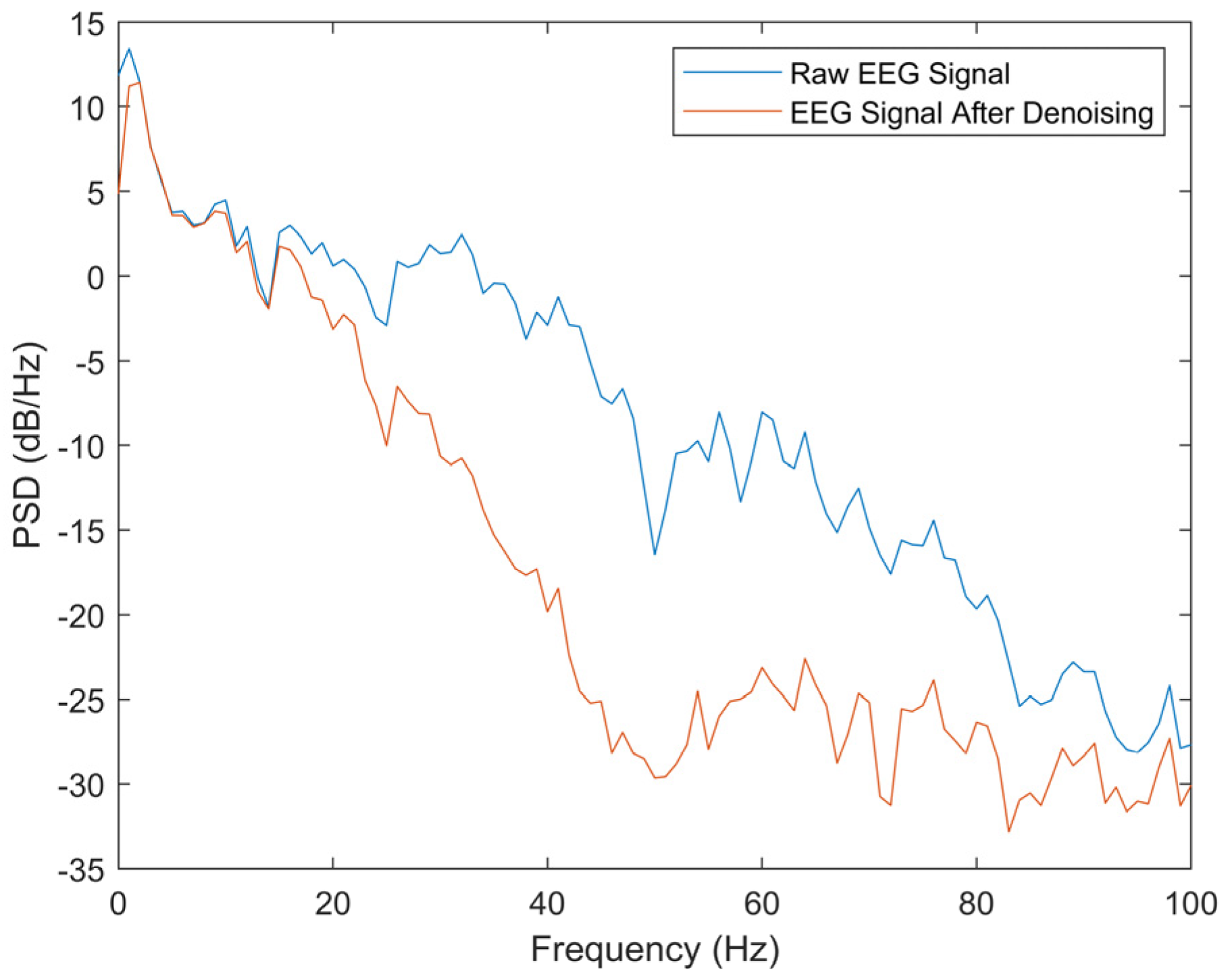
Preprocessing is an essential part of EEG signal processing that plays an important role in removing noise and improving the signal-to-noise ratio (SNR) of the signal. It is generally believed that the noise in EEG signals exists at frequencies above 30 Hz, so using a low-pass filter to remove signals higher than 30 Hz is the traditional method of noise removal, but the processing of signals by this method is relatively rough. In order to further improve the signal quality and lay a good foundation for following processing, a signal denoising method based on complementary ensemble empirical modal decomposition (CEEMD) and independent component analysis (ICA) is designed in this paper, with CEEMD responsible for decomposing the signal into several frequency bands and ICA responsible for separating out the noise components in the expectation of achieving more detailed denoising. CEEMD decomposes the signal into several intrinsic mode functions (IMFs) and a residual signal without modal aliasing and auxiliary noise residuals [
9,
10]. ICA is a common signal processing method that can decompose signals into a few independent components (ICs) [
11].
The feature extraction of EEG signals is also a key factor affecting the effectiveness of the model. The fatigue degree can eventually be accurately recognized only when the selected features contain valid information that helps to classify. EEG features associated with band power (e.g., power spectral density, band power ratio) have been widely used in the study of fatigue degree recognition. Hendrawan et al. extracted power percentage features for each EEG segment and used the LDA algorithm to recognize fatigue with an accuracy of 92.82% [
12]. Zeng et al. used band power ratios (β/θ and α/θ) as features and domain-adversarial neural networks (DANN) as classifiers in their fatigue recognition study, achieving a recognition accuracy of 91.63% [
13]. These studies have suggested that features related to the band power of EEG signals can be useful for fatigue recognition. Therefore, in this paper, the relative band power of EEG signals is employed as a feature for fatigue recognition. Relative band power is defined as the band power ratio of δ, θ, α and β frequency bands relative to the total frequency band.
A number of studies have shown a correlation between fatigue and attention. Van der Linden et al. suggested that mental fatigue can also be manifested as a decrease in attention [
14]. Azarnoosh et al. found that mental fatigue leads to a decrease in overall brain activity and a decrease in attention as one of the regular activities of the brain in response to incoming stimuli [
15]. Boksem et al. showed that mental fatigue reduces attention levels [
16]. Meanwhile, fuzzy entropy is a commonly used EEG feature that has shown its effectiveness in attention recognition [
17,
18]. Therefore, this study considers the introduction of features that have been proven effective in attention recognition studies, and fuzzy entropy satisfies this condition. With the help of the relationship between attention level and fatigue degree, effective features of attention recognition (fuzzy entropy) are added to the fatigue recognition task as a way to aid the recognition of fatigue degree.
In this work, eight subjects were invited to participate in an N-back experiment to induce fatigue, while the corresponding EEG signals were recorded through the process. A denoising method based on CEEMD and ICA was designed to remove noise from the raw EEG signals. Relative band power and fuzzy entropy were extracted as features of the EEG signal. Based on these features, the extreme gradient boosting (XGBoost) algorithm was employed to classify three different degrees of mental fatigue.
The paper is organized as follows.
Section 2 describes the methods of EEG preprocessing, feature extraction, and classification.
Section 3 describes the experiment results.
Section 4 is the discussion, and
Section 5 concludes this paper.
2. Materials and Methods
2.1. N-Back Experiment
The N-back experiment, which is one of the most common experimental paradigms in brain load elicitation experiments, was first proposed by Kirchner in 1958 to study the short-term memory capacity of people of different ages for rapidly changing information [
19].
At the beginning of the experiment, a cross symbol appears in the center of the screen to remind subjects to focus. During the experiment, a capital letter will appear randomly on the screen, and the subject needs to determine whether the current letter is consistent with the letter presented N items ago, and if it is consistent, press the “1” key, otherwise press the “0” key. The 1-back experiment requires the subject to determine whether the current letter is the same as the previous letter, while the 2-back experiment requires a comparison with the letter two items ago. The process of the 2-back experiment is shown in
Figure 1.
2.2. EEG Data Acquisition
Studies have demonstrated that 30 min of continuous 1-back or 2-back tasks can effectively induce fatigue [
20]. In order to obtain fatigue-related EEG signals, eight healthy subjects (five males and three females) were invited to participate in the N-back experiment. The subjects were all university students aged 20 to 26 years old, with normal vision. The students had no history of neurological disorders and had not taken any medication within 24 h prior to the experiment.
The experiment was divided into three stages: Stage 1, where the EEG signals were recorded in the subject’s calm state for 5 min; Stage 2: where the subject completed a 30 min 1-back task and recorded the EEG signals in the last 5 min of the task; Stage 3, where the subject completed a 30 min 2-back task and recorded the EEG signals in the last 5 min. The EEG signals acquired in the experiment contained 19 channels, the electrodes were arranged according to the international 10–20 system, the right and left earlobes were selected as reference electrodes, and the sampling rate was 512 Hz.
After each stage of the experiment, subjects were guided to fill out a subjective fatigue scale, as shown in
Table 1. Subjects were also instructed to rate their degree of attention on a scale of 1 to 10, with higher scores representing more confused thinking and distraction.
2.3. Preprocessing
Preprocessing has two main tasks: segmentation of the acquired raw EEG signal for further processing, and removal of noise from the raw EEG.
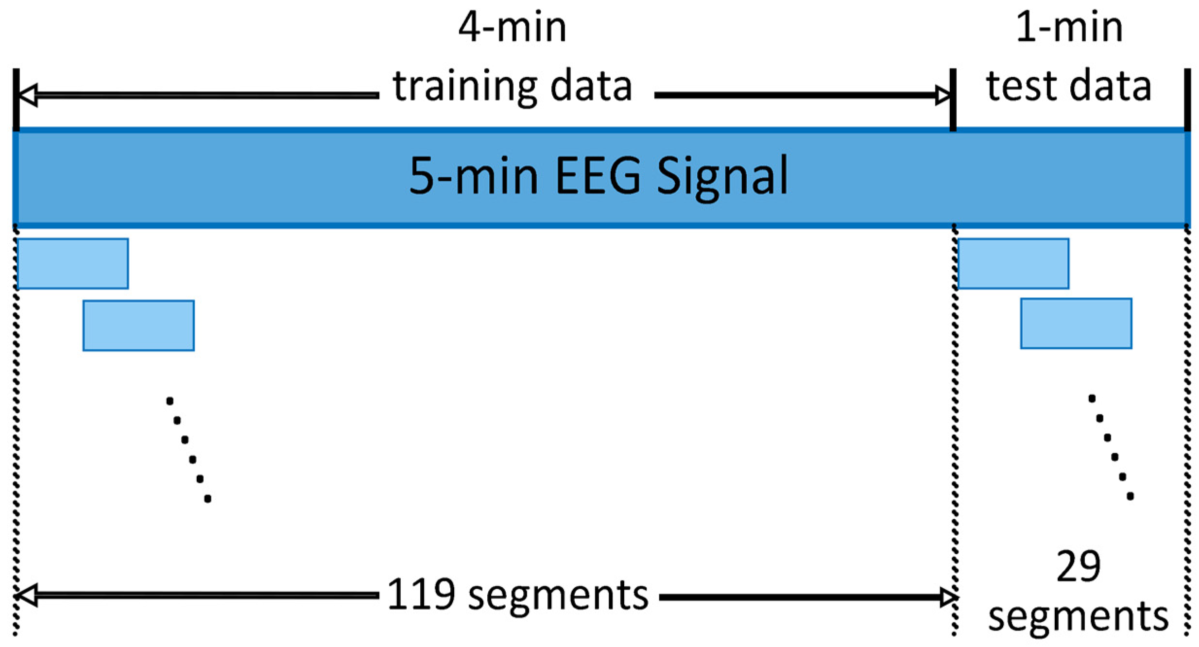
The EEG signals were first segmented as shown in
Figure 2. For each of the three experimental stages, a 5 min EEG signal was recorded, the first 4 min of which were used for training, and the last 1 min for testing. Both training and test data were divided into segments of 4 s with a 2 s overlap between two adjacent segments. To avoid data leakage, there was no overlap between the last segment of the training data and the first segment of the test data. For each 5 min EEG signal, a total of 119 segments of training data and 29 segments of test data were obtained.
Each segment of the EEG signal then needed to be denoised. In this paper, a method based on CEEMD and ICA was designed for EEG denoising. It is first desired to decompose the EEG signal into several frequency bands and then analyze which of them contains more noise. The noise-containing band should be denoised and then the signal of each frequency band is reconstructed to obtain the denoised EEG signal.
According to the frequency range, EEG is divided into five frequency bands: delta, theta, alpha, beta, and gamma, but this way of division is rather rough and is not suitable for a more detailed analysis of where the noise lies in the frequency range. Wavelet analysis can decompose the signals into multiple frequency bands in a more detailed way, but its effect is affected by the wavelet function used, so it needs to analyze which wavelet function to use to obtain the best results. CEEMD, on the other hand, performs an adaptive decomposition of the signal into a number of IMFs, each representing a frequency component, without any other analysis. Therefore, this paper used CEEMD to decompose the EEG signal. The value of sample entropy is positively correlated with the noise power [
21]. That is, a larger sample entropy indicates a more complex signal, indicating that the signal contains more noise; conversely, a smaller sample entropy indicates a higher degree of autocorrelation, suggesting that the signal contains less noise. Therefore, this paper used sample entropy to find the IMFs that contained more noise. In conventional preprocessing methods, IMFs that are considered to contain noise are usually discarded directly, which usually results in the loss of some useful signal components. To improve this problem, a further step with ICA was performed on the noisy IMFs. More specifically, the IMFs containing noise were processed by ICA to obtain a number of ICs, and then the sample entropy of each IC was calculated to find the ICs that contained more noise. The first IC with the highest value of sample entropy was regarded as a noise-containing component and set to zero. Then, the inverse ICA was performed on all ICs to obtain the noise-removed IMFs. Finally, the denoised IMFs and the IMFs considered to contain no noise were reconstructed, and the reconstructed signal was the preprocessed denoised signal. The preprocessing process is shown in
Figure 3.
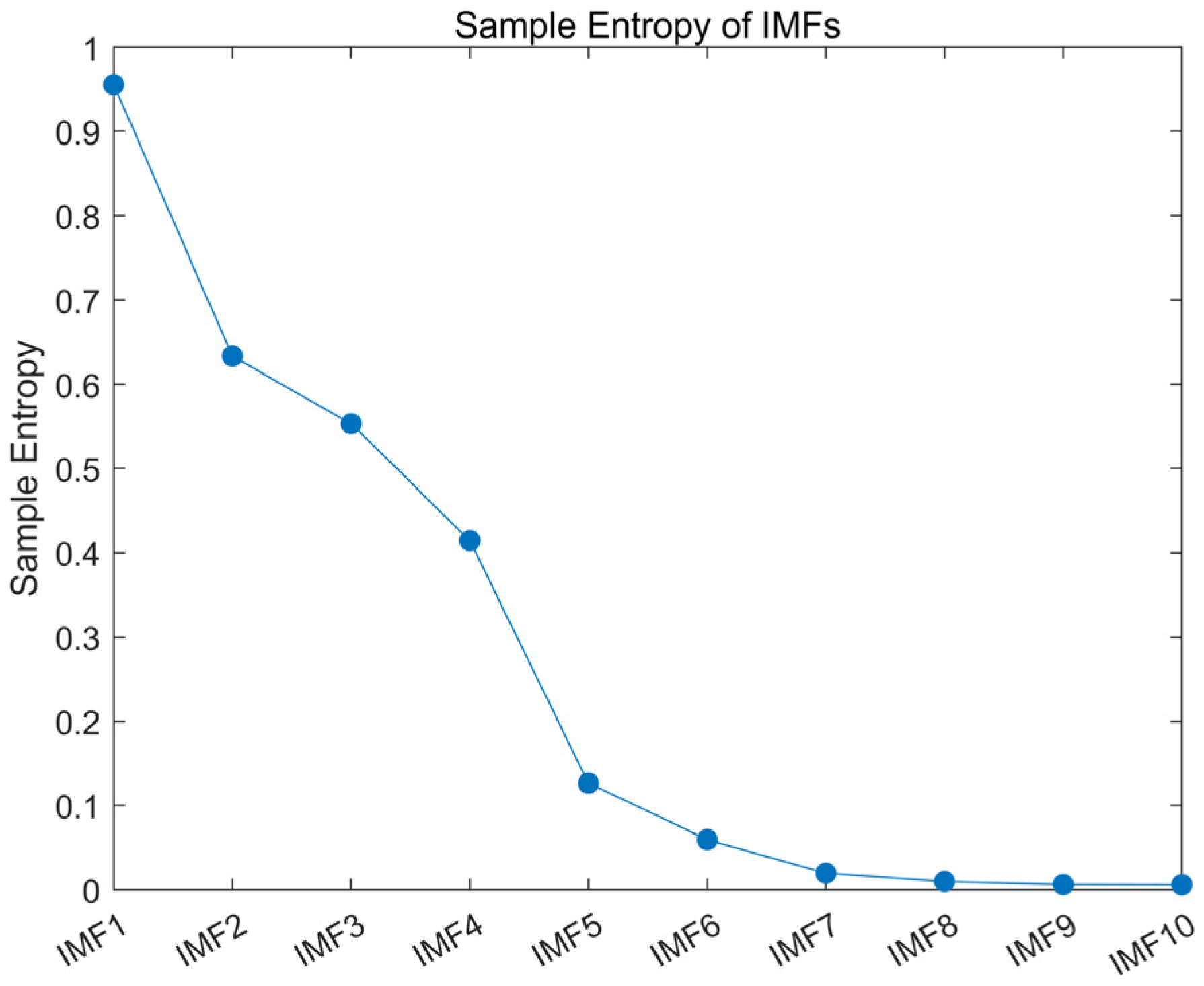
The raw EEG signal was decomposed into several IMFs using CEEMD, and the sample entropy of each IMF was calculated.
Figure 4 shows the calculated sample entropy of each IMF after the signal was decomposed by CEEMD. The sample entropy of IMFs decreased in order, and the first six IMFs had larger sample entropy values, while the remaining IMFs had smaller sample entropy. After several repeated experiments, the acquired EEG signals all showed this characteristic. Therefore, the first six IMFs of the raw signal were considered to contain more noise, while the rest of the IMFs did not. Then, the ICA was performed on the first six IMFs to obtain a number of ICs, and the IC with the highest sample entropy value was set to zero; then, the inverse ICA was applied to obtain the denoised IMF1–IMF6. The denoised EEG signal was obtained by reconstructing all IMFs.
2.4. Feature Extraction
In this work, the relative band power and the fuzzy entropy of EEG signals were extracted as features for fatigue recognition. Relative band power has been widely used in fatigue recognition studies. We assumed here that this feature would play an important role in recognizing fatigue degrees. Considering that an increase in fatigue level is often accompanied by a decrease in attention level, this paper introduced fuzzy entropy, which is a feature commonly used for attention level recognition, to help improve the accuracy of fatigue level recognition. The details of these features are described as follows.
Relative band power (RBP) is the ratio of the power of the EEG signal in a particular frequency band to the total power. The PSD of the EEG signal was first estimated using the Welch method, and then the power percentage of the different frequency bands was calculated according to (1).
where P(f) represents the PSD of the EEG signal, f
1 and f
2 represent the minimum and maximum frequencies of the EEG signal, respectively, and
represents the frequency range of α band. Similarly, we could calculate the power percentage of the β, θ and δ frequency bands [
22]. In particular, the δ band had a frequency range between 0 and 3 Hz, and the θ band was between 4 and 7 Hz, while the α and β bands were located between 8 and 13 Hz and 14 and 30 Hz, respectively.
Fuzzy entropy is used to measure the complexity and irregularity of a time series. It is calculated as follows [
23]. Let the signal sequence containing N samples be {u(i): 1 ≤ i ≤ N}; this sequence forms a set of m dimensional vectors
as shown in (2).
where
is the i-th point to the i + m − 1 point of sequence u(i), and
is their mean value.
is defined as the distance between vector
and
, and its value represents the maximum difference between the corresponding elements of the two vectors.
where i, j = 1~N − m and i ≠ j.
The fuzzy entropy function u(
, n, r) is used to define the similarity of vectors
and
, as shown in (5).
where n and r denote the gradient and width of the fuzzy function boundary. The function
is defined according to (6).
Ultimately, fuzzy entropy is defined as (7).
The raw EEG signal was divided into 4 s segments after pre-processing, and each segment contained 19 channels. The above five features were calculated for each channel, so a total of 95 feature values were obtained for each segment.
2.5. Classification
XGBoost is currently one of the most advanced algorithms in the field of ensemble learning. It follows the general modelling process of the Boosting algorithm, i.e., building the base estimators in turn, computing a loss function based on the output of the previous base estimators, and adaptively influencing the construction of the next base estimators. The final output of the ensemble algorithm is influenced by all of the base estimators. Specifically, suppose the XGBoost algorithm has a total of
base estimators (typically decision tree); for a certain sample x
i, the final output H(x
i) is expressed as (8).
where f
k(x
i) denotes the output of sample x
i on the
base estimator, and
is the learning rate, which is a hyperparameter in the XGBoost algorithm. In addition, as the iteration progresses, the ensemble output result for each sample is continuously computed. The output of sample x
i when building the k-th base estimator is shown in (9).
Unlike other Boosting algorithms, XGBoost adaptively influences the creation of weak evaluators by fitting pseudo-residuals. Specifically, before each new base estimator is built, the first-order derivative
and the second-order derivative
of the current loss function with respect to the output are computed, as shown in (10) and (11).
where
is the loss function, and
is the true label of sample
. Then, the current pseudo-residuals are
When building a new base estimator, the model needs to fit the pseudo-residuals rather than the true label value of the sample. This approach allows XGBoost to ensure that the loss function is minimized at each iteration.
When performing the classification task, XGBoost feeds the ensemble output into the Softmax function and obtains the classification probability. The final classification decision is made by choosing the class with the highest probability. In this work, we employed XGBoost to identify fatigue degrees due to its fast modeling speed and high resistance to over-fitting.
In this paper, the relative band power of the , and bands and the fuzzy entropy were extracted as features of the EEG signals. The above features were calculated for each of the three fatigue degrees, and the feature matrix was fed into the XGBoost classifier. Considering the inter-subject variability, a subject-specific model was used in this paper, i.e., a separate classification model was trained for each subject. EEG signals representing each of the three fatigue degrees were recorded in each of the three stages of the data acquisition experiment, with each recording lasting 5 min. According to the proposed data segmentation method, each 4 s segment was defined as a sample. Each subject had 119 training samples and 29 test samples at each fatigue degree, and each subject recorded EEG at three different fatigue degrees. This led to a total of 357 training samples and 87 test samples for each subject.
4. Discussion
4.1. Comparison with Other Preprocessing Methods
In this paper, an EEG preprocessing method based on CEEMD and ICA was proposed. To further illustrate the effectiveness of the method, three other methods were used to preprocess the data in this paper separately. Both features, relative band power and sample entropy, were also calculated, and the XGBoost classifier was also used to compare their classification performance. The three preprocessing methods were low-pass filtering, wavelet threshold denoising, and CEEMD, where low-pass filtering used a low-pass filter with a passband of less than 30 Hz, wavelet threshold denoising used a 4-layer decomposition of the db4 wavelet with a soft threshold function and a fixed threshold, and CEEMD decomposed the signal into several IMFs and reconstructed it after discarding the first six IMFs.
Table 4 shows the performance of the classification for the three fatigue degrees using different preprocessing methods. The results show that the classification results obtained with the preprocessing method of this paper were better.
4.2. Comparison with Other Methods
Table 5 shows a comparison of the method proposed in this paper with some of the existing methods. Similarly to our study, the studies in
Table 5 also designed their own experiments to induce mental fatigue in subjects, extracting multiple features of EEG signals and recognizing the degree of fatigue using certain classification methods. It should be noted that the results listed in the table were from different EEG data, so the comparison does not directly indicate that the method proposed in this study is superior to other existing methods. However, it can still show to a certain extent that our method can reach a good level in the related field.
4.3. Limitations of the Current Study
This paper proposed a fatigue recognition method based on EEG signals that includes four parts: data acquisition, preprocessing, feature extraction and classification. Experimental results demonstrated that our method is able to recognize three different degrees of mental fatigue with high accuracy. However, there were certain limitations of the current study that need to be improved in further studies.
First, the method may suffer from feature redundancy. Relative band power of four frequency bands and fuzzy entropy were selected as EEG features. Therefore, five feature values needed to be calculated for each segment of EEG. Since there were 19 channels per segment of EEG, there were a total of 95 feature values per sample. This led to a computationally intensive algorithm, which is not conducive to practical applications, especially for real-time detection of fatigue degree. The use of feature selection algorithms to select the features that contribute most to the classification helps to reduce the redundancy of features, and the design of channel optimization algorithms to find the most important channels can also further reduce the computational effort.
Second, how the features of the EEG signal affect the classification results remains to be further studied. In this paper, a variety of features were extracted, and the classification results showed that these features together were effective in distinguishing different fatigue levels. However, it is unclear whether there is a significant difference between the features at three fatigue levels. For example, the same method obtained an accuracy of 100.00% on subject S01, but only 83.91% on S03. Is this because one or more features did not differ significantly between the three levels of fatigue for S03? More in-depth analysis is needed to understand the effect of each feature on the classification results.
Admittedly, the method proposed in this paper is still not representative of the state-of-the-art. Designing a more rational experimental paradigm to more effectively induce mental fatigue in subjects, optimizing the preprocessing process to further improve signal quality, screening and using features that are valid across all subjects, and using a classification model with greater learning ability are all areas for improvement in this study. The resolution of these issues will contribute further to the practical application of fatigue recognition technology.


