Case Report

Cone Beam Computed Tomography in the Assessment of the Effectiveness of Physical Therapy with the Use of the Electromagnetic Field Combined with Light Radiation Emitted by LEDs in the Treatment of Inflammation of the Paranasal Sinuses—A Case Study

1
Student Scientific Society, Department of Internal Medicine, Angiology and Physical Medicine, Medical University of Silesia in Katowice, 40-055 Katowice, Poland
2
Department of Internal Medicine, Angiology and Physical Medicine, Medical University of Silesia in Katowice, 40-055 Katowice, Poland
3
Provincial Specialist Hospital No. 4 in Bytom, 41-902 Bytom, Poland
4
Department of Propaedeutic, Physical Diagnostics and Dental Physiotherapy, Faculty of Medicine and Dentistry, Pomeranian Medical University, 70-204 Szczecin, Poland
*
Author to whom correspondence should be addressed.
Int. J. Environ. Res. Public Health 2022, 19(20), 13570; https://doi.org/10.3390/ijerph192013570
Submission received: 4 September 2022 / Revised: 9 October 2022 / Accepted: 11 October 2022 / Published: 20 October 2022

Abstract

:
Modern high-resolution volumetric tomography, commonly known as cone beam computed tomography (CBCT), is one of the most innovative imaging techniques which can provide views of anatomical structures not attainable by conventional techniques. Magnetic field LED therapy is a physical therapy method, combining the effects of the Extremely Low Frequency-Electromagnetic Field (ELF-EMF) and high-power light radiation emitted by Light Emitting Diodes (LEDs). The method has been widely applied in the treatment and rehabilitation of complications of many medical conditions, including in dentistry. The aim of this study was to use CBCT to assess the effectiveness of the simultaneous use of electromagnetic field and LED light in the physical therapy of paranasal sinusitis. Treatments employing the electromagnetic field combined with LED light were administered to a 39-year-old female outpatient of the physiotherapy ward for rehabilitation therapy of paranasal sinusitis. Normal sinus pneumatization was restored almost completely. Reduction in the swelling of the sinus mucosa was so significant that even the pneumatization of the ethmoid bulla was restored. Physical therapy with the simultaneous use of ELF-EMF and LED light was found to be effective in the rehabilitation of the patient with paranasal sinusitis. Positive effects of the treatment were confirmed by CBCT findings.

1. Introduction

Different X-ray imaging techniques used in dentistry differ in terms of accuracy in the reproduction of anatomical details of the diagnosed area [1,2,3]. Cone beam computed tomography (CBCT) imaging has an instrumental advantage in that it can be used to reconstruct a three-dimensional image, providing a detailed insight into the topography of skeletal structures in a chosen projection [3,4,5,6]. CBCT is also successfully used in otolaryngology. Due to the accuracy of this imaging technique, it is possible to simultaneously diagnose and plan treatment for paranasal sinusitis. Importantly, this technique is safe for the patient since the dose of ionizing radiation in CBCT is reduced, about 20–30 times lower than the conventional multi-slice CT. The average dose of radiation in CBCT is in the range of 20–650 µSv, depending on the size of the field of view and image resolution [7,8,9,10]. In turn, magnetic field LED therapy is a popular physical therapy modality with a high therapeutic efficacy, due to the combined effects of Extremely Low Frequency-Electromagnetic Field (ELF-EMF) and high-power light emitted by Light Emitting Diodes (LEDs), producing red light (R) (wavelength 630 nm) and infrared light (IR) (wavelength 855 nm) radiation [11,12,13,14,15]. The electromagnetic field used in this method usually has magnetic flux density of 15 μT, and frequency in the range of 180–195 Hz [16,17]. In dental applications, it has been shown to provide regenerative effects and nerve stimulation. The method can be used in the treatment of large inflammatory lesions of periapical tissues, soft tissue cysts, dry socket, neuralgia, lingual nerve regeneration, tooth replantation sites and mandibular fractures. The electromagnetic field combined with high-power LED light activates calcium channels, transmembrane ion transport, and stimulates enzyme activity. Regenerative effects primarily come from the intensification of oxygen supply and tissue respiration associated with increased diffusion and oxygen uptake by hemoglobin and cytochromes. Increased oxygen uptake stimulates tissue respiration, DNA synthesis, and accelerates the mitotic cycle. The anti-inflammatory effect is associated with the upregulation of c-AMP and prostaglandin E synthesis, which promotes the accumulation of c-AMP and reduces the secretion of inflammatory mediators from neutrophils, basophils, mast cells and lymphocytes [16,17,18,19,20,21,22]. In turn, the analgesic effect of this method can be attributed to the reduction of nerve impulse conduction in afferent nerve fibers due to the hyperpolarization of neuron membranes, as well as to the increase in synthesis and secretion of endogenous opiates (beta-endorphins) in the central nervous system, consequently raising the pain threshold [23,24,25]. Magnetic field LED therapy has found many applications in the treatment and rehabilitation of complications of many medical conditions [26,27,28,29,30,31], including in dentistry [32].

2. Aim of Study

The aim of this study was to use cone beam computed tomography with a large field of view to assess the effectiveness of the simultaneous use of electromagnetic field and LED light in the physical therapy of paranasal sinusitis.

3. Material and Methods

A 39-year-old female, A.F., presented to the outpatient clinic with recurrent upper respiratory tract infections, which had been treated for 2.5 years with antibiotics and steroids. Periodically, the patient was prescribed an intranasal glucocorticoid—Mometasone nasal spray, to be administered once daily, at 200 µg (two actuations) of the drug into each nostril, for no longer than 3 weeks. Concurrently, the patient took an antibiotic, amoxicillin (750 mg) with clavulanic acid (125 mg), twice daily for no longer than 2 weeks. During the treatment, the patient also rinsed her nose with a saline solution. The main symptoms reported by the patient were: a congested nose making it difficult to breathe, frequent nasal discharge, facial pain, a feeling of swelling, tenderness and discomfort around the eyes, forehead and cheeks. Olfactory loss was also observed. These symptoms had persisted for more than 2 weeks. Pharmacological treatment was unsuccessful, and the patient was offered an otolaryngological surgical intervention. The patient did not consent to the surgery. After ruling out contraindications, the patient was qualified for the study. The patient did not use any pharmacological agents during physical therapy treatments.
To make an accurate diagnosis and establish the etiology of the patient’s symptoms, we used cone beam computed tomography (CBCT), which enabled us to examine both hard tissue structures and soft elements of the mucous membrane in the paranasal sinuses. Tomography also ruled out odontogenic sinusitis. Volumetric tomographic imaging of the facial skeleton was performed using an i-CAT Next Generation (ISI) system with a wide field of view (FOV) (size 17 cm × 23 cm). Based on personal observation of selected cases, we analyzed the suitability and effectiveness of this radiographic technique for the diagnosis of pathological lesions in the mucous membrane of maxillary sinuses. The patient was examined in a sitting position with a stabilized head to avoid motion-induced artefacts, and Quantum IQ software was used to acquire high-quality images of soft tissues. All scans were taken in the same radiography laboratory using the same apparatus and exposure parameters: 110 kV, 5 mA, time 17 s and −0.3 voxel resolution.
Volumetric tomographic imaging revealed significantly decreased aeration of maxillary sinuses (Figure 1).
CBCT also showed mucosal thickening in ethmoid cells and at the bottom of the sphenoid sinus (Figure 2).
A detailed analysis of the images made it possible to rule out the dental origin of pathological lesions (Figure 3).
The Viofor JPS System (Med and Life, Komorów, Poland) was used in the treatment under discussion. The system features a control unit and proprietary applicators for low frequency magnetic therapy and combination therapy, namely Pulsed Magnetic Field Therapy (Magnetotherapy), Pulsed Magnetic Field Stimulation (Magnetostimulation), and Pulsed Magnetic Field LED Therapy. With the wide range of applicators, it is possible to perform topical treatments on small and mid-sized surfaces, as well as whole-body treatments. The patient underwent a series of physical therapy treatments, with 30 daily sessions lasting 20 min each, and each using two panel applicators of the VIOFOR JPS Magnetic Light device containing 560 LEDs emitting mixed radiation: red light (wavelength 630 nm, power 500 mW) and infrared light (wavelength 855 nm, power 3400 mW), with a frequency of light impulses of 181.8 Hz. This generated a simultaneous electromagnetic field with magnetic flux density of 15 μT and magnetic field frequency in the range of 180–195 Hz (Figure 4).
The effectiveness of the physical therapy was assessed by comparing the CBCT images acquired before and after the series of treatments, with a focus on the condition of the paranasal sinuses. The contraindications for this therapy included: pregnancy and breastfeeding, acute inflammation, skin cancer, autoimmune diseases, light sensitivity, epilepsy, treatment with vitamin A derivatives, photophobia, porphyria, use of medications prone to photodegradation, tuberculosis, (uncontrolled) diabetes, severe viral, bacterial and fungal infections and electronic implants. During the procedure, the patient wore protective glasses.

4. Results

Follow-up imaging was performed at the end of the 30-day therapy. CBCT scans revealed a marked regression of inflammation in all paranasal sinuses, which was correlated with improved patient comfort. Normal sinus pneumatization was restored almost completely (Figure 5).
Reduction in the swelling of the sinus mucosa was so significant that even ethmoid bulla pneumatization was restored. Pathological lesions in the sphenoid sinus also resolved completely (Figure 6). At the bottom of the maxillary sinuses, mucosal thickening was still visible. In the right maxillary sinus, the maximum mucosal thickness was 3–4 mm. In the left maxillary sinus, the maximum mucosal thickness was 3–7 mm. The patient follow-up is continuing. She will require a second round of physical therapy. After the series of treatments discussed in this paper, the patient reported manifest improvement, including elimination of frontal headaches, eye and nose pain, nasal discharge and congestion.

5. Discussion

Sinusitis is a broad clinical syndrome with the common feature of mucosal inflammation. It is a complex of symptoms rather than a specific disease [33,34]. Objective evidence of chronic rhinosinusitis can be obtained by physical examination (anterior rhinoscopy and endoscopy) or radiography, preferably computed tomography of the sinuses [35,36]. The primary methods of treatment are pharmacological, using corticosteroids and antibiotics. First line pharmacological treatment mainly consists of topical intranasal and oral corticosteroids. Corticosteroids, which have very broad anti-inflammatory properties, also have the strongest evidence for efficacy. If the treatment fails, surgical options are used. Antibiotics are likely to be effective in a subpopulation of patients, but the different phenotypes and endotypes that make up sinusitis have been poorly defined to date. For some phenotypes, early surgery, as well as biologic therapy, may be more effective and cost-effective [33,34,37]. With intranasal corticosteroids, caution is advised, especially in children, pregnant women and elderly patients, and even more so in patients with comorbidities such as asthma, whose total steroid intake may be high due to the administration of both intranasal and inhaled corticosteroids [37]. In the present case study, physical therapy using LED light and electromagnetic field produced very good therapeutic results. This method does not have the limitations of corticosteroids and is not invasive like surgical procedures. The two physical factors have been observed to counteract inflammation by providing regenerative, analgesic, anti-inflammatory and antiedematous, as well as antimicrobial, angiogenetic and vasodilatory effects. There are reports of an improvement in tissue oxygen utilization and favorable modification of the immune function and coagulation system [38,39,40,41,42,43]. The above is a case report, and so the authors cannot explain why mucosal thickening in certain areas of the paranasal sinuses was reduced to a greater extent. Perhaps this is related to the different volume of the sinuses. Even after volume restoration of the maxillary sinuses, mucosa thickening was still seen.

6. Conclusions

Cone beam computed tomography can identify the most inconspicuous pathological changes in the paranasal sinuses and help exclude the dental origin of sinus inflammation.
Physiotherapy with the simultaneous use of ELF-EMF and LEDs light proved to be effective in the rehabilitation of a sinusitis patient. High therapeutic efficacy, lack of significant side effects and good tolerance of the treatment make this therapy potentially useful as a valuable adjunct to conventional symptomatic treatment. The obtained results will encourage further studies in larger patient samples and with longer therapy and follow-up periods.

Funding

This research received no external funding.

Institutional Review Board Statement

The study was approved by the Bioethics Committee, Pomeranian Medical University in Szczecin; Poland: No. KB-0012/32/13.

Informed Consent Statement

Informed consent was obtained from all subjects involved in the study.

Conflicts of Interest

The authors declare that there is no conflict of interest regarding the publication of this manuscript.

Abbreviations

CBCTCone Beam Computed Tomography
ELF-EMFExtremely Low Frequency—Electromagnetic Field
LEDLight Emitting Diode
IRinfrared light
Rred light

References

  1. Jager, L.; Rammelsberg, P.; Reiser, M. Bildgebende Diagnostik der Normalanatomie des Temporomandibulargelenks. Radiologe 2001, 41, 734–740. [Google Scholar] [CrossRef] [PubMed]
  2. Young, H.M.; Sun Park, C.K.; Chau, O.W.; Lee, T.Y.; Gaede, S. Technical Note: Volumetric computed tomography for radiotherapy simulation and treatment planning. J. Appl. Clin. Med. Phys. 2021, 22, 295–302. [Google Scholar] [CrossRef] [PubMed]
  3. Howerton, W.B., Jr.; Mora, M.A. Advancements in digital imaging. What is new and on the horizon? J. Am. Dent. Assoc. 2008, 139 (Suppl. 3), S20–S24. [Google Scholar] [CrossRef]
  4. Loubele, M.; Bogaerts, R.; Van Dijck, E.; Pauwels, R.; Vanheusden, S.; Suetens, P.; Marchal, G.; Sanderink, G.; Jacobs, R. Comparison between effective radiation dose of CBCT and MSCT scanners for dentomaxillofacialis applications. Eur. J. Radiol. 2009, 71, 461–468. [Google Scholar] [CrossRef]
  5. Bargan, S.; Merrill, R.; Tetradis, S. Cone beam computed tomography imaging in the evaluation of the temporomandibular joint. J. Calif Dent Assoc. 2010, 38, 33–39. [Google Scholar]
  6. Anjos Pontual, M.L.; Freire, J.S.; Barbosa, J.M.; Frazão, M.A.; dos Anjos Pontual, A. Evaluation of bone changes in the temporomandibular joint using cone beam CT. Dentomaxillofac. Radiol. 2012, 41, 24–29. [Google Scholar] [CrossRef] [Green Version]
  7. Phothikhun, S.; Suphanantachat, S.; Chuenchompoonut, V.; Nisapakultorn, K. Cone-beam computed tomographic evidence of the association between periodontal bone loss and mucosal thickening of the maxillary sinus. J. Periodontol. 2012, 83, 557–564. [Google Scholar] [CrossRef] [PubMed]
  8. Roberts, J.A.; Drage, N.A.; Davies, J.; Thomas, D.W. Effective dose from cone beam CT examinations in dentistry. Brit. J. Radiol. 2009, 82, 35–40. [Google Scholar] [CrossRef]
  9. Loubbele, M.; Jacobs, R.; Maes, F.; Denis, K.; White, S.; Coudyzer, W.; Lambrichts, I.; van Steenberghe, D.; Suetens, P. Image quality vs. radiation dose of four cone beam computed tomography scaners. Dentomaxillofac. Radiol. 2008, 37, 309–319. [Google Scholar] [CrossRef] [PubMed]
  10. Scarfe, W.C.; Farman, A.G. What is cone-beam CK and how does it work? Dent Clin. N. Am. 2008, 52, 707–730. [Google Scholar] [CrossRef]
  11. Calderhead, R.G.; Won-Serk, K.; Ohshiro, T.; Trelles, M.A.; Vasily, D.B. Adjunctive 830 nm light-emitting diode therapy can improve the results following aesthetic procedures. Laser Ther. 2015, 24, 277–289. [Google Scholar] [CrossRef] [PubMed]
  12. Barolet, D.B. Light-emitting diodes (LEDs) in dermatology. Semin Cutan Med. Surg. 2008, 27, 227–238. [Google Scholar] [CrossRef] [PubMed]
  13. Paolillo, F.R.; Borghi-Silva, A.; Parizotto, N.A.; Curachi, C.; Bagnato, V.S. New treatment of cellulite with infrared-LED illumination applied during high-intensity treadmill training. J. Cosmet Laser Ther. 2011, 13, 166–171. [Google Scholar] [CrossRef] [PubMed]
  14. Russell, B.A.; Kellett, N.; Reilly, L.R. A study to determine the efficacy of combination LED light therapy (633nm and 830nm) in facial skin rejuvenation. J. Cosmet Laser Ther. 2005, 7, 196–200. [Google Scholar] [CrossRef] [PubMed]
  15. von Felbert, V.; Kernland-Lang, K.; Hoffmann, G.; Wienert, V.; Simon, D.; Hunziker, T. Irradiation with water-filtered infrared A plus visible light improves cutaneous scleroderma lesions in a series of cases. Dermatology 2011, 222, 347–357. [Google Scholar] [CrossRef] [PubMed]
  16. Pall, M.L. Electromagnetic fields act via activation of voltage-gated calcium channels to produce beneficial or adverse effects. J. Cell Mol. Med. 2013, 17, 958–965. [Google Scholar] [CrossRef]
  17. Fan, W.; Qian, F.; Ma, Q.; Zhang, P.; Chen, T.; Chen, C.; Zhang, Y.; Deng, P.; Zhou, Z.; Yu, Z. 50 Hz electromagnetic field exposure promotes proliferation and cytokine production of bone marrow mesen-chymal stem cells. Int. J. Clin. Exp. Med. 2015, 8, 7394–7404. [Google Scholar]
  18. Pilla, A.; Fitzsimmons, R.; Muehsam, D.; Wu, J.; Rohde, C.; Casper, D. Electromagnetic fields as first messenger in biological signaling: Application to calmodulin-dependent signaling in tissue repair. Biochim. Biophys. Acta 2011, 1810, 1236–1245. [Google Scholar] [CrossRef]
  19. Marcinkowska-Gapińska, A.; Nawrocka-Bogusz, H. Analysis of the magnetic field influence on the rheological properties of healthy person’s blood. BioMed Res. Int. 2013, 2013, 490410. [Google Scholar] [CrossRef] [Green Version]
  20. Vianale, G.; Reale, M.; Amerio, P.; Stefanachi, M.; Di Luzio, S.; Muraro, R. Extremely low frequency electromagnetic field enhances human keratinocyte cell growth and decreases pro-inflammatory chemokine production. Br. J. Dermatol. 2008, 158, 1189–1196. [Google Scholar] [CrossRef]
  21. Patruno, A.; Amerio, P.; Pesce, M.; Vianale, G.; Di Luzio, S.; Tulli, A.; Franceschelli, S.; Grilli, A.; Muraro, R.; Reale, M. Extremely low frequency electromagnetic fields modulate expression of inducible nitric oxide syn-thase, endothelial nitric oxide synthase and cyclooxygenase–2 in the human keratinocyte cell line HaCaT: Potential therapeutic effects in wound healing. Br. J. Dermatol. 2010, 162, 258–266. [Google Scholar] [CrossRef] [PubMed]
  22. Ross, C.L.; Teli, T.; Harrison, B.S. Effect of electromagnetic field on cyclic adenosine monophosphate (cAMP) in a human mu-opioid receptor cell model. Electromagn Biol Med. 2016, 35, 206–213. [Google Scholar] [CrossRef] [PubMed]
  23. Lietz-Kijak, D.; Ardan, R. Physiotherapeutic reduction of orofacial pain using extremely low-frequency electromagnetic field and light-emitting diode therapy-a pilot study. Pain Res. Manag. 2022, 2022, 3115154. [Google Scholar] [CrossRef] [PubMed]
  24. Kopacz, Ł.; Ciosek, Ż.; Gronwald, H.; Skomro, P.; Ardan, R.; Lietz-Kijak, D. Comparative analysis of the influence of selected physical factors on the level of pain in the course of temporomandibular joint disorders. Pain Res. Manag. 2020, 2020, 1036306. [Google Scholar] [CrossRef] [PubMed]
  25. Cieślar, G.; Mrowiec, J.; Kasperczyk, S.; Sieroń-Stoltny, K.; Sieroń, A. Analgesic effect of simultaneous exposure to infrared laser radiation and μT magnetic field in rats. In Mechanisms for Low-Light Therapy III; Hamblin, M.R., Waynant, R.W., Anders, J., Eds.; SPIE: Bellingham, WA, USA, 2008; Volume 6846, pp. 74–80. [Google Scholar]
  26. Gualdi, G.; Costantini, E.; Reale, M.; Amerio, P. Wound repair and extremely low frequency-electromagnetic field: Insight from in vitro study and potential clinical application. Int J. Mol. Sci. 2021, 22, 5037. [Google Scholar] [CrossRef] [PubMed]
  27. Pasek, J.; Pasek, T.; Sieroń-Stołtny, K.; Cieślar, G.; Sieroń, A. Electromagnetic fields in medicine–The state of art. Electromagn. Biol. Med. 2016, 35, 170–175. [Google Scholar] [CrossRef]
  28. Gordon, G.A. Designed electromagnetic pulsed therapy: Clinical applications. J. Cell Physiol. 2007, 212, 579–582. [Google Scholar] [CrossRef]
  29. Nelson, F.R.; Zvirbulis, R.; Pilla, A.A. Non-invasive electromagnetic field therapy produces rapid and substantial pain reduction in early knee osteoarthritis: A randomized double-blind pilot study. Rheumatol. Int. 2013, 33, 2169–2173. [Google Scholar] [CrossRef]
  30. Pesce, M.; Patruno, A.; Speranza, L.; Reale, M. Extremely low frequency electromagnetic field and wound healing: Implication of cytokines as biological mediators. Eur. Cytokine Netw. 2013, 24, 1–10. [Google Scholar] [CrossRef] [Green Version]
  31. Cieślar, G.; Rozmus-Kuczia, I.; Łatka, U.; Matyszkiewicz, B.; Krzeszkowiak, T.; Mrowiec, J.; Sieroń-Stołtny, K.; Sieroń, A. Estimation of clinical efficacy of Viofor JPS System Magnetic & Light Therapy–device for magnetostimulation connected with light energy in the treatment of degenerative and inflammatory diseases of joints. Balneol. Pol. 2004, 46, 42–58. [Google Scholar]
  32. Lietz-Kijak, D.; Kijak, E.; Śliwiński, Z.; Opalko, K. The use of physiotherapy in the regeneration of periapical bone structures of the teeth, prepared to load the prosthetic. Adv. Hyg. Exper. Med. 2013, 67, 643–647. [Google Scholar] [CrossRef] [PubMed]
  33. Ghogomu, N.; Kern, R. Chronic rhinosinusitis: The rationale for current treatments. Expert Rev. Clin. Immunol. 2017, 13, 259–270. [Google Scholar] [CrossRef] [PubMed]
  34. Guilemany, J.M.; Alobid, I.; Mullo, J. Controversies in the treatment of chronic rhinosinusitis. Expert Rev. Respir Med. 2010, 4, 463–477. [Google Scholar] [CrossRef] [PubMed]
  35. Sedaghat, A.R. Chronic Rhinosinusitis. Am. Fam Physician. 2017, 96, 500–506. [Google Scholar] [PubMed]
  36. Rosenfeld, R.M.; Piccirillo, J.F.; Chandrasekhar, S.S.; Brook, I.; Kumar, A.K.; Kramper, M.; Orlandi, R.R.; Palmer, J.N.; Patel, Z.M.; Peters, A.; et al. Clinical practice guideline (update): Adult sinusitis. Otolaryngol. Head Neck Surg. 2015, 152 (Suppl. 2), S1–S39. [Google Scholar] [CrossRef] [Green Version]
  37. Mullol, J.; Obando, A.; Pujols, L.; Alobid, I. Corticosteroid treatment in chronic rhinosinusitis: The possibilities and the limits. Immunol Allergy Clin. N. Am. 2009, 29, 657–668. [Google Scholar] [CrossRef]
  38. Pasek, J.; Misiak, A.; Mucha, R.; Pasek, T.; Sieroń, A. New possibilities in physical therapy-magnetolaserotherapy. Physioter. Pol. 2008, 8, 1–10. [Google Scholar]
  39. Sieroń, A.; Pasek, J.; Mucha, R. Magnetic field and light energy in medicine and rehabilitation-magnetoledotherapy. Balneol. Pol. 2007, 49, 1–7. [Google Scholar]
  40. Pasek, J.; Pasek, T.; Sieroń, A. Some practical recommendations in the use of magnetic fields and light in physical medicine. Acta Bio-Opt. Inform. Med. 2007, 13, 284–285. [Google Scholar]
  41. Orien, A.; Pasek, J.; Mucha, R. Low-energy light in medicine and rehabilitation. Rehabil. Pract. 2007, 2, 25–27. [Google Scholar]
  42. Sieroń, A.; Cieślar, G.; Krawczyk-Krupka, A.; Biniszkiewicz, T.; Bilska, A.; Adamek, M. Application of Magnetic Fields in Medicine, 2nd ed.; Į-Medica Press: Bielsko, Biała, 2002. [Google Scholar]
  43. Reddy, G.K. Biological basis and clinical role of low-intensity lasers in biology and medicine. J. Clin. Laser Med. Surg. 2004, 22, 141–150. [Google Scholar] [CrossRef] [PubMed]
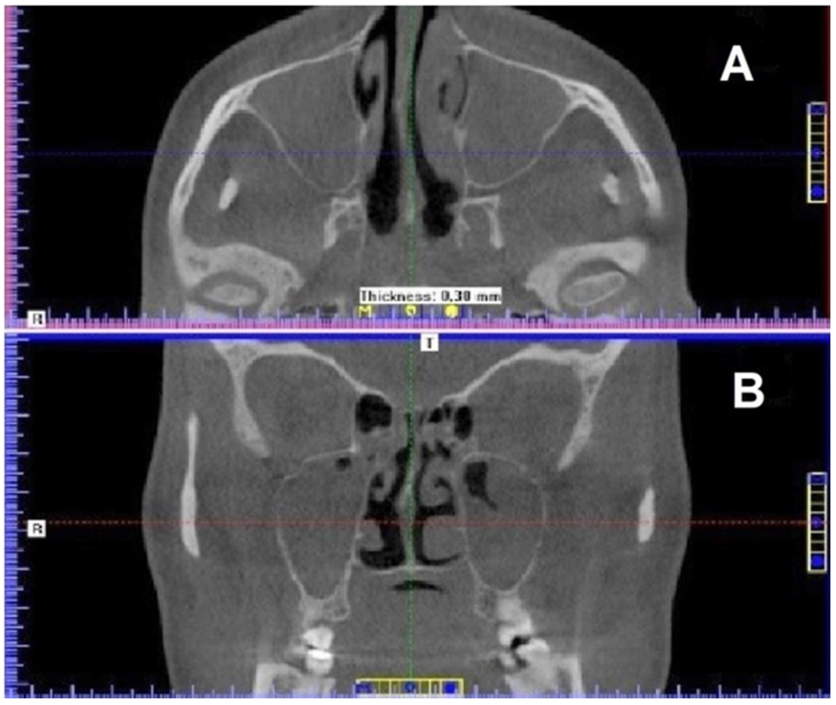
Figure 1. Maxillary sinuses visualized as dark (low-density) areas. (A) axial view; (B) coronal view (CBCT scans).
Ijerph 19 13570 g001
Figure 2. Mucosal thickening in ethmoid cells and at the bottom of the sphenoid sinus—CBCT scan—sagittal view.
Ijerph 19 13570 g002
Figure 3. Maxillary sinuses in sagittal view: (A) Right and (B) left. (CBCT scans).
Ijerph 19 13570 g003
Figure 4. Therapeutic treatment with the simultaneous use of electromagnetic field and high-power light emitted by LEDs, applied by means of two panel applicators of the Viofor JPS Magnetic Light device.
Ijerph 19 13570 g004
Figure 5. Image of maxillary sinuses after 30 sessions of electromagnetic field and LED light therapy. (A) coronal view; (B) axial view. (CBCT scans).
Ijerph 19 13570 g005
Figure 6. Ethmoid bulla and the sphenoid sinus—status after a series of 30 physical therapy sessions. (CBCT scan).
Ijerph 19 13570 g006
Publisher’s Note: MDPI stays neutral with regard to jurisdictional claims in published maps and institutional affiliations.