Review

Inanimate Surfaces as a Source of Hospital Infections Caused by Fungi, Bacteria and Viruses with Particular Emphasis on SARS-CoV-2

by
Agata Jabłońska-Trypuć
1,*,
Marcin Makuła
2,
Maria Włodarczyk-Makuła
3,
Elżbieta Wołejko
1,
Urszula Wydro
1,
Lluis Serra-Majem
4 and
Józefa Wiater
5
1
Department of Chemistry, Biology and Biotechnology, Faculty of Civil Engineering and Environmental Sciences, Bialystok University of Technology, Wiejska 45E Street, 15-351 Białystok, Poland
2
Faculty of Medical Sciences in Zabrze, Medical University of Silesia, Traugutta sq.2, 41-800 Zabrze, Poland
3
Faculty of Infrastructure and Environment, Częstochowa University of Technology, 69 Dabrowskiego Str., 42-201 Częstochowa, Poland
4
Research Institute of Biomedical and Health Sciences, University of Las Palmas de Gran Canaria, 35001 Las Palmas de Gran Canaria, Spain
5
Department of Agri-Food Engineering and Environmental Management, Faculty of Civil Engineering and Environmental Sciences, Bialystok University of Technology, Wiejska 45E Street, 15-351 Białystok, Poland
*
Author to whom correspondence should be addressed.
Int. J. Environ. Res. Public Health 2022, 19(13), 8121; https://doi.org/10.3390/ijerph19138121
Submission received: 9 June 2022 / Revised: 28 June 2022 / Accepted: 30 June 2022 / Published: 1 July 2022
(This article belongs to the Collection COVID-19 Research)

Abstract

:
The carriers of nosocomial infections are the hands of medical personnel and inanimate surfaces. Both hands and surfaces may be contaminated as a result of contact with the patient, their body fluids, and touching contaminated surfaces in the patient’s surroundings. Visually clean inanimate surfaces are an important source of pathogens. Microorganisms have properties thanks to which they can survive in unfavorable conditions, from a few days to several months. Bacteria, viruses and fungi are able to transmit from inanimate surfaces to the skin of the patient and the medical staff. These pathogens include SARS-CoV-2, which can survive on various types of inanimate surfaces, being a potential source of infection. By following the recommendations related to washing and disinfecting hands and surfaces, and using appropriate washing and disinfecting agents with a broad biocidal spectrum, high material compatibility and the shortest duration of action, we contribute to breaking the chain of nosocomial infections.

1. Introduction

Healthcare-associated infections remain one of the leading causes of increased morbidity and mortality among patients. It is estimated that in the United States, about 1.7 million people develop nosocomial-related illness each year, and approximately 99,000 of them die [1]. The patient’s endogenous bacterial flora is considered to be the main source of nosocomial infections, however 20 to 40% of nosocomial infections are caused by cross-contamination, where the vector of pathogen transmission is the hands of medical personnel. Contamination of the hands of medical personnel may result from direct contact with the patient or be caused by touching infected surfaces in the patient’s environment. On the other hand, a patient can become infected by pathogens that cause nosocomial infections by coming into contact with contaminated surfaces in a medical facility [2,3,4]. All surfaces in healthcare facilities should be visibly clean, i.e., free of visible residues, e.g., body fluids. However, apparent purity does not always correlate with microbiological purity. Surfaces that are considered clean may be microbiologically contaminated and constitute a reservoir of infectious agents.
The risk of nosocomial infections is related to microbial contamination of the surface by Gram (−) bacteria, e.g., Acinetobacter, Gram (+), e.g., Staphylococcus aureus, viruses, such as corona-, noro- and rotaviruses, and fungi, e.g., Candida. Even a single contact of human skin with a contaminated surface can contribute to the transmission of the pathogen. The most easily transmitted diseases from inanimate surfaces to the skin are: Escherichia coli, Salmonella spp., Staphylococcus aureus (100% of cases), Candida albicans (90%), rhinoviruses (61%), HAV (33%) and rotaviruses (16%) [4,5,6,7]. Microorganisms on the hands can be transferred to various surfaces, from which they can re-infect other people, both patients and medical staff. Given the very low hand-washing compliance rate among healthcare professionals, the risks associated with contaminated surfaces cannot be overlooked.
Different countries have different rules for cleaning and disinfecting of surfaces, but surface disinfection always increases the level of microbiological cleanliness of the patient’s surroundings, thus preventing the occurrence of additional infectious complications, breaking the chain of infections and contributing to the prevention of infectious diseases. Therefore, there is a strong need to constantly improve the procedures of cleaning and disinfecting surfaces in healthcare facilities. Very high concentrations of some pathogens in body fluids, such as blood, which can remain on surfaces in the patient’s environment in very small amounts on the order of a few µL, pose a serious risk of infection. Therefore, disinfection procedures, not simply cleaning procedures, are important, because even the most effective washing does not completely remove microscopic residues of body fluids, which can be a carrier of infectious agents [8,9].
Currently, both due to their way of life, the specificity of work and epidemic threats, people spend most of their time indoors. Therefore, they are exposed to constant contact with potentially contaminated surfaces, both in workplaces and in hospitals, where, due to the epidemiological situation, a large number of people have stayed/are staying. Detailed knowledge of the survival of pathogens on inanimate surfaces is essential for the understanding and concomitant control of infectious diseases. Hygiene measures are an important element in infection prevention procedures, which reduce the risk of transmission of pathogens. Infection control to prevent hospital epidemics is mandatory and essential, especially in hospitals and other healthcare settings. Therefore, the occurrence and survival of various pathogens on various inanimate surfaces was analyzed in many experimental studies.
The purpose of this review is to provide a comprehensive summary of recent experimental data on the survival of various pathogenic microorganisms with a special emphasis on SARS-CoV-2, and to compile a review of the current scientific literature and evidence on the persistence of various nosocomial pathogens on inanimate surfaces.

2. Methods

Literature research was carried out as follows: scientific papers written on the basis of experimental studies were found and analyzed in PubMed using a combination of the following search terms: (persistence OR survival) AND (inanimate surface) AND (bacteria OR virus OR fungi OR SARS-CoV-2 OR COVID 19), (maintenance OR survival) AND (surface OR disease) AND hospital. Additionally, “Species: humans” was used as a filter. Authors manually selected the publications used. Literature research was conducted between January and December 2021. Only human pathogens were selected and grouped into the following groups: enveloped viruses and non-enveloped, SARS-CoV-2, bacteria and fungi. Survival of pathogens was determined as follows: from each publication, data were separated and grouped according to the group of pathogens (non-enveloped and enveloped viruses; gram-positive, gram-negative and other bacteria; and fungi), area—type and material, temperature and relative humidity (RH).

3. Discussion

3.1. Factors Influencing the Persistence of Pathogens
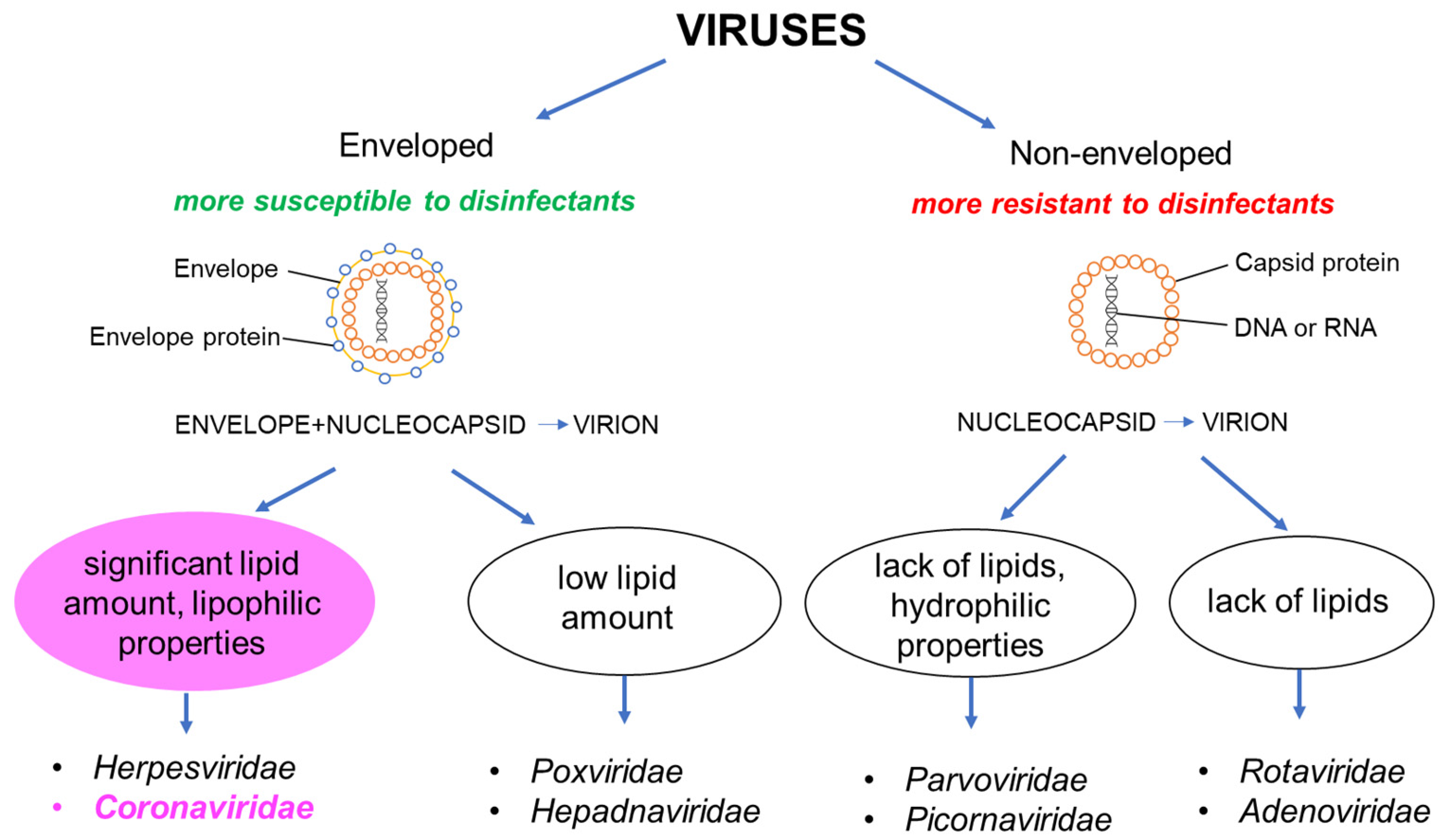
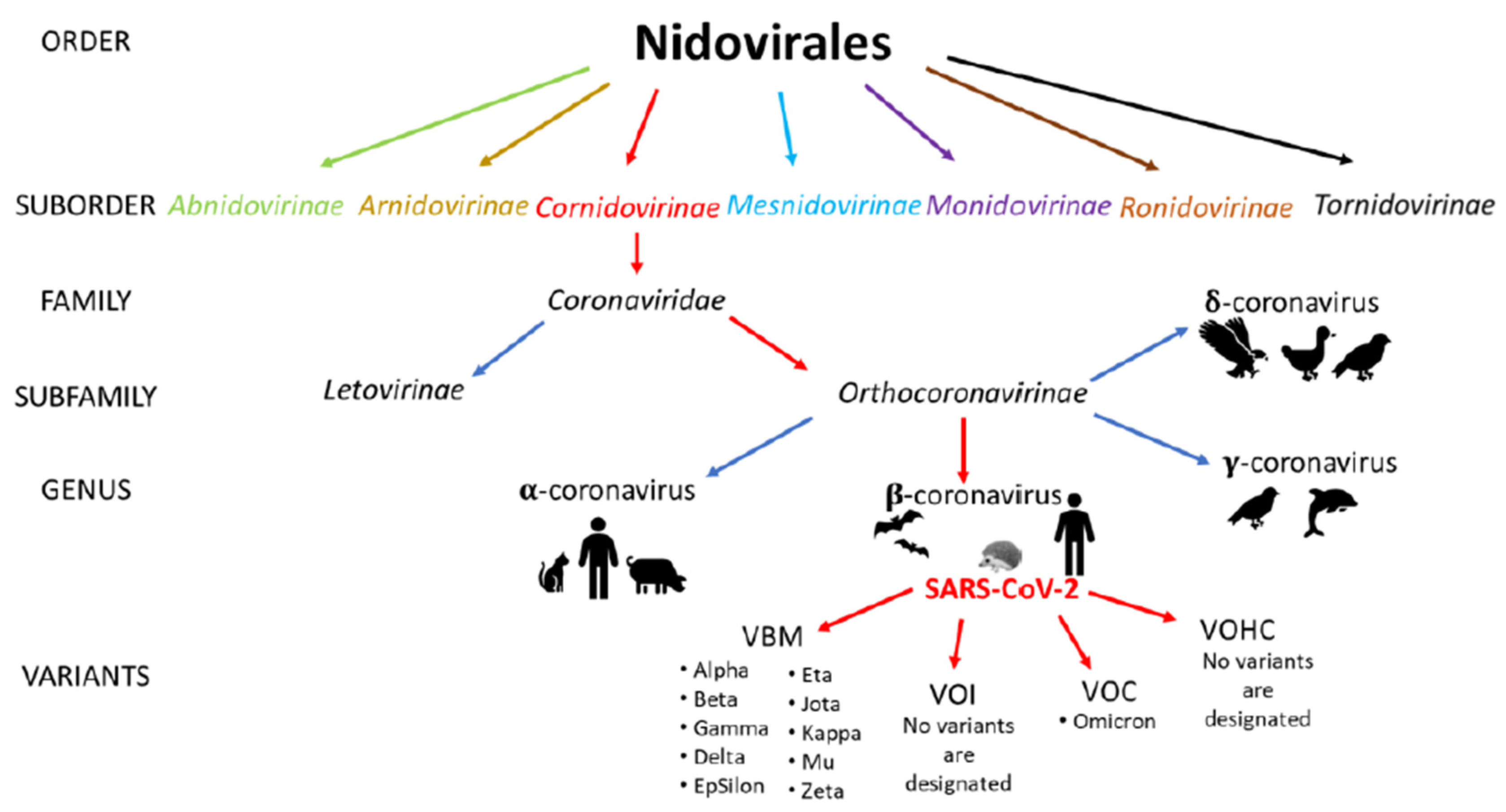
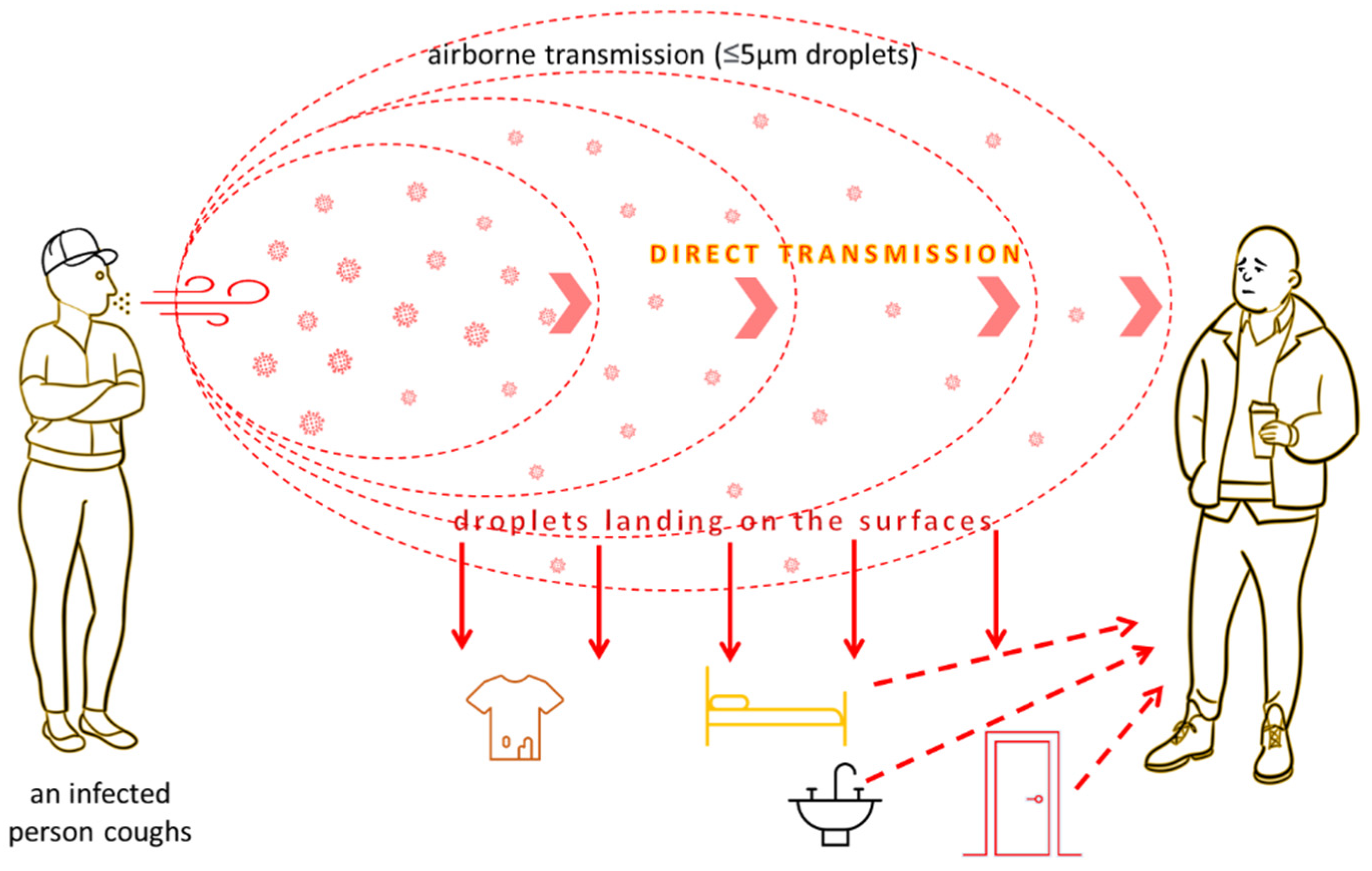
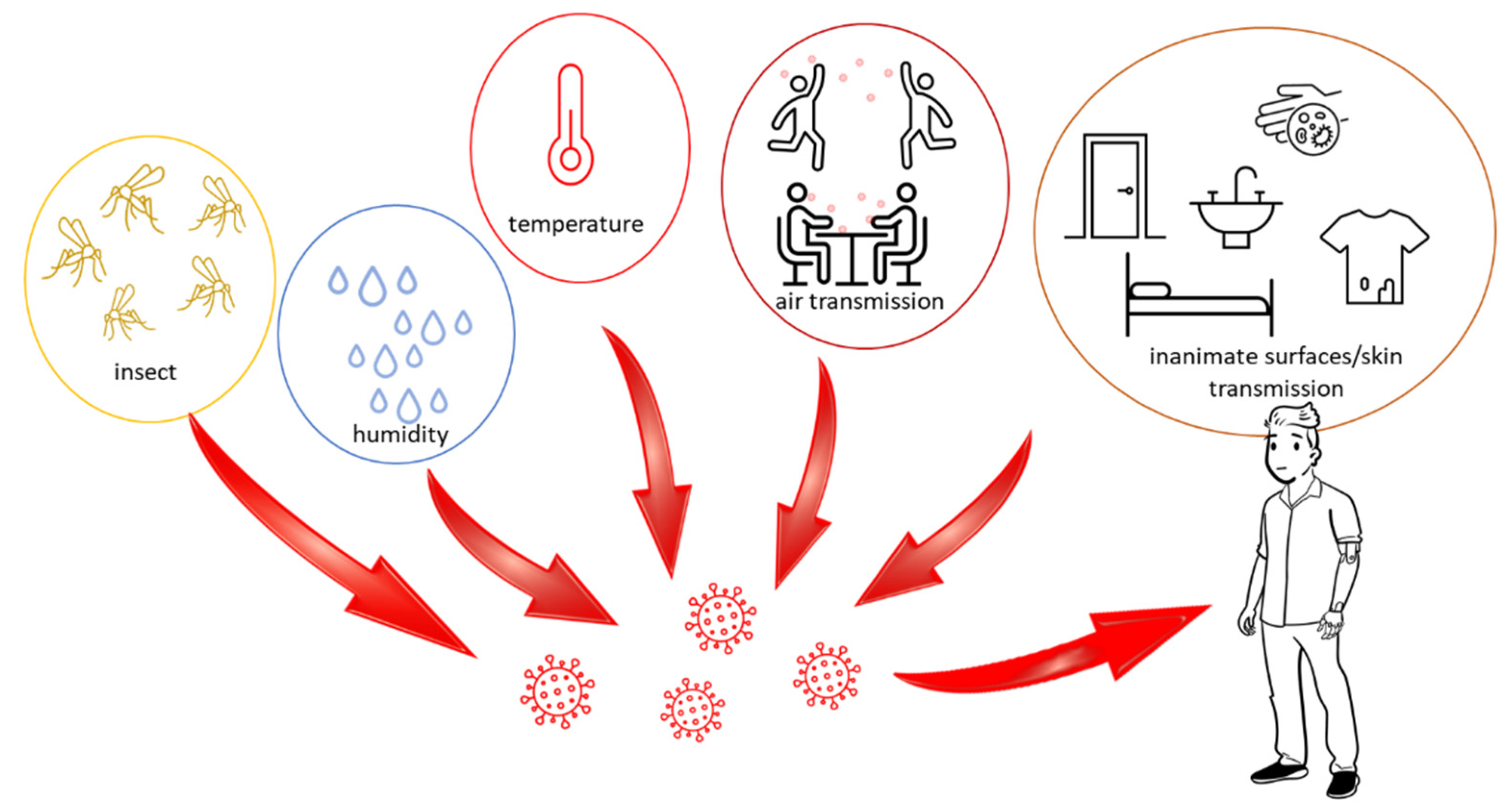
In hospitals, viruses, bacteria and their spores as well as fungi are transmitted not only by infected patients, but also by hospital staff, visitors and inanimate hospital environments, including surfaces frequently touched by hands, called “high-touch surfaces”, which include, among others, door handles, light switches, surfaces in toilets and in the area where the patient is in the room [10]. The risk of nosocomial transmission depends on the ability of pathogens to persist on surfaces. The longer a microorganism can remain on an inanimate surface, the greater the risk of its transmission to the patient or hospital staff [11]. The ability of hospital pathogens to colonize and survive on inanimate surfaces depends on several factors, including relative humidity (RH), temperature, ability to form biofilm and properties of the material from which the surface is made, including its porosity and orientation (horizontal or vertical). Hand hygiene and personal protection against infections are likewise important. The characteristics of the microorganism itself are also crucial, including its ability to create spores (bacteria), cell structure and other individual characteristics for a given microorganism [10,12,13].
One of the main factors determining the survival of microorganisms in the hospital environment is relative humidity (RH). For most viruses, the ability to survive depends mainly on the presence of a lipid envelope, which increases its resistance to low RH [12]. In the case of bacteria, some Gram-negative strains (Pseudomonas sp., Klebsiella sp., Enterobacter sp.) prefer higher relative humidity and lower temperature. On the other hand, bacteria such as E. coli, some species of Salmonella sp. and Proteus vulgaris have a lower survival rate at an intermediate relative humidity (50–70%). Among Gram-positive bacteria, for example, S. aureus can survive at low relative humidity, while Staphylococcus epidermidis, Bacillus subtilis and Streptococcus haemolyticus have a lower survival at 50–70% humidity. The different survival rate of bacteria depending on the humidity depends mainly on the structure of the cell wall. Consequently, Gram-positive bacteria, unlike Gram-negative bacteria, are more protected from physical stress and need less moisture to survive. In the case of fungi, it is argued that they prefer a moist environment [10].
Temperature is another important factor determining the survival of nosocomial pathogens. As reported by Kim et al. and Williams et al., lower temperatures result in longer survival times [14,15]. Often, the survival of microorganisms at low temperature depends on humidity, which can reduce drying stress [16]. In addition, high temperatures may reduce the activity of enzymes or damage the protein envelope of both the viral genome (RNA or DNA) and bacterial cells [4].
The factor that undoubtedly causes the persistence of microorganisms on inanimate surfaces is the ability to form a biofilm. It is the dominant life form of microorganisms in an ecosystem rich in nutrients [11]. The growth of pathogens in the biofilm makes them resistant to disinfection, which is associated with an increased production of extracellular substances such as proteins and polysaccharides after attachment to the surface. Biofilms maintain humidity and nutrients, and at the same time protect microorganisms against unfavorable physical and chemical factors, including disinfection. In a hospital environment, biofilm can be created on a variety of surfaces and objects such as blind cords, rubber sink stoppers or plastic parts of doors, windows, tables [17].
The survival of microorganisms in hospital conditions also depends on the type of surface and its characteristics. Polyvinyl chloride, ceramic tiles, and stainless steel are typical surfaces found in hospitals. Their features, such as porosity, hydrophobicity and free energy of the surface, determine the adhesion of microorganisms and the possibility of creating a biofilm [18,19,20]. As reported by Lagha et al., stainless steel is a material that promotes biofilm formation and can promote cross-contamination. Other materials such as ceramics or PVC can also be a source of disease transmitted from inanimate surfaces [21].
Pathogens with specific microbiological properties take part in the contamination of hospital environments. Such microorganisms must be able to survive long periods on an inanimate surface and to maintain virulence after exposure to environmental agents. In addition to the ability to contaminate surfaces in a hospital environment, they should also be able to easily colonize patients and temporarily colonize the hands of medical workers, which are the main vector of infection. Moreover, such microorganisms are infectious even in low doses and show relative resistance to active substances commonly used in surface disinfection preparations [22,23]. As mentioned earlier, survival on inanimate surfaces may be determined by the structure of the cell (in the case of Gram-negative and Gram-positive bacteria) or the presence of a lipid envelope (viruses). Other interesting features of microorganisms determining their persistence include increased survival of Haemophilus influenzae due to the activity of urease at low pH, or the presence of protein A, responsible for virulence and immunogenicity in Staphylococcus aureus [24,25].

3.2. Viruses

Viral infections can cause very different clinical courses, ranging from asymptomatic infections to severe, potentially fatal diseases. Viruses have the ability to contaminate and survive on inanimate surfaces. More than forty years ago, the potential role of inanimate surfaces in the transmission of certain viruses was highlighted by examining their ability to persist on several types of surfaces [26]. Hygiene and surface disinfection are particularly important when dealing with influenza, parainfluenza, intestinal viruses, hepatitis B and the coronaviruses causing SARS. It is believed that the SARS-causing coronaviruses are sprayed mainly by droplets; however, oral-fecal transmission and transmission via contaminated surfaces cannot be ruled out. These viruses can survive 24–72 h on plastic-laminated surfaces [27]. According to Sizun et al., human coronaviruses may survive on aluminum, cotton and latex a few h after drying, which could be linked with the possibility of person-to-person transmission through hand contamination from inanimate surfaces [28]. This was also confirmed by Kramer (2006), who pointed out that a significant number of viruses strains infecting the respiratory tract, such as coronaviruses, influenza or coxsackie viruses, consist of a potential source of transmission existing on surfaces even for a few days [4].
The survival of selected viruses (surrogate coronaviruses, gastroenteritis virus (TGEV) and mouse hepatitis virus (MHV)) on hard, non-porous surfaces under various conditions of air temperature (AT) and relative humidity (RH) was also investigated. TGEV and MHV can survive for up to several days on surfaces in AT and RH typical of healthcare environments. Generally, the genomes of RNA viruses are more sensitive to temperature than DNA viruses, but high temperatures can also affect DNA integrity. For some types of viruses, such as polioviruses and adenoviruses, low temperature increases their durability in a hospital environment [4]. Most respiratory viruses (e.g., influenza virus, coronavirus), herpes virus, rubella, measles and others that have a lipid envelope have the ability to survive in an environment with a lower relative humidity, ranging from 20 to 30%. In turn, viruses without a lipid envelope (enteroviruses, adenoviruses, rhinoviruses) can survive at a higher RH of 70–80% [12]. This means that enveloped viruses remain infectious on surfaces long enough to pose a risk of exposure, and lead to infection and disease transmission [29].
Viruses can be classified according to their structure, which also determines their susceptibility to chemical disinfectants. Viruses consist of two components: the viral genome (RNA or DNA) and the virus-encoded protein capsid surrounding the genome. If a virus contains these two elements, it is called a non-enveloped virus. If a virus particle contains an additional external lipid bilayer membrane surrounding the protein capsid, it is called an enveloped virus. Non-enveloped viruses such as polio virus, hepatitis A virus (HAV) and parvovirus are characterized by strong hydrophilic properties and, subsequently, the highest resistance to disinfectants. On the other hand, viruses with reduced hydrophilic properties, such as rotaviruses, noroviruses and adenoviruses, are a little more susceptible to disinfection. The sensitivity of enveloped viruses to disinfectants depends on their lipid content. Those with low lipid content are more resistant than those with high lipid content. This is due to the fact that the lipid envelope is easily destroyed by chemical compounds that affect lipids such as alcohol, ether, chlohexidine, etc. The group of viruses most sensitive to chemical disinfectants includes enveloped viruses such as HIV, herpes virus, HCV hepatitis, and, importantly in the context of the pandemic, coronaviruses (Figure 1) [30,31].
SARS-CoV-2 is one of the viruses from the family Coronaviridae and order Nidovirales. The family Coronaviridae is divided into two subfamilies, Letovirinae and Orthocoronavirinae, and SARS-CoV-2 belongs to the latter (Figure 2) [32]. According to the CDC, only Omicron was selected as a SARS-CoV-2 Variant of Concern, and no variants were designated as variants of interest or variants of high consequences. In April 2022, the Delta variant was downgraded from a Variant of Concern to a Variant Being Monitored because of evidence indicating that it does not currently pose a significant risk to public health in the United States [33].
By the end of 2019, only six different coronaviruses dangerous to humans were recorded, four of which resulted in mild symptoms of the common cold (HCoV-NL63, HCoV-229E, HCoV-OC43 and HKU1), while two have caused a pandemic in the past 20 years. The severe acute respiratory syndrome coronavirus (SARS-CoV) caused the SARS epidemic (10% mortality, 2002–2003). In contrast, the Middle East Respiratory Syndrome (MERS-CoV) coronavirus caused a pandemic in 2012 (37% mortality) [34]. All viruses from the group of coronaviruses that turned out to be pathogenic to humans were characterized by zoonotic origin. Coronaviruses are positive-strand RNA viruses found in many species of animals, often showing no disease symptoms in their hosts. On the basis of genetic and serological characteristics, four types of them have been distinguished: alphacoronavirus (alpha-CoV), betacoronavirus (beta-CoV), gamma-coronavirus (gamma-CoV) and deltacoronavirus (delta-CoV) (Figure 2). Gammacoronaviruses and deltacoronaviruses are mainly found in birds, although gammacoronaviruses also infect some cetaceans, including beluga whales and bottlenose dolphins. In contrast, alphacoronaviruses and betacoronaviruses occur mainly in mammals. SARS-CoV and MERS-CoV have spread to humans from civet cats and dromedary camels [35]. The closest known relative strain of SARS-CoV-2 is the coronavirus strain RaTG13 found in a bat Rhinolophus affinis from Yunnan Province, China in 2013. The degree of similarity at 96% may suggest a relatively close relationship between the two virus strains [36]. One of the possibilities is that the SARS-CoV-2 ancestor has been incubating for years inside bats, accumulating mutations, and probably through a random event, the virus was transmitted in humans. It should be also emphasized that both civets and racoon dogs, which are susceptible to SARS-CoV-2 infections, were sold live in Wuhan in 2019, and a market in Wuhan was an epicenter of SARS-CoV-2 infection [37]. SARS-CoV-2 turned out to be much more contagious than other viruses in the family, as evidenced by its extremely rapid spread in over 180 countries around the world. This feature, combined with the often severe course of the infection, has raised concerns about the possible breakdown of healthcare systems that will not be able to treat and save a large number of cases at the same time. [38]. Therefore, both governments and public health sectors are trying to contain this pandemic and avoid a catastrophic scenario. As there are no effective and safe antiviral drugs for SARS-CoV-2, infection control is currently the only available method, apart from vaccination, to limit the spread of the virus. The choice of preventive measures to control infection depends largely on knowing and understanding the routes of transmission of the virus. COVID-19-related pneumonia diagnosed in both hospital and family settings provided the basis for describing the direct route of person-to-person transmission of the virus [39,40]. However, indirect transmission pathways, such as fecal-oral, hospital, airborne, and contact with contaminated surfaces, are also believed to play an important role (Figure 3) [41].
The dependence of the stability and rate of spread of the virus on environmental conditions such as temperature, relative and absolute humidity and sunlight is poorly understood. Coronaviruses, due to the fact that they are enveloped viruses and are more sensitive to heat, are able to survive longer in lower temperatures and relative humidity [42]. The study of these dependencies is necessary to be able to construct a robust and long-term protocol to interrupt the indirect transmission of SARS-CoV-2 to the environment, limit its spread and thus minimize the risk. Research has also been carried out on the potential for transmission of SARS-CoV-2 by house flies (Musca domestica). Flies generally can transmit microbes from waste-contaminated breeding habitats to food consumed by humans. Although flies theoretically can transfer SARS-CoV-2 virus mechanically, the results of the research indicate that flies do not play a significant role in the transmission of SARS-CoV-2 to humans and animals [43]. Mosquitoes also do not play a fundamental role in the transmission of SARS-CoV-2 to humans (Figure 4) [44,45].
The ability of a given virus to survive in an inanimate environment is a prerequisite for its spread. Important factors determining the level of infectivity and the extent and speed of spreading a virus are its characteristics, but also the biotic and abiotic properties of the environmental surface it contaminates, as well as environmental conditions. Therefore, SARS-CoV-2 is considered to persist for a long time on environmental surfaces. The adhesion mechanism used by the SARS-CoV-2 virus on various types of inanimate surfaces has not yet been fully elucidated. However, electrostatic interactions, hydrophobic effects and, to a lesser extent, non-covalent bonds such as van der Waals forces are believed to play a large role. All these factors can influence the adhesion of the S protein to the surface [46,47]. Literature data indicate that the load on the surface of the virus varies with the pH of the environment. Viral particles are characterized by high stability in a wide range of pH, however, they are more stable at alkaline pH. Protein E, which is part of the hydrophobic layer surrounding the viral particle, also plays an important role in viral adhesion [48,49].
SARS-CoV-2 virus RNA has been detected on the surface of door handles, cell phones, and other items in the homes of confirmed cases. To date, limited data are available on the survival of SARS-CoV-2 in the environment [50,51]. The durability of SARS-CoV-2 on surfaces made of plastic was tested in two experiments. In the first study, SARS-CoV-2 maintained its infectivity for 4 days and completely degraded after 7 days on a plastic surface at room temperature and 65% relative humidity. The second study showed that SARS-CoV-2 maintained its infectivity for 3 days on the plastic surface at room temperature. There was also no difference between the persistence of SARS-CoV-2 and SARS-CoV-1 on plastic surfaces, and both viruses lost their infectivity completely after 4 days. Literature data show that the survival rate of coronaviruses on metal surfaces differs depending on the type of metal. They survive shorter on copper, nickel and brass surfaces than on stainless steel and zinc surfaces. SARS-CoV-2 was shown to persist on the surface of stainless steel for 3 days and only after 4 days did it become undetectable. This virus showed a lower survival on copper (4 h) compared to SARS-CoV-1 (8 h). This data was also confirmed by the study of Aboubakr et al. (2021), who concluded that the survival rate of SARS-CoV-1 and SARS-CoV-2 was significantly lower on copper, latex, and less porous fabrics than on surfaces such as metals (stainless steel and zinc), glass. Based on the obtained results, the authors suggested that the use of contact surfaces made of copper in hospitals may be a factor reducing the transmission of SARS-CoV-2. However, it should be noted that the coronavirus may have different survival rates on the same surface but under different temperature and relative humidity conditions [42]. The influence of temperature, relative humidity and droplet size on the stability of SARS-CoV-2 was tested in laboratory conditions on specially created matrices imaging non-porous surfaces. It was shown that the rate of decomposition of SARS-CoV-2 increased with increasing humidity or temperature, but the droplet volume (1–50 μL) and the type of surface (stainless steel, plastics or nitrile) did not have a significant effect on the parameter tested. Consequently, potential transmission can linger for hours to days in indoor areas, making it difficult to assess the risk of surface contamination [52].
The survival of SARS-CoV-2 on glass at room temperature and 65% relative humidity was also investigated. The virus remained contagious for 2 days and became completely undetectable after 4 days. The stability of SARS-CoV-2 drops significantly with increasing temperature and humidity. Values above 38 °C and 95% cause the adhesion of virus particles to the surface to be much weaker. Sunlight also acts as a natural virus inactivating agent on surfaces [48,53]. Of the swab samples taken from inanimate surfaces in the infectious disease emergency department, only two were positive for low SARS-CoV-2 levels, and none caused a cytopathic effect on day 7 of the study. Therefore, it is believed that contact with inanimate surfaces in contaminated areas may indicate the possibility of infection, but not as extensively as is believed [54].
The survival rate of SARS-CoV-2 on cardboard compared to SARS-CoV-1 was also analyzed. SARS-CoV-2 survived longer (1 day) than SARS-CoV-1, which only survived 8 h under the same conditions. Comparing surfaces with different porosity, it was shown that SARS-CoV-2 survived longer (days) on surfaces with greater porosity than those with lower porosity (hours). Virus survival times on the inner and outer layers of the surgical masks were 4 and 7 days, respectively, and 2 days on fabric and 1 day on banknotes. On paper, the virus only survived 30 min with a total degradation time of 3 h [48,55]. Studies conducted and published by Kampf et al., van Doremalen et al., Ren et al., and Rawlinson et al., confirmed that SARS-CoV-2, similarly to other coronaviruses, could persist on inanimate surfaces such as plastic, glass and metals for a few days: from 72 h to even 9 days. However, they also pointed out that SARS-CoV-2 as an enveloped virus is susceptible to surface disinfection procedures conducted with the use of most cleaning agents [55,56,57,58].
Influenza virus is also an infectious agent in the environment even after thorough drying of the surfaces on which it is located, and it can be re-sprayed as an aerosol when cleaning floors. It can survive 24–48 h on non-porous surfaces and be reapplied to the skin, causing staff–patient and patient–staff cross-contamination. Parainfluenza virus exhibits similar resistance to desiccation and can survive up to 10 h on non-porous surfaces and up to 6 h on clothes [59]. A 2016 study found that influenza A (H1N1) viruses can persist and remain infectious on stainless steel surfaces for 7 days [60].
Enteric viruses such as noroviruses belong to caliciviruses, single-stranded non-enveloped RNA viruses that are common pathogens in humans and animals. Viruses from the human norovirus group are the leading cause of epidemic and sporadic acute gastroenteritis (AGE) worldwide. Inanimate surfaces are considered to be the key carriers of the spread of human norovirus during the epidemic. It should also be mentioned that the literature data indicate that the virus easily spreads between inanimate surfaces and human skin [61,62]. Noroviruses cause extensive contamination of surfaces and it is possible to spread them in the form of aerosols when cleaning floors or other larger surfaces. They can survive on large surfaces for up to 12 days [63]. They can also be found on surfaces such as door handles, buttons in elevators, and toilet flushing cisterns. In the case of the presence of noroviruses in the environment, it is very important to disinfect the entire environment of the patient properly, because due to the long survival time of the virus and the small inoculum sufficient for infection, another outbreak may occur. In addition, these viruses are very resistant to chemical disinfection, so it is worth disinfecting contaminated surfaces with washing and disinfecting preparations several times, each time preparing a new solution of the preparation. Rotaviruses are very common food-borne gastroenteritis viruses, but hands and contact with contaminated surfaces are essential for the transmission of infection. These viruses are very infectious and remain on surfaces for a long time, up to 60 days [64,65,66].
HBV virus is very infectious and highly resistant to environmental factors. It does not lose infectivity for up to 6 months at room temperature, while at 60 °C it can survive up to 4 h. It is inactivated at 100 °C after 20 min and in an autoclave (121 °C) after 15 min. Due to the exceptional infectivity of the virus, surfaces should be considered as a possible source of HBV infection. The blood of an infected person is a high-risk factor, as 0.0001 mL of an infected person’s blood is enough to transmit an infection. Therefore, compliance with the rules of disinfection breaks the chain of infection to some extent [67,68,69].

3.3. Bacteria

In the hospital environment, the most common bacterial strains causing difficult-to-control diseases include: Clostridium difficile, Klebsiella pneumoniae, Acinetobacter species, Escherichia coli, Enterobacter sakazakii, Enterobacter cloacae, Pseudomonas aeruginosa and Staphylococcus aureus, subdivided into two groups (MRSA-methicillin-resistant Staphylococcus aureus and VRSA-vancomycin-resistant Staphylococcus aureus).
Katzenberger et al. described the percentage share of bacteria which play the main roles in outbreak events in hospitals, e.g., S. aureus (11.9%), K. pneumoniae (7.9%), P. aeruginosa (7.1%), A. baumannii (7.0%), S. marcescens (4.6%), E. faecium (3.6%), E. coli (2.4%) and E. cloacae (2.3%) [70]. According to Kramer et al., single-handed contact with a contaminated surface will have a variable degree of transmission depending on the bacteria. It was observed that transmission from contaminated surfaces to hands was most effective in the case of S. aureus MRSA or VRE, E. coli and Salmonella spp. [71]. The multiresistance of bacteria to antibiotics is a serious problem not only in terms of their prolonged persistence on surfaces, but also in limiting the possibilities of appropriate treatment. The longevity of bacteria increases the risk of spreading them in the hospital environment, especially in the conditions that exist in routine care of patients [72]. According to Katzenberger et al., 2021, the most susceptible bacteria to spread in the hospital environment are strains that are resistant to antibiotics, because they can more easily form biofilms on various surfaces, which may increase the likelihood of prolonged life and facilitate further spread [70].
It is widely recognized that these pathogens are not able to be completely eliminated from the hospital environment, but they can be reduced to a minimum. In order to decrease their prevalence, an in-depth knowledge of the resistance mechanisms of these pathogens is essential, as is a precise definition of universal principles of infection prevention, hygiene practice and a correct antibiotic policy.
Clostridium difficile is an anaerobic, Gram (+), spore-forming and toxin-producing rod. It is part of the physiological human intestinal flora in almost 3% of the healthy population and in 20–30% of those hospitalized. It exists in a vegetative form of spores in the intestine or outside of it. The vegetative form can survive on a dry surface at room temperature for up to 15 min, and on a wet surface for up to 6 h. Clostridium difficile spores are characterized by high resistance to alcohol-based disinfectants and other chemicals used in hospitals, UV radiation, high temperatures, etc. (Table 1).
The high resistance results from the stratified structure of the spore. The core inside contains dehydrated cytosol, DNA, RNA and enzymes. The core is covered with an inner membrane, a wall, and then a cortical layer (a modified peptidoglycan that will later be involved in the formation of the cell wall of the vegetative form). Outside, it is covered with a mantle containing mainly proteins, dipicolinic acid and calcium ions, which form calcium dipicolinate. Therefore, Clostridium difficile spores are very tough and resistant to traditional surface cleaning methods [73]. C. difficile spores have a strong ability to adhere to a variety of surfaces, including plastic laboratory equipment. They can survive on the surfaces of hospital equipment for up to 5 weeks, and on hospital floors for up to 5 months. Literature data show that the storage of spore-contaminated materials at 4 °C and the freezing and re-thawing cycles also did not reduce their number [94,95].
Contamination of the inanimate environment by C. difficile has been reported in close proximity to an infected patient. The contamination rate was up to 58%, and beds, cabinets, pressure measuring devices, walls, floors, washbasins and furniture were particularly frequently infected. Hospital floors can remain infected with C. difficile for up to 5 months, and the rate of contamination increases in the presence of colonized patients or those patients with diarrhea [96]. Molecular techniques provide compelling evidence of C. difficile transmission from environmental surfaces to the patient. According to the data obtained with their help, it was found that surfaces in the patient’s environment serve as a reservoir of the germ and allow cross-colonization of patients after their contact with healthy healthcare professionals [97,98]. Among the hospital surfaces where Clostridium difficile has been found, in addition to the walls and floors, there are also cabinets, beds, swimming pools, blood pressure cuffs, sinks, and occasionally shoes and stethoscopes [99,100].
Staphylococci, which are microorganisms that easily contaminate the hospital environment, can be divided into two groups: coagulase-positive, e.g., Staphylococcus aureus, and coagulase-negative, e.g., Staphylococcus epidermis and Staphylococcus saprophyticus. Staphylococcus aureus is especially common in healthcare professionals, diabetics and people with venous catheters. However, the greatest threat is MRSA, or methicillin-resistant Staphylococcus aureus, which causes nosocomial infections characterized by particularly high morbidity and mortality rates [101]. Staphylococcus aureus was one of the most common pathogens associated with nosocomial infections reported to the NHSN (National Healthcare Safety Network) from January 2006 to October 2007. MRSA constitutes 56% of all S. aureus isolates from hospital equipment and supplies. The most important reservoir of MRSA in a hospital is colonized or infected patients, who easily contaminate medical and electronic equipment in their environment. MRSA can survive on dry surfaces for up to several months [102,103,104]. Although the primary mode of MRSA transmission to patients is through the colonized hands of healthcare professionals, there is evidence that exposure to MRSA contaminated surfaces may also cause patient infections [105,106]. Literature data indicate that S. aureus remains viable on dry surfaces for periods from 1 week up to even 3 years [90]. While there are data showing the survival of S. aureus in home and healthcare settings, little attention has been paid to the spread of these organisms, including MRSA, across the community [107]. Table 1 shows quite varied survival of S. aureus on various inanimate surfaces, e.g., polyethylene for 90 days, sterile packages for 266 days, screw cap bottles for 318 days and polypropylene for over 1097 days [77,89,108,109]. It seems, however, that these findings are the outcomes of the optimization of the experimental conditions. They used staphylococcal strains with a high level of resistance to desiccation and an inoculum of 107–109 CFU. These results were clear and consistent under laboratory conditions, but they may not accurately reflect S. aureus survival in the community. Domon H. demonstrated that hazardous and pathogenic S. aureus strains, including MRSA, which cause healthcare-associated infections, are rarely isolated from inanimate surfaces in the community, because of their poor survival on a dry surface for more than 24 h [110].
Acinetobacter is a group of Gram (−) bacteria that do not ferment glucose. It includes 32 species, of which Acinetobacter baumannii is the most important, constituting up to 70% of isolates. In recent years, an increase in Acinetobacter resistance and an increasing number of nosocomial infections caused by these microorganisms have been observed. This is due to their ability to survive in the environment both on dry and wet surfaces for a long period of time (weeks) in a wide range of temperatures and environmental pH. In vitro studies showed that Acinetobacter can survive on ceramic surfaces, stainless steel, rubber and polyvinyl chloride, and relatively higher humidity promotes bacterial growth [111,112,113]. Therefore, in a hospital environment, Acinetobacter can most often be found in humid places such as bathrooms, mops, respirators and air humidifiers. These strains have the ability to produce a biofilm that hinders the penetration of disinfectants and increases bacterial resistance to decontamination.
The microorganisms of the genus Enterococcus colonizing the human gastrointestinal tract belong to the physiological flora, and are actually resistant to many antibiotics, e.g., penicillin, aminoglycosides and glycopeptides. There are 43 species here, but the most common are Enterococcus faecalis and Enterococcus faecium. Vancomycin-resistant enterococcal (VRE) strains were first isolated in 1986. Currently, they account for 1/3 of all nosocomial infections caused by contamination of the hospital environment, especially surfaces and medical equipment. VRE causes serious infections in immunocompromised patients. Transmission of VRE in hospitals is usually associated with transient colonization of the hands of medical personnel. However, there are literature reports showing the role of hospital surfaces and medical equipment as vectors for VRE [114]. Katzenberger et al. studied the survival kinetics of the selected microorganisms on four different types of surfaces, such as polyvinyl chloride, glass, stainless steel and aluminum (Table 1). The scholar stated that A. baumannii and E. faecium showed the highest survival capability regardless of the material of the surface. Viable bacteria of those two species remained detectable even at the end of the entire observation period of one month [70]. Transmission of both A. baumannii and E. faecium through contaminated surfaces can easily occur if appropriate preventive infection control measures are not taken. These pathogens, being able to survive for several days on inanimate surfaces, pose a significant risk of transmission, e.g., in hospitals. Thus, in the event of high disease rates, qualified personnel should carefully search for previously unidentified areas or violations of standard decontamination procedures if the spread of the pathogen continues despite extensive cleaning and disinfecting efforts.
Many studies confirm the presence of Pseudomonas aeruginosa in sinks and drains in plumbing, but it is still unclear whether using sinks leads to an increased risk of the spread of this microorganism. P. aeruginosa isolates from inanimate surfaces were not always compatible with patient isolates. Most infections tend to be caused by the patient’s endogenous flora, but surfaces and medical equipment as possible sources of infection cannot be ruled out [115,116]. The literature data presenting experiments regarding survival of different bacterial species on various surfaces, like glass, stainless steel, polyvinyl chloride, and aluminum (Table 1), indicate that P. aeruginosa was completely inactivated in less than two days [70]. Neely AM presented the results regarding the survival rate of clinical and environmental strains of P. aeruginosa survival rate on different textiles such as cotton, polyester and polyethylene. It amounted to 2 h–7 days when inoculum was 104–105 CFU [77].

3.4. Fungi

Since the 1980s, there has been a continuous increase in the number of fungal infections, also proving that fungi have become a common pathogen causing hospital infections. Therefore, there is a need to evaluate the mycological flora in studies involving personnel, patients, walls, floors and equipment.
Nosocomial fungi have a wide range of temperature (0–60 °C) and pH (2–8.5) tolerances and grow more effectively on wet surfaces. Candida fungi are primarily isolated from hospital surfaces and can survive for up to 4 months. Although the vast majority of infections caused by Candida result from endogenous sources, molecular studies of yeast obtained from patients collected by medical personnel and from the medical community proved the transmission of Candida albicans, Candida glabrata and Candida parapsilosis among patients [117,118].
Candida infections are a serious problem in hospitals due to high patient mortality, especially in patients with low immunity, as well as long-term and costly treatment. Systemic infections caused by Candida are the fourth leading cause of nosocomial bloodstream infections. Candidiasis caused by Candida sp. is mainly the result of long-term antibiotic therapy with a broad spectrum of activity. In addition, one of the features that causes Candida infection and the possibility of its transmission between hospital patients is the ability to form biofilm, which at the same time makes it difficult to combat, both by drugs and by disinfecting the hospital space. In recent years, in hospitals in many countries, special attention has been paid to the strain C. auris, which is characterized by multi-drug resistance and is difficult to control due to its long-term survival ability on both wet and dry surfaces [119]. According to Shield et al. and Schelenz et al., the causes of Candida infections in hospitals are, apart from the hands of healthcare workers, hospital surfaces and medical tools and equipment [120,121]
The surfaces can be intensively and permanently contaminated by fungi of various species, as shown by experimental inoculation of dry surfaces with C. albicans (3 days) and C. parapsilosis (14 days) [122]. Molecular typing of Candida isolates obtained from the hospital environment and from patients proves that the endemic species of C. albicans and C. glabrata constitute the environmental reservoir of Candida. Candida strains obtained from patients were identical to those found on hospital surfaces [123,124]. According to Traore O et al., Candida parapsilosis survives much better than C. albicans on non-porous surfaces such as glass and stainless steel. C. albicans was undetectable at the end of the third day of the experiment, while C. parapsilosis was detectable even after 14 days of incubation under ambient conditions. C. albicans survived also on textiles (polyester + cotton and 100% cotton) significantly better than on glass and metal, while C. parapsilosis survived on both fabrics as well as on non-porous supports. Colony forming units of both Candida species remained detectable even after 14 days of storage under ambient conditions [122].
Both the reservoirs and the methods of transmission of Aspergillus and Zygomycetes are similar. In hospitals, the sources of Aspergillus are contaminated air filtering systems, ventilation systems contaminated with dust accumulated during renovation or construction, carpets, food and plants [125,126]. The species of the genus Aspergillus most commonly causing nosocomial infections are A. fumigatus, A. flavus and A. terreus. They are filamentous fungi which are characterized by a great variety and the ability to colonize various habitats around the world. They can cause a disease called aspergillosis, which presents with non-invasive infections of the respiratory tract, ears, or eyes. Invasive Aspergillus sp. infections may occur after immunosuppression or surgery, which may even result in death [126]. Research by Neely A. and Orloff M. indicates plastics and materials commonly used in hospitals as important reservoirs and vectors for the transmission of fungal infections. The tested species of fungi were able to survive on inanimate surfaces for at least one day, and very often for several weeks [127]. The method recommended for preventing fungal nosocomial infections is constant monitoring of air pollution, but so far there is no standardization of the methodology used to identify fungal contamination. As traditional methodologies are characterized by low precision and overly-long measurement time, flow cytometry is recommended as a method that can be validated for a specific measurement [128]. The most effective method of fighting fungal nosocomial infections is the constant involvement of people working in healthcare facilities in widely understood preventive measures, removing potential causes of infections and monitoring the possibility of pathogens in the patient’s environment. According to Baudisch et al., Mold fungi are commonly present, resistant to temperature and easily adsorbed on dust particles, which makes them durable for up to several months [22,129,130]

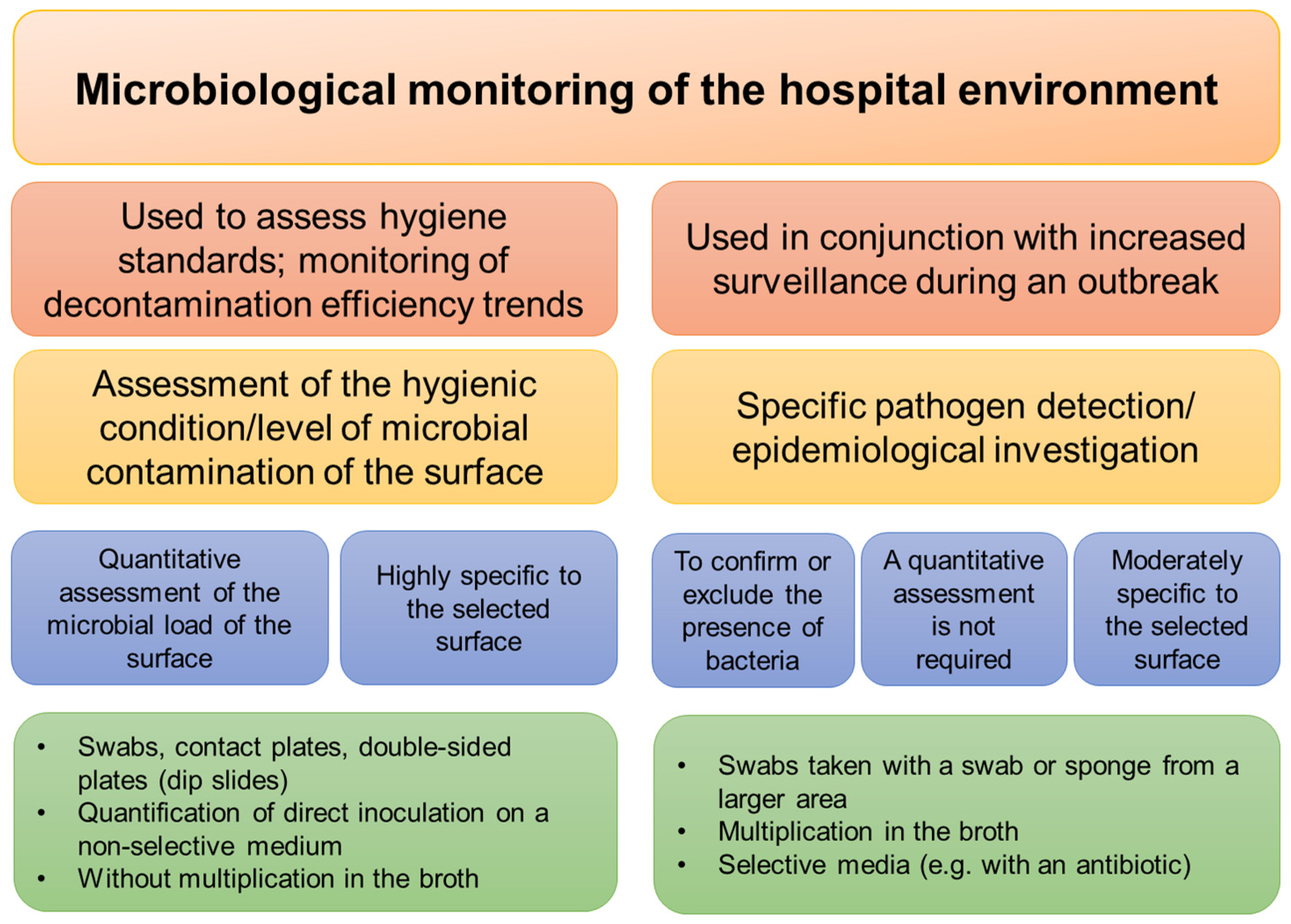
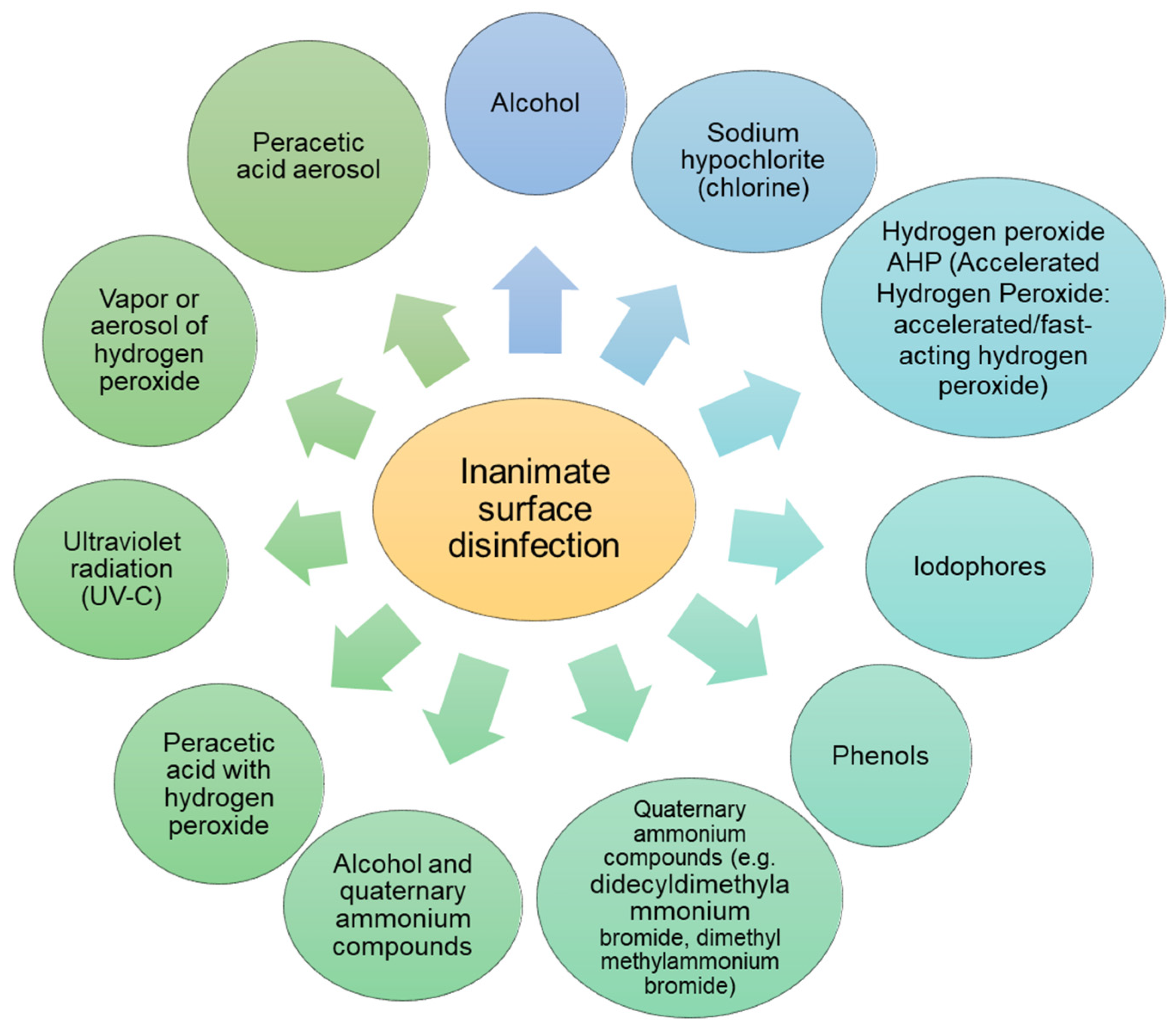
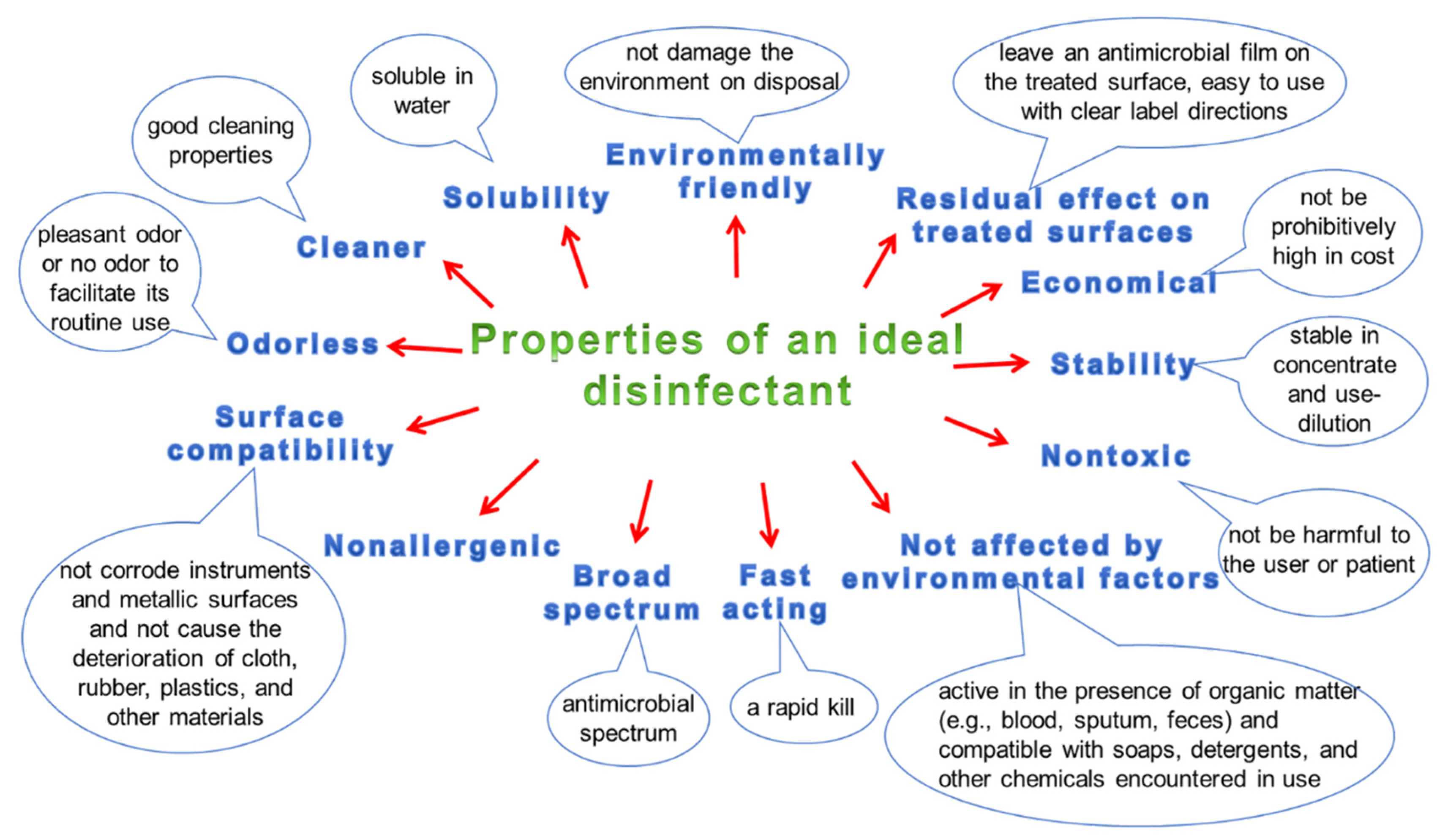
3.5. Hygiene and Disinfection of Surfaces

Contaminated surfaces and medical equipment can clearly contribute to the spread of nosocomial infections by infecting the hands of healthcare professionals and, indirectly, patients, or they can infect patients directly. Therefore, both an implementation of microbiological monitoring of the hospital environment and cleaning/disinfection of surfaces is essential to prevent cross-contamination (Figure 5).
Cleaning and disinfection recommendations include, among others, implementing protocols based on empirically proven guidelines for cleaning and disinfecting hospital surfaces and medical equipment [131]. The second recommendation is cleaning and disinfecting of surfaces that are easily and frequently contaminated during routine activities, especially frequently touched surfaces, such as: beds, bedside cabinets, door handles, bathroom fittings in patients’ rooms and equipment in the patient’s immediate vicinity. For the disinfection of small surfaces, preparations based on hydrogen peroxide, most often in sprays, are recommended, which are easy to use and, if they show sporicidal activity, can also be used in the environment of a patient infected with C. difficile. Since C. difficile spores are resistant to alcohol and various commonly used disinfectants, in the case of C. difficile infection, the use of chlorine-based disinfectants on medical equipment in close proximity to a patient infected with C. difficile is recommended. Due to their antimicrobial activity, preparations containing per-acetic acid and hydrogen peroxide are also recommended [132]. In cases of possible SARS-CoV-2 contamination, the WHO recommends cleaning surfaces frequently with water, detergents and disinfectants. Environmental cleaning and disinfection procedures should be conducted consistently and correctly with the use of ethanol at >70% concentration, povidone iodine, sodium hypochlorite and quaternary ammonium compounds with alcohol [56,133,134]. (Figure 6). One of the innovative methods of surface disinfection is the use of the non-thermal plasma sterilization technique, also known as “cold plasma”. The following technique types can be distinguished: direct current (DC) corona discharge, atmospheric pressure plasma jet (APPJ) microwave, dielectric barrier discharge (DBD), micro-hollow cathode discharge (MHCD) jet and pin-to-hole spark discharge (PHD) plasma. Non-thermal plasma techniques can be applied to medical devices without noticeably affecting their structure [135]. The action of plasma on microorganisms is based on the denaturation of proteins, inactivation of enzymes and DNA mutation. For example, treatment of E. coli with an atmospheric pressure plasma jet (APPJ) can cause cell-membrane damage and consequent cell lysis [84]. In the case of S. aureus, the use of plasma as a pre-sterilization step increases the susceptibility of Staphylococcus aureus to antibiotics [136]. Studies were also carried out on the effect of plasma on the destruction of enveloped and non-enveloped viruses, in which the inactivation of, e.g., influenza and RSV (enveloped viruses) and adenovirus (non-enveloped) [135]. The use of plasma seems to be a future tool in surface disinfection, but the influence of plasma on biomacromolecules (cell membranes, cell walls or membrane proteins and polysaccharides) or the mechanism of mutation and cell death is still unexplained and is the subject of research.
On the other hand, noroviruses are resistant to the activity of many commonly used disinfectants; therefore, a preparation based on chlorine compounds or another preparation in the spectrum which also includes noroviruses should be used (CDC). Properties of an ideal disinfectant are presented in Figure 7. When choosing a disinfectant, one should take into account both the type of disinfected surface and the degree and type of its contamination. A very important aspect is also the possibly ecological, environmentally friendly formula of the preparation and its possibly non-allergenic properties. The desirable properties of the biocide also include quick action, good water solubility (which allows for its easy preparation), as well as a broad spectrum of activity allowing the elimination of many types of pathogens at the same time. Considering surface disinfection, an additional positive aspect is the simultaneous cleaning and disinfecting effect of the preparation, which saves time and reduces disinfection costs, because it allows for the simultaneous conduct of two processes: washing and disinfection.

4. Conclusions

Scientific research has confirmed that contaminated hospital surfaces can be the cause of infection; therefore, cleaning and disinfection procedures should be carried out very carefully, selecting appropriate washing, disinfecting or washing-disinfecting agents with a broad biocidal spectrum and high efficiency, as well as in accordance with applicable standards and recommendations. Different microorganisms are characterized by different resistances to disinfectants, depending on the active substance used in the disinfectant. Bacterial spores (e.g., Bacillus, Clostridium) are generally the most difficult to combat with standard disinfectants. Their resistance due to their structure, which makes them resistant to disinfectants such as chlorhexidine, glutaraldehyde and QACs. The second most difficult microorganism to control with disinfection is Mycobacteria, followed by small non-enveloped viruses such as norovirus and poliovirus. Gram-negative bacteria are more resistant to disinfection than Gram-positive bacteria due to the presence of the cell wall composed of lipopolysaccharide and proteins in the former. Fungi and large non-enveloped viruses are more resistant to disinfection than lipid-enveloped viruses, which include SARS-CoV-2. Among all the above-mentioned microorganisms, SARS-CoV-2 belongs to the group that is most susceptible to removal from both inanimate surfaces and from the skin using traditional methods of washing and disinfecting. The above-mentioned literature data confirm the presence and the ability to survive on inanimate surfaces of the SARS-CoV-2 virus. Even in the first months of the global pandemic, its ability to travel through contact with contaminated surfaces was investigated as one of the possible contaminants. It turned out that it can be active and infectious on surfaces for a time range from hours to several days. However, because it is easily removable with commonly available surfactants and disinfectants, regular disinfection and proper hand washing can reduce viral transmission of both SARS-CoV-2 and other pathogens commonly found on inanimate surfaces in hospitals. Currently, one of the problems faced by hospitals is cross-transmission of pathogens, which at the same time poses a challenge in the search for methods of adequate control of surface contamination and the search for effective methods of its disinfection. The use of the correct disinfectant and following an effective cleaning procedure are key to preventing health and safety risks. A properly selected preparation for a specific surface and the degree of its contamination ensures that the microorganisms will be completely removed, not spread accidentally, and any additional threats will be minimized.

Author Contributions

Conceptualization, visualization, writing—original draft and writing—review and editing, A.J.-T., E.W. and U.W.; writing—review and editing, J.W., M.W.-M., L.S.-M. and M.M.; methodology, A.J.-T., U.W. and E.W. All authors have read and agreed to the published version of the manuscript.

Funding

This work was financially supported by the Ministry of Science and Higher Education, Poland, under the research project number WZ/WB-IIŚ/6/2022.

Institutional Review Board Statement

Not applicable.

Informed Consent Statement

Not applicable.

Data Availability Statement

Not applicable.

Conflicts of Interest

The authors declare no conflict of interest. The funders had no role in the design of the study; in the collection, analyses, or interpretation of data; in the writing of the manuscript; or in the decision to publish the results.

References

  1. Klevens, R.M.; Edwards, J.R.; Richards, C.L., Jr.; Horan, T.C.; Gaynes, R.P.; Pollock, D.A.; Cardo, D.M. Estimating Health Care-Associated Infections and Deaths in U.S. Hospitals, 2002. Public Health Rep. 2007, 122, 160–166. [Google Scholar] [CrossRef] [PubMed]
  2. Daneman, N.; Sarwar, S.; Fowler, R.A.; Cuthbertson, B.H.; SuDDICU Canadian Study Group. Effect of Selective Decontamination on Antimicrobial Resistance in Intensive Care Units: A Systematic Review and Meta-Analysis. Lancet Infect. Dis. 2013, 13, 328–341. [Google Scholar] [CrossRef]
  3. Zaragoza, R.; Ramírez, P.; López-Pueyo, M.J. Infección nosocomial en las unidades de cuidados intensivos. Enferm. Infecc. Microbiol. Clin. 2014, 32, 320–327. [Google Scholar] [CrossRef] [PubMed]
  4. Kramer, A.; Schwebke, I.; Kampf, G. How Long Do Nosocomial Pathogens Persist on Inanimate Surfaces? A Systematic Review. BMC Infect. Dis. 2006, 6, 130. [Google Scholar] [CrossRef] [PubMed] [Green Version]
  5. Wißmann, J.E.; Kirchhoff, L.; Brüggemann, Y.; Todt, D.; Steinmann, J.; Steinmann, E. Persistence of Pathogens on Inanimate Surfaces: A Narrative Review. Microorganisms 2021, 9, 343. [Google Scholar] [CrossRef] [PubMed]
  6. Moccia, G.; Motta, O.; Pironti, C.; Proto, A.; Capunzo, M.; De Caro, F. An Alternative Approach for the Decontamination of Hospital Settings. J. Infect. Public Health 2020, 13, 2038–2044. [Google Scholar] [CrossRef]
  7. Trudel-Ferland, M.; Jubinville, E.; Jean, J. Persistence of Hepatitis A Virus RNA in Water, on Non-Porous Surfaces, and on Blueberries. Front. Microbiol. 2021, 12, 618352. [Google Scholar] [CrossRef]
  8. Dancer, S.J.; King, M.-F. Systematic Review on Use, Cost and Clinical Efficacy of Automated Decontamination Devices. Antimicrob. Resist. Infect. Control 2021, 10, 34. [Google Scholar] [CrossRef]
  9. Mao, N.; An, C.K.; Guo, L.Y.; Wang, M.; Guo, L.; Guo, S.R.; Long, E.S. Transmission Risk of Infectious Droplets in Physical Spreading Process at Different Times: A Review. Build. Environ. 2020, 185, 107307. [Google Scholar] [CrossRef]
  10. Suleyman, G.; Alangaden, G.; Bardossy, A.C. The Role of Environmental Contamination in the Transmission of Nosocomial Pathogens and Healthcare-Associated Infections. Curr. Infect. Dis. Rep. 2018, 20, 12. [Google Scholar] [CrossRef]
  11. Kramer, A.; Assadian, O. Survival of Microorganisms on Inanimate Surfaces. In Use of Biocidal Surfaces for Reduction of Healthcare Acquired Infections; Springer International Publishing: Berlin/Heidelberg, Germany, 2014; pp. 7–26. [Google Scholar]
  12. Tang, J.W. The Effect of Environmental Parameters on the Survival of Airborne Infectious Agents. J. R. Soc. Interface 2009, 6, S737–S746. [Google Scholar] [CrossRef] [PubMed] [Green Version]
  13. Igo, M.J.; Schaffner, D.W. Quantifying the Influence of Relative Humidity, Temperature, and Diluent on the Survival and Growth of Enterobacter Aerogenes. J. Food Prot. 2019, 82, 2135–2147. [Google Scholar] [CrossRef] [PubMed]
  14. Kim, H.; Bang, J.; Beuchat, L.R.; Ryu, J.-H. Fate of Enterobacter sakazakii Attached to or in Biofilms on Stainless Steel upon Exposure to Various Temperatures or Relative Humidities. J. Food Prot. 2008, 71, 940–945. [Google Scholar] [CrossRef] [PubMed]
  15. Williams, A.P.; Avery, L.M.; Killham, K.; Jones, D.L. Persistence of Escherichia coli O157 on Farm Surfaces under Different Environmental Conditions. J. Appl. Microbiol. 2005, 98, 1075–1083. [Google Scholar] [CrossRef] [PubMed]
  16. Levin, P.D.; Shatz, O.; Sviri, S.; Moriah, D.; Or-Barbash, A.; Sprung, C.L.; Moses, A.E.; Block, C. Contamination of Portable Radiograph Equipment with Resistant Bacteria in the ICU. Chest 2009, 136, 426–432. [Google Scholar] [CrossRef] [Green Version]
  17. Vickery, K.; Deva, A.; Jacombs, A.; Allan, J.; Valente, P.; Gosbell, I.B. Presence of Biofilm Containing Viable Multiresistant Organisms despite Terminal Cleaning on Clinical Surfaces in an Intensive Care Unit. J. Hosp. Infect. 2012, 80, 52–55. [Google Scholar] [CrossRef]
  18. Donlan, R.M. Biofilm Formation: A Clinically Relevant Microbiological Process. Clin. Infect. Dis. 2001, 33, 1387–1392. [Google Scholar] [CrossRef] [Green Version]
  19. Donlan, R.M. Biofilms: Microbial Life on Surfaces. Emerg. Infect. Dis. 2002, 8, 881–890. [Google Scholar] [CrossRef]
  20. Sinde, E.; Carballo, J. Attachment of Salmonella spp. and Listeria monocytogenes to Stainless Steel, Rubber and Polytetrafluorethylene: The Influence of Free Energy and the Effect of Commercial Sanitizers. Food Microbiol. 2000, 17, 439–447. [Google Scholar] [CrossRef]
  21. Lagha, R.; Bellon-Fontaine, M.-N.; Renault, M.; Briandet, R.; Herry, J.-M.; Mrabet, B.; Bakhrouf, A.; Chehimi, M.M. Impact of Long-Term Starvation on Adhesion to and Biofilm Formation on Stainless Steel 316 L and Gold Surfaces of Salmonella Enterica Serovar Typhimurium. Ann. Microbiol. 2015, 65, 399–409. [Google Scholar] [CrossRef]
  22. Hota, B. Contamination, Disinfection, and Cross-Colonization: Are Hospital Surfaces Reservoirs for Nosocomial Infection? Clin. Infect. Dis. 2004, 39, 1182–1189. [Google Scholar] [CrossRef] [PubMed] [Green Version]
  23. Boyce, J.M. Environmental Contamination Makes an Important Contribution to Hospital Infection. J. Hosp. Infect. 2007, 65, 50–54. [Google Scholar] [CrossRef]
  24. Murphy, T.F.; Brauer, A.L. Expression of Urease by Haemophilus Influenzae during Human Respiratory Tract Infection and Role in Survival in an Acid Environment. BMC Microbiol. 2011, 11, 183. [Google Scholar] [CrossRef] [PubMed] [Green Version]
  25. Kobayashi, S.D.; DeLeo, F.R. Staphylococcus aureus Protein A Promotes Immune Suppression. MBio 2013, 4, e00764-13. [Google Scholar] [CrossRef] [PubMed] [Green Version]
  26. Mahl, M.C.; Sadler, C. Virus Survival on Inanimate Surfaces. Can. J. Microbiol. 1975, 21, 819–823. [Google Scholar] [CrossRef]
  27. World Health Organization. First Data on Stability and Resistance of SARS Coronavirus Compiled by Members of WHO Laboratory Network. Available online: http://www.who.int/csr/sars/survival_2003_05_04/en/index.html (accessed on 28 February 2021).
  28. Sizun, J.; Yu, M.W.; Talbot, P.J. Survival of Human Coronaviruses 229E and OC43 in Suspension and after Drying Onsurfaces: A Possible Source Ofhospital-Acquired Infections. J. Hosp. Infect. 2000, 46, 55–60. [Google Scholar] [CrossRef]
  29. Casanova, L.M.; Jeon, S.; Rutala, W.A.; Weber, D.J.; Sobsey, M.D. Effects of Air Temperature and Relative Humidity on Coronavirus Survival on Surfaces. Appl. Environ. Microbiol. 2010, 76, 2712–2717. [Google Scholar] [CrossRef] [Green Version]
  30. Eggers, M.; Schwebke, I.; Suchomel, M.; Fotheringham, V.; Gebel, J.; Meyer, B.; Morace, G.; Roedger, H.J.; Roques, C.; Visa, P.; et al. The European Tiered Approach for Virucidal Efficacy Testing-Rationale for Rapidly Selecting Disinfectants against Emerging and Re-Emerging Viral Diseases. Euro. Surveill. 2021, 26, 2000708. [Google Scholar] [CrossRef]
  31. Tarka, P.; Nitsch-Osuch, A. Evaluating the Virucidal Activity of Disinfectants According to European Union Standards. Viruses 2021, 13, 534. [Google Scholar] [CrossRef]
  32. King, A.M.Q.; Lefkowitz, E.J.; Mushegian, A.R.; Adams, M.J.; Dutilh, B.E.; Gorbalenya, A.E.; Harrach, B.; Harrison, R.L.; Junglen, S.; Knowles, N.J.; et al. Changes to Taxonomy and the International Code of Virus Classification and Nomenclature Ratified by the International Committee on Taxonomy of Viruses. Arch. Virol. 2018, 163, 2601–2631. [Google Scholar] [CrossRef] [Green Version]
  33. SARS-CoV-2 Variant Classifications and Definitions. Available online: https://www.cdc.gov/coronavirus/2019-ncov/variants/variant-classifications.html (accessed on 24 June 2022).
  34. Rabi, F.A.; Al Zoubi, M.S.; Kasasbeh, G.A.; Salameh, D.M.; Al-Nasser, A.D. SARS-CoV-2 and Coronavirus Disease 2019: What We Know so Far. Pathogens 2020, 9, 231. [Google Scholar] [CrossRef] [PubMed]
  35. Fan, Y.; Zhao, K.; Shi, Z.-L.; Zhou, P. Bat Coronaviruses in China. Viruses 2019, 11, 210. [Google Scholar] [CrossRef] [PubMed] [Green Version]
  36. Zhou, P.; Yang, X.-L.; Wang, X.G.; Hu, B.; Zhang, L.; Zhang, W.; Si, H.R.; Zhu, Y.; Li, B.; Huang, C.-L.; et al. A pneumonia outbreak associated with a new coronavirus of probable bat origin. Nature 2020, 579, 270–273. [Google Scholar] [CrossRef] [Green Version]
  37. Holmes, E.C.; Goldstein, S.A.; Rasmussen, A.L.; Robertson, D.L.; Crits-Christoph, A.; Wertheim, J.O.; Anthony, S.J.; Barclay, W.S.; Boni, M.F.; Doherty, P.C.; et al. The origins of SARS-CoV-2: A critical review. Cell 2021, 184, 4848–4856. [Google Scholar] [CrossRef] [PubMed]
  38. DeCapprio, D.; Gartner, J.; McCall, C.J.; Burgess, T.; Kothari, S.; Sayed, S. Building a COVID-19 Vulnerability Index. arXiv 2020, arXiv:2003.07347. [Google Scholar] [CrossRef] [Green Version]
  39. Lai, C.-C.; Shih, T.-P.; Ko, W.-C.; Tang, H.-J.; Hsueh, P.-R. Severe Acute Respiratory Syndrome Coronavirus 2 (SARS-CoV-2) and Coronavirus Disease-2019 (COVID-19): The Epidemic and the Challenges. Int. J. Antimicrob. Agents 2020, 55, 105924. [Google Scholar] [CrossRef]
  40. Chan, J.F.-W.; Yuan, S.; Kok, K.-H.; To, K.K.-W.; Chu, H.; Yang, J.; Xing, F.; Liu, J.; Yip, C.C.-Y.; Poon, R.W.-S.; et al. A Familial Cluster of Pneumonia Associated with the 2019 Novel Coronavirus Indicating Person-to-Person Transmission: A Study of a Family Cluster. Lancet 2020, 395, 514–523. [Google Scholar] [CrossRef] [Green Version]
  41. Aydogdu, M.O.; Altun, E.; Chung, E.; Ren, G.; Homer-Vanniasinkam, S.; Chen, B.; Edirisinghe, M. Surface Interactions and Viability of Coronaviruses. J. R. Soc. Interface 2021, 18, 20200798. [Google Scholar] [CrossRef]
  42. Aboubakr, H.A.; Sharafeldin, T.A.; Goyal, S.M. Stability of SARS-CoV-2 and Other Coronaviruses in the Environment and on Common Touch Surfaces and the Influence of Climatic Conditions: A Review. Transbound. Emerg. Dis. 2021, 68, 296–312. [Google Scholar] [CrossRef]
  43. Balaraman, V.; Drolet, B.S.; Mitzel, D.N.; Wilson, W.C.; Owens, J.; Gaudreault, N.N.; Meekins, D.A.; Bold, D.; Trujillo, J.D.; Noronha, L.E.; et al. Mechanical Transmission of SARS-CoV-2 by House Flies. Parasit. Vectors 2021, 14, 214. [Google Scholar] [CrossRef]
  44. Balaraman, V.; Drolet, B.S.; Gaudreault, N.N.; Wilson, W.C.; Owens, J.; Bold, D.; Swanson, D.A.; Jasperson, D.C.; Noronha, L.E.; Richt, J.A.; et al. Susceptibility of Midge and Mosquito Vectors to SARS-CoV-2. J. Med. Entomol. 2021, 58, 1948–1951. [Google Scholar] [CrossRef] [PubMed]
  45. Valsamatzi-Panagiotou, A.; Penchovsky, R. Environmental Factors Influencing the Transmission of the Coronavirus 2019: A Review. Environ. Chem. Lett. 2022, 20, 1603–1610. [Google Scholar] [CrossRef] [PubMed]
  46. Jones, L.; Walsh, K.; Willcox, M.; Morgan, P.; Nichols, J. The COVID-19 Pandemic: Important Considerations for Contact Lens Practitioners. Cont. Lens Anterior Eye 2020, 43, 196–203. [Google Scholar] [CrossRef] [PubMed]
  47. Vasickova, P.; Pavlik, I.; Verani, M.; Carducci, A. Issues Concerning Survival of Viruses on Surfaces. Food Environ. Virol. 2010, 2, 24–34. [Google Scholar] [CrossRef]
  48. Chin, A.W.H.; Chu, J.T.S.; Perera, M.R.A.; Hui, K.P.Y.; Yen, H.-L.; Chan, M.C.W.; Peiris, M.; Poon, L.L.M. Stability of SARS-CoV-2 in Different Environmental Conditions. Lancet Microbe 2020, 1, e10. [Google Scholar] [CrossRef]
  49. Siddiquie, R.Y.; Agrawal, A.; Joshi, S.S. Surface Alterations to Impart Antiviral Properties to Combat COVID-19 Transmission. Trans Indian Natl. Acad. Eng. 2020, 5, 343–347. [Google Scholar] [CrossRef]
  50. Wolff, M.H.; Sattar, S.A.; Adegbunrin, O.; Tetro, J. Environmental Survival and Microbicide Inactivation of Coronaviruses. In Coronaviruses with Special Emphasis on First Insights Concerning SARS; Birkhäuser: Basel, Switzerland, 2005; pp. 201–212. [Google Scholar]
  51. WHO Surface Sampling of Coronavirus Disease (COVID-19): A Practical “How to” Protocol for Health Care and Public Health Professionals. Who.int. Available online: https://www.who.int/publications/i/item/surface-sampling-of-coronavirus-disease-(-covid-19)-a-practical-how-to-protocol-for-health-care-and-public-health-professionals (accessed on 9 June 2022).
  52. Biryukov, J.; Boydston, J.A.; Dunning, R.A.; Yeager, J.J.; Wood, S.; Reese, A.L.; Ferris, A.; Miller, D.; Weaver, W.; Zeitouni, N.E.; et al. Increasing Temperature and Relative Humidity Accelerates Inactivation of SARS-CoV-2 on Surfaces. mSphere 2020, 5, e00441-20. [Google Scholar] [CrossRef]
  53. Ratnesar-Shumate, S.; Williams, G.; Green, B.; Krause, M.; Holland, B.; Wood, S.; Bohannon, J.; Boydston, J.; Freeburger, D.; Hooper, I.; et al. Simulated Sunlight Rapidly Inactivates SARS-CoV-2 on Surfaces. J. Infect. Dis. 2020, 222, 214–222. [Google Scholar] [CrossRef]
  54. Colaneri, M.; Seminari, E.; Novati, S.; Asperges, E.; Biscarini, S.; Piralla, A.; Percivalle, E.; Cassaniti, I.; Baldanti, F.; Bruno, R.; et al. Severe Acute Respiratory Syndrome Coronavirus 2 RNA Contamination of Inanimate Surfaces and Virus Viability in a Health Care Emergency Unit. Clin. Microbiol. Infect. 2020, 26, 1094.e1–1094.e5. [Google Scholar] [CrossRef]
  55. Van Doremalen, N.; Bushmaker, T.; Morris, D.H.; Holbrook, M.G.; Gamble, A.; Williamson, B.N.; Tamin, A.; Harcourt, J.L.; Thornburg, N.J.; Gerber, S.I.; et al. Aerosol and Surface Stability of SARS-CoV-2 as Compared with SARS-CoV-1. N. Engl. J. Med. 2020, 382, 1564–1567. [Google Scholar] [CrossRef]
  56. Kampf, G.; Todt, D.; Pfaender, S.; Steinmann, E. Persistence of Coronaviruses on Inanimate Surfaces and Their Inactivation with Biocidal Agents. J. Hosp. Infect. 2020, 104, 246–251. [Google Scholar] [CrossRef] [PubMed] [Green Version]
  57. Rawlinson, S.; Ciric, L.; Cloutman-Green, E. COVID-19 Pandemic–Let’s Not Forget Surfaces. J. Hosp. Infect. 2020, 105, 790–791. [Google Scholar] [CrossRef]
  58. Ren, S.-Y.; Wang, W.-B.; Hao, Y.-G.; Zhang, H.-R.; Wang, Z.-C.; Chen, Y.-L.; Gao, R.-D. Stability and Infectivity of Coronaviruses in Inanimate Environments. World J. Clin. Cases 2020, 8, 1391–1399. [Google Scholar] [CrossRef] [PubMed]
  59. Henrickson, K.J. Parainfluenza Viruses. Clin. Microbiol. Rev. 2003, 16, 242–264. [Google Scholar] [CrossRef] [PubMed] [Green Version]
  60. Perry, K.A.; Coulliette, A.D.; Rose, L.J.; Shams, A.M.; Edwards, J.R.; Noble-Wang, J.A. Persistence of Influenza A (H1N1) Virus on Stainless Steel Surfaces. Appl. Environ. Microbiol. 2016, 82, 3239–3245. [Google Scholar] [CrossRef] [PubMed] [Green Version]
  61. Glass, R.I.; Parashar, U.D.; Estes, M.K. Norovirus Gastroenteritis. N. Engl. J. Med. 2009, 361, 1776–1785. [Google Scholar] [CrossRef] [Green Version]
  62. Lopman, B.; Gastañaduy, P.; Park, G.W.; Hall, A.J.; Parashar, U.D.; Vinjé, J. Environmental Transmission of Norovirus Gastroenteritis. Curr. Opin. Virol. 2012, 2, 96–102. [Google Scholar] [CrossRef]
  63. Kuusi, M.; Nuorti, J.P.; Maunula, L.; Tran Minh, N.N.; Ratia, M.; Karlsson, J.; Von Bonsdorff, C.-H. A Prolonged Outbreak of Norwalk-like Calicivirus (NLV) Gastroenteritis in a Rehabilitation Centre Due to Environmental Contamination. Epidemiol. Infect. 2002, 129, 133–138. [Google Scholar] [CrossRef]
  64. Kuchar, E.; Nitsch-Osuch, A.; Szenborn, L.; Ołdak, E. Rotaviruses as a cause of nosocomial infections in Poland--systematic review with metaanalysis of 11 studies. Przegl. Epidemiol. 2012, 66, 409–415. [Google Scholar]
  65. Smith, M.J.; Clark, H.F.; Lawley, D.; Bell, L.M.; Hodinka, R.L.; DiStefano, D.J.; Kulnis, G.; Zaoutis, T.E.; Coffin, S.E. The Clinical and Molecular Epidemiology of Community- and Healthcare-Acquired Rotavirus Gastroenteritis. Pediatr. Infect. Dis. J. 2008, 27, 54–58. [Google Scholar] [CrossRef]
  66. Gervasi, G.; Capanna, A.; Mita, V.; Zaratti, L.; Franco, E. Nosocomial Rotavirus Infection: An up to Date Evaluation of European Studies. Hum. Vaccin. Immunother. 2016, 12, 2413–2418. [Google Scholar] [CrossRef] [PubMed] [Green Version]
  67. Goldmann, D.A. Blood-Borne Pathogens and Nosocomial Infections. J. Allergy Clin. Immunol. 2002, 110, S21–S26. [Google Scholar] [CrossRef]
  68. Sin, W.W.; Lin, A.W.; Chan, K.C.; Wong, K.H. Management of Health Care Workers Following Occupational Exposure to Hepatitis B, Hepatitis C, and Human Immunodeficiency Virus. Hong Kong Med. J. 2016, 22, 472–477. [Google Scholar] [CrossRef] [PubMed] [Green Version]
  69. Askarian, M.; Yadollahi, M.; Kuochak, F.; Danaei, M.; Vakili, V.; Momeni, M. Precautions for Health Care Workers to Avoid Hepatitis B and C Virus Infection. Int. J. Occup. Environ. Med. 2011, 2, 191–198. [Google Scholar] [PubMed]
  70. Katzenberger, R.H.; Rösel, A.; Vonberg, R.-P. Bacterial Survival on Inanimate Surfaces: A Field Study. BMC Res. Notes 2021, 14, 97. [Google Scholar] [CrossRef]
  71. Kramer, A.; Guggenbichler, P.; Heldt, P.; Jünger, M.; Ladwig, A.; Thierbach, H.; Weber, U.; Daeschlein, G. Hygienic Relevance and Risk Assessment of Antimicrobial-Impregnated Textiles. Curr. Probl. Dermatol. 2006, 33, 78–109. [Google Scholar] [CrossRef]
  72. Woodford, N.; Turton, J.F.; Livermore, D.M. Multiresistant Gram-negative bacteria: The role of high-risk clones in the dissemination of antibiotic resistance. FEMS Microbiol. Rev. 2011, 35, 736–755. [Google Scholar] [CrossRef] [Green Version]
  73. Paredes-Sabja, D.; Shen, A.; Sorg, J.A. Clostridium difficile Spore Biology: Sporulation, Germination, and Spore Structural Proteins. Trends Microbiol. 2014, 22, 406–416. [Google Scholar] [CrossRef] [Green Version]
  74. Jump, R.L.P.; Pultz, M.J.; Donskey, C.J. Vegetative Clostridium Difficile Survives in Room Air on Moist Surfaces and in Gastric Contents with Reduced Acidity: A Potential Mechanism to Explain the Association between Proton Pump Inhibitors and C. Difficile -Associated Diarrhea? Antimicrob. Agents Chemother. 2007, 51, 2883–2887. [Google Scholar] [CrossRef] [Green Version]
  75. Metcalf, D.S.; Costa, M.C.; Dew, W.M.V.; Weese, J.S. Clostridium difficile in Vegetables, Canada: Clostridium difficile in Vegetables. Lett. Appl. Microbiol. 2010, 51, 600–602. [Google Scholar] [CrossRef] [Green Version]
  76. Otter, J.A.; French, G.L. Survival of Nosocomial Bacteria and Spores on Surfaces and Inactivation by Hydrogen Peroxide Vapor. J. Clin. Microbiol. 2009, 47, 205–207. [Google Scholar] [CrossRef] [PubMed] [Green Version]
  77. Neely, A.N. A Survey of Gram-Negative Bacteria Survival on Hospital Fabrics and Plastics. J. Burn Care Rehabil. 2000, 21, 523–527. [Google Scholar] [CrossRef] [PubMed]
  78. Esteves, D.C.; Pereira, V.C.; Souza, J.M.; Keller, R.; Simões, R.D.; Winkelstroter Eller, L.K.; Rodrigues, M.V.P. Influence of Biological Fluids in Bacterial Viability on Different Hospital Surfaces and Fomites. Am. J. Infect. Control 2016, 44, 311–314. [Google Scholar] [CrossRef] [PubMed]
  79. Pathogen Safety Data Sheets: Infectious Substances–Klebsiella spp. Available online: https://www.canada.ca/en/public-health/services/laboratory-biosafety-biosecurity/pathogen-safety-data-sheets-risk-assessment/klebsiella.html (accessed on 15 April 2022).
  80. Lanjri, S.; Uwingabiye, J.; Frikh, M.; Abdellatifi, L.; Kasouati, J.; Maleb, A.; Bait, A.; Lemnouer, A.; Elouennass, M. In Vitro Evaluation of the Susceptibility of Acinetobacter Baumannii Isolates to Antiseptics and Disinfectants: Comparison between Clinical and Environmental Isolates. Antimicrob. Resist. Infect. Control 2017, 6, 36. [Google Scholar] [CrossRef]
  81. Siroli, L.; Patrignani, F.; Serrazanetti, D.I.; Chiavari, C.; Benevelli, M.; Grazia, L.; Lanciotti, R. Survival of Spoilage and Pathogenic Microorganisms on Cardboard and Plastic Packaging Materials. Front. Microbiol. 2017, 8, 2606. [Google Scholar] [CrossRef] [PubMed] [Green Version]
  82. Scott, E.; Bloomfield, S.F. The Survival and Transfer of Microbial Contamination via Cloths, Hands and Utensils. J. Appl. Bacteriol. 1990, 68, 271–278. [Google Scholar] [CrossRef]
  83. Public Health Agency of Canada. Pathogen Safety Data Sheets: Infectious Substances–Escherichia coli, enterohemorrhagic. Canada.ca. Available online: https://www.canada.ca/en/public-health/services/laboratory-biosafety-biosecurity/pathogen-safety-data-sheets-risk-assessment/escherichia-coli-enterohemorrhagic.html (accessed on 8 June 2022).
  84. Waman, P.; Swapnil, S.; Krishna, K.; Rajshree, C.; Chetan, K.; Naresh, B.; Udit, P.; Ram, P.; Ghanshyam, L.B. Future prospects of plasma treatment technology for disinfection. In Engineering Technology of the 21st Century, 1st ed.; Roy, A.K., Ed.; New India Publishing Agency: New Delhi, India, 2015; Chapter 10. [Google Scholar]
  85. Pina Pérez, M.C.; Rodrigo Aliaga, D.; Saucedo Reyes, D.; Martínez López, A. Pressure Inactivation Kinetics of Enterobacter sakazakii in Infant Formula Milk. J. Food Prot. 2007, 70, 2281–2289. [Google Scholar] [CrossRef]
  86. Weterings, V.; Veenemans, J.; Kleefman, A.; den Bergh, M.K.; Mulder, P.; Verhulst, C.; Willemsen, I.; Kluytmans, J. Evaluation of an in Vitro Model with a Novel Statistical Approach to Measure Differences in Bacterial Survival of Extended-Spectrum β-Lactamase-Producing Escherichia coli on an Inanimate Surface. Antimicrob. Resist. Infect. Control 2019, 8, 106. [Google Scholar] [CrossRef]
  87. Koca, O.; Altoparlak, U.; Ayyildiz, A.; Kaynar, H. Persistence of Nosocomial Pathogens on Various Fabrics. Eurasian J. Med. 2012, 44, 28–31. [Google Scholar] [CrossRef]
  88. Nasr, A.M.; Mostafa, M.S.; Arnaout, H.H.; Elshimy, A.A.A. The Effect of Exposure to Sub-Inhibitory Concentrations of Hypochlorite and Quaternary Ammonium Compounds on Antimicrobial Susceptibility of Pseudomonas aeruginosa. Am. J. Infect. Control 2018, 46, e57–e63. [Google Scholar] [CrossRef] [Green Version]
  89. Wagenvoort, J.H.; Sluijsmans, W.; Penders, R.J. Better Environmental Survival of Outbreak vs. Sporadic MRSA Isolates. J. Hosp. Infect. 2000, 45, 231–234. [Google Scholar] [CrossRef] [PubMed] [Green Version]
  90. Chaibenjawong, P.; Foster, S.J. Desiccation Tolerance in Staphylococcus aureus. Arch. Microbiol. 2011, 193, 125–135. [Google Scholar] [CrossRef] [PubMed]
  91. Neely, A.N.; Maley, M.P. Survival of Enterococci and Staphylococci on Hospital Fabrics and Plastic. J. Clin. Microbiol. 2000, 38, 724–726. [Google Scholar] [CrossRef] [PubMed] [Green Version]
  92. Moesby, L.; Hansen, E.W.; Christensen, J.D.; Høyer, C.H.; Juhl, G.L.; Olsen, H.B. Dry and Moist Heat Sterilisation Cannot Inactivate Pyrogenicity of Gram Positive Microorganisms. Eur. J. Pharm. Sci. 2005, 26, 318–323. [Google Scholar] [CrossRef] [PubMed]
  93. Ayliffe, G.A.J. Control of Staphylococcus aureus and Enterococcal Infections. In Disinfection, Sterilization, and Preservation, 5th ed.; Block, S.S., Ed.; Lippincott Williams & Wilkins: Philadelphia, PA, USA, 2001; pp. 491–504. [Google Scholar]
  94. Broukhanski, G.; Budylowski, P. Laboratory Plasticware–Use at Your Own Risk: Suitability of Microcentrifuge Tubes for Spores’ Analysis of Clostridium difficile. Anaerobe 2019, 55, 61–66. [Google Scholar] [CrossRef]
  95. Weber, D.J.; Anderson, D.J.; Sexton, D.J.; Rutala, W.A. Role of the Environment in the Transmission of Clostridium difficile in Health Care Facilities. Am. J. Infect. Control 2013, 41, S105–S110. [Google Scholar] [CrossRef]
  96. Weber, D.J.; Rutala, W.A.; Miller, M.B.; Huslage, K.; Sickbert-Bennett, E. Role of Hospital Surfaces in the Transmission of Emerging Health Care-Associated Pathogens: Norovirus, Clostridium difficile, and Acinetobacter species. Am. J. Infect. Control 2010, 38, S25–S33. [Google Scholar] [CrossRef]
  97. Lessa, F.C.; Gould, C.V.; McDonald, L.C. Current Status of Clostridium difficile Infection Epidemiology. Clin. Infect. Dis. 2012, 55, S65–S70. [Google Scholar] [CrossRef] [Green Version]
  98. Soyfoo, R.; Shaw, K. How to Cut Clostridium difficile Infection. Nurs. Times 2008, 104, 42–44. [Google Scholar]
  99. Claro, T.; Cahill, O.J.; O’Connor, N.; Daniels, S.; Humphreys, H. Cold-Air Atmospheric Pressure Plasma against Clostridium difficile Spores: A Potential Alternative for the Decontamination of Hospital Inanimate Surfaces. Infect. Control Hosp. Epidemiol. 2015, 36, 742–744. [Google Scholar] [CrossRef]
  100. Claro, T.; Daniels, S.; Humphreys, H. Detecting Clostridium difficile Spores from Inanimate Surfaces of the Hospital Environment: Which Method Is Best? J. Clin. Microbiol. 2014, 52, 3426–3428. [Google Scholar] [CrossRef] [PubMed] [Green Version]
  101. Lin, D.; Ou, Q.; Lin, J.; Peng, Y.; Yao, Z. A Meta-Analysis of the Rates of Staphylococcus aureus and Methicillin-Resistant S Aureus Contamination on the Surfaces of Environmental Objects That Health Care Workers Frequently Touch. Am. J. Infect. Control 2017, 45, 421–429. [Google Scholar] [CrossRef] [PubMed]
  102. O’Hara, L.M.; Calfee, D.P.; Miller, L.G.; Pineles, L.; Magder, L.S.; Johnson, J.K.; Morgan, D.J.; Harris, A.D. Optimizing Contact Precautions to Curb the Spread of Antibiotic-Resistant Bacteria in Hospitals: A Multicenter Cohort Study to Identify Patient Characteristics and Healthcare Personnel Interactions Associated with Transmission of Methicillin-Resistant Staphylococcus aureus. Clin. Infect. Dis. 2019, 69, S171–S177. [Google Scholar] [CrossRef] [PubMed] [Green Version]
  103. Lei, H.; Jones, R.M.; Li, Y. Exploring Surface Cleaning Strategies in Hospital to Prevent Contact Transmission of Methicillin-Resistant Staphylococcus aureus. BMC Infect. Dis. 2017, 17, 85. [Google Scholar] [CrossRef] [Green Version]
  104. Cimolai, N. MRSA and the Environment: Implications for Comprehensive Control Measures. Eur. J. Clin. Microbiol. Infect. Dis. 2008, 27, 481–493. [Google Scholar] [CrossRef]
  105. Hardy, K.J.; Oppenheim, B.A.; Gossain, S.; Gao, F.; Hawkey, P.M. A Study of the Relationship between Environmental Contamination with Methicillin-Resistant Staphylococcus aureus (MRSA) and Patients’ Acquisition of MRSA. Infect. Control Hosp. Epidemiol. 2006, 27, 127–132. [Google Scholar] [CrossRef]
  106. Aldeyab, M.A.; McElnay, J.C.; Elshibly, S.M.; Hughes, C.M.; McDowell, D.A.; McMahon, M.A.S.; Scott, M.G.; Kearney, M.P. Evaluation of the Efficacy of a Conventional Cleaning Regimen in Removing Methicillin-Resistant Staphylococcus aureus from Contaminated Surfaces in an Intensive Care Unit. Infect. Control Hosp. Epidemiol. 2009, 30, 304–306. [Google Scholar] [CrossRef] [Green Version]
  107. Davis, M.F.; Iverson, S.A.; Baron, P.; Vasse, A.; Silbergeld, E.K.; Lautenbach, E.; Morris, D.O. Household Transmission of Meticillin-Resistant Staphylococcus aureus and Other Staphylococci. Lancet Infect. Dis. 2012, 12, 703–716. [Google Scholar] [CrossRef]
  108. Dietze, B.; Rath, A.; Wendt, C.; Martiny, H. Survival of MRSA on Sterile Goods Packaging. J. Hosp. Infect. 2001, 49, 255–261. [Google Scholar] [CrossRef]
  109. Hu, D.-L.; Nakane, A. Mechanisms of Staphylococcal Enterotoxin-Induced Emesis. Eur. J. Pharmacol. 2014, 722, 95–107. [Google Scholar] [CrossRef]
  110. Domon, H.; Uehara, Y.; Oda, M.; Seo, H.; Kubota, N.; Terao, Y. Poor Survival of Methicillin-Resistant Staphylococcus aureus on Inanimate Objects in the Public Spaces. Microbiologyopen 2016, 5, 39–46. [Google Scholar] [CrossRef] [PubMed] [Green Version]
  111. Greene, C.; Wu, J.; Rickard, A.H.; Xi, C. Evaluation of the Ability of Acinetobacter Baumannii to Form Biofilms on Six Different Biomedical Relevant Surfaces. Lett. Appl. Microbiol. 2016, 63, 233–239. [Google Scholar] [CrossRef] [PubMed] [Green Version]
  112. Chapartegui-González, I.; Lázaro-Díez, M.; Bravo, Z.; Navas, J.; Icardo, J.M.; Ramos-Vivas, J. Acinetobacter Baumannii Maintains Its Virulence after Long-Time Starvation. PLoS ONE 2018, 13, e0201961. [Google Scholar] [CrossRef] [PubMed]
  113. Shimoda, T.; Okubo, T.; Enoeda, Y.; Yano, R.; Nakamura, S.; Thapa, J.; Yamaguchi, H. Effect of Thermal Control of Dry Fomites on Regulating the Survival of Human Pathogenic Bacteria Responsible for Nosocomial Infections. PLoS ONE 2019, 14, e0226952. [Google Scholar] [CrossRef]
  114. Drees, M.; Snydman, D.R.; Schmid, C.H.; Barefoot, L.; Hansjosten, K.; Vue, P.M.; Cronin, M.; Nasraway, S.A.; Golan, Y. Prior Environmental Contamination Increases the Risk of Acquisition of Vancomycin-Resistant Enterococci. Clin. Infect. Dis. 2008, 46, 678–685. [Google Scholar] [CrossRef]
  115. Harris, A.D.; Jackson, S.S.; Robinson, G.; Pineles, L.; Leekha, S.; Thom, K.A.; Wang, Y.; Doll, M.; Pettigrew, M.M.; Johnson, J.K. Pseudomonas aeruginosa Colonization in the Intensive Care Unit: Prevalence, Risk Factors, and Clinical Outcomes. Infect. Control Hosp. Epidemiol. 2016, 37, 544–548. [Google Scholar] [CrossRef] [Green Version]
  116. Garvey, M.I.; Bradley, C.W.; Tracey, J.; Oppenheim, B. Continued Transmission of Pseudomonas aeruginosa from a Wash Hand Basin Tap in a Critical Care Unit. J. Hosp. Infect. 2016, 94, 8–12. [Google Scholar] [CrossRef]
  117. Badiee, P.; Alborzi, A.; Joukar, M. Molecular Assay to Detect Nosocomial Fungal Infections in Intensive Care Units. Eur. J. Intern. Med. 2011, 22, 611–615. [Google Scholar] [CrossRef]
  118. Katiyar, S.; Edlind, T. New Locus for Candida Glabrata Sequence-Based Strain Typing Provides Evidence for Nosocomial Transmission. J. Clin. Microbiol. 2021, 59, e02933-20. [Google Scholar] [CrossRef]
  119. Pristov, K.E.; Ghannoum, M.A. Resistance of Candida to Azoles and Echinocandins Worldwide. Clin. Microbiol. Infect. 2019, 25, 792–798. [Google Scholar] [CrossRef]
  120. Shields, R.K.; Kline, E.G.; Healey, K.R.; Kordalewska, M.; Perlin, D.S.; Nguyen, M.H.; Clancy, C.J. Spontaneous Mutational Frequency and FKS Mutation Rates Vary by Echinocandin Agent against Candida Glabrata. Antimicrob. Agents Chemother. 2019, 63, e01692-18. [Google Scholar] [CrossRef] [PubMed] [Green Version]
  121. Schelenz, S.; Hagen, F.; Rhodes, J.L.; Abdolrasouli, A.; Chowdhary, A.; Hall, A.; Ryan, L.; Shackleton, J.; Trimlett, R.; Meis, J.F.; et al. First Hospital Outbreak of the Globally Emerging Candida Auris in a European Hospital. Antimicrob. Resist. Infect. Control 2016, 5, 35. [Google Scholar] [CrossRef] [Green Version]
  122. Traoré, O.; Springthorpe, V.S.; Sattar, S.A. A Quantitative Study of the Survival of Two Species of Candida on Porous and Non-Porous Environmental Surfaces and Hands. J. Appl. Microbiol. 2002, 92, 549–555. [Google Scholar] [CrossRef] [PubMed] [Green Version]
  123. Sakita, K.M.; Faria, D.R.; da Silva, E.M.; Tobaldini-Valério, F.K.; Kioshima, E.S.; Svidzinski, T.I.E.; Bonfim-Mendonça, P. de S. Healthcare Workers’ Hands as a Vehicle for the Transmission of Virulent Strains of Candida spp.: A Virulence Factor Approach. Microb. Pathog. 2017, 113, 225–232. [Google Scholar] [CrossRef]
  124. Paluchowska, P.; Tokarczyk, M.; Bogusz, B.; Skiba, I.; Budak, A. Molecular Epidemiology of Candida Albicans and Candida Glabrata Strains Isolated from Intensive Care Unit Patients in Poland. Mem. Inst. Oswaldo Cruz 2014, 109, 436–441. [Google Scholar] [CrossRef]
  125. Gerson, S.L.; Parker, P.; Jacobs, M.R.; Creger, R.; Lazarus, H.M. Aspergillosis Due to Carpet Contamination. Infect. Control Hosp. Epidemiol. 1994, 15, 221–223. [Google Scholar] [CrossRef] [PubMed]
  126. Cristina, M.L.; Sartini, M.; Spagnolo, A.M. Health Care-Acquired Aspergillosis and Air Conditioning Systems. J. Prev. Med. Hyg. 2009, 50, 3–8. [Google Scholar] [CrossRef] [PubMed]
  127. Neely, A.N.; Orloff, M.M. Survival of Some Medically Important Fungi on Hospital Fabrics and Plastics. J. Clin. Microbiol. 2001, 39, 3360–3361. [Google Scholar] [CrossRef] [Green Version]
  128. Prigione, V.; Lingua, G.; Marchisio, V.F. Development and Use of Flow Cytometry for Detection of Airborne Fungi. Appl. Environ. Microbiol. 2004, 70, 1360–1365. [Google Scholar] [CrossRef] [Green Version]
  129. Baudisch, C.; Assadian, O.; Kramer, A. Evaluation of Errors and Limits of the 63-Μm House-Dust-Fraction Method, a Surrogate to Predict Hidden Moisture Damage. BMC Res. Notes 2009, 2, 218. [Google Scholar] [CrossRef] [Green Version]
  130. Biswal, M.; Rudramurthy, S.M.; Jain, N.; Shamanth, A.S.; Sharma, D.; Jain, K.; Yaddanapudi, L.N.; Chakrabarti, A. Controlling a Possible Outbreak of Candida Auris Infection: Lessons Learnt from Multiple Interventions. J. Hosp. Infect. 2017, 97, 363–370. [Google Scholar] [CrossRef] [PubMed] [Green Version]
  131. Rutala, W.A.; Weber, D.J. Disinfection and Sterilization in Health Care Facilities: An Overview and Current Issues. Infect. Dis. Clin. North. Am. 2016, 30, 609–637. [Google Scholar] [CrossRef] [PubMed]
  132. Dubberke, E.R.; Reske, K.A.; Noble-Wang, J.; Thompson, A.; Killgore, G.; Mayfield, J.; Camins, B.; Woeltje, K.; McDonald, J.R.; McDonald, L.C.; et al. Prevalence of Clostridium difficile Environmental Contamination and Strain Variability in Multiple Health Care Facilities. Am. J. Infect. Control 2007, 35, 315–318. [Google Scholar] [CrossRef] [PubMed]
  133. WHO. Infection Prevention and Control during Health Care When Novel Coronavirus (nCoV) Infection Is Suspected. Who.int. Available online: https://www.who.int/publications/i/item/10665-331495 (accessed on 9 June 2022).
  134. Dev Kumar, G.; Mishra, A.; Dunn, L.; Townsend, A.; Oguadinma, I.C.; Bright, K.R.; Gerba, C.P. Biocides and Novel Antimicrobial Agents for the Mitigation of Coronaviruses. Front. Microbiol. 2020, 11, 1351. [Google Scholar] [CrossRef] [PubMed]
  135. Sakudo, A.; Yagyu, Y.; Onodera, T. Disinfection and Sterilization Using Plasma Technology: Fundamentals and Future Perspectives for Biological Applications. Int. J. Mol. Sci. 2019, 20, 5216. [Google Scholar] [CrossRef] [PubMed] [Green Version]
  136. Guo, L.; Xu, R.; Zhao, Y.; Liu, D.; Liu, Z.; Wang, X.; Chen, H.; Kong, M.G. Gas Plasma Pre-Treatment Increases Antibiotic Sensitivity and Persister Eradication in Methicillin-Resistant Staphylococcus aureus. Front. Microbiol. 2018, 9, 537. [Google Scholar] [CrossRef]
Figure 1. Virus classification based on structure.
Ijerph 19 08121 g001
Figure 2. Virus classification (VBM—Variants Being Monitored; VOI—Variant of Interest; VOC—Variant of Concern; VOHC—Variant of High Consequence).
Ijerph 19 08121 g002
Figure 3. Potential routes of COVID-19 spread from coughing by an infected person.
Ijerph 19 08121 g003
Figure 4. Environmental factors that play a potential role in the spread of SARS-CoV-2: Temperature, humidity, insects, air transmission, inanimate surfaces and human skin.
Ijerph 19 08121 g004
Figure 5. Microbiological monitoring of the hospital environment.
Ijerph 19 08121 g005
Figure 6. Inanimate surface disinfection.
Ijerph 19 08121 g006
Figure 7. An ideal disinfectant.
Ijerph 19 08121 g007
Table 1. Selected bacterial strain survival on inanimate surfaces and their resistance to disinfectants.
PathogenMaterialSurvival on SurfacesSusceptibility/Resistance to DisinfectantsPhysical InactivationRef.
Clostridium difficileStainless steel>6 weeksClostridium spores are resistant to ethyl and propyl alcohols;
high level disinfectants such as 2% glutaraldehyde, 8% formaldehyde and 20 ppm sodium hypochlorite can kill spores within 20 min
inactivated by moist heat at 121 °C for 15–30 min[73,74,75]
Glass15 min
Flooring material5 months
Klebsiella pneumoniaePlastics surfaces9–32 daysGram-negative bacteria are susceptible to disinfectants including phenolic compounds, alcohols (70% ethanol), hypochlorites (1% sodium hypochlorite), glutaraldehyde, iodines (0.075 g/L) and formaldehyde (18.5 g/L; 5% formalin in water).Reduction in the growth and metabolic activity at temperatures > 35 °C and significant growth reduction at 60 °C;[76,77,78,79]
Stainless steel3–6 weeks
Ceramics/Flooring material2 weeks
fabrics<1 h–4 weeks
Acinetobacter sp.Glass 7–20 daysSusceptible to disinfectants such as povidone-iodine, 0.5% chlorhexidine digluconate, 70% ethyl alcohol and didecyl dimethyl ammonium chloride in combination with N-(3-aminopropyl)-N-dodecylpropane-1, 3-diamine;
glutaraldehyde-based product has a high-level disinfection claim of 5 min at 35 °C.
successfully survived at −20 to 44 °C;
inactivated by moist heat at 70 °C for 30 min and dry heat (160–170 °C for 1–2 h)
[80]
Fabrics25 days
Paper 6 days
Escherichia coliGlass1–≥14 daysSusceptible to many disinfectants—1% sodium hypochlorite, 70% ethanol, phenolics, glutaraldehyde, iodines, formaldehydeHeat sensitive, inactivated by moist heat (121 °C for at least 15 min) and dry heat (160–170 °C for at least 1 h)
Dielectric-barrier discharges (DBDs) plasma
[77,78,81,82,83,84]
Steel14–>60 days
Fabrics 4 h–>8 weeks
Plastics surfaces24 h–>300 days
Flooring materials1 h–>8 weeks
Enterobacter sakazakiistainless steel1–24 daysSusceptible to 70–80% ethanol, 1% sodium hypochlorite, formaldehyde, glutaraldehyde, hydrogen peroxide, iodines, peracetic acid, and quaternary ammonium compoundsInactivated by pulsed electric fields and high hydrostatic pressure; can persist longer at higher relative humidity and low temperature[12,85]
Enterobacter cloacaestainless steel1–24 daysSusceptible to 70–80% ethanol, 1% sodium hypochlorite, formaldehyde, glutaraldehyde, hydrogen peroxide, iodines, peracetic acid, and quaternary ammonium compoundsCan persist longer at higher relative humidity and low temperature;
inactivated by moist heat (121 °C for 15 min–30 min) and dry heat (160–170 °C for 1–2 h)
[12,86]
Pseudomonas aeruginosafabrics1 h–>8 weeksSusceptibility has been shown for 1% sodium hypochlorite, 70% ethanol, 2% glutaraldehyde, and formaldehydeInactivation and sterilization by moist heat at 121 °C for 15 min or longer, dry heat at 170–250 °C or higher for 30 min or more; can persist longer at higher relative humidity and low temperature[77,78,82,87,88]
Plastics surfaces9 h–10 days
Flooring materials1 h–>8 weeks
Stainless steel5 days
Staphylococcus aureus (MRSA)Glass15–25 daysSusceptible to 70% ethanol, clorhexidine, 1% sodium hypochlorite, 2% glutaraldehyde, 0.25% benzalkonium chloride, and formaldehydeCan persist longer at low humidity; can be inactivated by dry heat (160–170 °C for 1 h) but not to moist heat treatment[77,78,82,89,90]
fabrics1–>70 days
Plastics surfaces21 days–>3 years
Flooring materials>4 h–8 weeks
Stainless steel6 h–>6 weeks
polyethylene90–1097 days
Staphylococcus aureus (VRSA)fabrics 1–2 weeksSusceptible to 70% ethanol, clorhexidine, 1% sodium hypochlorite, 2% glutaraldehyde, 0.25% benzalkonium chloride, and formaldehydeCan persist longer at low humidity, grow in a pH of 4.2 to 9.3 and in salt concentrations of up to 15%;
can be inactivated by dry heat (160–170 °C for 1 h) but not to moist heat treatment
[91,92]
polyethylene>90 days
countertop2-month
Enterococcus sp. fabrics 5 to 7 daysSusceptible to 70% isopropyl alcohol, 70% ethanol, 5.25% sodium hypochlorite, phenolic and quaternary ammonia compounds, and glutaraldehyde. Resistant to 3% hydrogen peroxideEnterococci are killed by temperatures in excess of 80 °C[91,93]
Plastics surfaces1 day
polyethylene5 days–2 months
Publisher’s Note: MDPI stays neutral with regard to jurisdictional claims in published maps and institutional affiliations.

