Article

Epicardial Adipose Tissue and Cardiovascular Risk Assessment in Ultra-Marathon Runners: A Pilot Study

by
Michał Konwerski
1,
Marek Postuła
2,
Marzena Barczuk-Falęcka
3,
Anna Czajkowska
4,
Anna Mróz
5,
Katarzyna Witek
5,
Wawrzyniec Bakalarski
5,
Aleksandra Gąsecka
1,
Łukasz A. Małek
6,† and
Tomasz Mazurek
1,*,†
1
1st Chair and Department of Cardiology, Medical University of Warsaw, 02-097 Warsaw, Poland
2
Center for Preclinical Research and Technology CEPT, Department of Experimental and Clinical Pharmacology, Medical University of Warsaw, 02-097 Warsaw, Poland
3
Department of Pediatric Radiology, Medical University of Warsaw, 02-091 Warsaw, Poland
4
Faculty of Tourism and Recreation, Józef Piłsudski University of Physical Education in Warsaw, 00-968 Warsaw, Poland
5
Faculty of Physical Education, Józef Piłsudski University of Physical Education in Warsaw, 00-968 Warsaw, Poland
6
Department of Epidemiology, Cardiovascular Disease Prevention and Health Promotion Institute of Cardiology, 04-628 Warsaw, Poland
*
Author to whom correspondence should be addressed.
Senior authors.
Int. J. Environ. Res. Public Health 2021, 18(6), 3136; https://doi.org/10.3390/ijerph18063136
Submission received: 23 January 2021 / Revised: 12 March 2021 / Accepted: 14 March 2021 / Published: 18 March 2021
(This article belongs to the Special Issue Physical Activity and Exercise for Cardiovascular Health)

Abstract

:
Epicardial adipose tissue (EAT) volume is associated with cardiovascular disease (CVD). Data regarding the influence of extremely intensive training on CVD are scarce. We compared EAT volume among ultra-marathon runners and in the sedentary control group, and assessed the correlations between EAT and risk factors of coronary artery disease (CAD). EAT volume around three main coronary vessels and right ventricle (RV) was measured in 30 healthy amateur ultrarunners and 9 sex- and age-matched sedentary controls using cardiac magnetic resonance. In addition, body composition, lipid profile, interleukin-6 (IL-6) plasma concentration, and intima-media thickness (IMT) were measured as well. The EAT volume was lower in all measured locations in the ultrarunners’ group compared to control group (p < 0.001 for all). Ultrarunners had lower BMI and fat percentage (FAT%) and more favorable lipid profile compared to the control group (p < 0.05 for all). Ultrarunners had lower rate of pathologically high levels of plasma IL-6 (>1 pg/mL) compared to the control group (17% vs. 56%, p < 0.05). IMT was similar in both groups. In the ultrarunners’ group, there was a positive correlation between EAT surrounding left anterior descending artery, circumflex artery, and RV and FAT%, and between EAT around circumflex artery and LDL and non-HDL cholesterol (p < 0.05 for all). In summary, extremely intensive training may decrease the risk of cardiovascular events in adult population of amateur athletes by reducing the amount and pro-inflammatory activity of EAT. However, more research is needed to draw firm conclusions regarding the anti- and pro-inflammatory effects of intensive training.

1. Introduction

Despite the constant progress in pharmacological and interventional treatment, the incidence of cardiovascular diseases (CVDs) is still growing. In 2012 and 2013, CVDs were responsible for 17.3 million deaths worldwide, which makes them the leading cause of mortality [1,2,3]. A number of risk factors for CVDs have already been described and many of them are potentially reversible. In the worldwide INTERHEART study, there were nine potentially modifiable factors in patients with myocardial infarction: smoking, dyslipidemia, arterial hypertension, diabetes mellitus, abdominal obesity, psychosocial factors, daily consumption of fruits and vegetables, regular alcohol consumption, and regular physical activity [4].
Obesity and physical activity are potentially easily reversible risk factors. Particularly, the abdominal type of obesity is one of the most important factors predisposing to atherosclerosis and cardiovascular events [5,6]. The volume and thickness of epicardial adipose tissue (EAT) was showed to correlate with intra-abdominal fat mass, severity of obesity [7,8], and the incidence of cardiovascular events [9]. EAT is a visceral fat with high metabolic activity, located between the pericardium and the myocardium and not separated from myocardium and coronary vessels [10,11]. EAT activity solely depends on the metabolic state. In patients with metabolic syndrome, perivascular fat surrounding coronary arteries (pericoronary adipose tissue, PCAT), which is a subtype of EAT, loses its protective capability and becomes an aggressive, pro-inflammatory tissue, releasing cytokines and chemokines. Recently, it was showed that the carotid intima-media thickness (IMT), which is a very strong indicator of systemic atherosclerosis, was associated with EAT thickness measured by echocardiography, independently from body mass index (BMI) and waist circumference (WC) [11]. Hence, PCAT may be directly involved in the pathogenesis of coronary artery disease (CAD) [12,13,14].
Moderate physical activity has a protective effect against CAD and decreases all-cause mortality [4,15,16,17,18,19]. This effect can be caused by increasing HDL concentration in serum, lowering blood pressure, weight reduction, and a reduction in insulin resistance. Recently, the number of people practicing sports in developed countries has been growing geometrically. Especially, endurance running has gained popularity and probably is the most popular sport worldwide in middle-age adults [17].
Since little is known about the influence of extreme high-intensity training on cardiovascular risk factors, we sought to compare EAT volume assessed with cardiac magnetic resonance (CMR) among ultra-marathon runners and in the sedentary control group, and assess the correlations between EAT and risk factors of CAD (body composition, venous blood lipid profile, interleukin-6 (IL-6) plasma concentration, and IMT).

2. Materials and Methods

The study included a group of 30 healthy, male, amateur experienced ultra-marathon runners and 9 gender- and age-matched sedentary controls [20]. All study participants underwent CMR with 3T scanner (Siemens, Erlangen, Germany) in which the EAT area was measured in 4 points: (1) in the vicinity of the free right ventricle (RV) wall, (2) surrounding right coronary artery (RCA) and (3) circumflex artery (Cx) on 4-chamber cine balanced steady-state free processing (b-SSFP) images, and (4) in the neighborhood of the left anterior descending (LAD) coronary artery on the basal short axis b-SSFP image. Figure 1 shows an example of PCAT calculation with CMR by measurement of the area around the main coronary arteries and over the free wall of the RV.
Body composition was analyzed by Tanita body composition analyzer (Tanita Europe BV, Amsterdam, The Netherlands). Venous blood lipid profile was measured at fasting in the accredited hospital laboratory. Interleukin-6 (IL-6) plasma concentration were analyzed using the commercially available enzyme-linked immunosorbent assay (ELISA; Roche Diagnostics, Indianapolis, IN, USA). Intima-media thickness (IMT) was measured using carotid ultrasonography (Siemens ACUSON S1000 Ultrasound System, HELX Evolution with Touch Control). IMT was determined by B-mode ultrasound with a 5–14 MHz linear transducer. The measurements were performed using sagittal imaging of the common carotid artery (CCA). At least five measurements on each side were taken to obtain an average value. IMT was assessed on the posterior wall of the CCA on the right and on the left, 1 cm from the carotid bifurcation. According to the European Society of Cardiology (ESC) Cardiovascular Disease (CVD) Prevention in Clinical Practice Guidelines 2016, we set the cut-off point to values > 0.9 mm [21].

3. Results

Description of ultra-marathon runners is shown in Table 1.
The participants’ baseline characteristics and results are shown in Table 2.
The EAT volume was smaller in all localizations in the ultra-marathon runners as compared to control group (p <0.001 for all). Regarding body composition, the control group had higher mean body weight, BMI, and fat mass (FAT) (p < 0.001) as compared to ultra-marathon runners. The concentrations of total cholesterol, low-density lipoprotein (LDL) cholesterol, non-high-density lipoprotein (non- HDL) cholesterol, and triglycerides were higher, and the concentration of high-density lipoprotein (HDL) cholesterol was lower in the control group compared to ultra-marathon runners (p < 0.05 for all parameters). There were no differences in the IL-6 plasma concentration between the groups (p = 0.16). However, in the runner’s group, the rate of pathologically high levels of plasma IL-6 (>1 pg/mL) was 3-fold lower compared to the control group (17% vs. 56%; p < 0.05). IMT was similar in both groups.
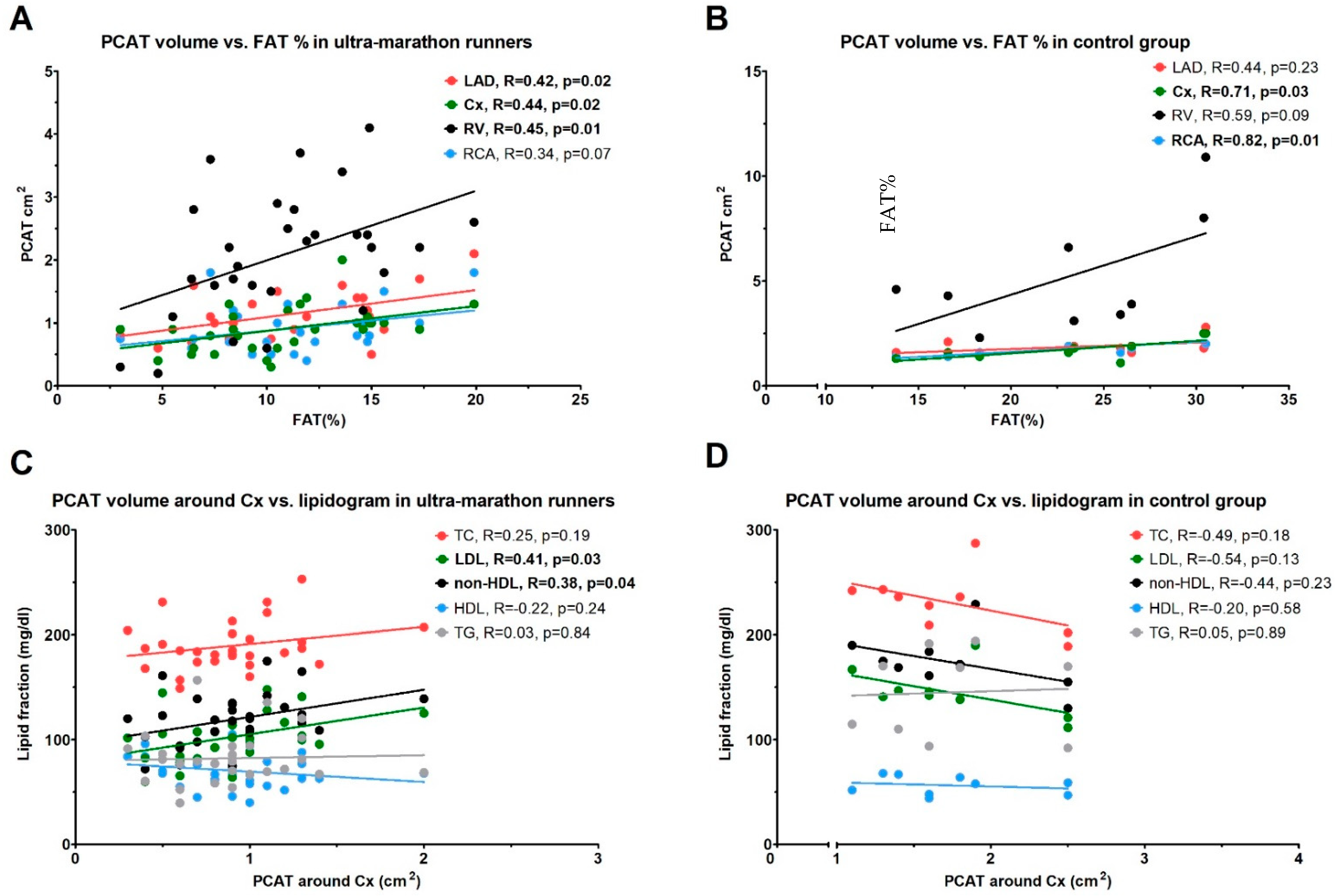
Figure 2 shows the correlations between body FAT% and PCAT, and between PCAT around the Cx artery and lipidogram parameters in the ultra-marathon runners and control group.
There was an intermediate, positive correlation between FAT% and PCAT around LAD, Cx, and RV in the runners’ group (R ≥ 0.42 for all; p ≤ 0.02 for all; Figure 2A) and a strong, positive correlation between FAT% and PCAT around Cx and RCA in the control group (R ≥ 0.71; p ≤ 0.03 for both; Figure 2B). Moreover, there was an intermediate position correlation between the PCAT around Cx artery and the concentration of LDL and non-HDL in the runners’ group (R ≥ 0.41; p ≤ 0.03 for both; Figure 2C), which was not observed in the control group (group (Figure 2D). There were no significant correlations between PCAT and FAT% with IL-6 plasma concentrations and IMT in both studied groups.

4. Discussion

The purpose of this study was to compare the EAT volume assessed with CMR among ultra-marathon runners and in the sedentary control group, and assess the correlations between EAT and risk factors of CAD, including body composition, venous blood lipid profile, IL-6 plasma concentration, and IMT. The main findings of this study are that (i) EAT volume is smaller in all measured localizations in the ultra-marathon runners compared to the control group, and (ii) there is a positive correlation between FAT% and PCAT surrounding LAD artery, Cx artery, and RV in the ultra-marathon runners, and a positive correlation between LDL and non-HDL concentration and PCAT around Cx artery.
CVD and obesity are significant health problems in developed and developing countries [1,2,3,5,6]. Strong connections between obesity, insulin resistance, and atherogenic lipids profile are well-documented [22]. Whereas subcutaneous adipose tissue (SAT) is not associated with cardiovascular risk, visceral fat seems to play a major role in atherosclerosis [23]. Recently, there is a growing interest in PCAT, which is a specific visceral tissue located in the direct neighborhood of the coronary arteries and therefore, in addition to the systemic effect, it also has paracrine and vasocrine effect on the surrounding vessels [10,24,25]. There are many differences at the anatomic, histologic, embryologic, and biomolecular levels between EAT/ PCAT and other fat depots [12,13]. Inflammatory processes underlying atherosclerosis are well-documented [26]. In addition, EAT was showed to participate in the pathogenesis of coronary atherosclerosis by secreting multiple cytokines and chemokines, thus serving as an active pro-inflammatory tissue [14,27]. In patients with advanced CAD undergoing coronary artery bypass grafting, EAT released more inflammatory markers than the SAT [27]. Moreover, the thickness of EAT measured using computed tomography reflected the stage of CAD. There was also a positive correlation between PCAT and vulnerability of atherosclerotic plaques based on their composition, assessed by intravascular ultrasound imaging [28]. Mazurek et al. analyzed the composition of atherosclerotic plaques using virtual histology intravascular ultrasonography and quantitatively assessed PCAT using positron emission tomography/computed tomography among patients with acute coronary syndrome without persistent ST-segment elevation. The inflammatory activity was measured by maximal standardized uptake value of 18-fluorodeoxyglucose. Inflammatory activity within PCAT was higher than in subcutaneous, visceral thoracic or epicardial adipose tissue, and there was a strong, positive correlation with pro-inflammatory activity of PCAT and both coronary plaque burden and its necrotic core component [29]. The association between EAT, atrial fibrillation [30], and impairment of cardiac function was also demonstrated [31]. Recently, it was demonstrated that among patients without known cardiovascular disease, the mean EAT thickness evaluated in magnetic resonance was higher in those with subclinical left ventricle impairment compared to those with normal left ventricle function, independently of traditional risk factors [32]. It has been also shown that patients with a familiar hypercholesterolemia had increased EAT thickness, and there was a positive correlation between EAT and plasma LDL concentration in these patients [33]. Volume and thickness of EAT correlates with intra-abdominal fat mass, obesity extent [7,8], and is independently associated with cardiovascular events [9]. Further, EAT is an independent predictor for the presence of carotid plaque [11]. It has been emphasized that EAT has a stronger association with systemic inflammation and subclinical atherosclerosis (IMT than BMI and waist circumference) [34,35].
The effect of very intense physical activity on physiological processes is still unknown. Both short-term and long-term endurance sport can induce both anti- and pro-inflammatory response. Long-term physical exercise was shown to reduce the level of C-reactive protein (CRP), especially when accompanied by a decrease in weight, especially in individuals with initially elevated CRP [36]. On the other hand, long-term exercise may also stimulate inflammatory processes, vascular oxidative stress, and fatigue of the elastic components of the vessel wall [37]. For example, obese non-elite runners had higher CRP plasma concentration than lean elite runners, measured 24 h after marathon. In addition, obese non-elite runners had a higher level of IL-6 and a lower level of IL-10 at baseline than lean elite and lean non-elite runners [38]. These results indicate the association between adipose tissue and inflammation.
In our population of extreme amateur runners, EAT volume was smaller in all measured locations compared to control group. As expected, runners had lower weight, BMI, fat free mass, body FAT%, concentrations of LDL and TG, higher concentration of HDL, and less frequently pathologically high levels of plasma IL-6 compared to controls. These findings suggest that high intensity training may have a favorable effect on cardiovascular risk factors. On the other hand, there was a correlation between EAT/PCAT surrounding LAD artery, Cx artery, and RV, and between PCAT surrounding Cx artery and lipid profile in the ultra-marathon runners’ group, which might potentially reflect increased cardiovascular risk in this population.

5. Limitations

The main limitation of this study is the small sample size. For example, likely the correlations between FAT% and PCAT around coronary arteries, which were not significant in our control group, would become significant in a larger cohort. The second limitations are the limited inclusion criteria of the study population, including homogenous individuals regarding gender (only male) and ethnicity (only Caucasian, Polish). Therefore, our results are not applicable to female and to any other ethnic group. Another limitation is the control group comprising sex- and age-matched sedentary controls with higher body weight, BMI, and FAT levels, which are known risk factors of CVD. Ideally, the control group would comprise individuals with moderate physical activity and similar body composition to the study group. However, such group was not available when we performed this study. Finally, the correlations showed in our study do not prove any causal relationship between intensive training, EAT, and CVD. Due to high risk of bias in this study, we may only speculate about the benefit and risk of extremely intensive training on cardiovascular health, and further research is required to ultimately answer this question.

6. Conclusions

In summary, extremely intensive training may decrease the risk of cardiovascular events in adult population of amateur athletes by reducing the amount and pro-inflammatory activity of EAT. More research is needed to draw firm conclusions regarding the anti- and pro-inflammatory effects of intensive training.

Author Contributions

Conceptualization, Ł.A.M. and M.P.; methodology, Ł.A.M. and M.P.; validation, Ł.A.M.; formal analysis, Ł.A.M. and M.K.; investigation, M.B.-F. and A.M. and A.C. and K.W. and W.B.; resources, Ł.A.M. and M.P.; data curation, Ł.A.M.; writing—original draft preparation, M.K.; writing—review and editing, M.K. and A.G. and T.M.; visualization, M.K. and A.G.; supervision, Ł.A.M. and T.M.; project administration, Ł.A.M.; funding acquisition, Ł.A.M. All authors have read and agreed to the published version of the manuscript.

Funding

This research received no external funding.

Institutional Review Board Statement

The study was conducted according to the guidelines of the Declaration of Helsinki, and approved by the Ethics Committee of the Regional Medical Chamber in Warsaw, Poland (protocol no. 52/17 and date of approval 12 October 2017).

Informed Consent Statement

Informed consent was obtained from all subjects involved in the study.

Data Availability Statement

The data presented in this study are available on request from the corresponding author. The data are not publicly available due to patients’ privacy.

Acknowledgments

We acknowledge the participants for their consent to take part in the study.

Conflicts of Interest

The authors report no conflict of interest related to this study.

References

  1. Laslett, L.J.; Alagona, P., Jr.; Clark, B.A., 3rd; Drozda, J.P., Jr.; Saldivar, F.; Wilson, S.R.; Poe, C.; Hart, M. The worldwide environment of cardiovascular disease: Prevalence, diagnosis, therapy, and policy issues: A report from the American College of Cardiology. J. Am. Coll. Cardiol. 2012, 60 (Suppl. 25), 1–49. [Google Scholar] [CrossRef] [Green Version]
  2. GBD 2013 Mortality and Causes of Death Collaborators. Global, regional, and national age-sex specific all-cause and cause-specific mortality for 240 causes of death, 1990–2013: A systematic analysis for the Global Burden of Disease Study 2013. Lancet 2015, 385, 117–171. [Google Scholar] [CrossRef]
  3. Roth, G.A.; Huffman, M.D.; Moran, A.E.; Feigin, V.; Mensah, G.A.; Naghavi, M.; Murray, C.J. Global and regional patterns in cardiovascular mortality from 1990 to 2013. Circulation 2015, 132, 1667–1678. [Google Scholar] [CrossRef] [PubMed] [Green Version]
  4. Yusuf, S.; Hawken, S.; Ounpuu, S.; Dans, T.; Avezum, A.; Lanas, F.; McQueen, M.; Budaj, A.; Pais, P.; Varigos, J.; et al. Interheart Study Investigators. Effect of potentially modifiable risk factors associated with myocardial infarction in 52 countries (the INTERHEART study): Case-control study. Lancet 2004, 364, 937–952. [Google Scholar] [CrossRef]
  5. Despres, J.P.; Lemieux, I. Abdominal obesity and metabolic syndrome. Nature 2006, 444, 881–887. [Google Scholar] [CrossRef] [PubMed]
  6. Mathieu, P.; Lemieux, I.; Despres, J.P. Obesity, inflammation, and cardiovascular risk. Clin. Pharmacol. Ther. 2010, 87, 407–416. [Google Scholar] [CrossRef] [PubMed]
  7. Rabkin, S.W. Epicardial fat: Properties, function and relationship to obesity. Obes. Rev. 2007, 8, 253–261. [Google Scholar] [CrossRef] [PubMed]
  8. Iacobellis, G.; Willens, H.J.; Barbaro, G.; Sharma, A.M. Threshold values of high-risk echocardiographic epicardial fat thickness. Obesity 2008, 16, 887–892. [Google Scholar] [CrossRef] [PubMed]
  9. Mahabadi, A.; Berg, M.H.; Lehmann, N.; Kälsch, H.; Bauer, M.; Kara, K.; Dragano, N.; Moebus, S.; Jockel, K.-H.; Erbel, R.; et al. Association of epicardial fat with cardiovascular risk factors and incident myocardial infarction in the general population. J. Am. Coll. Cardiol. 2013, 61, 1388–1395. [Google Scholar] [CrossRef] [PubMed] [Green Version]
  10. Sacks, H.S.; Fain, J.N. Human epicardial adipose tissue: A review. Am. Heart J. 2007, 153, 907–917. [Google Scholar] [CrossRef]
  11. Erdogan, T.; Durakoglukil, M.E.; Cetin, M.; Altan Kocaman, S.; Duman, H.; Çiçek, Y.; Şatıroğlu, Ö. Epicardial Adipose tissue predicts carotid intima-media thickness independently of body mass index and waist circumference. Acta Cardiol. Sin. 2019, 35, 32–41. [Google Scholar]
  12. Mazurek, T. Proinflammatory capacity of adipose tissue: New insights in the pathophysiology of atherosclerosis. Kardiol. Pol. 2009, 67, 1119–1124. [Google Scholar]
  13. Mazurek, T.; Opolski, G. Pericoronary adipose tissue: A novel therapeutic target in obesity-related coronary atherosclerosis. J. Am. Coll. Nutr. 2015, 34, 244–254. [Google Scholar] [CrossRef] [PubMed]
  14. Mazurek, T.; Zhang, L.; Zalewski, A.; Mannion, J.D.; Diehl, J.T.; Arafat, H.; Sarov-Blat, L.; O’Brien, S.; Keiper, E.A.; Johnson, A.G.; et al. Human epicardial adipose tissue is a source of inflammatory mediators. Circulation 2003, 108, 2460–2466. [Google Scholar] [CrossRef] [Green Version]
  15. Paffenbarger, R.S., Jr.; Hyde, R.T.; Wing, A.L.; Lee, I.M.; Jung, D.L.; Kampert, J.B. The association of changes in physical-activity level and other lifestyle characteristics with mortality among men. N. Engl. J. Med. 1993, 328, 538–545. [Google Scholar] [CrossRef]
  16. Leon, A.S.; Connett, J.; Jacobs, D.R., Jr.; Rauramaa, R. Leisure-time physical activity levels and risk of coronary heart disease and death. The Multiple Risk Factor Intervention Trial. JAMA 1987, 258, 2388–2395. [Google Scholar] [CrossRef] [PubMed]
  17. Lee, D.C.; Sui, X.; Artero, E.G.; Lee, I.M.; Church, T.S.; McAuley, P.A.; Stanford, F.C.; Kohl, H.W., 3rd; Blair, S.N. Long-term effects of changes in cardiorespiratory fitness and body mass index on all-cause and cardiovascular disease mortality in men: The Aerobics Center Longitudinal Study. Circulation 2011, 124, 2483–2490. [Google Scholar] [CrossRef] [Green Version]
  18. Kubota, Y.; Evenson, K.R.; Maclehose, R.F.; Roetker, N.S.; Joshu, C.E.; Folsom, A.R. Physical Activity and Lifetime Risk of Cardiovascular Disease and Cancer. Med. Sci. Sports Exerc. 2017, 49, 1599–1605. [Google Scholar] [CrossRef] [PubMed]
  19. Mazurek, K.; Żmijewski, P.; Kozdroń, E.; Fojt, A.; Czajkowska, A.; Szczypiorski, P.; Mazurek, T. Cardiovascular risk reduction in sedentary postmenopausal women during organized physical activity. Kardiol. Pol. 2017, 75, 476–485. [Google Scholar] [CrossRef] [PubMed]
  20. Małek, Ł.A.; Barczuk-Falęcka, M.; Werys, K.; Czajkowska, A.; Mróz, A.; Witek, K.; Burrage, M.; Bakalarski, W.; Nowicki, D.; Roik, D.; et al. Cardiovascular magnetic resonance with parametric mapping in long-term ultra-marathon runners. Eur. J. Radiol. 2019, 117, 89–94. [Google Scholar] [CrossRef] [PubMed]
  21. Piepoli, M.F.; Hoes, A.W.; Agewall, S.; Albus, C.; Brotons, C.; Catapano, A.L.; Cooney, M.T.; Corrà, U.; Cosyns, B.; Deaton, C.; et al. 2016 European Guidelines on cardiovascular disease prevention in clinical practice: The Sixth Joint Task Force of the European Society of Cardiology and Other Societies on Cardiovascular Disease Prevention in Clinical Practice (constituted by representatives of 10 societies and by invited experts)Developed with the special contribution of the European Association for Cardiovascular Prevention & Rehabilitation (EACPR). Eur. Heart J. 2016, 37, 2315–2381. [Google Scholar]
  22. Nieves, D.J.; Cnop, M.; Retzlaff, B.; Walden, C.E.; Brunzell, J.D.; Knopp, R.H.; Kahn, S.E. The atherogenic lipoprotein profile associated with obesity and insulin resistance is largely attributable to intra-abdominal fat. Diabetes 2003, 52, 172–179. [Google Scholar] [CrossRef] [PubMed] [Green Version]
  23. Fox, C.S.; Massaro, J.M.; Hoffmann, U.; Pou, K.M.; Maurovich-Horvat, P.; Liu, C.Y.; Vasan, R.S.; Murabito, J.M.; Meigs, J.B.; Cupples, L.A.; et al. Abdominal visceral and subcutaneous adipose tissue compartments: Association with metabolic risk factors in the Framingham Heart Study. Circulation 2007, 116, 39–48. [Google Scholar] [CrossRef] [Green Version]
  24. Iacobellis, G. Local and systemic effects of the multifaceted epicardial adipose tissue depot. Nat. Rev. Endocrinol. 2015, 11, 363–371. [Google Scholar] [CrossRef] [PubMed]
  25. Niedziela, M.; Wojciechowska, M.; Zarębiński, M.; Cudnoch-Jędrzejewska, A.; Mazurek, T. Adiponectin promotes ischemic heart preconditioning–PRO and CON. Cytokine 2020, 127, 154981. [Google Scholar] [CrossRef]
  26. Hansson, G.K. Inflammation, atherosclerosis, and coronary artery disease. N. Engl. J. Med. 2005, 352, 1685–1695. [Google Scholar] [CrossRef] [PubMed] [Green Version]
  27. Kaartinen, M.; Penttila, A.; Kovanen, P.T. Mast cells accompany microvessels in human coronary atheromas: Implications for intimal neovascularisation and hemorrhage. Atherosclerosis 1996, 123, 123–131. [Google Scholar] [CrossRef]
  28. Yamashita, K.; Yamamoto, M.H.; Igawa, W.; Ono, M.; Kido, T.; Ebara, S.; Okabe, T.; Saito, S.; Amemiya, K.; Isomura, N.; et al. Association of Epicardial Adipose Tissue Volume and Total Coronary Plaque Burden in Patients with Coronary Artery Disease. Int. Heart J. 2018, 59, 1219–1226. [Google Scholar] [CrossRef] [PubMed] [Green Version]
  29. Mazurek, T.; Kochman, J.; Kobylecka, M.; Wilimski, R.; Filipiak, K.; Krolicki, L.; Opolski, G. Inflammatory activity of pericoronary adipose tissue may affect plaque composition in patients with acute coronary syndrome without persistent ST-segment elevation: Preliminary results. Kardiol. Pol. 2013, 72, 410–416. [Google Scholar] [CrossRef]
  30. Thanassoulis, G.; Massaro, J.M.; O’Donnell, C.J.; Hoffmann, U.; Levy, D.; Ellinor, P.T.; Wang, T.J.; Schnabel, R.B.; Vasan, R.S.; Fox, C.S.; et al. Pericardial fat is associated with prevalent atrial fibrillation: The Framingham Heart Study. Circ. Arrhythmia Electrophysiol. 2010, 3, 345–350. [Google Scholar] [CrossRef] [Green Version]
  31. Cavalcante, J.L.; Tamarappoo, B.K.; Hachamovitch, R.; Kwon, D.H.; Alraies, M.C.; Halliburton, S.; Schoenhagen, P.; Dey, D.; Berman, D.S.; Marwick, T.H. Association of epicardial fat, hypertension, subclinical coronary artery disease, and metabolic syndrome with left ventricular diastolic dysfunction. Am. J. Cardiol. 2012, 110, 1793–1798. [Google Scholar] [CrossRef] [Green Version]
  32. Rado, S.D.; Lorbeer, R.; Gatidis, S.; Machann, J.; Storz, C.; Nikolaou, K.; Rathmann, W.; Hoffmann, U.; Peters, A.; Bamberg, F.; et al. MRI-based assessment and characterization of epicardial and paracardial fat depots in the context of impaired glucose metabolism and subclinical left-ventricular alterations. Br. J. Radiol. 2019, 92, 20180562. [Google Scholar] [CrossRef] [PubMed]
  33. Dönmez, Y.; Bulut, A. Epicardial fat thickness is significantly increased and related to LDL cholesterol level in patients with familial hypercholesterolemia. J. Ultrasound 2019, 22, 309–314. [Google Scholar] [CrossRef] [PubMed]
  34. Natale, F.; Tedesco, M.A.; Mocerino, R.; de Simone, V.; Di Marco, G.M.; Aronne, L.; Credendino, M.; Siniscalchi, C.; Calabrò, P.; Cotrufo, M.; et al. Visceral adiposity and arterial stiffness: Echocardiographic epicardial fat thickness reflects, better than waist circumference, carotid arterial stiffness in a large population of hypertensives. Eur. J. Echocardiogr. 2009, 10, 549–555. [Google Scholar] [CrossRef]
  35. Sengul, C.; Cevik, C.; Ozveren, O.; Oduncu, V.; Sunbul, A.; Akgun, T.; Can, M.M.; Semiz, E.; Dindar, I. Echocardiographic epicardial fat thickness is associated with carotid intima-media thickness in patients with metabolic syndrome. Echocardiography 2011, 28, 853–858. [Google Scholar] [CrossRef]
  36. Barros, E.S.; Nascimento, D.C.; Prestes, J.; Nóbrega, O.T.; Córdova, C.; Sousa, F.; Boullosa, D.A. Acute and Chronic Effects of Endurance Running on Inflammatory Markers: A Systematic Review. Front. Physiol. 2017, 8, 779. [Google Scholar] [CrossRef] [PubMed] [Green Version]
  37. Radtke, T.; Schmidt-Trucksäss, A.; Brugger, N.; Schäfer, D.; Saner, H.; Wilhelm, M. Ultra-endurance sports have no negative impact on indices of arterial stiffness. Eur. J. Appl. Physiol. 2014, 114, 49–57. [Google Scholar] [CrossRef]
  38. Nickel, T.; Emslander, I.; Sisic, Z.; David, R.; Schmaderer, C.; Marx, N.; Schmidt-Trucksäss, A.; Hoster, E.; Halle, M.; Weis, M.; et al. Modulation of dendritic cells and toll-like receptors by marathon running. Eur. J. Appl. Physiol. 2012, 112, 1699–1708. [Google Scholar] [CrossRef] [PubMed]
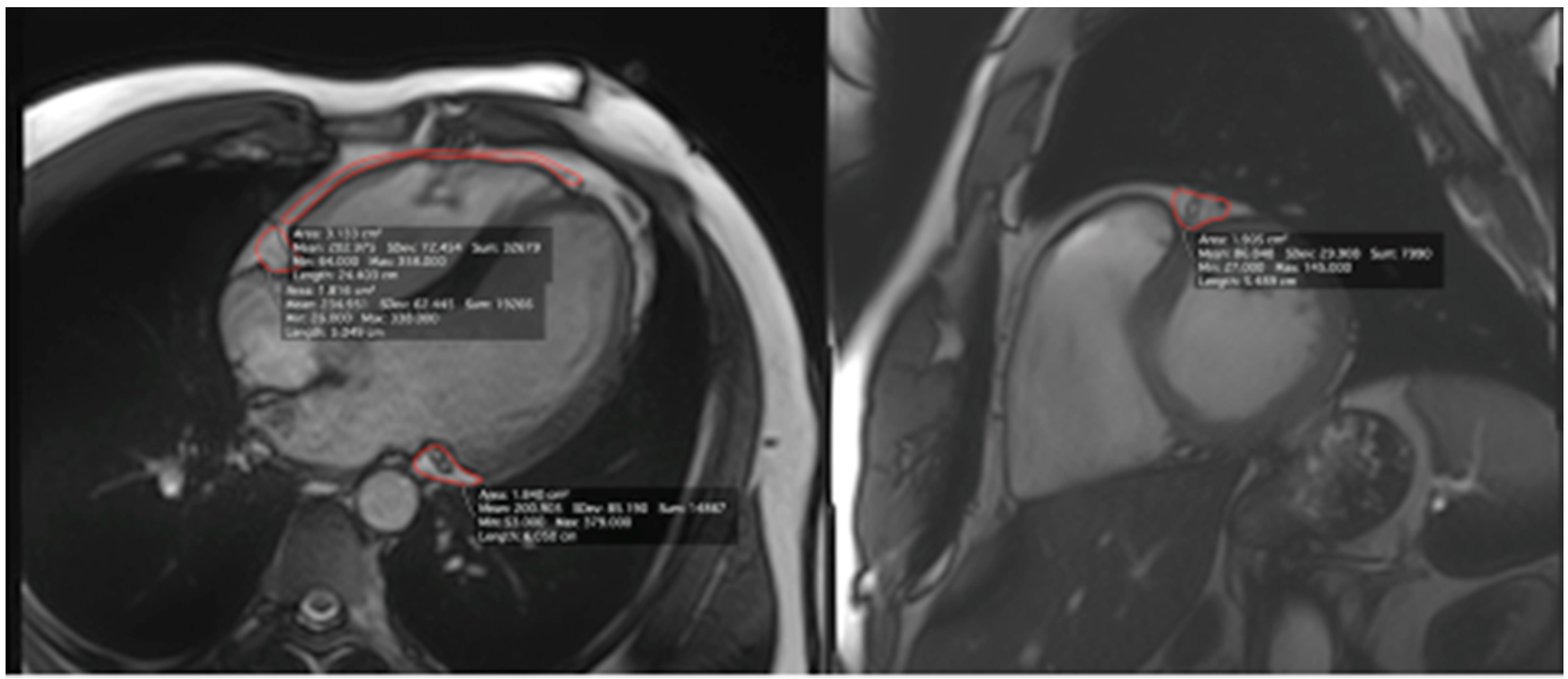
Figure 1. Example of pericoronary adipose tissue calculation with cardiac magnetic resonance by measurement of the area around the main coronary arteries and over the free wall of the right ventricle (epicardial adipose tissue).
Ijerph 18 03136 g001
Figure 2. (A,B) Correlations between body fat percentage (FAT%) and pericoronary adipose tissue (PCAT) in the ultra-marathon runners and control group. (C,D) Correlations between PCAT around the circumflex artery (Cx) and lipidogram parameters. LAD—left anterior descending artery; RV—right ventricle; RCA—right coronary artery; LDL—low-density lipoproteins; HDL—high-density lipoproteins; non-HDL—non-high-density lipoproteins; TC—total cholesterol; TG—triglycerides.
Ijerph 18 03136 g002
Table 1. Description of ultra-marathon runners. IQR—interquartile range.
ParameterUltra-Marathon Runners n = 30, Median (IQR)
Years of running9 (7–15)
Age at start of ultra-running34 (29–39)
Total covered distance (km)25,000 (20,000-40,000)
Weekly running distance (km)80 (70–90)
Number of ultra-races completed15 (10–27.5)
Number of ultra-races during last 2 years5.5 (4–9)
Number of completed ultra-races >100 km3.5 (2–7)
Best place achieved in an ultra-race5 (1–13)
Table 2. Participants’ baseline characteristics and results.
Baseline Characteristics
ParameterUltra-marathon Runners (n = 30)Control Group (n = 9)p
Age (years)40.93 ± 6.5740.78 ± 8.320.95
Heigth (cm)172.02 ± 32.49179.89 ± 6.430.48
Weight (kg)72.73 ± 5.1996.10 ± 19.39<0.001
BMI (cm/m2)23.09 ± 1.5430.00 ± 5.41<0.001
FAT % (%)10.78 ± 4.0123.17 ± 5.91<0.001
FAT mass (kg)8.02 ± 3.3223.16 ± 10.16<0.001
FFM (kg)65.30 ± 3.4474.34 ± 14.860.004
TBW (kg)47.81 ± 2.5253.40 ± 7.17<0.001
Blood Test Results
ParameterUltra-marathon Runners (n = 30)Control Group (n = 9)p
TC (mg/dl)189.66 ± 23.38230.22 ± 28.60<0.001
HDL (mg/dl)70.52 ± 15.9656.33 ± 8.990.016
LDL (mg/dl)102.68 ± 22.45144.86 ± 23.12<0.001
non-HDL (mg/dl)119.14 ± 25.07173.89 ± 27.10<0.001
TG (mg/dl)82.27 ± 24.73145.07 ± 41.82<0.001
IL-6 (pg/mL)1.29 ± 0.751.70 ± 0.720.156
Results of Imaging Tests
Cardiac Magnetic Resonance
ParameterUltra-marathon Runners (n = 30)Control Group (n = 9)p
LAD (cm2)1.12 ± 0.41.86 ± 0.41<0.001
RCA (cm2)0.88 ± 0.391.78 ± 0.34<0.001
Cx (cm2)0.90 ± 0.361.74 ± 0.49<0.001
RV (cm2)2.07 ± 0.97 5.23 ± 2.77 <0.001
Ultrasonography
Left IMT (cm)0.07 ± 0.020.07 ± 0.010.99
Right IMT (cm)0.08 ± 0.030.08 ± 0.010.68
FAT%—body fat percentage, BMI—body mass index, FFM—fat free mass, TBW—total body water, TC—total cholesterol concentration; HDL—high-density lipoprotein concentration; LDL—low-density lipoprotein concentration; non-HDL—non-high-density lipoprotein concentration; TG—triglyceride concentration; IL-6—interleukin-6 concentration; LAD—PCAT area around left anterior descending artery; RCA—PCAT area around right coronary artery; Cx—PCAT area around circumflex artery; RV—EAT area over right ventricle; IMT—carotid intima-media thickness.
Publisher’s Note: MDPI stays neutral with regard to jurisdictional claims in published maps and institutional affiliations.