Article

Multi- and Single-Joint Resistance Exercises Promote Similar Plantar Flexor Activation in Resistance Trained Men

1
College of Physical Education and Dance, Federal University of Goiás, Goiânia 74690-900, Brazil
2
College of Physical Education, Federal University of Rio Grande do Sul, Porto Alegre 90040-060, Brazil
3
College of Physical Education, University of Brasília, Brasília 70910-900, Brazil
*
Author to whom correspondence should be addressed.
Int. J. Environ. Res. Public Health 2020, 17(24), 9487; https://doi.org/10.3390/ijerph17249487
Submission received: 11 November 2020 / Revised: 8 December 2020 / Accepted: 14 December 2020 / Published: 18 December 2020
(This article belongs to the Section Exercise and Health)

Abstract

:
The present study aimed to compare soleus, lateral, and medial gastrocnemius muscles activation during leg press and calf raise exercises in trained men. The study involved 22 trained men (27.1 ± 3.6 years, 82.7 ± 6.6 kg, 177.5 ± 5.2 cm, 3.6 ± 1.4 experience years) who performed one set of each exercise using a 10-repetition maximum (10RM) load in a counterbalanced randomized order and separated by 10 min of rest. The electromyographic signal was measured for the three major plantar flexors: soleus, medial, and lateral gastrocnemius. A comparison between exercises showed that the mean adjusted by peak values during the leg press were 49.20% for the gastrocnemius lateralis, 51.31% for the gastrocnemius medialis, and 50.76% for the soleus. Values for calf raise were 50.70%, 52.19%, and 51.34% for the lateral, medial gastrocnemius, and soleus, respectively. There were no significant differences between exercises for any muscle (lateral gastrocnemius (p = 0.230), medial gastrocnemius (p = 0.668), and soleus (p = 0.535)). The present findings suggest that both leg press and calf raises can be used with the purpose to recruit triceps surae muscles. This bring the suggestion that one can chose between exercises based on personal preferences and practical aspects, without any negative impact on muscle activation.

1. Introduction

Resistance training (RT) is a popular mode of physical exercise often associated with voluntary muscle contraction against an external load, and its regular practice has been advised as an essential part of physical conditioning programs [1,2]. RT benefits are often associated with improvements in the musculoskeletal system such as increased muscle strength and hypertrophy, improved bone health [3,4], reduced low-back pain [5], and better functionality [6,7,8]. Such adaptations are influenced at some extend by the mechanical stimuli provided by repeated muscle activation in a process called mechanotransduction [9,10]. Muscle activation and subsequently mechanical stimuli might be affected by the manipulation and combination of RT variables such as load [11], rest between sets [12], and exercise selection [13,14].
Regarding exercise selection, RT can be performed with single (SJ) or multi-joint exercises (MJ), depending on the number of joints involved. For upper body muscles, it has been well established that SJ and MJ promote similar levels of muscle activation [15,16], as well as similar increases in muscle size and strength [17,18]. Moreover, other studies showed that, in general, the addition of SJ exercises to an RT program involving MJ exercises might not be necessary to bring optimal results in terms of muscle size and strength [19,20,21,22,23,24]. This brought the suggestion that one can chose between SJ and MJ based on personal preferences and practical aspects, without any negative impact on the results obtained from the intervention. Although less evidence is available for the lower body, studies about muscle activation [25,26], muscle strength [18], and hypertrophy [27] showed the same trends for thighs and hip muscles. However, information for calf muscles is scarce and controversial.
Bryanton et al. [28] reported near maximum efforts of the plantar flexors during the squat, when compared to the torque produced during a maximum isometric contraction. Interestingly, the relative muscle effort of the plantar flexors during the squat was similar to the ones of the hip and knee extensors, suggesting that the plantar flexors have an important involvement in the exercise performance. On the other hand, Escamilla et al. [29] reported that the activation of the plantar flexors was relatively low during different variations of the squat and leg press (9–17%), when normalized to a maximal isometric contraction. In addition to the controversial findings, these previous studies only analyzed MJ exercises and did not compare MJ and SJ under similar load conditions. Therefore, the current literature does not allow to conclude if plantar flexor muscle activation during a MJ exercise would be equivalent to the activation obtained during SJ exercises.
Some studies suggest that activation might be important for muscle adaptations [30], since mechanotransduction is likely to occur only in muscle fibers activated during exercise [31]. This suggests that high levels of muscle activation produced from repeated contractions can provide stimulation to the muscle, especially when combined with other factors, such as mechanical and metabolic stress, muscle swelling, etc. [31,32,33,34,35]. Therefore, considering that electromyographic (EMG) activity may reflect greater challenges to the musculoskeletal system, the investigation of plantar flexors muscle activity during MJ and SJ exercise using EMG analysis might contribute to close the gap regarding this topic.
Although plantar flexors are not commonly studied, their morphology and function might be important in clinical conditions such as chronic venous insufficiency and venous leg ulcers [36,37]. Moreover, strengthening the plantar flexors might have important applications in rehabilitation [38], to maintain orthostatic tolerance during exposure to microgravity [39], to improve balance [40,41], decrease the risk of falls [40], treat Achilles tendinopathy [42], and improve functionality [41]. Therefore, knowledge about how different exercises recruit these muscles might be of clinical and practical relevance. Based on the lack of information about plantar flexors’ activity during SJ and MJ exercises and the clinical and practical importance of the plantar flexors, the purpose of the present study was to compare soleus, lateral, and medial gastrocnemius muscles activation during leg press and calf raises in trained men. Our hypothesis is that calf raises would elicit superior activation of all muscles analyzed in comparison to leg press.

2. Materials and Methods

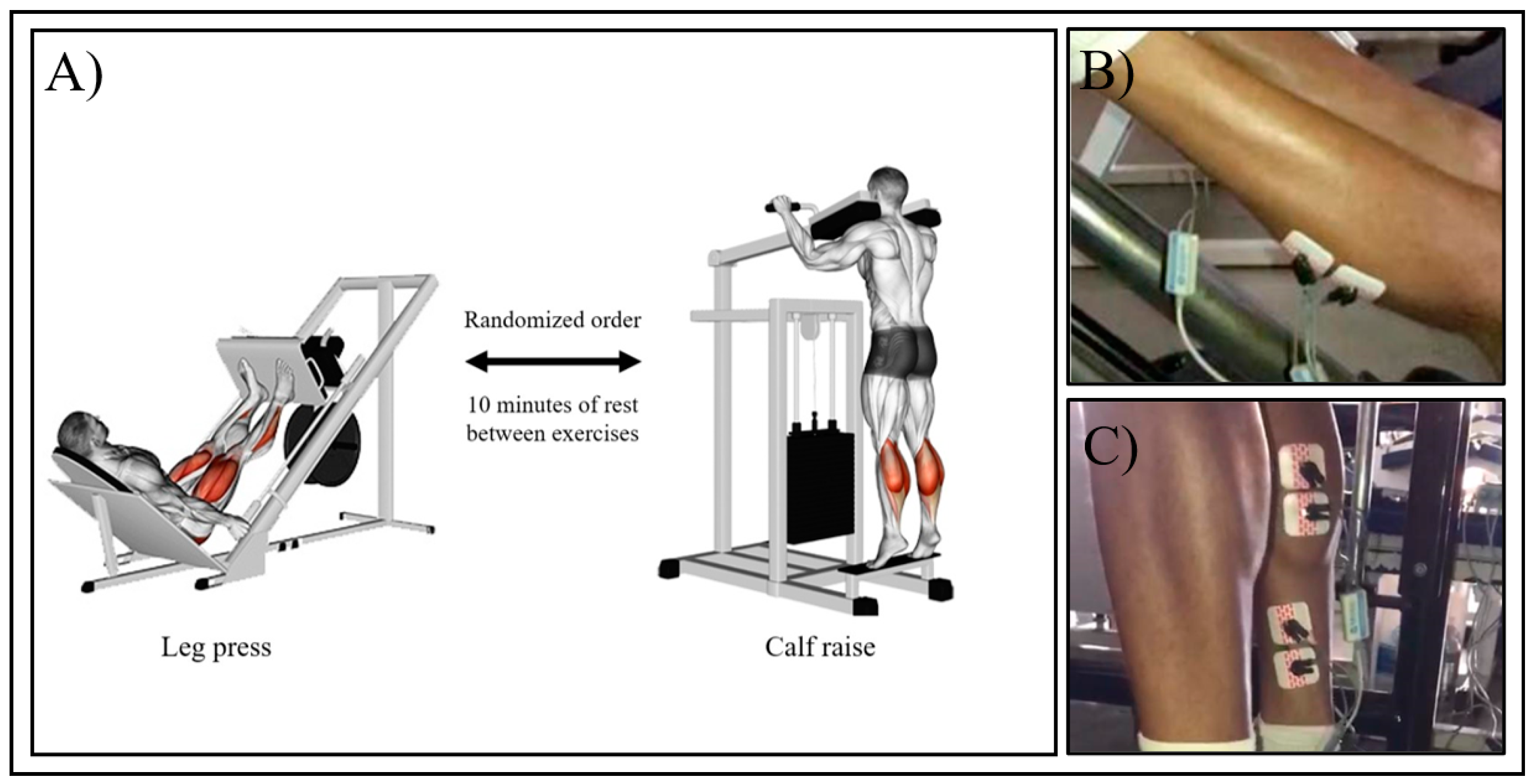
2.1. Experimental Approach

The study involved 22 trained men who visited the laboratory three times. The first two visits involved 10RM testing in the leg press and standing calf raise exercises and the third involved the performance of one set in each exercise using the 10RM load. Each visit was separated by 48–72 h and the volunteers were oriented to not perform any exercise or strenuous activity involving the studied muscles for 48 h before the first visit and between the subsequent visits. During the third visit, the exercises were performed in a counterbalanced randomized order and separated by 10 min of rest. Electromyographic signal was measured for the three major plantar flexors: soleus, medial, and lateral gastrocnemius.

2.2. Participants

Volunteers were recruited by social media and personal invitation among college students and the attendees of the Univesity’s RT facility. To be eligible to participate, the volunteers must have had at least one year of experience with RT, including the exercises tested. The performance of two RT sessions per week, involving 4 sets of exercise for the major muscle groups, was defined as minimum criteria to regular RT participation in accordance with the American College of Sports Medicine recommendations [1]. The volunteers were not allowed to participate if they had any clinical condition or medical problem that could be aggravated by the study protocol. After being informed about the study protocol, its risks and benefits, the participants signed and informed consent form in accordance with the Declaration of Helsinki. The study was approved by the relevant Ethics Committee (Protocol N°.56907716.5.0000.5083).

2.3. 10-Repetition Maximum (10RM) Testing

Participants performed 10RM tests on the leg press and calf raise in the first and second sessions in accordance with the National Strength and Conditioning Association (NSCA) recommendations [43]. Before the tests, the participants warmed up with 10 reps at a comfortable self-selected load and then rested for 5 min. Then, the initial load was defined based on the participants’ training history. If the volunteer could not perform 10 repetitions or performed more than 10 repetitions, the load was adjusted by 5–10% and another attempt was performed after 5 min of rest. No more than three attempts were necessary in any occasion. The tests were performed at a controlled velocity. A digital metronome was used, and the volunteers were oriented to perform each muscle action (concentric and eccentric phases) in two seconds, with no pause between contractions. The range of motion was controlled for each movement.
The leg press was performed in a 45-degree sled machine (Hammer Strength; Life Fitness, São Paulo, Brazil). The back pad was adjusted to provide a hip angle of 90 degree when the knees were fully extended. The movement went from full knee extension to 100 degrees of knee flexion. Calf raises was performed in standing position using a specific machine (Hammer Strength; Life Fitness, São Paulo, Brazil) with the knees fully extended during all time. Range of motion went from full ankle extension to full flexion. The movement was interrupted when the participants could not perform the movement with the defined range of motion or could not adhere to the proposed cadence [44].

2.4. Exercise Testing

The exercises were performed in the third visit using the load obtained in the 10RM testing and following the same procedures, with an especial attention to range of motion and movement velocity. We adopted a controlled movement velocity by controlling cadence, to standardize the time windows of the EMG signal and because a previous study showed that this could predict proximity to maximum effort [44]. The participants performed both exercises in the same day with the same electrode positioning. The exercises were performed in a randomized counterbalance order and were separated by 10 min of rest, as illustrated in Figure 1A. All tests and executions were recorded and reviewed to certify that they adhered to the procedures.

2.5. Electromyography

Electromyographic activity was recorded from the soleus, medial, and lateral gastrocnemius muscles. After skin preparation, including shaving and abrasion with alcohol to minimize impedance, pairs of electrodes were positioned in a bipolar configuration (distance of 20 mm between electrodes) along the direction of the muscle fibers according to the SENIAM recommendations (www.seniam.org) (Figure 1B,C). The reference electrode was placed on the knee patella. Electromyographic activity was measured using a system with 4 channels (Miotool400, 14-bit resolution, Miotec-Biomedical Equipment), and with a sampling frequency of 2000 Hz per channel. After measurement, electromyographic signals were filtered using the Butterworth filter with 20 Hz and 500 Hz cut-off frequencies for the lower and upper bandpass, respectively; and the adjusted by peak values were calculated while performing all repetitions. The means values were normalized using the maximum value obtained during the tests for each muscle.

2.6. Statistical Analysis
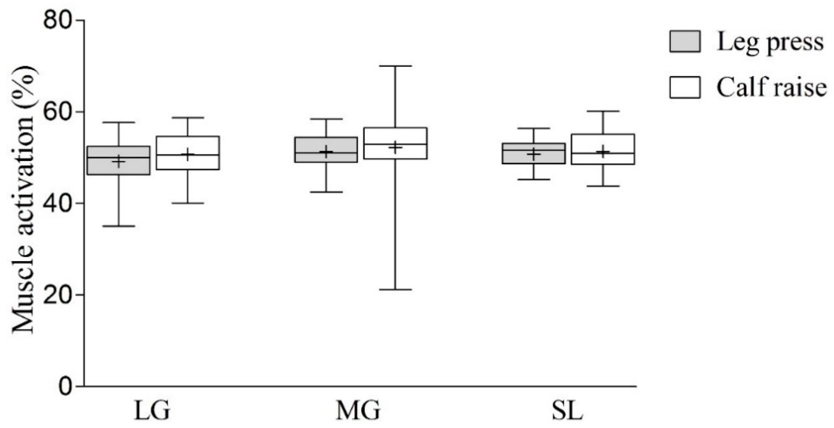
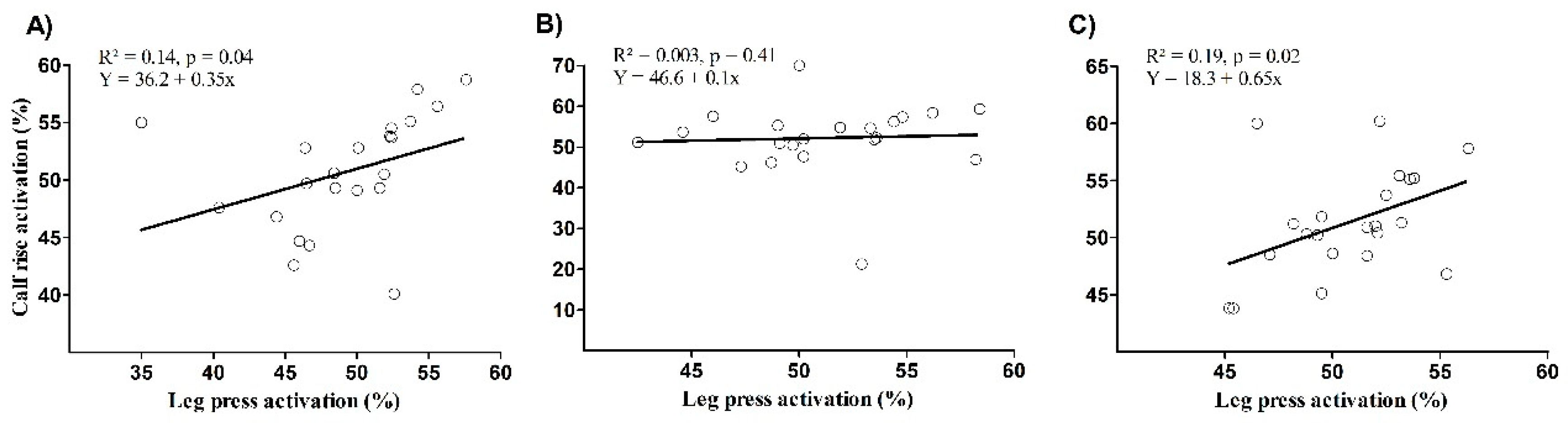
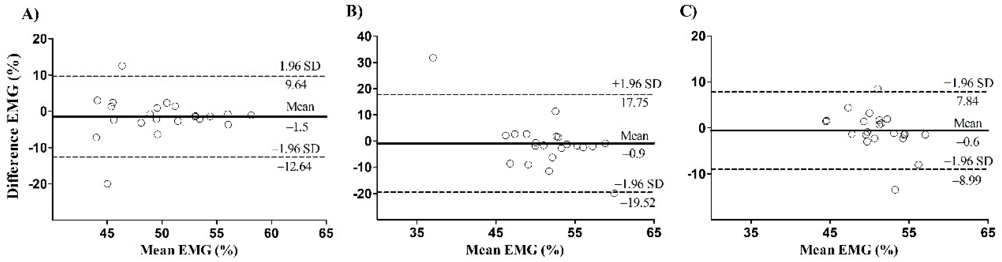
The normality of the data was confirmed by the Shapiro–Wilk test. Data are presented as means ± standard deviation. Paired T-tests were performed to compare mean EMG values between exercises. The effect size (ES) of the mean difference between exercise conditions was assessed by Cohen’s d effect size (mean exercise 1—mean exercise 2/SDpooled). An ES < 0.20 is considered trivial, small ≥ 0.20 and ≤ 0.60, moderate > 0.60 and ≤ 1.20, large > 1.20 and ≤ 2.00, and extremely large > 2.00 [45]. In addition, the agreement for the flexors plantar mean EMG values in leg press and calf raise was assessed by the simple linear regression and the Bland–Altman plot. An ES is considered as very weak if R2 < 0.04, weak if 0.04 ≤ R2 < 0.16, moderate if 0.16 ≤ R2 < 0.49, high if 0.49 ≤ R2 < 0.81 and very high if 0.81 ≤ R2 < 1.0 [46]. Statistical significance was defined as p < 0.05. Statistical analyses were performed using the Statistical Package for the Social Sciences 20.0 software (SPSS, Chicago, IL, USA).

3. Results

Participant characteristics are presented in Table 1. The load used for 10RM were (282.1 ± 32.7) in the leg press and (78.5 ± 7.8) in the calf raise. The estimated 1 repetition maximum [47] was 376 kg for the leg press and 105 kg for the calf raise.
Means values are illustrated in Figure 2. Comparison between groups showed that mean EMG values during the leg press were 49.2% for the lateral gastrocnemius, 51.31% for the medial gastrocnemius, and 50.76% for the soleus. Values for calf raises were 50.7%, 52.19%, and 51.34% for the lateral, medial gastrocnemius, and soleus, respectively. There were no differences between exercises between groups for any muscle and the ES of differences were trivial (gastrocnemius lateralis (p = 0.230, ES = 0.18), gastrocnemius medialis (p = 0.668, ES = 0.08), and soleus (p = 0.535, ES = 0.07)).
There is a significant correlation between the leg press and calf raise mean EMG for the lateral gastrocnemius (p = 0.04) and the soleus (p = 0.02), but not for the medialis gastrocnemius (p = 0.41) (Figure 3). The Bland–Altman plot showed systematic bias of −1.5 ± 5.7, −0.9 ± 9.5 and −0.6 ± 4.3 for the lateral, medial gastrocnemius, and soleus, respectively (Figure 4). The presence of proportional bias was verified for the medial gastrocnemius (p < 0.01), but not for the lateral gastrocnemius (p = 0.84) and soleus (p = 0.50).

4. Discussion

The purpose of the present study was to compare plantar flexors muscle activation during SJ and MJ exercises in trained men, here represented by calf raise and leg press exercises, respectively. Our main finding is that there was no difference for muscle activation for any muscle analyzed. This agrees with previous findings in upper and lower body muscles [15,16,25,26] and confirms the suggestion that plantar flexors have a great involvement in lower body MJ exercises [28,48]; however, the results were, to some extent, unexpected.
Plantar flexion is performed mainly by the three muscles that compose the triceps surae: soleus, medial, and lateral gastrocnemius. Soleus is originated in the tibia and fibula and inserted in the calcaneus, crossing only one joint. Considering that it acts mainly in the ankle and that both the leg press and calf raises exercises have a similar range of motion in that joint, the similar levels of muscle activation was not surprising. However, the medial and lateral gastrocnemius cross the knee and ankle joints, as they are originated in the femur and inserted in the calcaneus. Therefore, the gastrocnemius would have antagonistic actions during the leg press, as they flex the knee and extend the ankle, which is known as the Lombard Paradox [49]. Considering that the shortening in one joint is accompanied by the lengthening at the other, it was expected that the gastrocnemius activation would be decreased during the leg press. However, this was not the case. Considering that the calf muscles might act as dynamic knee stabilizers [50], one possible explanation is that the muscle contracted mainly isometrically, which can increase activation, as previously suggested [51].
To the best of our knowledge, this the first study to compare calf muscle activation between MJ and SJ exercises. The present results have many potentially important applications, since exercises oriented for the plantar flexors might be useful in chronic venous insufficiency and venous leg ulcers [36,37], rehabilitation [38], orthostatic tolerance [39], balance [40,41], prevention of falls [40], and functionality [41]. Based on our results, one can choose between MJ and SJ when aiming at activating the plantar flexors. It is interesting to note that previous studies showed that MJ exercises, like leg press and squat, are able to promote adaptations in quadriceps, hamstrings, and gluteus [52,53,54]. If lower body MJ exercises prove to be efficient also for plantar flexors, they can provide a time-efficient approach, since their performance can be sufficient to train most lower body muscles. In addition, a significant correlation was verified between exercise conditions for the lateralis gastrocnemius and the soleus, respectively, while no correlation was verified between exercise conditions for the medialis gastrocnemius. To the authors’ knowledge, there are no clear reasons to explain these differences between muscle groups.
We acknowledge that caution is required when extrapolating an acute effect onto a chronic change. Whilst some studies suggest that activation might be important for muscle adaptations [30], others suggest that electromyography cannot be necessarily linked to gains in muscle size and strength [55]. However, it has been suggested that mechanotransduction is likely to occur only in muscle fibers activated during exercise [31]. This suggests that high levels of muscle activation produced from repeated contractions can provide stimulation to the muscle, especially when combined with other factors, such as mechanical and metabolic stress, muscle swelling, etc. [31,32,33,34,35]. Therefore, the present study can provide a rationale for further studies aimed at comparing the long-term effects of MJ and SJ in plantar flexors’ function and morphology.
Based on the results of the present study, the leg press exercise can be recommended to promote muscle activation of the plantar flexors similarly to calf raises. Considering that MJ exercise involves other muscles such as quadriceps, hamstrings, and gluteus, exercise professionals should consider their use as a time efficient approach for exercise prescription.

Author Contributions

Conceptualization, P.G. and M.S.; methodology, P.G., D.S., M.S. and R.R.A.; formal analysis, D.S., R.R.A. and M.H.C.; resources M.B.; writing—original draft preparation, P.G. and M.S.; writing—review and editing, P.G., D.S., M.S., R.R.A., M.H.C., R.P. and M.B.; Visualization, P.G., D.S., R.R.A. and R.P. All authors have read and agreed to the published version of the manuscript

Funding

This research received no external funding.

Conflicts of Interest

The authors declare no conflict of interest.

References

  1. Garber, C.E.; Blissmer, B.; Deschenes, M.R.; Franklin, B.A.; Lamonte, M.J.; Lee, I.M.; Nieman, D.C.; Swain, D.P. Quantity and quality of exercise for developing and maintaining cardiorespiratory, musculoskeletal, and neuromotor fitness in apparently healthy adults: Guidance for prescribing exercise. Med. Sci. Sports Exerc. 2011, 43, 1334–1359. [Google Scholar] [CrossRef]
  2. Steele, J.; Fisher, J.; Skivington, M.; Dunn, C.; Arnold, J.; Tew, G.; Batterham, A.M.; Nunan, D.; O’Driscoll, J.M.; Mann, S.; et al. A higher effort-based paradigm in physical activity and exercise for public health: Making the case for a greater emphasis on resistance training. BMC Public Health 2017, 17. [Google Scholar] [CrossRef] [Green Version]
  3. Martyn-St James, M.; Carroll, S. High-intensity resistance training and postmenopausal bone loss: A meta-analysis. Osteoporos. Int. 2006, 17, 1225–1240. [Google Scholar] [CrossRef]
  4. Souza, D.; Barbalho, M.; Ramirez-Campillo, R.; Martins, W.; Gentil, P. High and low-load resistance training produce similar effects on bone mineral density of middle-aged and older people: A systematic review with meta-analysis of randomized clinical trials. Exp. Gerontol. 2020, 138, 110973. [Google Scholar] [CrossRef]
  5. Carpenter, D.M.; Nelson, B.W. Low back strengthening for the prevention and treatment of low back pain. Med. Sci. Sports Exerc. 1999, 31, 18–24. [Google Scholar] [CrossRef] [PubMed]
  6. Fiatarone, M.A.; Marks, E.C.; Ryan, N.D.; Meredith, C.N.; Lipsitz, L.A.; Evans, W.J. High-intensity strength training in nonagenarians. Effects on skeletal muscle. JAMA 1990, 263, 3029–3034. [Google Scholar] [CrossRef] [PubMed]
  7. Barbalho, M.D.S.M.; Gentil, P.; Izquierdo, M.; Fisher, J.; Steele, J.; Raiol, R.D.A. There are no no-responders to low or high resistance training volumes among older women. Exp. Gerontol. 2017, 99, 18–26. [Google Scholar] [CrossRef] [PubMed]
  8. Souza, D.; Barbalho, M.; Vieira, C.A.; Martins, W.R.; Cadore, E.L.; Gentil, P. Minimal dose resistance training with elastic tubes promotes functional and cardiovascular benefits to older women. Exp. Gerontol. 2018, 115, 132–138. [Google Scholar] [CrossRef]
  9. Hornberger, T.A. Mechanotransduction and the regulation of mTORC1 signaling in skeletal muscle. Int. J. Biochem. Cell Biol. 2011, 43, 1267–1276. [Google Scholar] [CrossRef] [Green Version]
  10. Maestroni, L.; Read, P.; Bishop, C.; Papadopoulos, K.; Suchomel, T.J.; Comfort, P.; Turner, A. The Benefits of Strength Training on Musculoskeletal System Health: Practical Applications for Interdisciplinary Care. Sports Med. 2020, 50, 1431–1450. [Google Scholar] [CrossRef]
  11. Jenkins, N.D.M.; Housh, T.J.; Bergstrom, H.C.; Cochrane, K.C.; Hill, E.C.; Smith, C.M.; Johnson, G.O.; Schmidt, R.J.; Cramer, J.T. Muscle activation during three sets to failure at 80 vs. 30% 1RM resistance exercise. Eur. J. Appl. Physiol. 2015, 115, 2335–2347. [Google Scholar] [CrossRef] [PubMed]
  12. Miranda, H.; Maia, M.; de Oliveira, C.G.; Farias, D.; da Silva, J.B.; Lima, V.P.; Willardson, J.M.; Paz, G.A. Myoeletric indices of fatigue adopting different rest intervals during leg press sets. J. Bodyw. Mov. Ther. 2018, 22, 178–183. [Google Scholar] [CrossRef]
  13. Ebben, W.P. Hamstring activation during lower body resistance training exercises. Int. J. Sports Physiol. Perform. 2009, 4, 84–96. [Google Scholar] [CrossRef] [PubMed] [Green Version]
  14. Gentil, P.; Fisher, J.; Steele, J. A Review of the Acute Effects and Long-Term Adaptations of Single- and Multi-Joint Exercises during Resistance Training. Sports Med. 2017, 47, 843–855. [Google Scholar] [CrossRef] [PubMed]
  15. Welsch, E.A.; Bird, M.; Mayhew, J.L. Electromyographic activity of the pectoralis major and anterior deltoid muscles during three upper-body lifts. J. Strength Cond. Res. 2005, 19, 449–452. [Google Scholar] [CrossRef]
  16. Rocha, V.D.A., Jr.; Gentil, P.; Oliveira, E.; Do Carmo, J. Comparison among the EMG activity of the pectoralis major, anterior deltoidis and triceps brachii during the bench press and peck deck exercises. Rev. Bras. Med. Esporte 2007, 13, 51–54. [Google Scholar]
  17. Gentil, P.; Soares, S.; Bottaro, M. Single vs. Multi-Joint Resistance Exercises: Effects on Muscle Strength and Hypertrophy. Asian J. Sports Med. 2015, 6, e24057. [Google Scholar] [CrossRef] [Green Version]
  18. Paoli, A.; Gentil, P.; Moro, T.; Marcolin, G.; Bianco, A. Resistance training with single vs. multi-joint exercises at equal total load volume: Effects on body composition, cardiorespiratory fitness, and muscle strength. Front. Physiol. 2017, 8, 1–6. [Google Scholar] [CrossRef] [Green Version]
  19. Gentil, P.; Soares, S.R.S.; Pereira, M.C.; da Cunha, R.R.; Martorelli, S.S.; Martorelli, A.S.; Bottaro, M. Effect of adding single-joint exercises to a multi-joint exercise resistance-training program on strength and hypertrophy in untrained subjects. Appl. Physiol. Nutr. Metab. 2013, 38, 341–344. [Google Scholar] [CrossRef]
  20. de França, H.S.; Branco, P.A.N.; Guedes Junior, D.P.; Gentil, P.; Steele, J.; Teixeira, C.V.L.S. The Effects of Adding Single-Joint Exercises To a Multi-Joint Exercise Resistance Training Program on Upper Body Muscle Strength and Size in Trained Men. Appl. Physiol. Nutr. Metab. 2015, 826, 150409143403004. [Google Scholar] [CrossRef] [Green Version]
  21. Steele, J.; Coswig, V.S.; Fisher, J.P.; Raiol, R.; Paoli, A.; Gentil, P.; Bianco, A.; Barbalho, M. Does the addition of single joint exercises to a resistance training program improve changes in performance and anthropometric measures in untrained men? Eur. J. Transl. Myol. 2018, 28. [Google Scholar] [CrossRef]
  22. Fisher, J.; Steele, J.; Raiol, R.; Gentil, P.; Barbalho, M.; Coswig, V. Influence of Adding Single-Joint Exercise to a Multijoint Resistance Training Program in Untrained Young Women. J. Strength Cond. Res. 2018, 1. [Google Scholar] [CrossRef]
  23. Barbalho, M.; Coswig, V.; Raiol, R.; Fisher, J.; Steele, J.; Bianco, A.; Gentil, P. Single joint exercises do not provide benefits in performance and anthropometric changes in recreational bodybuilders. Eur. J. Sport Sci. 2019, 1–8. [Google Scholar] [CrossRef] [PubMed]
  24. Barbalho, M.; Coswig, V.; Raiol, R.; Steele, J.; Fisher, J.; Paoli, A.; Gentil, P. Effects of Adding Single Joint Exercises to a Resistance Training Programme in Trained Women. Sports 2018, 6, 160. [Google Scholar] [CrossRef] [PubMed] [Green Version]
  25. Wilk, K.E.; Escamilla, R.F.; Fleisig, G.S.; Barrentine, S.W.; Andrews, J.R.; Boyd, M.L. A comparison of tibiofemoral joint forces and electromyographic activity during open and closed kinetic chain exercises. Am. J. Sports Med. 1996, 24, 518–527. [Google Scholar] [CrossRef]
  26. Signorile, J.F.; Weber, B.; Roll, B.; Caruso, J.F.; Lowensteyn, I.; Perry, A.C. An Electromyographical Comparison of the Squat and Knee Extension Exercises. J. Strength Cond. Res. 1994, 8, 178–183. [Google Scholar] [CrossRef]
  27. Barbalho, M.; Coswig, V.; Souza, D.; Serrão, J.C.; Campos, M.H.; Gentil, P. Back Squat vs. Hip Thrust Resistance-training Programs in Well-trained Women. Int. J. Sports Med. 2020. [Google Scholar] [CrossRef]
  28. Bryanton, M.A.; Kennedy, M.D.; Carey, J.P.; Chiu, L.Z. Effect of Squat Depth and Barbell Load on Relative Muscular Effort in Squatting. J. Strength Cond. Res. 2012. [Google Scholar] [CrossRef]
  29. Escamilla, R.F.; Fleisig, G.S.; Zheng, N.; Lander, J.E.; Barrentine, S.W.; Andrews, J.R.; Bergemann, B.W.; Moorman, C.T., 3rd. Effects of technique variations on knee biomechanics during the squat and leg press. Med. Sci. Sports Exerc. 2001, 33, 1552–1566. [Google Scholar] [CrossRef]
  30. Kohiruimaki, R.; Maeo, S.; Kanehisa, H. Suspended Push-up Training Augments Size of not only Upper Limb but also Abdominal Muscles. Int. J. Sports Med. 2019, 40, 789–795. [Google Scholar] [CrossRef]
  31. Rennie, M.J.; Wackerhage, H.; Spangenburg, E.E.; Booth, F.W. Control of the Size of the Human Muscle Mass. Annu. Rev. Physiol. 2004, 66, 799–828. [Google Scholar] [CrossRef] [PubMed]
  32. Counts, B.R.; Buckner, S.L.; Dankel, S.J.; Jessee, M.B.; Mattocks, K.T.; Mouser, J.G.; Laurentino, G.C.; Loenneke, J.P. The acute and chronic effects of “NO LOAD” resistance training. Physiol. Behav. 2016, 164, 345–352. [Google Scholar] [CrossRef] [PubMed]
  33. Rudroff, T.; Staudenmann, D.; Enoka, R.M. Electromyographic measures of muscle activation and changes in muscle architecture of human elbow flexors during fatiguing contractions. J. Appl. Physiol. 2008, 104, 1720–1726. [Google Scholar] [CrossRef] [Green Version]
  34. Farup, J.; de Paoli, F.; Bjerg, K.; Riis, S.; Ringgard, S.; Vissing, K. Blood flow restricted and traditional resistance training performed to fatigue produce equal muscle hypertrophy. Scand. J. Med. Sci. Sports 2015. [Google Scholar] [CrossRef]
  35. Yasuda, T.; Loenneke, J.P.; Thiebaud, R.S.; Abe, T. Effects of Blood Flow Restricted Low-Intensity Concentric or Eccentric Training on Muscle Size and Strength. PLoS ONE 2012, 7, 1–7. [Google Scholar] [CrossRef] [PubMed] [Green Version]
  36. Orr, L.; Klement, K.A.; McCrossin, L.; Drombolis, D.O.; Houghton, P.E.; Spaulding, S.; Burke, S. A systematic review and meta-Analysis of exercise intervention for the treatment of calf muscle pump impairment in individuals with chronic venous insufficiency. Ostomy Wound Manag. 2017, 63, 30–43. [Google Scholar] [CrossRef]
  37. Orsted, H.L.; Radke, L.; Gorst, R. The impact of musculoskeletal changes on the dynamics of the calf muscle pump. Ostomy. Wound. Manag. 2001, 47, 18–24. [Google Scholar]
  38. Brorsson, A.; Grävare Silbernagel, K.; Olsson, N.; Nilsson Helander, K. Calf Muscle Performance Deficits Remain 7 Years After an Achilles Tendon Rupture. Am. J. Sports Med. 2018, 46, 470–477. [Google Scholar] [CrossRef]
  39. Convertino, V.A.; Doerr, D.F.; Stein, S.L. Changes in size and compliance of the calf after 30 days of simulated microgravity. J. Appl. Physiol. 1989, 66, 1509–1512. [Google Scholar] [CrossRef]
  40. Cattagni, T.; Scaglioni, G.; Laroche, D.; Van Hoecke, J.; Gremeaux, V.; Martin, A. Ankle muscle strength discriminates fallers from non-fallers. Front. Aging Neurosci. 2014, 6. [Google Scholar] [CrossRef] [Green Version]
  41. Spink, M.J.; Fotoohabadi, M.R.; Wee, E.; Hill, K.D.; Lord, S.R.; Menz, H.B. Foot and ankle strength, range of motion, posture, and deformity are associated with balance and functional ability in older adults. Arch. Phys. Med. Rehabil. 2011, 92, 68–75. [Google Scholar] [CrossRef] [PubMed]
  42. Gatz, M.; Betsch, M.; Dirrichs, T.; Schrading, S.; Tingart, M.; Michalik, R.; Quack, V. Eccentric and Isometric Exercises in Achilles Tendinopathy Evaluated by the VISA-A Score and Shear Wave Elastography. Sports Health 2020, 12, 373–381. [Google Scholar] [CrossRef] [PubMed]
  43. National Strength and Conditioning Association. Exercise Technique Manual for Resistance Training; Human Kinetics, Inc.: Champaign, IL, USA, 2016; ISBN 9781492506928. [Google Scholar]
  44. Gentil, P.; Marques, V.A.; Neto, J.P.P.; Santos, A.C.G.; Steele, J.; Fisher, J.P.; Paoli, A.; Bottaro, M. Using velocity loss for monitoring resistance training effort in a real world setting. Appl. Physiol. Nutr. Metab. 2018. [Google Scholar] [CrossRef] [PubMed]
  45. Hopkins, W.G.; Marshall, S.W.; Batterham, A.M.; Hanin, J. Progressive statistics for studies in sports medicine and exercise science. Med. Sci. Sports Exerc. 2009, 41, 3–12. [Google Scholar] [CrossRef] [PubMed] [Green Version]
  46. Forte, P.; Marinho, D.A.; Nikolaidis, P.T.; Knechtle, B.; Barbosa, T.M.; Morais, J.E. Analysis of cyclist’s drag on the aero position using numerical simulations and analytical procedures: A case study. Int. J. Environ. Res. Public Health 2020, 17, 3430. [Google Scholar] [CrossRef] [PubMed]
  47. Brzycki, M. Strength Testing—Predicting a One-Rep Max from Reps-to-Fatigue. J. Phys. Educ. Recreat. Danc. 1993, 64, 88–90. [Google Scholar] [CrossRef]
  48. Da Silva, E.M.; Brentano, M.A.; Cadore, E.L.; De Almeida, A.P.V.; Kruel, L.F.M. Analysis of muscle activation during different leg press exercises at submaximum effort levels. J. Strength Cond. Res. 2008, 22, 1059–1065. [Google Scholar] [CrossRef] [Green Version]
  49. Lombard, W.P. The Action of Two-Joint Muscles. Am. Phys. Educ. Rev. 1903, 8, 141–145. [Google Scholar] [CrossRef]
  50. Sherbondy, P.S.; Queale, W.S.; McFarland, E.G.; Mizuno, Y.; Cosgarea, A.J. Soleus and gastrocnemius muscle loading decreases anterior tibial translation in anterior cruciate ligament intact and deficient knees. J. Knee Surg. 2003, 16, 152–158. [Google Scholar]
  51. Babault, N.; Pousson, M.; Ballay, Y.; Van Hoecke, J. Activation of human quadriceps femoris during isometric, concentric, and eccentric contractions. J. Appl. Physiol. 2001, 91, 2628–2634. [Google Scholar] [CrossRef]
  52. Goncalves, A.; Gentil, P.; Steele, J.; Giessing, J.; Pauli, A.; Fisher, J.P. Comparison of single- and multi-joint lower body resistance training upon strength increases in recreationally active males and females: A within-participant unilateral training study. Eur. J. Transl. Myol. 2019, 29, 304–308. [Google Scholar] [CrossRef] [PubMed]
  53. Popov, D.V.; Tsvirkun, D.V.; Netreba, A.I.; Tarasova, O.S.; Prostova, A.B.; Larina, I.M.; Borovik, A.S.; Vinogradova, O.L.; Swirkun, D.V.; Netreba, A.I.; et al. Hormonal adaptation determines the increase in muscle mass and strength during low-intensity strength training without relaxation. Hum. Physiol. 2006, 32, 121–127. [Google Scholar] [CrossRef]
  54. Yasuda, T.; Fukumura, K.; Fukuda, T.; Uchida, Y.; Iida, H.; Meguro, M.; Sato, Y.; Yamasoba, T.; Nakajima, T. Muscle size and arterial stiffness after blood flow-restricted low-intensity resistance training in older adults. Scand. J. Med. Sci. Sports 2014, 24, 799–806. [Google Scholar] [CrossRef] [PubMed]
  55. Vigotsky, A.D.; Beardsley, C.; Contreras, B.; Steele, J.; Ogborn, D.; Phillips, S.M. Greater Electromyographic Responses Do Not Imply Greater Motor Unit Recruitment and ‘Hypertrophic Potential’ Cannot Be Inferred. J. Strength Cond. Res. 2017, 31, e1–e4. [Google Scholar] [CrossRef] [Green Version]
Figure 1. Illustration of exercise testing procedure (A), and electrode position during leg press (B) and calf raise (C).
Ijerph 17 09487 g001
Figure 2. Comparison of muscle activation between leg press and calf raise. LG, lateral gastrocnemius; MG, medial gastrocnemius; SL, soleus. Values are presented as median (lines) with interquartile range (boxes) ± range (minimum and maximum) and + indicates mean.
Ijerph 17 09487 g002
Figure 3. Correlation between calf raise and leg press mean EMG for the lateralis gastrocnemius (A), the medialis gastrocnemius (B), and the soleus (C).
Ijerph 17 09487 g003
Figure 4. Bland–Altman plot of difference EMG for the lateralis gastrocnemius (A), the medialis gastrocnemius (B), and the soleus (C). The dotted line represents the limits of agreement upper and lower boundary. The continued line on the center of plot represents the systematic bias. The continued line on the Y axis represents the mean difference between leg press and calf raise mean EMG, and on the X axis represents the mean of leg press and calf raise mean EMG.
Ijerph 17 09487 g004
Table 1. Characteristics of the participants.
VariablesMean ± Standard Deviation
Age (years)27.1 ± 3.6
Weight (kg)82.7 ± 6.6
Height (cm)177.5 ± 5.2
Resistance training experience (years)3.6 ± 1.4
Publisher’s Note: MDPI stays neutral with regard to jurisdictional claims in published maps and institutional affiliations.

