Article

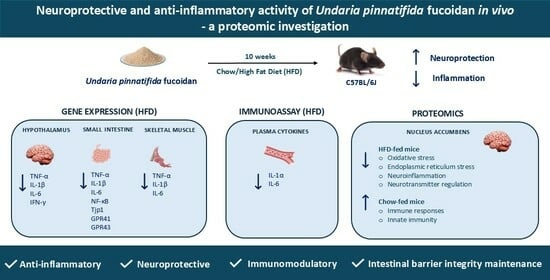
Neuroprotective and Anti-Inflammatory Activity of Undaria pinnatifida Fucoidan In Vivo—A Proteomic Investigation

by
Cheng Yang
1,
Corinna Dwan
2,
Barbara C. Wimmer
2,
Maurizio Ronci
3,
Richard Wilson
4,
Luke Johnson
5 and
Vanni Caruso
1,*
1
School of Pharmacy and Pharmacology, University of Tasmania, Hobart, TAS 7005, Australia
2
Marinova Pty Ltd., 249 Kennedy Drive, Cambridge, TAS 7170, Australia
3
Department of Medical, Oral and Biotechnology Sciences “G.D’Annunzio”, 66100 Chieti, Italy
4
Central Science Laboratory, College of Science and Engineering, University of Tasmania, Hobart, TAS 7001, Australia
5
School of Psychological Sciences, Psychology, University of Tasmania, Launceston, TAS 7248, Australia
*
Author to whom correspondence should be addressed.
Mar. Drugs 2025, 23(5), 189; https://doi.org/10.3390/md23050189
Submission received: 17 March 2025 / Revised: 18 April 2025 / Accepted: 23 April 2025 / Published: 27 April 2025

Abstract

:
Undaria pinnatifida fucoidan (UPF), a bioactive sulphated polysaccharide, is widely recognised for its anti-inflammatory, antioxidant, antitumor, anticoagulant, antiviral, and immunomodulatory properties. However, the precise mechanisms by which UPF regulates inflammation and neuronal health remain unclear. This study aimed to investigate the effects of UPF supplementation on pro-inflammatory cytokines in skeletal muscle, small intestine, and the hypothalamus, as well as plasma cytokine levels. Additionally, a brain proteomic investigation in the nucleus accumbens (NAc) was performed to assess UPF’s impact on neuronal protein expression in mice. A total of 64 C57BL/6J mice were administered either a standard chow or high-fat diet (HFD) with or without UPF (400 mg/kg/day) for 10 weeks. In HFD-fed mice, UPF significantly reduced the expression of pro-inflammatory cytokines (TNF-α, IL-1β, and IL-6) in skeletal muscle, small intestine, and hypothalamus, while also lowering circulating IL-1α and IL-6 levels. Proteomic analysis of the NAc revealed that UPF modulated proteins involved in oxidative stress, neuroinflammation, neurotransmitter regulation, and endoplasmic reticulum stress. In contrast, in chow-fed mice, UPF had no effect on the neuroinflammatory–oxidative stress markers but influenced the abundance of proteins associated with immune response and innate immunity. These findings suggest that UPF modulates stress-response pathways in a diet-dependent manner, supporting its potential neuroprotective role in inflammation-related disorders and brain health.

Graphical Abstract

1. Introduction

Marine-derived compounds have demonstrated promising neuroprotective effects in preclinical studies, targeting key pathways involved in neuroinflammation and neurodegenerative disorders [1,2]. Among these compounds, fucoidan extracted from Undaria pinnatifida (UPF) has gained widespread recognition as a functional food and nutraceutical due to its diverse biological activities and significant contributions to human health. Extensive scientific evidence highlights the potent anticancer [3,4], antioxidant [5], antiarthritic [6] antibacterial [7], and antiviral [8,9] properties of UPF. Additionally, recent studies have demonstrated that UPF can modulate inflammatory pathways by suppressing pro-inflammatory cytokine expression, reducing oxidative stress, and regulating immune cell infiltration in both in vitro and in vivo investigations [6,10,11,12]. However, the precise mechanisms by which UPF exerts its anti-inflammatory effects remain an area of active investigation.
Animal models, particularly rodents, serve as valuable tools for studying inflammation due to their ability to closely mimic human dietary patterns and metabolic responses [13]. Specifically, high-fat diet (HFD) consumption provides critical insights into the complex relationship between inflammation and the mechanisms driving obesity-related metabolic disorders. Rodents consuming HFD develop low-grade, chronic inflammation in adipose tissue, characterised by the infiltration of macrophages and other immune cells, and this condition is consistently linked to elevated systemic inflammation [14,15]. Several investigations have demonstrated that adipocyte hypertrophy, a common consequence of HFD feeding, triggers the release of pro-inflammatory cytokines such as TNF-α, IL-1 β and IL-6, and monocyte chemoattractant protein-1 (MCP-1), promoting chronic low-grade inflammation that affects multiple distant organs including muscle, intestine, and brain [16,17].
Little is known about the effects of UPF supplementation in conjunction with long-term HFD consumption, particularly its influence on inflammatory markers in high-metabolism organs and tissues such as brain, muscles, and the intestine. In our previous investigation, we found that ongoing UPF supplementation enhanced exercise performance, improved muscle function, and positively modulated the gut microbiome in mice, regardless of dietary condition [18]. In vitro studies demonstrated that a 4 h pre-treatment with UPF (10, 50, and 100 µg/mL) significantly suppressed lipopolysaccharide (LPS)-induced upregulation of pro-inflammatory cytokines, including tumour necrosis factor-alpha (TNF-α) and interleukins 1β and 6 (IL-1 β and IL-6), in human macrophages and peripheral blood mononuclear cells [12]. UPF (5–40 μg/mL) also protects hypothalamic neurons from endoplasmic reticulum (ER) stress-induced apoptosis through the Akt/mTOR pathway, highlighting its neuroprotective potential role [19]. In both in vitro and in vivo models of neurodegenerative diseases, UPF exhibited potent antioxidant and neuroprotective effects, significantly reducing amyloid-beta (Aβ1–42) and amyloid-beta (Aβ25–35) aggregation and cytotoxicity in neuronal PC-12 cells while enhancing neurite outgrowth [20,21,22].
In humans, clinical studies further support the anti-inflammatory effects of UPF. One clinical trial showed that a single dose of UPF (1 g) modulated microRNA expression related to immune response and inflammation, highlighting its systemic regulatory potential [23]. In another double-blind randomised placebo-controlled clinical trial, 2 weeks of UPF administration (1 g/day) significantly suppressed the upregulation of inflammatory cytokines induced by high-intensity exercise [24]. Similarly, UPF combined with green-lipped mussel mitigated joint pain and prediabetes, demonstrating its antioxidant and anti-inflammatory effects in a clinical setting [25].
Building on UPF’s known bioactive properties, this study aimed to investigate its potential to counteract the negative effects of HFD consumption on peripheral metabolic organs and in the brain, specifically focusing on its neuroprotective and anti-inflammatory properties.

2. Results

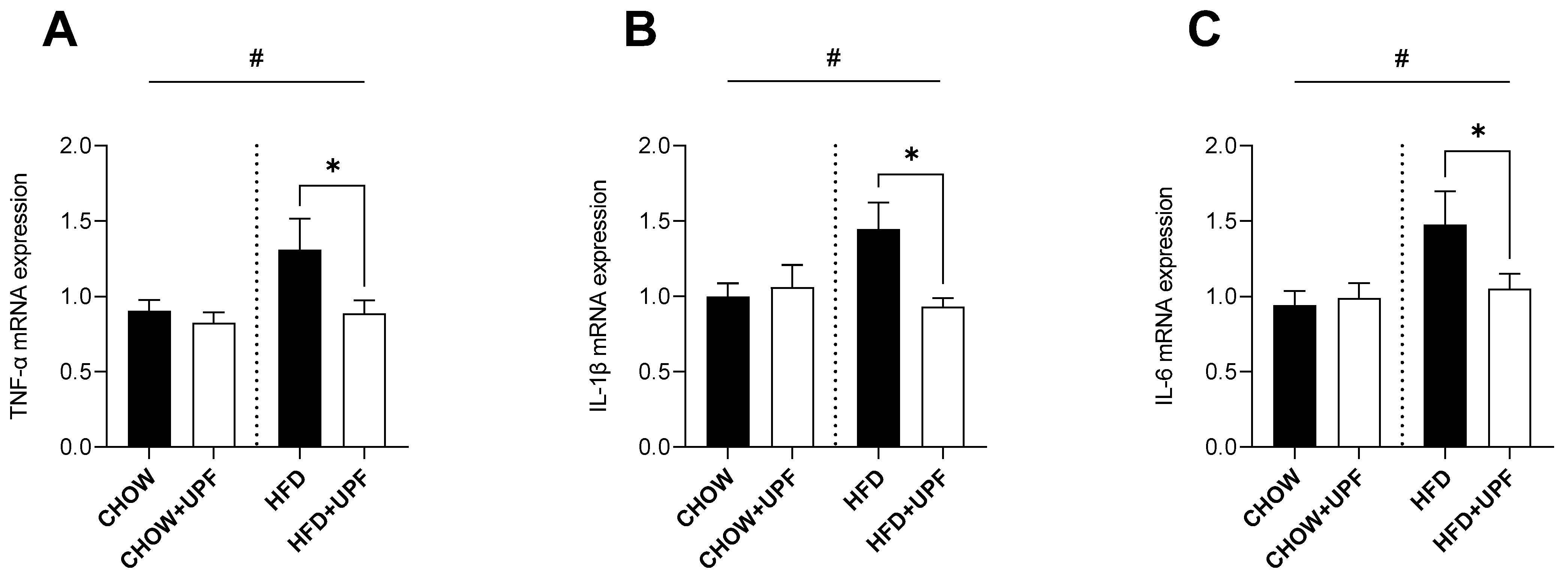
2.1. Effects of HFD and UPF on Skeletal Muscle Gene Expression

High-fat diet (HFD) consumption increased the muscular mRNA expression of TNF-α (Figure 1A), IL-1β (Figure 1B), and IL-6 (Figure 1C) compared to a standard chow diet. However, in mice consuming HFD, UPF supplementation significantly downregulated the mRNA expression of TNF-α (−32.2%; p < 0.05, Figure 1A), IL-1β (−35.6%; p < 0.05, Figure 1B), and IL-6 (−28.9%; p < 0.05, Figure 1C). No significant effects of UPF supplementation were observed in the mRNA expression of these cytokines in mice consuming a standard chow diet.

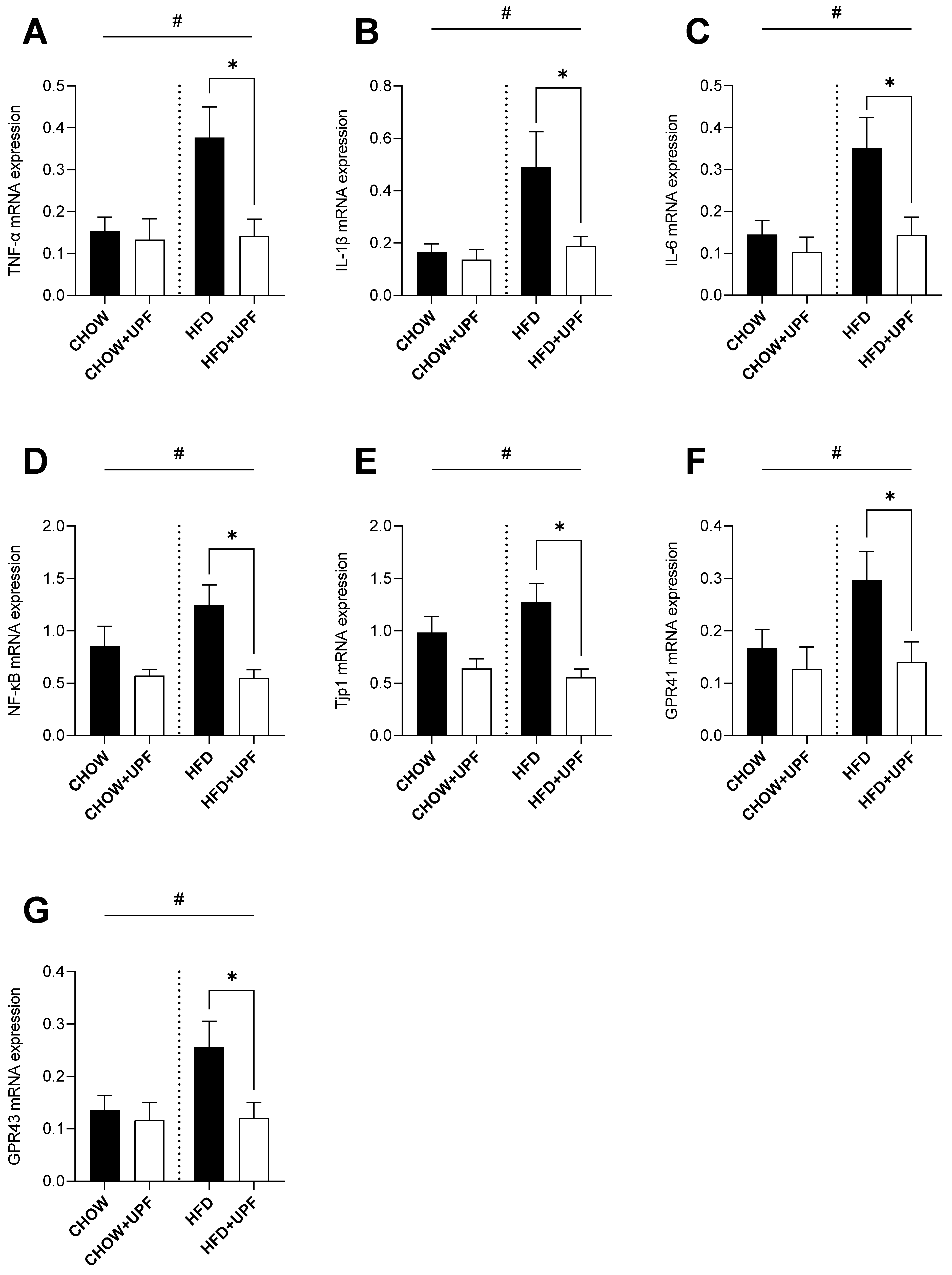
2.2. Effects of HFD and UPF on Small Intestine Gene Expression

Overall, HFD increased the intestinal mRNA expression of TNF-α (Figure 2A), IL-1β (Figure 2B), IL-6 (Figure 2C), nuclear factor kappa B (NF-κB, Figure 2D), Tjp1 (Figure 2E), and G-protein coupled receptors 41 and 43 (GPR41 and GPR43, Figure 2F,G) compared to the standard chow diet. However, in mice consuming HFD, UPF supplementation significantly downregulated the mRNA expression of TNF-α (−62.3%; p < 0.05, Figure 2A), IL-1β (−62.8%; p < 0.05, Figure 2B), IL-6 (−58.9%; p < 0.05, Figure 2C), NF-κB (−55.6%; p < 0.05, Figure 2D), Tjp1 (−56.5%; p < 0.05, Figure 2E), GPR41 (−52.8%; p < 0.05, Figure 2F), and GPR43 (−52.9%; p < 0.05, Figure 2G). No significant effects of UPF supplementation were observed for these genes in mice consuming a standard chow diet.

2.3. Effects of HFD and UPF on Hypothalamic Gene Expression

HFD consumption significantly increased the hypothalamic mRNA expression of TNF-α (Figure 3A) and IL-1β (Figure 3B) compared to a standard chow diet.
In HFD-fed mice, UPF supplementation significantly downregulated the mRNA expression of TNF-α (−58.8%; p < 0.05, Figure 3A) and IL-1β (−66.8%; p < 0.05, Figure 3B). Additionally, UPF administration significantly reduced the expression levels of IL-6 (−51.9%; p < 0.05, Figure 3C) and IFN-γ (−41.2%; p < 0.05, Figure 3D) in the hypothalamus. In contrast, UPF supplementation had no statistically significant effect on the mRNA expression of TNF-α (Figure 3A), IL-1β (Figure 3B), IL-6 (Figure 3C), or IFN-γ (Figure 3D) in chow-fed mice.

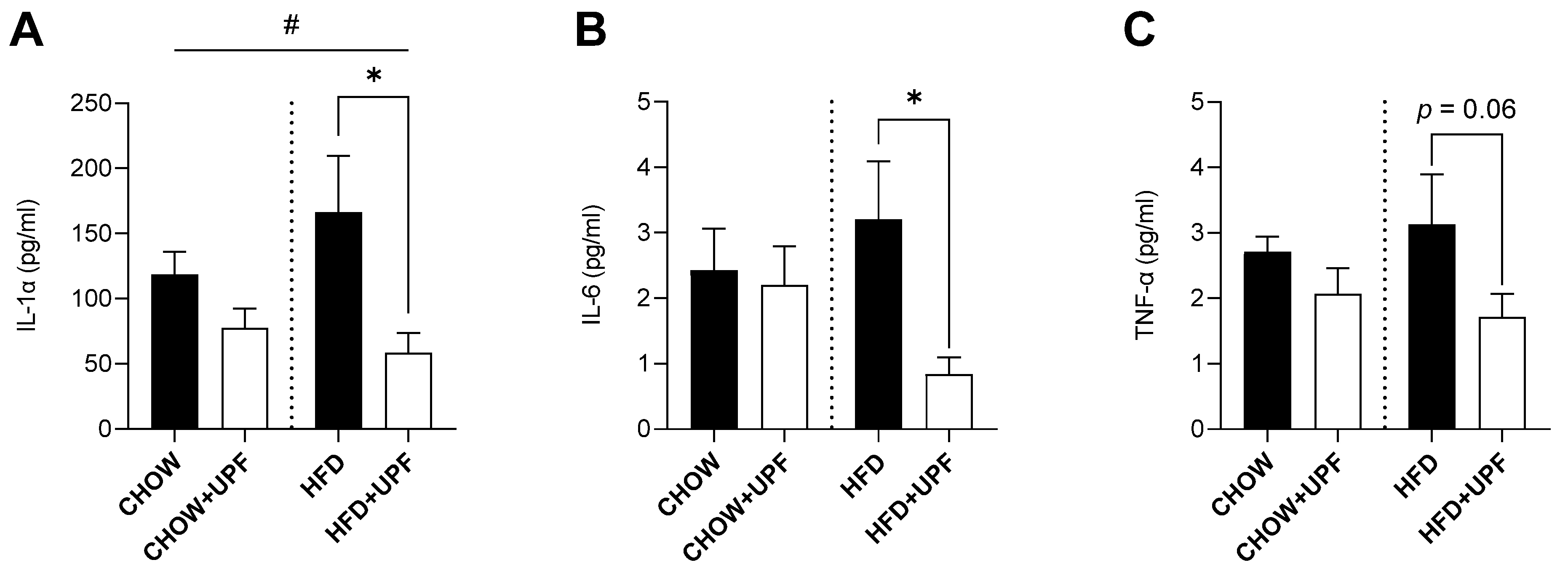
2.4. Effects of HFD and UPF on Pro-Inflammatory Plasma Cytokine Levels

HFD consumption elevated the plasma levels of IL-1α (Figure 4A) compared to a standard chow diet. In HFD-fed mice, UPF supplementation significantly decreased IL-1α by 61.7% (p < 0.05, Figure 4A), with no statistically significant effect observed in the chow group. Additionally, UPF supplementation significantly downregulated the plasma levels of IL-6 (Figure 4B) by 73.9% (p < 0.05, Figure 4B) in mice consuming HFD, while no effect was observed in the chow group.
Although UPF supplementation led to a reduction in TNF-α levels (−45.2%; Figure 4C), this effect was not statistically significant (p = 0.06). No statistically significant changes were observed in mice consuming a chow diet.

2.5. Effects of UPF on NAc Protein Abundance

A total of 5423 proteins were identified in the NAc of mice consuming HFD or a standard chow diet. In the CHOW group, 23 proteins showed significant differences in expression between UPF-treated and control groups (FDR-adjusted p-value < 0.05, Figure 5A). In the HFD group, UPF significantly regulated the expression of 36 proteins compared to the control group (FDR-adjusted p-value < 0.05, Figure 5B). Volcano plots (Figure 5) highlight these differences, showing proteins with log2 fold changes (FC) greater than ±0.57 and FDR-adjusted p-values below 0.05.

2.5.1. Expression Profiles of Differentially Expressed Proteins (DEPs) in the NAc

The differentially expressed proteins (DEPs) are detailed in Table 1 (CHOW+UPF vs. CHOW) and Table 2 (HFD+UPF vs. HFD). Among the 23 DEPs in the CHOW group (Table 1), 3 proteins were upregulated and 20 were downregulated in the NAc of UPF-treated mice. In the HFD group, 4 proteins showed upregulation and 32 downregulation in the NAc of UPF-treated mice (Table 2).

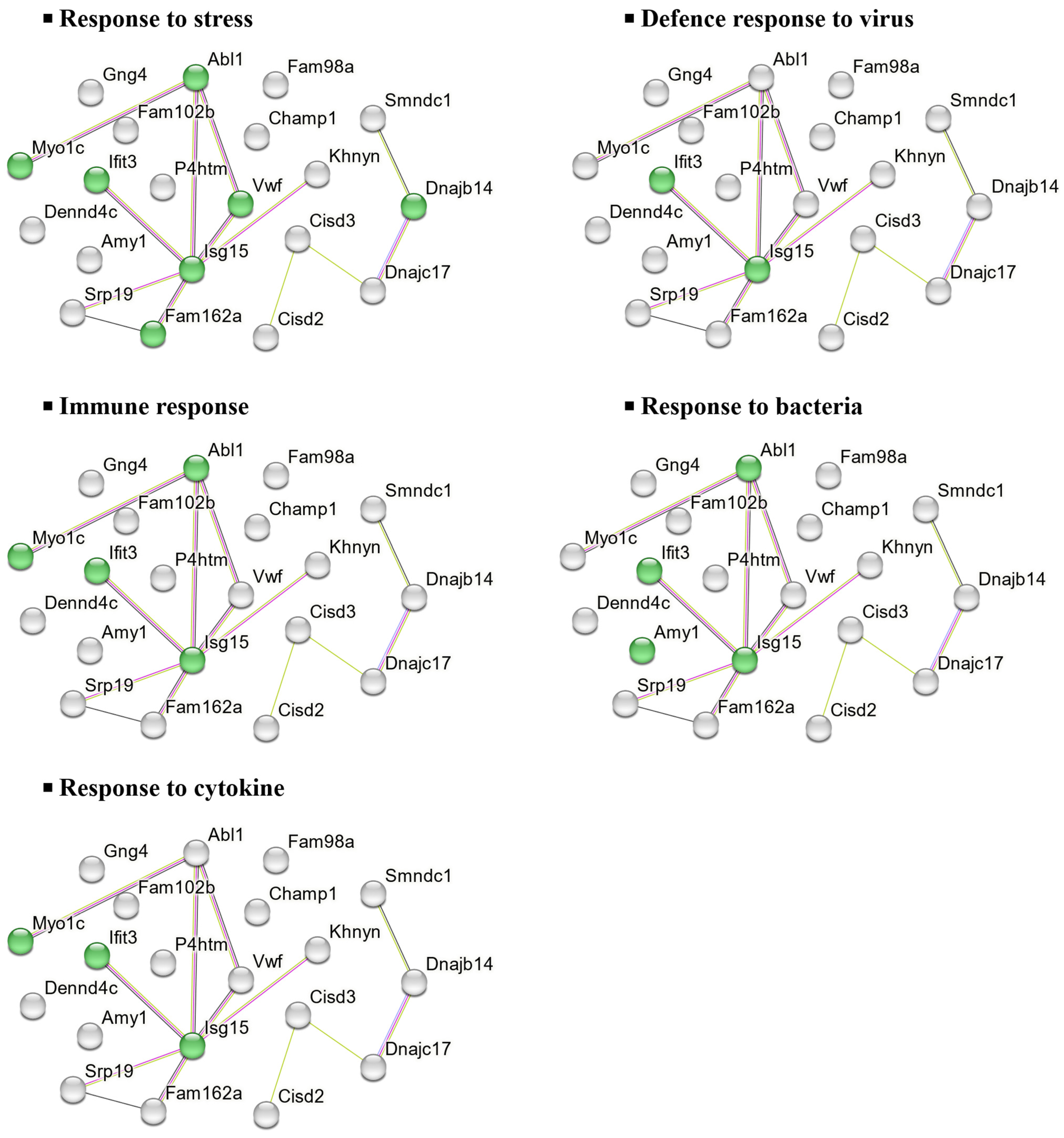
2.5.2. Molecular Functions and Biological Implications of DEPs in the NAc

To further investigate the molecular functions and biological implications of these DEPs, in silico analysis was performed using STRING, a protein–protein interaction (PPI) network tool. This analysis revealed the key biological processes associated with the 20 downregulated DEPs in the CHOW group, including stress response, viral defence, cytokine response, immune response, and bacterial defence (Figure 6).
Similarly, the 32 downregulated DEPs in the HFD group were associated with processes such as stress response, neurotransmitter regulation, neurogenesis, cell death regulation, and synaptic transmission, as shown in Figure 7.
Additionally, in the three upregulated DEPs in the NAc of mice consuming a standard chow diet, Cenpv and Nup210 were associated with cellular component biogenesis and organisation, with no PPI detected in Cops2. In contrast, among the four upregulated DEPs in mice consuming a HFD, Ppm1j and Mlip were involved in cellular metabolic processes. However, no PPI was observed in Fhl2 and Yjefn3.
Furthermore, we carried out a search for biological processes and functions of upregulated and downregulated DEPs by using DAVID. In the CHOW group, the upregulated DEPs were only enriched in the cellular component of the nuclear membrane and nuclear envelope. In contrast, the downregulated DEPs were linked to type II interferon production, stress response, bacterial defence, and iron-sulfur cluster binding (Figure 8A). In the HFD group, the upregulated DEPs were mainly enriched in the biological process of regulation of transcription by RNA polymerase II, DNA-templated transcription, and RNA biosynthetic process, as well as the molecular function of transcription corepressor activity and coregulator activity, while no cellular component was enriched. However, the downregulated DEPs were principally associated with signaling, neurotransmitter transport regulation, oxidative stress, and synapse-related functions (Figure 8B).

3. Discussion

This study identified potential mechanisms by which UPF exerts its anti-inflammatory effects in vivo. Specifically, we investigated how the compound modulated plasma pro-inflammatory cytokine protein levels and alterations in gene expression across multiple tissues, such as skeletal muscle, the small intestine, and the hypothalamus. Additionally, we conducted a proteomic analysis of the nucleus accumbens to further investigate UPF brain-specific anti-inflammatory effects and potential neuroprotective activities.

3.1. Inflammatory Markers and Gene Expression Studies

Extensive research in animal models indicates that prolonged HFD consumption is typically accompanied by overproduction of pro-inflammatory cytokines including IL-1 and its superfamily members, which results in the activation of the NF-κB pathway, leading to IL-6 and TNF-α secretion [26]. This cascade promotes apoptosis and organ damage and ultimately increases mortality [27,28,29,30,31]. In this study, and consistent with the literature, HFD mice exhibited increased pro-inflammatory cytokine expression (IL-1β, IL-6, TNF-α) in skeletal muscle (Figure 1), the small intestine (Figure 2), and the hypothalamus (Figure 3), while mice consuming standard chow showed no changes in inflammatory markers. Over the 10-week period, UPF supplementation (400 mg/kg/day) significantly reduced plasma levels of IL-1α and IL-6 (Figure 4) as well as mRNA expression of pro-inflammatory cytokines (IL-1β, IL-6, TNF-α) in the skeletal muscle, small intestine, and the hypothalamus of mice consuming HFD (Figure 1, Figure 2 and Figure 3).
Chronic inflammation is a major contributor to muscle wasting and reduced athletic function [32]. Previous investigations on UPF supplementation demonstrated that the compound enhanced exercise performance and improved muscle function in mice, regardless of the diet [18]. In addition, in a clinical trial, acute treatment with a single dose of UPF (1 g) modulated the expression of serum microRNA and affected several biological pathways including inflammation [23]. Our results are also in line with previous research conducted on other species of low-molecular-weight fucoidan [33,34]. Specifically, a 3-week supplementation with Sargassum hemiphyllum-derived fucoidan (160 mg/kg/day) reduced NF-κB activation and decreased the expression of pro-inflammatory cytokine (TNF-α, IL-6, and IL-1β) [33], and similarly, a 4-week administration of fucoidan extracted from Laminaria japonica (20, 40, and 80 mg/kg/day) protected the gastrocnemius muscles from inflammatory injury in diabetic rats [34].
In the small intestine, several investigations reported that fucoidan extracts showed anti-inflammatory effects by enhancing intestinal barrier function and improving the symptoms of ulcerative colitis while lowering pro-inflammatory cytokine levels [35,36,37,38,39]. In a mouse model of colitis using dextran sodium sulfate (DSS), a chemical colitogen with anticoagulant properties, to induce disease, 28-day oral administration of fucoidan derived from Scytosiphon lomentaria (100 and 300 mg/kg/day) inhibited the NF-κB/MAPK pathways, reduced pro-inflammatory cytokine TNF-α, and increased anti-inflammatory IL-10 production [38]. Similarly, in our experiment, suppression of NF-κB activity was accompanied by a reduced TNF-α, IL-1β, and IL-6 expression in mice consuming HFD. In mice consuming a chow diet and treated with UPF, pro-inflammatory cytokine levels were normal.
Tight junction proteins play a crucial role in maintaining the integrity of the intestinal barrier. When dysregulated, they contribute to increased intestinal permeability and chronic inflammation [40,41]. In our study, we observed an upregulation of tight junction protein 1 (Tjp1) in mice fed an HFD. However, UPF supplementation significantly suppressed this dysregulation, suggesting that fucoidan may restore tight junction protein levels to a baseline that supports optimal barrier function helping to maintain intestinal integrity [35,38,42]. Additionally, our study found that UPF treatment inhibited HFD-induced upregulation of GPR 41 and GPR43 in the small intestine. These receptors interact with short-chain fatty acids (SCFAs) such as acetate, propionate, and butyrate—microbial metabolites with a dual role in gut health [43,44]. While SCFA-mediated activation of GPR41 and GPR43 can enhance gut barrier integrity and immune regulation, excessive activation under inflammatory conditions may worsen cytokine production and immune cell recruitment [43,44,45,46,47]. This suggests that UPF-induced downregulation of these receptors could serve as a protective mechanism, preventing hyperactivation and reducing inflammation.
In the hypothalamus of HFD-fed mice, UPF significantly reduced the mRNA expression of pro-inflammatory cytokines, including TNF-α, IL-1, IL-6, and IFN-γ. The hypothalamus is a key regulator of energy balance and is particularly vulnerable to inflammation induced by HFD [48,49]. Hypothalamic inflammation has been associated with disruption of neuronal function and development of systemic metabolic dysfunction [50,51,52]. By suppressing the expression of hypothalamic pro-inflammatory cytokines, UPF may enhance neuronal resilience, further suggesting potential benefits in mitigating diet-induced metabolic disturbances [53,54]. However, further studies are needed to elucidate the mechanisms through which UPF exerts its systemic anti-inflammatory effects, particularly in plasma, skeletal muscle, the small intestine, and the hypothalamus.
To further investigate the anti-inflammatory role of UPF in the brain, we conducted a proteomic analysis of the NAc, a brain region involved in motivation, reward, and addiction [55]. Overall, our findings revealed that UPF modulated the expression of proteins related to immune response, neuronal stability, oxidative and endoplasmic reticulum stress, neurotransmitter regulation, and neuroinflammation, with effects varying based on diet. Specifically, we identified 23 differentially expressed proteins (DEPs) (Table 1) between the CHOW+UPF and CHOW groups (Figure 5A), and 36 DEPs (Table 2) between the HFD+UPF and HFD groups (Figure 5B). For clarity, we structured the discussion of the proteomic results based on the effects of UPF in relation to the specific diet consumed.

3.2. Proteomics: Effects of UPF in Mice Consuming Standard Chow

In mice fed a standard chow diet, UPF reduced the abundance of 20 proteins primarily involved in immune responses and cellular stress regulation in the NAc (Figure 6). Regarding immune modulation, two key interferon-stimulated proteins, ubiquitin-like protein interferon-stimulated gene 15 (Isg15) and interferon-induced protein with tetratricopeptide repeats 3 (Ifit3), were downregulated in the NAc of UPF-treated mice. Isg15, known for its antiviral and immunomodulatory functions, is induced in CNS cells during immune activation [56,57,58]. Its expression, associated with type I interferons (IFN-α and IFN-β) and other inflammatory mediators (IFN-γ and IL-10), possibly implies a role for UPF in brain immune regulation and anti-inflammatory processes [56,59] (Figure 8A). Likewise, Ifit3, which is activated by type I interferons, enhances antiviral defenses and modulates inflammatory pathways [60] (Figure 8A).
UPF also decreased Abl1 (Figure 6), a tyrosine-protein kinase involved in synaptic signaling and neuronal plasticity [61,62]. Overactivation of Abl1 has been linked to neurodegenerative diseases such as Parkinson’s and Alzheimer’s by facilitating α-synuclein accumulation and tau phosphorylation [62,63], suggesting a possible neuroprotective function of UPF in mitigating excitotoxic stress.
Additionally, UPF reduced von Willebrand factor (Vwf) and Myo1c (Figure 6), proteins essential for cerebrovascular stability and cytoskeletal regulation [64,65,66]. While Vwf is critical for hemostasis, it also contributes to vascular inflammation and CNS pathologies [67,68,69]. Myo1c, which stabilizes endothelial cells, prevents excessive Vwf release [66,70]. Their suppression indicates UPF’s potential role in reducing vascular inflammation and preserving neuronal integrity.
Conversely, Cops2 was the most overexpressed protein in UPF-treated, chow-fed mice (Figure 6). As a key component of the COPS complex, it regulates protein degradation, DNA repair, and cellular signaling [71]. Cops2 downregulates Oct-3/4 mRNA, facilitating neuronal differentiation and function [72], while also protecting cells from stress and supporting neuronal health [72,73,74]. Its increased expression points to UPF’s role in enhancing cellular repair and counteracting neurodegeneration associated with DNA damage, though its precise function remains to be fully elucidated.
Similarly, Nup210, the second most overexpressed protein (Figure 6), maintains nuclear–cytoplasmic transport and regulates gene expression [75]. Its elevated levels indicate improved nuclear integrity and transcriptional efficiency, reinforcing neuronal resilience and potentially alleviating neuroinflammation and aging-related impairments [76].
Overall, in mice consuming a chow diet, UPF modulated proteins associated with neuroinflammation, synaptic regulation, and cellular stress responses, highlighting its diverse neuroprotective potential. These findings suggest that UPF may contribute to maintaining brain homeostasis and could play a role in reducing the risk of inflammation-driven neurodegeneration.

3.3. Proteomics: Effects of UPF in Mice Consuming HFD

In mice consuming HFD, UPF treatment downregulated 32 proteins linked to oxidative stress responses and apoptosis, neurotransmitter regulation, neurogenesis, synaptic transmission, and behaviour. Among those proteins involved in oxidative stress responses, UPF reduced the abundance of leucine-rich repeat serine/threonine-protein kinase 2 (Lrrk2), wolframin (Wfs1), and neuroglobin (Ngb) (Figure 7).
Lrrk2, a multifunctional protein kinase, is expressed in immune cells like microglia and macrophages, where it regulates inflammatory responses, including the production of pro-inflammatory cytokines [77]. Lrrk2 kinase activates MAPK signalling and increases the production of reactive oxygen species (ROS) and pro-inflammatory cytokines, such as TNF-α, to promote neuroinflammation and apoptosis [77,78,79,80]. Other studies have also reported that mutations in Lrrk2 are linked to oxidative stress conditions and neurodegeneration in PD [77,78].
Wfs1 is a transmembrane protein primarily localised to the endoplasmic reticulum (ER) [81]. Its dysregulation induces ER stress, activating inflammation and apoptotic pathways, and has been linked to β-cell dysfunction, resulting in impaired insulin production and secretion [82]. Additionally, Wfs1 malfunctioning has been associated with neurodegenerative disorders [81,82,83,84].
Our findings showed reduction in levels of Ngb protein, an oxygen-binding globin protein, which has been associated with protection against oxidative stress and neuroprotection by scavenging ROS and supporting oxygen homeostasis [85]. Ngb expression is upregulated under several oxidative stress conditions, such as chronic inflammation, neurodegenerative diseases, and neurotoxin exposure, with its overexpression shown to reduce oxidative damage and improve mitochondrial function in neurons [86,87,88,89]. Mice fed with HFD alone had significantly lower Ngb levels compared to those receiving UPF alongside HFD. This reduction in Ngb under HFD-induced stress might be attributed to the antioxidant property of UPF, which is able to alleviate oxidative stress responses [90,91] (Figure 8B). These findings suggest that UPF might restore neuronal homeostasis and offer potential as a therapeutic strategy for HFD-induced neuroinflammatory and neurodegenerative conditions. Further research is needed to elucidate the precise mechanisms and clinical relevance.
In this study, we found an increase in protein abundance of four and a half LIM domains protein 2 (Fhl2) in the NAc of UPF-treated mice consuming HFD, which suggests significant regulatory effects of UPF on cellular stress responses and inflammatory pathways. Fhl2 plays a significant anti-inflammatory role across various tissues by modulating key pathways like NF-κB and p38 MAPK, balancing immune responses, and promoting tissue repair [92,93,94]. Fhl2 also acts as a scaffolding protein by interacting with various structural and signalling molecules, facilitating cellular processes such as gene transcription, cytoskeletal organisation, and signal transduction [95,96] (Figure 8B). These findings suggest that the ability of UPF to increase Fhl2 protein abundance may underline its neuroprotective and anti-inflammatory properties, highlighting UPF’s potential to modulate cellular and molecular pathways involved in stress adaptation, neurogenesis, and immune regulation.
Similarly, UPF also increased the levels of protein phosphatase 1J (Ppm1j), a member of the metal-dependent Ppm phosphatase family, which possesses key cellular functions such as cell metabolism, oxidative stress response, and immune signalling [97] (Figure 8B). Dysregulation of these phosphatases can lead to abnormal stress responses and metabolic disorders [97]. These findings highlight the potential significance of UPF-induced Ppm1j upregulation in neuronal resilience and stress response; however, additional research is required to uncover the specific mechanisms and functional roles of Ppm1j in brain neurons, particularly in relation to its regulatory pathways and potential implications for neuronal function and neurodegenerative diseases.

3.4. Limitations

Future studies should aim to delineate the specific signalling pathways involved in UPF-mediated cytokine modulation, with a particular focus on identifying any direct interactions between UPF and cytokine gene promoters or other elements of the transcriptional machinery. Additionally, extending these investigations to models of chronic inflammation, neurodegeneration, and autoimmune diseases could elucidate the therapeutic scope of UPF in clinical settings. Our study focused on evaluating the intrinsic biological activity of fucoidan without direct comparison to established neuroprotective or anti-inflammatory drugs. Incorporating reference drugs in specifically designed comparative studies could provide further insights into its therapeutic relevance. Moreover, while our study investigated fucoidan’s effects at a specific dose based on the existing literature, exploring multiple doses and their correlation with established data will be essential to fully characterize its anti-inflammatory and neuroprotective potential.

3.5. Conclusions

This study provides a descriptive mechanism through which 10 weeks of UPF administration reduced systemic inflammation and supported neuronal health in mice. UPF significantly downregulated pro-inflammatory cytokines (TNF-α, IL-1β, IL-6) in the skeletal muscle, small intestine, and hypothalamus, while lowering circulating IL-1α and IL-6 in HFD-fed mice (Figure 1, Figure 2, Figure 3 and Figure 4). Our findings highlight UPF’s context-dependent effects, modulating distinct protein sets based on metabolic and inflammatory conditions. In chow-fed mice, UPF affected proteins (Isg15, Ifit3, Abl1, Vwf, Myo1c) involved in immune regulation and neuronal stability [56,57,58,59,60,61,62,63,64,65,66,67,68,69,70,71,72,73,74] (Table 1). In HFD-fed mice, UPF influenced stress-response proteins (Lrrk2, Wfs1, Ngb, Slc6a3, Penk) linked to oxidative stress, neuroinflammation, and metabolic dysfunction [85,86,87,88,89,90,91] (Table 2).
Collectively, the findings of this study highlight the diet-dependent effects of UPF, demonstrating its ability to mitigate inflammation and modulate stress-related pathways in HFD-fed mice while exerting distinct immune-related effects in chow-fed mice. The observed changes in cytokine expression and proteomic profiles suggest UPF’s potential neuroprotective support for inflammation-related disorders and brain health, particularly under metabolic stress conditions.

4. Materials and Methods

4.1. Ethics Statement

This investigation was authorised by the Animal Ethics Committee of the University of Tasmania (A0027164). The animal work and procedures in this study were executed in strict adherence to the provisions delineated in the Tasmanian Animal Welfare Act (1993/63) and the Australian Code of Practice for the Care and Use of Animals for Scientific Purposes 8 edition 2013 [98].

4.2. Animals and Diet

Four-week-old male and female C57BL/6J mice (n = 32 males and n = 32 females, Animal Services, University of Tasmania) were housed under controlled conditions at a temperature of 20 ± 2 °C and maintained on a standard 12:12 h light/dark cycle. Following a one-week acclimatization period, 32 mice were provided with ad libitum access to a commercial high-fat pelleted diet (HFD) (19.4 MJ/kg, 23.5% fat, 23% protein, 5.4% crude fibre, SF16-059, Specialty Feeds, Perth, Australia), while the control group (32 mice) received a standard chow diet (12.8 MJ/kg, 6% fat, 20% protein, 3.2% crude fibre, product code 102108, Ridly Agri-Products) for ten weeks. During this time, mice were singularly housed with unrestricted access to food and drinking water ad libitum.

4.3. Fucoidan Administration and Experimental Groups

A fucoidan extract (≥85% fucoidan) derived from Undaria pinnatifida (UPF, Batch No.: UPF2022532) was provided by Marinova Pty Ltd. (Tasmania, Australia) and stored at ambient temperature (21–29 °C). The chemical composition of fucoidan used in this study is described below (Table 3 and Table 4).
The chemical composition of fucoidan used in this study is described in the Supplementary Information. Within each dietary cohort, half of the mice received UPF (400 mg/kg/day) in a form incorporated into artificially flavoured and sweetened jelly for voluntary oral administration, while the control group received the vehicle (non-UPF) jelly. Prior to commencing the study, mice were trained to consume the jelly using a previously reported method to ensure they were accustomed to its taste and texture [99]. The successful use of this method has already been described elsewhere [18]. This method yielded four experimental groups (n = 16 per group): CHOW, CHOW+UPF, HFD, and HFD+UPF. Dietary and UPF intervention continued for ten weeks.

4.4. Sample Collection

Mice were fasted overnight (12 h) and euthanised via carbon dioxide inhalation for plasma and tissue collection. Blood samples were collected in a 1.5 mL Eppendorf tube containing 7.5 µL of Heparin (1000 U/mL) by cardiac puncture and kept on ice. Plasma supernatants were collected by centrifugation (12,000 rpm/10 min, R.T.). Plasma was aliquoted and stored at −80 °C until use. Plasma levels of inflammatory cytokines IL-1α, IL-6, and TNF- α were measure with a commercial MILLIPLEX® mouse high-sensitivity T cell magnetic bead panel (MHSTCMAG-70K, Millipore, Cannon Hill, Australia) according to the manufacturer’s instructions. Mice small intestines, skeletal muscles (gastrocnemius), hypothalamus, and NAc were dissected, snap-frozen in liquid nitrogen, and stored at −80 °C for RNA extraction in gene expression studies and for protein extraction in proteomic assays.

4.5. Real-Time Quantitative PCR (RT-qPCR) Assay

Total mRNA was extracted from the mice’s small intestine, skeletal muscle, and hypothalamus tissues using an RNeasy Mini Kit (Cat# 74106, Qiagen, Chuo-ku, Japan) according to the manufacturer’s recommendations. A NanodropTM 8000 Spectrophotometer (NanoDrop Technologies Inc., Minato-ku, Japan) was used to measure the RNA concentration and purity ratios (A260/280 and A260/230), and only samples with an absorbance ratio of ~2.0 were used for complementary DNA (cDNA) synthesis. One microgram of RNA template from each sample was reverse-transcribed into cDNA using a High-Capacity cDNA Reverse Transcription Kit (Cat# 4368814, Applied Biosystem, Waltham, MA, USA). All primers were purchased from Kicqstart SYBR Green Primers (Sigma-Aldrich, St. Louis, MO, USA). The sequence of the primers used for quantification of gene expression of TNF-α, IL-1β, IL-6, IL-10, NF-κB, Tjp1, GPR41, GPR43, TLR4, β-actin, and GAPDH can be found in the Supplementary Information. RT-qPCR reactions were performed using PowerUp SYBR green master mix (Cat# A25918, Applied Biosystems, Waltham, MA, USA) in the CFX Connect TM Real-Time PCR Detection system (Bio-Rad, South Granville, Australia) following the manufacturer’s instructions. The amplification program included an initial denaturation step at 95 °C for 5 min, followed by 40 cycles of 10 s at 95 °C, 10 s at 60 °C, and 20 s at 72 °C. After each amplification, a melting curve study was conducted to validate the reliability of the results and product specificity. Target gene expression was normalised against β-actin and GAPDH housekeeping genes using the average of control group as a calibrator. Analysis was performed using the comparative 2−ΔΔCt method [100]. Samples were included in the RT-qPCR analysis only if they met the quality control criteria, which involved RNA integrity assessment (RIN) ≥ 7.0, cDNA synthesis efficiency, and proper amplification performance. Samples exhibiting poor RNA quality, inconsistent reverse transcription efficiency, irregular amplification curves, or statistical outliers (identified using Grubbs’ test) were excluded from the final analysis. As a result, the number of biological replicates (n) varied across different experiments, reflecting these quality control measures [18,50,101,102,103].

4.6. Proteomic Analysis

Frozen NAc tissues were homogenised using a tissue protein extraction reagent (TPER, Thermo Fisher Scientific, Rockford, IL, USA) to efficiently lyse cells and solubilise proteins. Protein concentrations in the extracts were quantified using a Nanodrop Spectrophotometer (NanoDrop Technologies Inc., Minato-ku, Japan) at an absorbance at A660 nm to ensure uniform protein (30 μg/90 μL) quantities for downstream analysis.
Proteins were reduced with 10 mM dithiothreitol (DTT) overnight at 4 °C, alkylated with 55 mM iodoacetamide (IAA) for 2 hrs in the dark at room temperature, and then digested with 1.2 μg trypsin/rLysC (Promega, Madison, WI, USA) after sample clean-up according to the SP3 method [104]. Peptides were then desalted using C18 ZipTips (Millipore, Billerica, MA, USA) and analysed by nano-flow HPLC and data-independent acquisition mass spectrometry using a Q-Exactive HF and Ultimate 3000 RSLCnano, as previously described [105].

4.7. Proteomics Data Processing and Statistical Analysis

Raw DIA-MS files were imported into Spectronaut software version 18 (Biognosys) and searched against the Mus musculus UniProt proteome UP00000589 comprising 44,456 entries (accessed on 6 April 2021) using the Pulsar search engine according to Biognosys (BGS) factory settings. The resulting library of 37,622 proteotypic peptides, mapped to 6298 protein groups, was then used for targeted data re-extraction and cross-run protein quantitation/normalisation using BGS factory settings, with the exception that single-hit protein IDs were excluded. The Perseus software platform (version 2.0.11) was used for imputation of missing intensity values, statistical analysis, and data visualization.
Differentially abundant proteins were identified using pair-wise t-test comparisons of all replicates (n = 10) of both CHOW and HFD (CHOW vs. CHOW+UPF and HFD vs. HFD+UPF), with a false-discovery rate (FDR) of 0.05 and an s0 value of 0.1 used to define significant proteins. Only proteins with a statistically significant change (as defined by FDR ≤ 0.05) and a moderated t-test incorporating an s0 value of 0.1 were reported as differentially expressed. Functional analyses of the differentially abundant proteins were performed using DAVID (Database for Annotation Visualization and Integrated Discovery) version 2024 (https://david.ncifcrf.gov/, accessed on 12 September 2024) [106,107] to analyse biological processes, molecular functions, and cellular components. The STRING functional protein association network (https://string-db.org/, accessed on 12 September 2024) [108] was used to develop interaction maps.
Other statistical analyses were performed using GraphPad Prism version 8.3.0 for Windows (GraphPad Software, San Diego, CA, USA, www.graphpad.com, accessed on 12 September 2024), and results were expressed as mean ± SEM. The effects of UPF on mRNA gene expression in mice muscle, intestine, and hypothalamus, and plasma inflammatory cytokine levels were analysed by a one-way ANOVA. ANOVA results were then assessed by a post hoc analysis using Fisher’s least significant difference test (LSD) as appropriate. When no overall diet effect was observed between groups [(CHOW and CHOW+UPF) vs. (HFD and HFD+UPF)], data were analysed using an unpaired t-test. Results were considered statistically significant when p < 0.05.

Supplementary Materials

The following supporting information can be downloaded at: https://www.mdpi.com/article/10.3390/md23050189/s1. Table S1: Sequence of primers used for RT-qPCR assay in mouse skeletal muscle, small intestine, and hypothalamus.

Author Contributions

Conceptualization, C.Y. and V.C.; methodology, C.Y. and V.C.; software, C.Y., M.R., R.W. and V.C.; validation, C.Y., C.D., B.C.W., M.R., R.W. and V.C.; formal analysis, C.Y., C.D., B.C.W., M.R., R.W., L.J. and V.C.; investigation, C.Y., C.D., B.C.W. and V.C.; resources, C.D., B.C.W. and V.C.; data curation, C.Y., C.D., B.C.W., M.R., R.W. and V.C.; writing original draft preparation, C.Y. and V.C.; writing review and editing, C.Y., C.D., B.C.W., M.R., R.W., L.J. and V.C.; visualization, C.Y. and V.C.; supervision, R.W., L.J. and V.C.; project administration, C.D., B.C.W. and V.C.; funding acquisition, V.C., C.D. and B.C.W. All authors have read and agreed to the published version of the manuscript.

Funding

This study was funded by The Marine Bioproducts Cooperative Research Centre (MBCRC)-Project 301.

Institutional Review Board Statement

The study was approved by the Animal Ethics Committee of the University of Tasmania (approval number A0027164 on 19 April 2023).

Data Availability Statement

The original data presented in the study are openly available in the ProteomeXchange Consortium via the PRIDE partner repository (https://www.ebi.ac.uk/pride/) [109] with the dataset identifier PXD06086 [accessible via login reviewer_pxd060861@ebi.ac.uk; password 7Mwe5q83hJPo].

Acknowledgments

We would like to thank Harmanjeet Harmanjeet and Devika Gaire for providing technical support during the study and the Animal Service Team of UTAS for assisting with animal care and monitoring.

Conflicts of Interest

Corinna Dwan and Barbara C Wimmer are employed by Marinova Pty. Ltd., and the other authors declare that there are no potential conflicts of interest. Marinova Pty. Ltd. has no role in the study design, collection, analysis, interpretation of data, the writing of this article or the decision to submit it for publication.

References

  1. Silva, J.; Alves, C.; Soledade, F.; Martins, A.; Pinteus, S.; Gaspar, H.; Alfonso, A.; Pedrosa, R. Marine-Derived Components: Can They Be a Potential Therapeutic Approach to Parkinson’s Disease? Mar. Drugs 2023, 21, 451. [Google Scholar] [CrossRef]
  2. Elbandy, M. Anti-Inflammatory Effects of Marine Bioactive Compounds and Their Potential as Functional Food Ingredients in the Prevention and Treatment of Neuroinflammatory Disorders. Molecules 2023, 28, 2. [Google Scholar] [CrossRef] [PubMed]
  3. Wang, S.K.; Li, Y.; White, W.L.; Lu, J. Extracts from New Zealand Undaria pinnatifida Containing Fucoxanthin as Potential Functional Biomaterials against Cancer in Vitro. J. Funct. Biomater. 2014, 5, 29–42. [Google Scholar] [CrossRef] [PubMed]
  4. Yang, L.; Wang, P.; Wang, H.; Li, Q.; Teng, H.; Liu, Z.; Yang, W.; Hou, L.; Zou, X. Fucoidan Derived from Undaria pinnatifida Induces Apoptosis in Human Hepatocellular Carcinoma SMMC-7721 Cells via the ROS-Mediated Mitochondrial Pathway. Mar. Drugs 2013, 11, 1961–1976. [Google Scholar] [CrossRef]
  5. Mak, W.; Hamid, N.; Liu, T.; Lu, J.; White, W.L. Fucoidan from New Zealand Undaria pinnatifida: Monthly variations and determination of antioxidant activities. Carbohydr. Polym. 2013, 95, 606–614. [Google Scholar] [CrossRef] [PubMed]
  6. Phull, A.-R.; Majid, M.; Haq, I.U.; Khan, M.R.; Kim, S.J. In vitro and in vivo evaluation of anti-arthritic, antioxidant efficacy of fucoidan from Undaria pinnatifida (Harvey) Suringar. Int. J. Biol. Macromol. 2017, 97, 468–480. [Google Scholar] [CrossRef]
  7. Zhao, Y.; Zheng, Y.; Wang, J.; Ma, S.; Yu, Y.; White, W.L.; Yang, S.; Yang, F.; Lu, J. Fucoidan Extracted from Undaria pinnatifida: Source for Nutraceuticals/Functional Foods. Mar. Drugs 2018, 16, 321. [Google Scholar] [CrossRef]
  8. Lee, J.B.; Hayashi, K.; Hashimoto, M.; Nakano, T.; Hayashi, T. Novel antiviral fucoidan from sporophyll of Undaria pinnatifida (Mekabu). Chem. Pharm. Bull. 2004, 52, 1091–1094. [Google Scholar] [CrossRef]
  9. Cooper, R.; Dragar, C.; Elliot, K.; Fitton, J.H.; Godwin, J.; Thompson, K. GFS, A Preparation of Tasmanian Undaria pinnatifida Is Associated with Healing and Inhibition of Reactivation of Herpes. BMC Complement. Altern. Med. 2002, 2, 11. [Google Scholar] [CrossRef]
  10. Herath, K.H.I.N.M.; Kim, H.J.; Kim, A.; Sook, C.E.; Lee, B.-Y.; Jee, Y. The Role of Fucoidans Isolated from the Sporophylls of Undaria pinnatifida against Particulate-Matter-Induced Allergic Airway Inflammation: Evidence of the Attenuation of Oxidative Stress and Inflammatory Responses. Molecules 2020, 25, 2869. [Google Scholar] [CrossRef]
  11. Shanmugasundaram, D.; Dwan, C.; Wimmer, B.C.; Srivastava, S. Fucoidan Ameliorates Testosterone-Induced Benign Prostatic Hyperplasia (BPH) in Rats. Res. Rep. Urol. 2024, 16, 283–297. [Google Scholar] [CrossRef]
  12. Ahmad, T.; Eapen, M.S.; Ishaq, M.; Park, A.Y.; Karpiniec, S.S.; Stringer, D.N.; Sohal, S.S.; Fitton, J.H.; Guven, N.; Caruso, V.; et al. Anti-Inflammatory Activity of Fucoidan Extracts In Vitro. Mar. Drugs 2021, 19, 702. [Google Scholar] [CrossRef] [PubMed]
  13. Duan, Y.; Zeng, L.; Zheng, C.; Song, B.; Li, F.; Kong, X.; Xu, K. Inflammatory Links Between High Fat Diets and Diseases. Front. Immunol. 2018, 9, 2649. [Google Scholar] [CrossRef]
  14. Bae, H.R.; Shin, S.-K.; Yoo, J.-H.; Kim, S.; Young, H.A.; Kwon, E.-Y. Chronic inflammation in high-fat diet-fed mice: Unveiling the early pathogenic connection between liver and adipose tissue. J. Autoimmun. 2023, 139, 103091. [Google Scholar] [CrossRef] [PubMed]
  15. Wu, Y.; Wu, T.; Wu, J.; Zhao, L.; Li, Q.; Varghese, Z.; Moorhead, J.F.; Powis, S.H.; Chen, Y.; Ruan, X.Z. Chronic inflammation exacerbates glucose metabolism disorders in C57BL/6J mice fed with high-fat diet. J. Endocrinol. 2013, 219, 195–204. [Google Scholar] [CrossRef] [PubMed]
  16. Hotamisligil, G.S. Foundations of Immunometabolism and Implications for Metabolic Health and Disease. Immunity 2017, 47, 406–420. [Google Scholar] [CrossRef]
  17. Heydemann, A. An Overview of Murine High Fat Diet as a Model for Type 2 Diabetes Mellitus. J. Diabetes Res. 2016, 2016, 2902351. [Google Scholar] [CrossRef]
  18. Yang, C.; Dwan, C.; Wimmer, B.C.; Wilson, R.; Johnson, L.; Caruso, V. Fucoidan from Undaria pinnatifida Enhances Exercise Performance and Increases the Abundance of Beneficial Gut Bacteria in Mice. Mar. Drugs 2024, 22, 485. [Google Scholar] [CrossRef]
  19. Kim, J.; Moon, I.S.; Goo, T.W.; Moon, S.S.; Seo, M. Algae Undaria pinnatifida Protects Hypothalamic Neurons against Endoplasmic Reticulum Stress through Akt/mTOR Signaling. Molecules 2015, 20, 20998–21009. [Google Scholar] [CrossRef]
  20. Azzini, E.; Peña-Corona, S.I.; Hernández-Parra, H.; Chandran, D.; Saleena, L.A.K.; Sawikr, Y.; Peluso, I.; Dhumal, S.; Kumar, M.; Leyva-Gómez, G.; et al. Neuroprotective and anti-inflammatory effects of curcumin in Alzheimer’s disease: Targeting neuroinflammation strategies. Phytother. Res. 2024, 38, 3169–3189. [Google Scholar] [CrossRef]
  21. Wei, H.; Gao, Z.; Zheng, L.; Zhang, C.; Liu, Z.; Yang, Y.; Teng, H.; Hou, L.; Yin, Y.; Zou, X. Protective Effects of Fucoidan on Aβ25-35 and d-Gal-Induced Neurotoxicity in PC12 Cells and d-Gal-Induced Cognitive Dysfunction in Mice. Mar. Drugs 2017, 15, 77. [Google Scholar] [CrossRef] [PubMed]
  22. Alghazwi, M.; Smid, S.; Karpiniec, S.; Zhang, W. Comparative study on neuroprotective activities of fucoidans from Fucus vesiculosus and Undaria pinnatifida. Int. J. Biol. Macromol. 2019, 122, 255–264. [Google Scholar] [CrossRef]
  23. Gueven, N.; Spring, K.J.; Holmes, S.; Ahuja, K.; Eri, R.; Park, A.Y.; Fitton, J.H. Micro RNA Expression after Ingestion of Fucoidan; A Clinical Study. Mar. Drugs 2020, 18, 143. [Google Scholar] [CrossRef]
  24. McFadden, B.A.; Vincenty, C.S.; Chandler, A.J.; Cintineo, H.P.; Lints, B.S.; Mastrofini, G.F.; Arent, S.M. Effects of fucoidan supplementation on inflammatory and immune response after high-intensity exercise. J. Int. Soc. Sports Nutr. 2023, 20, 2224751. [Google Scholar] [CrossRef] [PubMed]
  25. Tay, A.; Jiang, Y.; Signal, N.; O’Brien, D.; Chen, J.; Murphy, R.; Lu, J. Combining mussel with fucoidan as a supplement for joint pain and prediabetes: Study protocol for a randomized, double-blinded, placebo-controlled trial. Front. Nutr. 2022, 9, 1000510. [Google Scholar] [CrossRef]
  26. Barbier, L.; Ferhat, M.; Salamé, E.; Robin, A.; Herbelin, A.; Gombert, J.-M.; Silvain, C.; Barbarin, A. Interleukin-1 Family Cytokines: Keystones in Liver Inflammatory Diseases. Front. Immunol. 2019, 10, 2014. [Google Scholar] [CrossRef]
  27. Bleau, C.; Karelis, A.D.; St-Pierre, D.H.; Lamontagne, L. Crosstalk between intestinal microbiota, adipose tissue and skeletal muscle as an early event in systemic low-grade inflammation and the development of obesity and diabetes. Diabetes/Metab. Res. Rev. 2015, 31, 545–561. [Google Scholar] [CrossRef] [PubMed]
  28. Andersen, K.; Kesper, M.S.; Marschner, J.A.; Konrad, L.; Ryu, M.; Kumar VR, S.; Kulkarni, O.P.; Mulay, S.R.; Romoli, S.; Demleitner, J.; et al. Intestinal Dysbiosis, Barrier Dysfunction, and Bacterial Translocation Account for CKD–Related Systemic Inflammation. J. Am. Soc. Nephrol. 2017, 28, 76–83. [Google Scholar] [CrossRef]
  29. Cole, C.L.; Kleckner, I.R.; Jatoi, A.; Schwarz, E.M.; Dunne, R.F. The Role of Systemic Inflammation in Cancer-Associated Muscle Wasting and Rationale for Exercise as a Therapeutic Intervention. JCSM Clin. Rep. 2018, 3, 1–19. [Google Scholar] [CrossRef]
  30. Burfeind, K.G.; Michaelis, K.A.; Marks, D.L. The central role of hypothalamic inflammation in the acute illness response and cachexia. Semin. Cell Dev. Biol. 2016, 54, 42–52. [Google Scholar] [CrossRef]
  31. Perry, V.H. The influence of systemic inflammation on inflammation in the brain: Implications for chronic neurodegenerative disease. Brain Behav. Immun. 2004, 18, 407–413. [Google Scholar] [CrossRef] [PubMed]
  32. Tuttle, C.S.L.; Thang, L.A.N.; Maier, A.B. Markers of inflammation and their association with muscle strength and mass: A systematic review and meta-analysis. Ageing Res. Rev. 2020, 64, 101185. [Google Scholar] [CrossRef] [PubMed]
  33. Chen, M.C.; Hsu, W.L.; Hwang, P.A.; Chen, Y.L.; Chou, T.C. Combined administration of fucoidan ameliorates tumor and chemotherapy-induced skeletal muscle atrophy in bladder cancer-bearing mice. Oncotarget 2016, 7, 51608–51618. [Google Scholar] [CrossRef] [PubMed]
  34. Wang, Z.; Liu, T.; Chen, X.; You, H.; Zhang, Q.; Xue, J.; Zheng, Y.; Luo, D. Low molecular weight fucoidan ameliorates hindlimb ischemic injury in type 2 diabetic rats. J. Ethnopharmacol. 2018, 210, 434–442. [Google Scholar] [CrossRef]
  35. Iraha, A.; Chinen, H.; Hokama, A.; Yonashiro, T.; Kinjo, T.; Kishimoto, K.; Nakamoto, M.; Hirata, T.; Kinjo, N.; Higa, F.; et al. Fucoidan enhances intestinal barrier function by upregulating the expression of claudin-1. World J. Gastroenterol. 2013, 19, 5500–5507. [Google Scholar] [CrossRef]
  36. Lean, Q.Y.; Eri, R.D.; Fitton, J.H.; Patel, R.P.; Gueven, N. Fucoidan Extracts Ameliorate Acute Colitis. PLoS ONE 2015, 10, e0128453. [Google Scholar] [CrossRef]
  37. Choi, J.-I.; Raghavendran, H.R.B.; Sung, N.-Y.; Kim, J.-H.; Chun, B.S.; Ahn, D.H.; Choi, H.-S.; Kang, K.-W.; Lee, J.-W. Effect of fucoidan on aspirin-induced stomach ulceration in rats. Chem. Biol. Interact. 2010, 183, 249–254. [Google Scholar] [CrossRef]
  38. Tang, S.; Wang, L.; Ren, X.; Song, S.; Ai, C. Fucoidan Alleviates Colitis and Metabolic Disorder by Protecting the Intestinal Barrier, Suppressing the MAPK/NF-κB Pathways, and Regulating the Gut Microbiota. J. Food Biochem. 2024, 2024, 7955190. [Google Scholar] [CrossRef]
  39. Selim, H.M.; Negm, W.A.; Hawwal, M.F.; Hussein, I.A.; Elekhnawy, E.; Ulber, R.; Zayed, A. Fucoidan mitigates gastric ulcer injury through managing inflammation, oxidative stress, and NLRP3-mediated pyroptosis. Int. Immunopharmacol. 2023, 120, 110335. [Google Scholar] [CrossRef]
  40. Zong, X.; Cao, X.; Wang, H.; Xiao, X.; Wang, Y.; Lu, Z. Cathelicidin-WA Facilitated Intestinal Fatty Acid Absorption Through Enhancing PPAR-γ Dependent Barrier Function. Front. Immunol. 2019, 10, 1674. [Google Scholar] [CrossRef]
  41. Landy, J.; Ronde, E.; English, N.; Clark, S.K.; Hart, A.L.; Knight, S.C.; Ciclitira, P.J.; Al-Hassi, H.O. Tight junctions in inflammatory bowel diseases and inflammatory bowel disease associated colorectal cancer. World J. Gastroenterol. 2016, 22, 3117–3126. [Google Scholar] [CrossRef] [PubMed]
  42. Sun, T.; Liang, H.; Xue, M.; Liu, Y.; Gong, A.; Jiang, Y.; Qin, Y.; Yang, J.; Meng, D. Protective effect and mechanism of fucoidan on intestinal mucosal barrier function in NOD mice. Food Agric. Immunol. 2020, 31, 939–953. [Google Scholar] [CrossRef]
  43. Kim, M.H.; Kang, S.G.; Park, J.H.; Yanagisawa, M.; Kim, C.H. Short-Chain Fatty Acids Activate GPR41 and GPR43 on Intestinal Epithelial Cells to Promote Inflammatory Responses in Mice. Gastroenterology 2013, 145, 396–406.e310. [Google Scholar] [CrossRef] [PubMed]
  44. Ang, Z.; Ding, J.L. GPR41 and GPR43 in Obesity and Inflammation - Protective or Causative? Front. Immunol. 2016, 7, 28. [Google Scholar] [CrossRef] [PubMed]
  45. Maslowski, K.M.; Vieira, A.T.; Ng, A.; Kranich, J.; Sierro, F.; Di, Y.; Schilter, H.C.; Rolph, M.S.; Mackay, F.; Artis, D.; et al. Regulation of inflammatory responses by gut microbiota and chemoattractant receptor GPR43. Nature 2009, 461, 1282–1286. [Google Scholar] [CrossRef]
  46. Chang, G.; Zhang, H.; Wang, Y.; Ma, N.; Chandra, R.A.; Ye, G.; Zhuang, S.; Zhu, W.; Shen, X. Microbial community shifts elicit inflammation in the caecal mucosa via the GPR41/43 signalling pathway during subacute ruminal acidosis. BMC Vet. Res. 2019, 15, 298. [Google Scholar] [CrossRef]
  47. Wang, J.J.; Zhang, Q.M.; Ni, W.W.; Zhang, X.; Li, Y.; Li, A.L.; Du, P.; Li, C.; Yu, S.S. Modulatory effect of Lactobacillus acidophilus KLDS 1.0738 on intestinal short-chain fatty acids metabolism and GPR41/43 expression in β-lactoglobulin-sensitized mice. Microbiol. Immunol. 2019, 63, 303–315. [Google Scholar] [CrossRef]
  48. Lee, C.H.; Suk, K.; Yu, R.; Kim, M.S. Cellular Contributors to Hypothalamic Inflammation in Obesity. Mol. Cells 2020, 43, 431–437. [Google Scholar] [CrossRef]
  49. Cavaliere, G.; Viggiano, E.; Trinchese, G.; De Filippo, C.; Messina, A.; Monda, V.; Valenzano, A.; Cincione, R.I.; Zammit, C.; Cimmino, F.; et al. Long Feeding High-Fat Diet Induces Hypothalamic Oxidative Stress and Inflammation, and Prolonged Hypothalamic AMPK Activation in Rat Animal Model. Front. Physiol. 2018, 9, 818. [Google Scholar] [CrossRef]
  50. Ishaq, M.; Tran, D.; Yang, C.; Ng, M.J.; Kackanattil, A.; Tata, K.; Deans, B.J.; Bleasel, M.; Vicenzi, S.; Randall, C.; et al. The Anti-Obesity Compound Asperuloside Reduces Inflammation in the Liver and Hypothalamus of High-Fat-Fed Mice. Endocrines 2022, 3, 641–653. [Google Scholar] [CrossRef]
  51. Jais, A.; Brüning, J.C. Hypothalamic inflammation in obesity and metabolic disease. J. Clin. Investig. 2017, 127, 24–32. [Google Scholar] [CrossRef]
  52. Le Thuc, O.; Stobbe, K.; Cansell, C.; Nahon, J.-L.; Blondeau, N.; Rovère, C. Hypothalamic Inflammation and Energy Balance Disruptions: Spotlight on Chemokines. Front. Endocrinol. 2017, 8, 197. [Google Scholar] [CrossRef] [PubMed]
  53. Wang, L.; Zhang, K.; Ding, X.; Wang, Y.; Bai, H.; Yang, Q.; Ben, J.; Zhang, H.; Li, X.; Chen, Q.; et al. Fucoidan antagonizes diet-induced obesity and inflammation in mice. J. Biomed. Res. 2020, 35, 197–205. [Google Scholar] [CrossRef] [PubMed]
  54. Huang, J.; Huang, J.; Li, Y.; Lv, H.; Yin, T.; Fan, S.; Zhang, C.; Li, H. Fucoidan Protects Against High-Fat Diet-Induced Obesity and Modulates Gut Microbiota in Institute of Cancer Research Mice. J. Med. Food. 2021, 24, 1058–1067. [Google Scholar] [CrossRef] [PubMed]
  55. Ambroggi, F.; Ghazizadeh, A.; Nicola, S.M.; Fields, H.L. Roles of Nucleus Accumbens Core and Shell in Incentive-Cue Responding and Behavioral Inhibition. J. Neurosci. 2011, 31, 6820–6830. [Google Scholar] [CrossRef]
  56. Perng, Y.-C.; Lenschow, D.J. ISG15 in antiviral immunity and beyond. Nat. Rev. Microbiol. 2018, 16, 423–439. [Google Scholar] [CrossRef]
  57. Przanowski, P.; Loska, S.; Cysewski, D.; Dabrowski, M.; Kaminska, B. ISG’ylation increases stability of numerous proteins including Stat1, which prevents premature termination of immune response in LPS-stimulated microglia. Neurochem. Int. 2018, 112, 227–233. [Google Scholar] [CrossRef]
  58. Hwang, M.; Bergmann, C.C. Alpha/Beta Interferon (IFN-α/β) Signaling in Astrocytes Mediates Protection against Viral Encephalomyelitis and Regulates IFN-γ-Dependent Responses. J. Virol. 2018, 92, e01901-17. [Google Scholar] [CrossRef]
  59. Swaim, C.D.; Scott, A.F.; Canadeo, L.A.; Huibregtse, J.M. Extracellular ISG15 Signals Cytokine Secretion through the LFA-1 Integrin Receptor. Mol. Cell. 2017, 68, 581–590.e585. [Google Scholar] [CrossRef]
  60. Mears, H.V.; Sweeney, T.R. Better together: The role of IFIT protein–protein interactions in the antiviral response. J. Gen. Virol. 2018, 99, 1463–1477. [Google Scholar] [CrossRef]
  61. Gutiérrez, D.A.; Chandía-Cristi, A.; Yáñez, M.J.; Zanlungo, S.; Álvarez, A.R. c-Abl kinase at the crossroads of healthy synaptic remodeling and synaptic dysfunction in neurodegenerative diseases. Neural Regen. Res. 2023, 18, 237–243. [Google Scholar] [CrossRef] [PubMed]
  62. Hebron, M.L.; Lonskaya, I.; Olopade, P.; Selby, S.T.; Pagan, F.; Moussa, C.E. Tyrosine Kinase Inhibition Regulates Early Systemic Immune Changes and Modulates the Neuroimmune Response in α-Synucleinopathy. J. Clin. Cell Immunol. 2014, 5, 259. [Google Scholar] [CrossRef]
  63. Brahmachari, S.; Ge, P.; Lee, S.H.; Kim, D.; Karuppagounder, S.S.; Kumar, M.; Mao, X.; Shin, J.H.; Lee, Y.; Pletnikova, O.; et al. Activation of tyrosine kinase c-Abl contributes to α-synuclein–induced neurodegeneration. J. Clin. Investig. 2016, 126, 2970–2988. [Google Scholar] [CrossRef]
  64. Petri, B.; Broermann, A.; Li, H.; Khandoga, A.G.; Zarbock, A.; Krombach, F.; Goerge, T.; Schneider, S.W.; Jones, C.; Nieswandt, B.; et al. von Willebrand factor promotes leukocyte extravasation. Blood 2010, 116, 4712–4719. [Google Scholar] [CrossRef] [PubMed]
  65. Gragnano, F.; Sperlongano, S.; Golia, E.; Natale, F.; Bianchi, R.; Crisci, M.; Fimiani, F.; Pariggiano, I.; Diana, V.; Carbone, A.; et al. The Role of von Willebrand Factor in Vascular Inflammation: From Pathogenesis to Targeted Therapy. Mediat. Inflamm. 2017, 2017, 5620314. [Google Scholar] [CrossRef] [PubMed]
  66. El-Mansi, S.; Mitchell, T.P.; Mobayen, G.; McKinnon, T.A.J.; Miklavc, P.; Frick, M.; Nightingale, T.D. Myosin-1C augments endothelial secretion of von Willebrand factor by linking contractile actomyosin machinery to the plasma membrane. Blood. Adv. 2024, 8, 4714–4726. [Google Scholar] [CrossRef]
  67. Suidan, G.L.; Brill, A.; De Meyer, S.F.; Voorhees, J.R.; Cifuni, S.M.; Cabral, J.E.; Wagner, D.D. Endothelial Von Willebrand factor promotes blood-brain barrier flexibility and provides protection from hypoxia and seizures in mice. Arter. Thromb. Vasc. Biol. 2013, 33, 2112–2120. [Google Scholar] [CrossRef]
  68. Bongers, T.N.; de Maat, M.P.M.; van Goor, M.-L.P.J.; Bhagwanbali, V.; van Vliet, H.H.D.M.; Gómez García, E.B.; Dippel, D.W.J.; Leebeek, F.W.G. High von Willebrand Factor Levels Increase the Risk of First Ischemic Stroke. Stroke 2006, 37, 2672–2677. [Google Scholar] [CrossRef]
  69. Zhu, X.; Cao, Y.; Wei, L.; Cai, P.; Xu, H.; Luo, H.; Bai, X.; Lu, L.; Liu, J.-R.; Fan, W.; et al. von Willebrand factor contributes to poor outcome in a mouse model of intracerebral haemorrhage. Sci. Rep. 2016, 6, 35901. [Google Scholar] [CrossRef]
  70. Maravillas-Montero, J.L.; Gillespie, P.G.; Patiño-López, G.; Shaw, S.; Santos-Argumedo, L. Myosin 1c Participates in B Cell Cytoskeleton Rearrangements, Is Recruited to the Immunologic Synapse, and Contributes to Antigen Presentation. J. Immunol. 2011, 187, 3053–3063. [Google Scholar] [CrossRef]
  71. Liu, J.; Han, D.; Xuan, J.; Xie, J.; Wang, W.; Zhou, Q.; Chen, K. COP9 signalosome complex is a prognostic biomarker and corresponds with immune infiltration in hepatocellular carcinoma. Aging 2024, 16, 5264–5287. [Google Scholar] [CrossRef] [PubMed]
  72. Akiyama, H.; Sugiyama, A.; Uzawa, K.; Fujisawa, N.; Tashiro, Y.; Tashiro, F. Implication of Trip15/CSN2 in early stage of neuronal differentiation of P19 embryonal carcinoma cells. Dev. Brain Res. 2003, 140, 45–56. [Google Scholar] [CrossRef]
  73. Zhang, W.; Ni, P.; Mou, C.; Zhang, Y.; Guo, H.; Zhao, T.; Loh, Y.-H.; Chen, L. Cops2 promotes pluripotency maintenance by Stabilizing Nanog Protein and Repressing Transcription. Sci. Rep. 2016, 6, 26804. [Google Scholar] [CrossRef] [PubMed]
  74. Lykke-Andersen, K.; Schaefer, L.; Menon, S.; Deng, X.W.; Miller, J.B.; Wei, N. Disruption of the COP9 signalosome Csn2 subunit in mice causes deficient cell proliferation, accumulation of p53 and cyclin E, and early embryonic death. Mol. Cell. Biol. 2003, 23, 6790–6797. [Google Scholar] [CrossRef] [PubMed]
  75. D’Angelo, M.A.; Gomez-Cavazos, J.S.; Mei, A.; Lackner, D.H.; Hetzer, M.W. A change in nuclear pore complex composition regulates cell differentiation. Dev. Cell. 2012, 22, 446–458. [Google Scholar] [CrossRef]
  76. Ding, B.; Sepehrimanesh, M. Nucleocytoplasmic Transport: Regulatory Mechanisms and the Implications in Neurodegeneration. Int. J. Mol. Sci. 2021, 22, 4165. [Google Scholar] [CrossRef]
  77. Moehle, M.S.; Webber, P.J.; Tse, T.; Sukar, N.; Standaert, D.G.; DeSilva, T.M.; Cowell, R.M.; West, A.B. LRRK2 Inhibition Attenuates Microglial Inflammatory Responses. J. Neurosci. 2012, 32, 1602–1611. [Google Scholar] [CrossRef]
  78. Mendivil-Perez, M.; Velez-Pardo, C.; Jimenez-Del-Rio, M. Neuroprotective Effect of the LRRK2 Kinase Inhibitor PF-06447475 in Human Nerve-Like Differentiated Cells Exposed to Oxidative Stress Stimuli: Implications for Parkinson’s Disease. Neurochem. Res. 2016, 41, 2675–2692. [Google Scholar] [CrossRef]
  79. Kim, J.; Pajarillo, E.; Rizor, A.; Son, D.S.; Lee, J.; Aschner, M.; Lee, E. LRRK2 kinase plays a critical role in manganese-induced inflammation and apoptosis in microglia. PLoS ONE 2019, 14, e0210248. [Google Scholar] [CrossRef]
  80. Wallings, R.L.; Tansey, M.G. LRRK2 regulation of immune-pathways and inflammatory disease. Biochem. Soc. Trans. 2019, 47, 1581–1595. [Google Scholar] [CrossRef]
  81. Takeda, K.; Inoue, H.; Tanizawa, Y.; Matsuzaki, Y.; Oba, J.; Watanabe, Y.; Shinoda, K.; Oka, Y. WFS1 (Wolfram syndrome 1) gene product: Predominant subcellular localization to endoplasmic reticulum in cultured cells and neuronal expression in rat brain. Hum. Mol. Genet. 2001, 10, 477–484. [Google Scholar] [CrossRef]
  82. Fonseca, S.G.; Fukuma, M.; Lipson, K.L.; Nguyen, L.X.; Allen, J.R.; Oka, Y.; Urano, F. WFS1 Is a Novel Component of the Unfolded Protein Response and Maintains Homeostasis of the Endoplasmic Reticulum in Pancreatic β-Cells*. J. Biol. Chem. 2005, 280, 39609–39615. [Google Scholar] [CrossRef]
  83. Rigoli, L.; Bramanti, P.; Di Bella, C.; De Luca, F. Genetic and clinical aspects of Wolfram syndrome 1, a severe neurodegenerative disease. Pediatr. Res. 2018, 83, 921–929. [Google Scholar] [CrossRef]
  84. Hotamisligil, G.S. Endoplasmic Reticulum Stress and the Inflammatory Basis of Metabolic Disease. Cell 2010, 140, 900–917. [Google Scholar] [CrossRef]
  85. Li, R.C.; Morris, M.W.; Lee, S.K.; Pouranfar, F.; Wang, Y.; Gozal, D. Neuroglobin protects PC12 cells against oxidative stress. Brain Res. 2008, 1190, 159–166. [Google Scholar] [CrossRef] [PubMed]
  86. Fiocchetti, M.; Fernandez, V.S.; Montalesi, E.; Marino, M. Neuroglobin: A Novel Player in the Oxidative Stress Response of Cancer Cells. Oxid. Med. Cell. Longev. 2019, 2019, 6315034. [Google Scholar] [CrossRef] [PubMed]
  87. Watanabe, S.; Takahashi, N.; Uchida, H.; Wakasugi, K. Human Neuroglobin Functions as an Oxidative Stress-responsive Sensor for Neuroprotection. J. Biol. Chem. 2012, 287, 30128–30138. [Google Scholar] [CrossRef] [PubMed]
  88. Wei, X.; Yu, Z.; Cho, K.-S.; Chen, H.; Malik, M.T.A.; Chen, X.; Lo, E.H.; Wang, X.; Chen, D.F. Neuroglobin Is an Endogenous Neuroprotectant for Retinal Ganglion Cells against Glaucomatous Damage. Am. J. Pathol. 2011, 179, 2788–2797. [Google Scholar] [CrossRef]
  89. Liu, J.; Yu, Z.; Guo, S.; Lee, S.R.; Xing, C.; Zhang, C.; Gao, Y.; Nicholls, D.G.; Lo, E.H.; Wang, X. Effects of neuroglobin overexpression on mitochondrial function and oxidative stress following hypoxia/reoxygenation in cultured neurons. J. Neurosci. Res. 2009, 87, 164–170. [Google Scholar] [CrossRef]
  90. Alrafiah, A. Thymoquinone Protects Neurons in the Cerebellum of Rats through Mitigating Oxidative Stress and Inflammation Following High-Fat Diet Supplementation. Biomolecules 2021, 11, 165. [Google Scholar] [CrossRef]
  91. Koh, H.S.A.; Lu, J.; Zhou, W. Structure characterization and antioxidant activity of fucoidan isolated from Undaria pinnatifida grown in New Zealand. Carbohydr. Polym. 2019, 212, 178–185. [Google Scholar] [CrossRef]
  92. van de Pol, V.; Vos, M.; DeRuiter, M.C.; Goumans, M.J.; de Vries, C.J.M.; Kurakula, K. LIM-only protein FHL2 attenuates inflammation in vascular smooth muscle cells through inhibition of the NFκB pathway. Vasc. Pharmacol. 2020, 125-126, 106634. [Google Scholar] [CrossRef] [PubMed]
  93. Wixler, V.; Cromme, C.; Retser, E.; Meyer, L.-H.; Smyth, N.; Mühlenberg, K.; Korb-Pap, A.; Koers-Wunrau, C.; Sotsios, Y.; Bassel-Duby, R.; et al. FHL2 regulates the resolution of tissue damage in chronic inflammatory arthritis. Ann. Rheum. Dis. 2015, 74, 2216–2223. [Google Scholar] [CrossRef] [PubMed]
  94. Alnajar, A.; Nordhoff, C.; Schied, T.; Chiquet-Ehrismann, R.; Loser, K.; Vogl, T.; Ludwig, S.; Wixler, V. The LIM-only protein FHL2 attenuates lung inflammation during bleomycin-induced fibrosis. PLoS ONE 2013, 8, e81356. [Google Scholar] [CrossRef] [PubMed]
  95. Johannessen, M.; Møller, S.; Hansen, T.; Moens, U.; Van Ghelue, M. The multifunctional roles of the four-and-a-half-LIM only protein FHL2. Cell. Mol. Life. Sci. 2006, 63, 268–284. [Google Scholar] [CrossRef]
  96. Verset, L.; Feys, L.; Trépant, A.L.; De Wever, O.; Demetter, P. FHL2: A scaffold protein of carcinogenesis, tumour-stroma interactions and treatment response. Histol. Histopathol. 2016, 31, 469–478. [Google Scholar] [CrossRef]
  97. Kamada, R.; Kudoh, F.; Ito, S.; Tani, I.; Janairo, J.I.B.; Omichinski, J.G.; Sakaguchi, K. Metal-dependent Ser/Thr protein phosphatase PPM family: Evolution, structures, diseases and inhibitors. Pharmacol. Ther. 2020, 215, 107622. [Google Scholar] [CrossRef]
  98. Health, N.; Council, M.R. Australian Code of Practice for the Care and Use of Animals for Scientific Purposes; Australian Government Publishing Service: Canberra City, Australia, 1997. [Google Scholar]
  99. Zhang, L. Method for voluntary oral administration of drugs in mice. STAR Protoc. 2021, 2, 100330. [Google Scholar] [CrossRef]
  100. Pfaffl, M.W. A new mathematical model for relative quantification in real-time RT-PCR. Nucleic Acids Res. 2001, 29, e45. [Google Scholar] [CrossRef]
  101. Caruso, V.; Chen, H.; Morris, M.J. Early hypothalamic FTO overexpression in response to maternal obesity–potential contribution to postweaning hyperphagia. PLoS ONE 2011, 6, e25261. [Google Scholar] [CrossRef]
  102. Bahari, H.; Caruso, V.; Morris, M.J. Late-Onset Exercise in Female Rat Offspring Ameliorates the Detrimental Metabolic Impact of Maternal Obesity. Endocrinology 2013, 154, 3610–3621. [Google Scholar] [CrossRef] [PubMed]
  103. Caruso, V.; Bahari, H.; Morris, M. The Beneficial Effects of Early Short-Term Exercise in the Offspring of Obese Mothers are Accompanied by Alterations in the Hypothalamic Gene Expression of Appetite Regulators and FTO (Fat Mass and Obesity Associated) Gene. J. Neuroendocrinol. 2013, 25, 742–752. [Google Scholar] [CrossRef] [PubMed]
  104. Hughes, C.S.; Moggridge, S.; Müller, T.; Sorensen, P.H.; Morin, G.B.; Krijgsveld, J. Single-pot, solid-phase-enhanced sample preparation for proteomics experiments. Nat. Protoc. 2019, 14, 68–85. [Google Scholar] [CrossRef]
  105. Chear, S.; Perry, S.; Wilson, R.; Bindoff, A.; Talbot, J.; Ware, T.L.; Grubman, A.; Vickers, J.C.; Pébay, A.; Ruddle, J.B.; et al. Lysosomal alterations and decreased electrophysiological activity in CLN3 disease patient-derived cortical neurons. Dis. Model. Mech. 2022, 15, dmm049651. [Google Scholar] [CrossRef]
  106. Huang, D.W.; Sherman, B.T.; Lempicki, R.A. Systematic and integrative analysis of large gene lists using DAVID bioinformatics resources. Nat. Protoc. 2009, 4, 44–57. [Google Scholar] [CrossRef] [PubMed]
  107. Sherman, B.T.; Hao, M.; Qiu, J.; Jiao, X.; Baseler, M.W.; Lane, H.C.; Imamichi, T.; Chang, W. DAVID: A web server for functional enrichment analysis and functional annotation of gene lists (2021 update). Nucleic Acids Res. 2022, 50, W216–W221. [Google Scholar] [CrossRef]
  108. Szklarczyk, D.; Morris, J.H.; Cook, H.; Kuhn, M.; Wyder, S.; Simonovic, M.; Santos, A.; Doncheva, N.T.; Roth, A.; Bork, P.; et al. The STRING database in 2017: Quality-controlled protein-protein association networks, made broadly accessible. Nucleic Acids Res. 2017, 45, D362–D368. [Google Scholar] [CrossRef]
  109. Perez-Riverol, Y.; Bandla, C.; Kundu, D.J.; Kamatchinathan, S.; Bai, J.; Hewapathirana, S.; John, N.S.; Prakash, A.; Walzer, M.; Wang, S.; et al. The PRIDE database at 20 years: 2025 update. Nucleic Acids Res. 2025, 53, D543–D553. [Google Scholar] [CrossRef]
Figure 1. Effects of UPF on skeletal muscle gene expression. mRNA levels of (A) TNF-α, (B) IL-1β, and (C) IL-6. n = 11–16. Results are expressed as mean ± SEM. Data were analysed by one-way ANOVA, followed by post hoc LSD tests. #, p < 0.05, overall difference between (CHOW and CHOW+UPF) vs. (HFD and HFD+UPF). *, p < 0.05, difference between CHOW vs. CHOW+UPF and HFD vs. HFD+UPF.
Marinedrugs 23 00189 g001
Figure 2. Effects of UPF on small intestine gene expression. mRNA levels of (A) TNF-α, (B) IL-1β, (C) IL-6, (D) NF-κB, (E) Tjp1, (F) GPR41, and (G) GPR43. n = 7–15. Results are expressed as mean ± SEM. Data were analysed by one-way ANOVA, followed by post hoc LSD tests. #, p < 0.05, overall difference between (CHOW and CHOW+UPF) vs. (HFD and HFD+UPF). *, p < 0.05, difference between CHOW vs. CHOW+UPF and HFD vs. HFD+UPF.
Marinedrugs 23 00189 g002
Figure 3. Effects of UPF on hypothalamic gene expression. mRNA levels of (A) TNF-α, and (B) IL-1β. n = 7–10. Results are expressed as mean ± SEM. Data were analysed by one-way ANOVA, followed by post hoc LSD tests. #, p < 0.05, overall difference between (CHOW and CHOW+UPF) vs. (HFD and HFD+UPF). *, p < 0.05, difference between CHOW vs. CHOW+UPF and HFD vs. HFD+UPF. (C) IL-6, and (D) IFN-γ. n = 7–10. Results are expressed as mean ± SEM. Data were analysed by unpaired t-test. *, p < 0.05, difference between HFD vs. HFD+UPF.
Marinedrugs 23 00189 g003
Figure 4. Effects of UPF on plasma cytokines. (A) IL-1α. n = 6–10. Results are expressed as mean ± SEM. Data were analysed by one-way ANOVA, followed by post hoc LSD tests. #, p < 0.05, overall difference between (CHOW and CHOW+UPF) vs. (HFD and HFD+UPF). *, p < 0.05, difference between HFD vs. HFD+UPF. (B) IL-6, n = 4–6. Results are expressed as mean ± SEM. Data were analysed by unpaired t-test. *, p < 0.05, difference between HFD vs. HFD+UPF. (C) TNF-α, n = 6–8. Results are expressed as mean ± SEM. Data were analysed by unpaired t-test.
Marinedrugs 23 00189 g004
Figure 5. Volcano plot of proteomic changes in mice NAc. (A) CHOW+UPF vs. CHOW, and (B) HFD+UPF vs. HFD. Proteins that were statistically significantly different relative to the control groups for their respective diets are shown above the horizontal line in red (reduced abundance in the UPF group) or blue (increased abundance in the UPF group).
Marinedrugs 23 00189 g005
Figure 6. Main biological processes of downregulated DEPs in CHOW groups. PPIs were visualised using the STRING interaction network. Downregulated proteins are represented as a cluster. The coloured proteins in the clusters are involved in the indicated biological process. Classification of proteins was based on Gene Ontology (GO) biological processes. All the biological processes that are referred to present a false-discovery rate of less than 0.05.
Marinedrugs 23 00189 g006
Figure 7. Main biological processes of downregulated DEPs in HFD groups. PPIs were visualised using the STRING interaction network. Downregulated proteins are represented as a cluster. The coloured proteins in the clusters are involved in the indicated biological process. Classification of proteins was based on GO biological processes. All the biological processes that are referred to present a false-discovery rate of less than 0.05.
Marinedrugs 23 00189 g007
Figure 8. Functional annotation histogram of DEPs. (A) CHOW+UPF vs. CHOW, and (B) HFD+UPF vs. HFD. Biological process (BP), molecular function (MF), and cellular component (CC) mainly related to the DEPs. The histograms represent the main categories for each GO term in which the upregulated and downregulated DEPs were involved (FDR-adjusted p-value < 0.05). The x-axis shows the biological activity, and the y-axis shows the percentage and count of proteins involved in this process among the total proteins in the database.
Marinedrugs 23 00189 g008
Table 1. Proteins with significantly differential expression between the CHOW+UPF and CHOW group using Perseus software platform.
ProteinUniProt
Accession
Gene NameFDRFC
COP9 signalosome complex subunit 2A2AQE4Cops20.0301.9
Nuclear pore membrane glycoprotein 210Q9QY81Nup2100.0311.8
Centromere protein VQ9CXS4Cenpv0.0471.7
Interferon-induced protein with tetratricopeptide repeats 3Q64345Ifit30.0040.7
DnaJ homolog subfamily C member 17Q91WT4Dnajc170.0020.7
Signal recognition particle 19 kDa proteinQ9D104Srp190.0130.7
DENN domain-containing protein 4CA6H8H2Dennd4c0.0320.7
Survival of motor neuron-related-splicing factor 30Q8BGT7Smndc10.0090.7
CDGSH iron–sulfur domain-containing protein 2Q9CQB5Cisd20.0040.7
Protein FAM102BQ8BQS4Fam102b0.0030.6
CDGSH iron–sulfur domain-containing protein 3, mitochondrialB1AR13Cisd30.0440.6
Guanine nucleotide-binding protein G(I)/G(S)/G(O) subunit gamma-4P50153Gng40.0210.6
Protein KHNYNQ80U38Khnyn0.0050.6
Alpha-amylase 1P00687Amy10.0460.6
Ubiquitin-like protein ISG15Q64339Isg150.0360.5
DnaJ homolog subfamily B member 14Q149L6Dnajb140.0500.5
Chromosome alignment-maintaining phosphoprotein 1Q8K327Champ10.0250.4
Protein FAM162AQ9D6U8Fam162a0.0110.4
Tyrosine-protein kinase ABL1P00520Abl10.0060.4
Transmembrane prolyl 4-hydroxylaseQ8BG58P4htm0.0340.3
Unconventional myosin-IcQ9WTI7Myo1c0.0370.3
von Willebrand factorQ8CIZ8Vwf0.0090.2
Protein FAM98AQ3TJZ6Fam98a0.0150.2
Fold changes (FCs) shown in upregulated proteins if FC > 1 or in downregulated proteins if FC < 1. Sample size: n = 10 in both CHOW+UPF and CHOW Groups.
Table 2. Proteins with significantly differential expression between the HFD+UPF and HFD group using Perseus software platform.
ProteinUniProt
Accession
Gene NameFDRFC
Protein phosphatase 1JQ149T7Ppm1j0.0042.1
Four and a half LIM domains protein 2O70433Fhl20.0431.9
YjeF N-terminal domain-containing protein 3F6W8I0Yjefn30.0441.9
Muscular LMNA-interacting proteinV9GWW6Mlip0.0471.7
Rho GTPase-activating protein 6, isoform CRA_bG3UZI7Arhgap60.0120.7
Synaptic vesicle glycoprotein 2CQ69ZS6Sv2c0.0220.7
Sodium-dependent dopamine transporterQ61327Slc6a30.0400.7
Dehydrogenase/reductase SDR family member 13Q5SS80Dhrs130.0260.7
Gamma-synucleinQ9Z0F7Sncg0.0010.7
HephaestinQ9Z0Z4Heph0.0030.6
WolframinQ3UN10Wfs10.0080.6
ATP-sensitive inward rectifier potassium channel 10Q9JM63Kcnj100.0140.6
Lipid phosphate phosphatase-related protein type 1Q8BFZ2Lppr10.0010.6
Epidermal growth factor receptor kinase substrate 8-like protein 1Q8R5F8Eps8l10.0200.6
Lysosome-associated membrane glycoprotein 5Q9D387Lamp50.0090.6
Synaptic vesicular amine transporterQ8BRU6Slc18a20.0490.6
Inter-alpha-trypsin inhibitor heavy chain H3Q61704Itih30.0020.6
Nucleosome assembly protein 1-like 5Q9JJF0Nap1l50.0010.6
ProSAASQ9QXV0Pcsk1n0.0040.6
Synaptic vesicle membrane protein VAT-1 homolog-likeQ80TB8Vat1l0.0070.6
Leucine-rich repeat serine/threonine-protein kinase 2Q5S006Lrrk20.0360.6
Proto-oncogene tyrosine-protein kinase receptor RetP35546Ret0.0190.6
Peptidyl-prolyl cis-trans isomerase-like 4Q9CXG3Ppil40.0420.6
ERC protein 2Q6PH08Erc20.0290.6
Proenkephalin-AP22005Penk0.0290.6
Cellular retinoic acid-binding protein 1P62965Crabp10.0200.5
Protein FAM102BQ8BQS4Fam102b0.0380.5
Protein AW551984Q8BGF0AW5519840.0130.5
3-hydroxybutyrate dehydrogenase type 2Q8JZV9Bdh20.0070.5
Leukocyte elastase inhibitor BQ8VHP7Serpinb1b0.0070.5
BAI1-associated protein 3S4R1E7Baiap30.0210.5
NeuroglobinQ9ER97Ngb0.0010.4
Protein Krt78E9Q0F0Krt780.0210.4
Neurotensin/neuromedin NQ9D3P9Nts0.0060.4
Immunoglobulin superfamily member 1Q7TQA1Igsf10.0100.3
Rab GTPase-binding effector protein 2Q91WG2Rabep20.0280.1
Fold changes (FCs) shown in upregulated proteins if FC > 1 or in downregulated proteins if FC < 1. Sample size: n = 10 in both HFD+UPF and HFD groups.
Table 3. Absolute mass percentages of Undaria pinnatifida fucoidan (UPF) extract.
Fucoidan ExtractNeutral Carbohydrates
(%)
Sulfate
(%)
Fucoidan
(%)
Polyphenols
(%)
UPF202253246.128.389.3<2
Table 4. Carbohydrate breakdown (mass %) of neutral carbohydrates in Undaria pinnatifida fucoidan (UPF) extract.
Fucoidan ExtractFucose
(%)
Xylose
(%)
Galactose
(%)
Arabinose
(%)
Rhamnose
(%)
UPF202253222.50.3190.60.6
Disclaimer/Publisher’s Note: The statements, opinions and data contained in all publications are solely those of the individual author(s) and contributor(s) and not of MDPI and/or the editor(s). MDPI and/or the editor(s) disclaim responsibility for any injury to people or property resulting from any ideas, methods, instructions or products referred to in the content.

