Current Research Status of Azaspiracids
Abstract
1. Introduction
2. Toxin Distribution
3. Toxicology and Pathology
4. Toxic Mechanism
5. AZA Analogs from Different Sources
6. Synthesis In Vivo
7. Synthesis In Vitro
8. Detection of Toxins
8.1. LC-MS
8.1.1. UHPLC/HPLC-MS
8.1.2. SPE/MSPE+LC-MS
8.1.3. LC-ESI-MS
8.1.4. LC-HRMS
8.1.5. LC-MS+NMR
8.1.6. LC-MS + Others
8.2. MBA and CBA
8.3. Biosensor
8.4. ELISA
8.5. Other Immunoassays
9. Conclusions
Author Contributions
Funding
Conflicts of Interest
References
- Huss, H. Control of indigenous pathogenic bacteria in seafood. Food Control 1997, 18, 91–98. [Google Scholar] [CrossRef]
- McMahon, T. Winter toxicity of unknown aetiology in mussels. Harmful Algae News 1996, 14, 2. [Google Scholar]
- Satake, M.; Ofuji, K.; Naoki, H.; James, K.J.; Furey, A.; McMahon, T.; Silke, J.; Yasumoto, T. Azaspiracid, a new marine toxin having unique spiro ring assemblies, isolated from Irish mussels, Mytilus edulis. J. Am. Chem. Soc. 1998, 120, 9967–9968. [Google Scholar] [CrossRef]
- Satake, M.; Ofuji, K.; James, K.J.; Furey, A.; Yasumoto, T. New toxic event caused by Irish mussels. In Proceedings of the Harmful Algae, Proceedings of the VIII International Conference on Harmful Algae, Vigo, Spain, 25–29 June 1999; pp. 468–469. [Google Scholar]
- Krock, B.; Tillmann, U.; John, U.; Cembella, A.D. Characterization of azaspiracids in plankton size-fractions and isolation of an azaspiracid-producing dinoflagellate from the North Sea. Harmful Algae 2009, 8, 254–263. [Google Scholar] [CrossRef]
- Tillmann, U.; Elbrächter, M.; Krock, B.; John, U.; Cembella, A. Azadinium spinosum gen. et sp. nov (Dinophyceae) identified as a primary producer of azaspiracid toxins. Eur. J. Phycol. 2009, 44, 63–79. [Google Scholar] [CrossRef]
- Tillmann, U.; Salas, R.; Gottschling, M.; Krock, B.; O’Driscoll, D.; Elbrächter, M. Amphidoma languida sp. Nov (Dinophyceae) reveals a close relationship between Amphidoma and Azadinium. Protist 2012, 163, 701–719. [Google Scholar] [CrossRef]
- Tillmann, U.; Salas, R.; Jauffrais, T.; Hess, P.; Silke, J. AZA: The producing organisms–biology and trophic transfer. In Seafood and Freshwater Toxins; Botana, L.M., Ed.; CRC Press: Boca Raton, FL, USA, 2014; pp. 773–798. [Google Scholar]
- Furey, A.; Braña-Magdalena, A.; Lehane, M.; Moroney, C.; James, K.J.; Satake, M.; Yasumoto, T. Determination of azaspiracids in shellfish using liquid chromatography/tandem electrospray mass spectrometry. Rapid Commun. Mass Spectrom. RCM 2002, 16, 238–242. [Google Scholar] [CrossRef]
- Furey, A.; Moroney, C.; Braña-Magdalena, A.; Saez, M.J.; Lehane, M.; James, K.J. Geographical, temporal, and species variation of the polyether toxins, azaspiracids, in shellfish. Environ. Sci. Technol. 2003, 37, 3078–3084. [Google Scholar] [CrossRef]
- Magdalena, A.B.; Lehane, M.; Moroney, C.; Furey, A.; James, K.J. Food safety implications of the distribution of azaspiracids in the tissue compartments of scallops (Pecten maximus). Food Addit. Contam. 2003, 20, 154–160. [Google Scholar] [CrossRef]
- Ofuji, K.; Satake, M.; McMahon, T.; Silke, J.; James, K.J.; Naoki, H.; Oshima, Y.; Yasumoto, T. Two analogs of azaspiracid isolated from mussels, Mytilus edulis, involved in human intoxication in Ireland. Nat. Toxins 1999, 7, 99–102. [Google Scholar] [CrossRef]
- Rossi, R.; Dell’Aversano, C.; Krock, B.; Ciminiello, P.; Percopo, I.; Tillmann, U.; Soprano, V.; Zingone, A. Mediterranean Azadinium dexteroporum (Dinophyceae) produces six novel azaspiracids and azaspiracid-35: A structural study by a multi-platform mass spectrometry approach. Anal. Bioanal. Chem. 2017, 409, 1121–1134. [Google Scholar] [CrossRef]
- Sandvik, M.; Miles, C.O.; Løvberg, K.L.E.; Kryuchkov, F.; Wright, E.J.; Mudge, E.M.; Kilcoyne, J.; Samdal, I.A. In Vitro Metabolism of Azaspiracids 1-3 with a Hepatopancreatic Fraction from Blue Mussels (Mytilus edulis). J. Agric. Food Chem. 2021, 69, 11322–11335. [Google Scholar] [CrossRef]
- Klontz, K.C.; Abraham, A.; Plakas, S.M.; Dickey, R.W. Mussel-associated azaspiracid intoxication in the United States. Ann. Intern. Med. 2009, 150, 361. [Google Scholar] [CrossRef]
- Alvarez, G.; Uribe, E.; Avalos, P.; Mariño, C.; Blanco, J. First identification of azaspiracid and spirolides in Mesodesma donacium and Mulinia edulis from Northern Chile. Toxicon Off. J. Int. Soc. Toxinol. 2010, 55, 638–641. [Google Scholar] [CrossRef]
- Taleb HVale, P.; Amanhir, R.; Benhadouch, A.; Sagou, R.; Chafik, A. First detection of azaspiraicds in mussels in north west Africa. J. Shellfish Res. 2006, 25, 1067–1070. [Google Scholar]
- James, K.J.; Furey, A.; Satake, M.; Yasumoto, T. Azaspiracid poisoning (AZP): A new shellfish toxic syndrome in Europe. In Proceedings of the Ninth International Conference on Harmful Algal Bloom, Tasmania, Australia, 7–11 February 2000; pp. 250–253. [Google Scholar]
- Wang, J.; Li, R.; Liu, B.; Zhang, Q.; Wang, X.; Zhu, Y.; Zhang, Y. Occurrence and distribution of lipophilic marine algal toxins in the coastal seawater of Southeast China and the South China Sea. Mar. Pollut. Bull. 2023, 187, 114584. [Google Scholar] [CrossRef]
- Pelin, M.; Kilcoyne, J.; Nulty, C.; Crain, S.; Hess, P.; Tubaro, A.; Sosa, S. Toxic equivalency factors (TEFs) after acute oral exposure of azaspiracid 1, -2 and -3 in mice. Toxicol. Lett. 2018, 282, 136–146. [Google Scholar] [CrossRef] [PubMed]
- Furey, A.; O’Doherty, S.; O’Callaghan, K.; Lehane, M.; James, K.J. Azaspiracid poisoning (AZP) toxins in shellfish: Toxicological and health considerations. Toxicon Off. J. Int. Soc. Toxinol. 2010, 56, 173–190. [Google Scholar] [CrossRef] [PubMed]
- Pelin, M.; Kilcoyne, J.; Florio, C.; Hess, P.; Tubaro, A.; Sosa, S. Azaspiracids Increase Mitochondrial Dehydrogenases Activity in Hepatocytes: Involvement of Potassium and Chloride Ions. Mar. Drugs 2019, 17, 276. [Google Scholar] [CrossRef] [PubMed]
- Giuliani, M.E.; Accoroni, S.; Mezzelani, M.; Lugarini, F.; Bacchiocchi, S.; Siracusa, M.; Tavoloni, T.; Piersanti, A.; Totti, C.; Regoli, F.; et al. Biological Effects of the Azaspiracid-Producing Dinoflagellate Azadinium dexteroporum in Mytilus galloprovincialis from the Mediterranean Sea. Mar. Drugs 2019, 17, 595. [Google Scholar] [CrossRef]
- Aasen, J.A.; Espenes, A.; Miles, C.O.; Samdal, I.A.; Hess, P.; Aune, T. Combined oral toxicity of azaspiracid-1 and yessotoxin in female NMRI mice. Toxicon Off. J. Int. Soc. Toxinol. 2011, 57, 909–917. [Google Scholar] [CrossRef]
- Aune, T.; Espenes, A.; Aasen, J.A.; Quilliam, M.A.; Hess, P.; Larsen, S. Study of possible combined toxic effects of azaspiracid-1 and okadaic acid in mice via the oral route. Toxicon Off. J. Int. Soc. Toxinol. 2012, 60, 895–906. [Google Scholar] [CrossRef]
- EFSA, Marine biotoxins in shellfish–azaspiracid group, scientific opinion of the panel on contaminants in the food chain. EFSA J. 2008, 723, 1–52.
- Twiner, M.J.; Doucette, G.J.; Rasky, A.; Huang, X.P.; Roth, B.L.; Sanguinetti, M.C. Marine algal toxin azaspiracid is an open-state blocker of hERG potassium channels. Chem. Res. Toxicol. 2012, 25, 1975–1984. [Google Scholar] [CrossRef] [PubMed]
- Boente-Juncal, A.; Raposo-García, S.; Costas, C.; Louzao, M.C.; Vale, C.; Botana, L.M. Partial blockade of human voltage-dependent sodium channels by the marine toxins Azaspiracids. Chem. Res. Toxicol. 2020, 33, 2593–2604. [Google Scholar] [CrossRef] [PubMed]
- Boente-Juncal, A.; Raposo-García, S.; Louzao, M.C.; Vale, C.; Botana, L.M. Targeting chloride ion channels: New insights into the mechanism of action of the marine toxin azaspiracid. Chem. Res. Toxicol. 2021, 34, 865–879. [Google Scholar] [CrossRef]
- Alfonso, A.; Román, Y.; Vieytes, M.R.; Ofuji, K.; Satake, M.; Yasumoto, T.; Botana, L.M. Azaspiracid-4 inhibits Ca2+ entry by stored operated channels in human T lymphocytes. Biochem. Pharmacol. 2005, 69, 1627–1636. [Google Scholar] [CrossRef] [PubMed]
- Chevallier, O.P.; Graham, S.F.; Alonso, E.; Duffy, C.; Silke, J.; Campbell, K.; Botana, L.M.; Elliott, C.T. New insights into the causes of human illness due to consumption of azaspiracid contaminated shellfish. Sci. Rep. 2015, 5, 9818. [Google Scholar] [CrossRef]
- Ferreiro, S.F.; Vilariño, N.; Carrera, C.; Louzao, M.C.; Santamarina, G.; Cantalapiedra, A.G.; Rodríguez, L.P.; Cifuentes, J.M.; Vieira, A.C.; Nicolaou, K.C.; et al. In vivo arrhythmogenicity of the marine biotoxin azaspiracid-2 in rats. Arch. Toxicol. 2014, 88, 425–434. [Google Scholar] [CrossRef]
- Ito, E. Toxicology of Azaspiracid-1: Acute and chronic poisoning, tumourigenicity and chemical structure relationship to toxicity in a mouse bioassay. In Seafood and Freshwater Toxins: Pharmacology, Physiology and Detection; CRC Press: Boca Raton, FL, USA, 2008; pp. 775–784. [Google Scholar]
- Ito, E.; Satake, M.; Ofuji, K.; Higashi, M.; Harigaya, K.; McMahon, T.; Yasumoto, T. Chronic effects in mice caused by oral administration of sublethal doses of azaspiracid, a new marine toxin isolated from mussels. Toxicon Off. J. Int. Soc. Toxinol. 2002, 40, 193–203. [Google Scholar] [CrossRef]
- Bodero, M.; Hoogenboom, R.; Bovee, T.F.H.; Portier, L.; de Haan, L.; Peijnenburg, A.; Hendriksen, P.J.M. Whole genome mRNA transcriptomics analysis reveals different modes of action of the diarrheic shellfish poisons okadaic acid and dinophysis toxin-1 versus azaspiracid-1 in Caco-2 cells. Toxicol. Vitr. Int. J. Public Assoc. BIBRA 2018, 46, 102–112. [Google Scholar] [CrossRef] [PubMed]
- Reale, O.; Huguet, A.; Fessard, V. Novel Insights on the Toxicity of Phycotoxins on the Gut through the Targeting of Enteric Glial Cells. Mar. Drugs 2019, 17, 429. [Google Scholar] [CrossRef] [PubMed]
- Ferron, P.J.; Dumazeau, K.; Beaulieu, J.F.; Le Hégarat, L.; Fessard, V. Combined effects of lipophilic phycotoxins (okadaic acid, azapsiracid-1 and yessotoxin) on human intestinal cells models. Toxins 2016, 8, 50. [Google Scholar] [CrossRef] [PubMed]
- Ferreiro, S.F.; Vilariño, N.; Carrera, C.; Louzao, M.C.; Santamarina, G.; Cantalapiedra, A.G.; Cifuentes, J.M.; Crespo, A.; Botana, L.M. In vivo cardiomyocyte response to YTX- and AZA-1-induced damage: Autophagy versus apoptosis. Arch. Toxicol. 2017, 91, 1859–1870. [Google Scholar] [CrossRef]
- Ferreiro, S.F.; Vilariño, N.; Carrera, C.; Louzao, M.C.; Cantalapiedra, A.G.; Santamarina, G.; Cifuentes, J.M.; Vieira, A.C.; Botana, L.M. Subacute cardiovascular toxicity of the marine phycotoxin azaspiracid-1 in rats. Toxicol. Sci. Off. J. Soc. Toxicol. 2016, 151, 104–114. [Google Scholar] [CrossRef]
- Twiner, M.J.; Hess, P.; Dechraoui, M.Y.; McMahon, T.; Samons, M.S.; Satake, M.; Yasumoto, T.; Ramsdell, J.S.; Doucette, G.J. Cytotoxic and cytoskeletal effects of azaspiracid-1 on mammalian cell lines. Toxicon Off. J. Int. Soc. Toxinol. 2005, 45, 891–900. [Google Scholar] [CrossRef]
- Twiner, M.J.; Hess, P.; Doucette, G.J. Azaspiracids: Toxicology, Pharmacology, and Risk Assessment, Seafood and Freshwater Toxins; CRC Press: Boca Raton, FL, USA, 2014; pp. 823–856. [Google Scholar]
- Salas, R.; Murphy, E.; Doohan, R.; Tillmann, U.; Thomas, O.P. Production of the dinoflagellate Amphidoma languida in a large scale photobioreactor and structure elucidation of its main metabolite AZA-39. Harmful Algae 2023, 127, 102471. [Google Scholar] [CrossRef]
- Tillmann, U.; Wietkamp, S.; Gu, H.; Krock, B.; Salas, R.; Clarke, D. Multiple new strains of amphidomataceae (Dinophyceae) from the north atlantic revealed a high toxin profile variability of Azadinium spinosum and a new non-toxigenic az. cf. spinosum. Microorganisms 2021, 9, 134. [Google Scholar] [CrossRef]
- Luo, Z.; Krock, B.; Giannakourou, A.; Venetsanopoulou, A.; Pagou, K.; Tillmann, U.; Gu, H. Sympatric occurrence of two Azadinium poporum ribotypes in the Eastern Mediterranean Sea. Harmful Algae 2018, 78, 75–85. [Google Scholar] [CrossRef]
- Krock, B.; Tillmann, U.; Tebben, J.; Trefault, N.; Gu, H. Two novel azaspiracids from Azadinium poporum, and a comprehensive compilation of azaspiracids produced by Amphidomataceae, (Dinophyceae). Harmful Algae 2019, 82, 1–8. [Google Scholar] [CrossRef]
- Silva, M.; Rodríguez, I.; Barreiro, A.; Kaufmann, M.; Neto, A.I.; Hassouani, M.; Sabour, B.; Alfonso, A.; Botana, L.M.; Vasconcelos, V. Lipophilic toxins occurrence in non-traditional invertebrate vectors from North Atlantic Waters (Azores, Madeira, and Morocco): Update on geographical tendencies and new challenges for monitoring routines. Mar. Pollut. Bull. 2020, 161 Pt B, 111725. [Google Scholar] [CrossRef]
- Mudge, E.M.; Miles, C.O.; Hardstaff, W.R.; McCarron, P. Fatty acid esters of azaspiracids identified in mussels (Mytilus edulis) using liquid chromatography-high resolution mass spectrometry. Toxicon X 2020, 8, 100059. [Google Scholar] [CrossRef]
- O’Driscoll, D.; Škrabáková, Z.; James, K.J. Confirmation of extensive natural distribution of azaspiracids in the tissue compartments of mussels (Mytilus edulis). Toxicon Off. J. Int. Soc. Toxinol. 2014, 92, 123–128. [Google Scholar] [CrossRef]
- Jauffrais, T.; Marcaillou, C.; Herrenknecht, C.; Truquet, P.; Séchet, V.; Nicolau, E.; Tillmann, U.; Hess, P. Azaspiracid accumulation, detoxification and biotransformation in blue mussels (Mytilus edulis) experimentally fed Azadinium spinosum. Toxicon Off. J. Int. Soc. Toxinol. 2012, 60, 582–595. [Google Scholar] [CrossRef]
- Li, X.; Yan, M.; Gu, J.; Lam, V.T.T.; Wai, T.C.; Baker, D.M.; Thompson, P.D.; Yiu, S.K.F.; Lam, P.K.S.; Leung, P.T.Y. The effect of temperature on physiology, toxicity and toxin content of the benthic dinoflagellate Coolia malayensis from a seasonal tropical region. Water Res. 2020, 185, 116264. [Google Scholar] [CrossRef]
- Kittler, K.; Preiss-Weigert, A.; These, A. Identification strategy using combined mass spectrometric techniques for elucidation of phase I and phase II in vitro metabolites of lipophilic marine biotoxins. Anal. Chem. 2010, 82, 9329–9335. [Google Scholar] [CrossRef]
- Kilcoyne, J.; McCarron, P.; Twiner, M.J.; Nulty, C.; Crain, S.; Quilliam, M.A.; Rise, F.; Wilkins, A.L.; Miles, C.O. Epimers of azaspiracids: Isolation, structural elucidation, relative LC-MS response, and in vitro toxicity of 37-epi-azaspiracid-1. Chem. Res. Toxicol. 2014, 27, 587–600. [Google Scholar] [CrossRef]
- Triantafyllakis, M.; Tofi, M.; Montagnon, T.; Kouridaki, A.; Vassilikogiannakis, G. Singlet oxygen-mediated synthesis of bis-spiroketals found in azaspiracids. Org. Lett. 2014, 16, 3150–3153. [Google Scholar] [CrossRef] [PubMed]
- Evans, D.A.; Kvaernø, L.; Dunn, T.B.; Beauchemin, A.; Raymer, B.; Mulder, J.A.; Olhava, E.J.; Juhl, M.; Kagechika, K.; Favor, D.A. Total synthesis of (+)-azaspiracid-1. An exhibition of the intricacies of complex molecule synthesis. J. Am. Chem. Soc. 2008, 130, 16295–16309. [Google Scholar] [CrossRef] [PubMed]
- Zhang, Z.; Chen, Y.; Adu-Ampratwum, D.; Okumu, A.A.; Kenton, N.T.; Forsyth, C.J. Synthesis of the C22-C40 Domain of the Azaspiracids. Org. Lett. 2016, 18, 1824–1827. [Google Scholar] [CrossRef] [PubMed]
- Kenton, N.T.; Adu-Ampratwum, D.; Okumu, A.A.; McCarron, P.; Kilcoyne, J.; Rise, F.; Wilkins, A.L.; Miles, C.O.; Forsyth, C.J. Stereochemical Definition of the Natural Product (6R,10R,13R, 14R,16R,17R,19S,20S,21R,24S,25S,28S,30S,32R,33R,34R,36S,37S,39R)-Azaspiracid-3 by Total Synthesis and Comparative Analyses. Angew. Chem. (Int. Ed. Engl.) 2018, 57, 810–813. [Google Scholar] [CrossRef] [PubMed]
- Byrne, B.; Dunne, G.; Lyng, J.; Bolton, D.J. The development of a ‘clean sheep policy’ in compliance with the new Hygiene Regulation (EC) 853/2004 (Hygiene 2). Food Microbiol. 2007, 24, 301–304. [Google Scholar] [CrossRef] [PubMed]
- Ben-Gigirey, B.; Rodríguez-Velasco, M.L.; Otero, A.; Vieites, J.M.; Cabado, A.G. A comparative study for PSP toxins quantification by using MBA and HPLC official methods in shellfish. Toxicon Off. J. Int. Soc. Toxinol. 2012, 60, 864–873. [Google Scholar] [CrossRef] [PubMed]
- García-Altares, M.; Diogène, J.; de la Iglesia, P. The implementation of liquid chromatography tandem mass spectrometry for the official control of lipophilic toxins in seafood: Single-laboratory validation under four chromatographic conditions. J. Chromatogr. A 2013, 1275, 48–60. [Google Scholar] [CrossRef] [PubMed]
- McCarron, P.; Giddings, S.D.; Reeves, K.L.; Hess, P.; Quilliam, M.A. A mussel (Mytilus edulis) tissue certified reference material for the marine biotoxins azaspiracids. Anal. Bioanal. Chem. 2015, 407, 2985–2996. [Google Scholar] [CrossRef] [PubMed]
- Wright, E.J.; McCarron, P. A mussel tissue certified reference material for multiple phycotoxins. Part 5: Profiling by liquid chromatography-high-resolution mass spectrometry. Anal. Bioanal. Chem. 2021, 413, 2055–2069. [Google Scholar] [CrossRef]
- Ofuji, K.; Satake, M.; Oshima, Y.; McMahon, T.; James, K.J.; Yasumoto, T. A sensitive and specific determination method for azaspiracids by liquid chromatography mass spectrometry. Nat. Toxins 1999, 7, 247–250. [Google Scholar] [CrossRef]
- Draisci, R.; Palleschi, L.; Ferretti, E.; Furey, A.; James, K.J.; Satake, M.; Yasumoto, T. Development of a method for the identification of azaspiracid in shellfish by liquid chromatography-tandem mass spectrometry. J. Chromatogr. A 2000, 871, 13–21. [Google Scholar] [CrossRef]
- Lehane, M.; Braña-Magdalena, A.; Moroney, C.; Furey, A.; James, K.J. Liquid chromatography with electrospray ion trap mass spectrometry for the determination of five azaspiracids in shellfish. J. Chromatogr. A 2002, 950, 139–147. [Google Scholar] [CrossRef]
- Lehane, M.; Fidalgo Sáez, M.J.; Magdalena, A.B.; Ruppén Cañás, I.; Díaz Sierra, M.; Hamilton, B.; Furey, A.; James, K.J. Liquid chromatography--multiple tandem mass spectrometry for the determination of ten azaspiracids, including hydroxyl analogues in shellfish. J. Chromatogr. A 2004, 1024, 63–70. [Google Scholar] [CrossRef]
- Skrabáková, Z.; O’Halloran, J.; van Pelt, F.N.; James, K.J. Food contaminant analysis at ultra-high mass resolution: Application of hybrid linear ion trap-orbitrap mass spectrometry for the determination of the polyether toxins, azaspiracids, in shellfish. Rapid Commun. Mass Spectrom. RCM 2010, 24, 2966–2974. [Google Scholar] [CrossRef]
- Yao, J.; Tan, Z.; Zhou, D.; Guo, M.; Xing, L.; Yang, S. Determination of azaspiracid-1 in shellfishes by liquid chromatography with tandem mass spectrometry. Chin. J. Chromatogr. 2010, 28, 363–367. [Google Scholar] [CrossRef]
- Blay, P.; Hui, J.P.; Chang, J.; Melanson, J.E. Screening for multiple classes of marine biotoxins by liquid chromatography-high-resolution mass spectrometry. Anal. Bioanal. Chem. 2011, 400, 577–585. [Google Scholar] [CrossRef] [PubMed]
- Wu, H.; Guo, M.; Tan, Z.; Cheng, H.; Li, Z.; Zhai, Y. Liquid chromatography quadrupole linear ion trap mass spectrometry for multiclass screening and identification of lipophilic marine biotoxins in bivalve mollusks. J. Chromatogr. A 2014, 1358, 172–180. [Google Scholar] [CrossRef] [PubMed]
- Fang, L.; Yao, X.; Wang, L.; Li, J. Solid-phase extraction-based ultra-sensitive detection of four lipophilic marine biotoxins in bivalves by high-performance liquid chromatography-tandem mass spectrometry. J. Chromatogr. Sci. 2015, 53, 373–379. [Google Scholar] [CrossRef]
- Orellana, G.; Van Meulebroek, L.; Van Vooren, S.; De Rijcke, M.; Vandegehuchte, M.; Janssen, C.R.; Vanhaecke, L. Quantification and profiling of lipophilic marine toxins in microalgae by UHPLC coupled to high-resolution orbitrap mass spectrometry. Anal. Bioanal. Chem. 2015, 407, 6345–6356. [Google Scholar] [CrossRef] [PubMed]
- Samdal, I.A.; Løvberg, K.E.; Briggs, L.R.; Kilcoyne, J.; Xu, J.; Forsyth, C.J.; Miles, C.O. Development of an ELISA for the detection of Azaspiracids. J. Agric. Food Chem. 2015, 63, 7855–7861. [Google Scholar] [CrossRef]
- Leonardo, S.; Rambla-Alegre, M.; Samdal, I.A.; Miles, C.O.; Kilcoyne, J.; Diogène, J.; O’Sullivan, C.K.; Campàs, M. Immunorecognition magnetic supports for the development of an electrochemical immunoassay for azaspiracid detection in mussels. Biosens. Bioelectron. 2017, 92, 200–206. [Google Scholar] [CrossRef]
- Samdal, I.A.; Løvberg, K.E.; Kristoffersen, A.B.; Briggs, L.R.; Kilcoyne, J.; Forsyth, C.J.; Miles, C.O. A Practical ELISA for Azaspiracids in Shellfish via Development of a New Plate-Coating Antigen. J. Agric. Food Chem. 2019, 67, 2369–2376. [Google Scholar] [CrossRef]
- Xu, F.; Liu, F.; Wang, C.; Wei, Y. Reversed-phase/weak anion exchange magnetic mesoporous microspheres for removal of matrix effects in lipophilic marine biotoxins analysis by ultrahigh-performance liquid chromatography coupled to tandem mass spectrometry. Food Chem. 2019, 294, 104–111. [Google Scholar] [CrossRef]
- Onofrio, M.D.; Mallet, C.R.; Place, A.R.; Smith, J.L. A screening tool for the direct analysis of marine and freshwater phycotoxins in organic SPATT extracts from the chesapeake bay. Toxins 2020, 12, 322. [Google Scholar] [CrossRef] [PubMed]
- Han, S.; Wang, P.; Liu, Y.; Gu, J.; Lü, M.; Wang, J. Determination of three azaspiracids in edible shellfishes by QuEChERS method combined with ultra high performance liquid chromatography-tandem mass spectrometry. Chin. J. Chromatogr. 2013, 31, 939–945. [Google Scholar] [CrossRef] [PubMed]
- Domènech, A.; Cortés-Francisco, N.; Palacios, O.; Franco, J.M.; Riobó, P.; Llerena, J.J.; Vichi, S.; Caixach, J. Determination of lipophilic marine toxins in mussels. Quantification and confirmation criteria using high resolution mass spectrometry. J. Chromatogr. A 2014, 1328, 16–25. [Google Scholar] [CrossRef] [PubMed]
- Yang, L.; Singh, A.; Lankford, S.K.; Stuart, J.; Rice, D.; Wu, W.H.; Hungerford, J.M. A rapid method for the detection of diarrhetic shellfish toxins and Azaspiracid shellfish toxins in Washington State shellfish by liquid chromatography tandem mass spectrometry. J. AOAC Int. 2020, 103, 792–799. [Google Scholar] [CrossRef]
- Stobo, L.A.; Lacaze, J.P.; Scott, A.C.; Gallacher, S.; Smith, E.A.; Quilliam, M.A. Liquid chromatography with mass spectrometry--detection of lipophilic shellfish toxins. J. AOAC Int. 2005, 88, 1371–1382. [Google Scholar] [CrossRef] [PubMed]
- Krock, B.; Tillmann, U.; John, U.; Cembella, A. LC-MS-MS aboard ship: Tandem mass spectrometry in the search for phycotoxins and novel toxigenic plankton from the North Sea. Anal. Bioanal. Chem. 2008, 392, 797–803. [Google Scholar] [CrossRef]
- James, K.J.; Sierra, M.D.; Lehane, M.; Braña Magdalena, A.; Furey, A. Detection of five new hydroxyl analogues of azaspiracids in shellfish using multiple tandem mass spectrometry. Toxicon Off. J. Int. Soc. Toxinol. 2003, 41, 277–283. [Google Scholar] [CrossRef]
- Kim, J.H.; Tillmann, U.; Adams, N.G.; Krock, B.; Stutts, W.L.; Deeds, J.R.; Han, M.S.; Trainer, V.L. Identification of Azadinium species and a new azaspiracid from Azadinium poporum in Puget Sound, Washington State, USA. Harmful Algae 2017, 68, 152–167. [Google Scholar] [CrossRef]
- James, K.J.; Furey, A.; Lehane, M.; Ramstad, H.; Aune, T.; Hovgaard, P.; Morris, S.; Higman, W.; Satake, M.; Yasumoto, T. First evidence of an extensive northern European distribution of azaspiracid poisoning (AZP) toxins in shellfish. Toxicon Off. J. Int. Soc. Toxinol. 2002, 40, 909–915. [Google Scholar] [CrossRef]
- Villar-González, A.; Rodríguez-Velasco, M.L.; Ben-Gigirey, B.; Botana, L.M. Lipophilic toxin profile in Galicia (Spain): 2005 toxic episode. Toxicon Off. J. Int. Soc. Toxinol. 2007, 49, 1129–1134. [Google Scholar] [CrossRef]
- Torgersen, T.; Bremnes, N.B.; Rundberget, T.; Aune, T. Structural confirmation and occurrence of azaspiracids in Scandinavian brown crabs (Cancer pagurus). Toxicon Off. J. Int. Soc. Toxinol. 2008, 51, 93–101. [Google Scholar] [CrossRef] [PubMed]
- Amzil, Z.; Sibat, M.; Royer, F.; Savar, V. First report on azaspiracid and yessotoxin groups detection in French shellfish. Toxicon Off. J. Int. Soc. Toxinol. 2008, 52, 39–48. [Google Scholar] [CrossRef] [PubMed]
- Stobo, L.A.; Lacaze, J.P.; Scott, A.C.; Petrie, J.; Turrell, E.A. Surveillance of algal toxins in shellfish from Scottish waters. Toxicon Off. J. Int. Soc. Toxinol. 2008, 51, 635–648. [Google Scholar] [CrossRef] [PubMed]
- Liu, R.; Liang, Y.; Wu, X.; Xu, D.; Liu, Y.; Liu, L. First report on the detection of pectenotoxin groups in Chinese shellfish by LC-MS/MS. Toxicon Off. J. Int. Soc. Toxinol. 2011, 57, 1000–1007. [Google Scholar] [CrossRef]
- Wu, H.; Yao, J.; Guo, M.; Tan, Z.; Zhou, D.; Zhai, Y. Distribution of marine lipophilic toxins in shellfish products collected from the chinese market. Mar. Drugs 2015, 13, 4281–4295. [Google Scholar] [CrossRef]
- Tillmann, U.; Borel, C.M.; Barrera, F.; Lara, R.; Krock, B.; Almandoz, G.O.; Witt, M.; Trefault, N. Azadinium poporum from the Argentine Continental Shelf, Southwestern Atlantic, produces azaspiracid-2 and azaspiracid-2 phosphate. Harmful Algae 2016, 51, 40–55. [Google Scholar] [CrossRef]
- Tillmann, U.; Jaén, D.; Fernández, L.; Gottschling, M.; Witt, M.; Blanco, J.; Krock, B. Amphidoma languida (Amphidomatacea, Dinophyceae) with a novel azaspiracid toxin profile identified as the cause of molluscan contamination at the Atlantic coast of southern Spain. Harmful Algae 2017, 62, 113–126. [Google Scholar] [CrossRef]
- Schirone, M.; Berti, M.; Visciano, P.; Chiumiento, F.; Migliorati, G.; Tofalo, R.; Suzzi, G.; Di Giacinto, F.; Ferri, N. Determination of lipophilic marine biotoxins in mussels harvested from the adriatic sea by LC-MS/MS. Front. Microbiol. 2018, 9, 152. [Google Scholar] [CrossRef] [PubMed]
- Wietkamp, S.; Krock, B.; Gu, H.; Voß, D.; Klemm, K.; Tillmann, U. Occurrence and distribution of Amphidomataceae (Dinophyceae) in Danish coastal waters of the North Sea, the Limfjord and the Kattegat/Belt area. Harmful Algae 2019, 88, 101637. [Google Scholar] [CrossRef] [PubMed]
- Otero, P.; Miguéns, N.; Rodríguez, I.; Botana, L.M. LC-MS/MS analysis of the emerging toxin Pinnatoxin-G and high levels of esterified OA group toxins in galician commercial mussels. Toxins 2019, 11, 394. [Google Scholar] [CrossRef]
- Vale, P.; Bire, R.; Hess, P. Confirmation by LC-MS/MS of azaspiracids in shellfish from the Portuguese north-western coast. Toxicon Off. J. Int. Soc. Toxinol. 2008, 51, 1449–1456. [Google Scholar] [CrossRef]
- Fux, E.; Rode, D.; Bire, R.; Hess, P. Approaches to the evaluation of matrix effects in the liquid chromatography-mass spectrometry (LC-MS) analysis of three regulated lipophilic toxin groups in mussel matrix (Mytilus edulis). Food Addit. Contam. Part A Chem. Anal. Control Exp. Risk Assess. 2008, 25, 1024–1032. [Google Scholar] [CrossRef]
- McCarron, P.; Giddings, S.D.; Quilliam, M.A. A mussel tissue certified reference material for multiple phycotoxins. Part 2: Liquid chromatography-mass spectrometry, sample extraction and quantitation procedures. Anal. Bioanal. Chem. 2011, 400, 835–846. [Google Scholar] [CrossRef] [PubMed]
- Gerssen, A.; Mulder, P.P.; McElhinney, M.A.; de Boer, J. Liquid chromatography-tandem mass spectrometry method for the detection of marine lipophilic toxins under alkaline conditions. J. Chromatogr. A 2009, 1216, 1421–1430. [Google Scholar] [CrossRef] [PubMed]
- Liu, Y.; Yu, R.C.; Kong, F.Z.; Li, C.; Dai, L.; Chen, Z.F.; Zhou, M.J. Lipophilic marine toxins discovered in the Bohai Sea using high performance liquid chromatography coupled with tandem mass spectrometry. Chemosphere 2017, 183, 380–388. [Google Scholar] [CrossRef] [PubMed]
- Rúbies, A.; Muñoz, E.; Gibert, D.; Cortés-Francisco, N.; Granados, M.; Caixach, J.; Centrich, F. New method for the analysis of lipophilic marine biotoxins in fresh and canned bivalves by liquid chromatography coupled to high resolution mass spectrometry: A quick, easy, cheap, efficient, rugged, safe approach. J. Chromatogr. A 2015, 1386, 62–73. [Google Scholar] [CrossRef] [PubMed]
- Alfonso, C.; Alfonso, A.; Otero, P.; Rodríguez, P.; Vieytes, M.R.; Elliot, C.; Higgins, C.; Botana, L.M. Purification of five azaspiracids from mussel samples contaminated with DSP toxins and azaspiracids. J. Chromatogr. B Anal. Technol. Biomed. Life Sci. 2008, 865, 133–140. [Google Scholar] [CrossRef] [PubMed]
- Bosch-Orea, C.; Sanchís, J.; Farré, M.; Barceló, D. Analysis of lipophilic marine biotoxins by liquid chromatography coupled with high-resolution mass spectrometry in seawater from the Catalan Coast. Anal. Bioanal. Chem. 2017, 409, 5451–5462. [Google Scholar] [CrossRef]
- D’Amore, T.; Lo Magro, S.; Vita, V.; Di Taranto, A. Optimization and validation of a high throughput UHPLC-MS/MS method for determination of the EU regulated lipophilic marine toxins and occurrence in fresh and processed shellfish. Mar. Drugs 2022, 20, 173. [Google Scholar] [CrossRef] [PubMed]
- Moroney, C.; Lehane, M.; Braña-Magdalena, A.; Furey, A.; James, K.J. Comparison of solid-phase extraction methods for the determination of azaspiracids in shellfish by liquid chromatography-electrospray mass spectrometry. J. Chromatogr. A 2002, 963, 353–361. [Google Scholar] [CrossRef]
- Gerssen, A.; McElhinney, M.A.; Mulder, P.P.; Bire, R.; Hess, P.; de Boer, J. Solid phase extraction for removal of matrix effects in lipophilic marine toxin analysis by liquid chromatography-tandem mass spectrometry. Anal. Bioanal. Chem. 2009, 394, 1213–1226. [Google Scholar] [CrossRef] [PubMed]
- Magdalena, A.B.; Lehane, M.; Krys, S.; Fernández, M.L.; Furey, A.; James, K.J. The first identification of azaspiracids in shellfish from France and Spain. Toxicon Off. J. Int. Soc. Toxinol. 2003, 42, 105–108. [Google Scholar] [CrossRef] [PubMed]
- Orellana, G.; Vanden Bussche, J.; Van Meulebroek, L.; Vandegehuchte, M.; Janssen, C.; Vanhaecke, L. Validation of a confirmatory method for lipophilic marine toxins in shellfish using UHPLC-HR-Orbitrap MS. Anal. Bioanal. Chem. 2014, 406, 5303–5312. [Google Scholar] [CrossRef] [PubMed]
- Costa, C.Q.V.; Afonso, I.I.; Lage, S.; Costa, P.R.; Canário, A.V.M.; Da Silva, J.P. Quantitation overcoming matrix effects of lipophilic toxins in Mytilus galloprovincialis by liquid chromatography-full scan high resolution mass spectrometry analysis (LC-HR-MS). Mar. Drugs 2022, 20, 143. [Google Scholar] [CrossRef] [PubMed]
- Brombacher, S.; Edmonds, S.; Volmer, D.A. Studies on azaspiracid biotoxins. II. Mass spectral behavior and structural elucidation of azaspiracid analogs. Rapid Commun. Mass Spectrom. RCM 2002, 16, 2306–2316. [Google Scholar] [CrossRef] [PubMed]
- Díaz Sierra, M.; Furey, A.; Hamilton, B.; Lehane, M.; James, K.J. Elucidation of the fragmentation pathways of azaspiracids, using electrospray ionisation, hydrogen/deuterium exchange, and multiple-stage mass spectrometry. J. Mass Spectrom. 2003, 38, 1178–1186. [Google Scholar] [CrossRef] [PubMed]
- Chen, X.; Huang, B.; Zhao, Q.; Wang, Z.; Liu, W.; Zhang, J.; Zhou, Y.; Sun, Q.; Huang, H.; Huang, X.; et al. Shellfish contamination with lipophilic toxins and dietary exposure assessments from consumption of shellfish products in Shenzhen, China. Ecotoxicol. Environ. Saf. 2021, 221, 112446. [Google Scholar] [CrossRef] [PubMed]
- Ozawa, M.; Uchida, H.; Watanabe, R.; Matsushima, R.; Oikawa, H.; Takahashi, K.; Iwataki, M.; Suzuki, T. Complex profiles of azaspiracid analogues in two culture strains of Azadinium poporum (Amphidomataceae, Dinophyceae) isolated from Japanese coastal waters determined by LC-MS/MS. Toxicon Off. J. Int. Soc. Toxinol. 2021, 199, 145–155. [Google Scholar] [CrossRef]
- Kilcoyne, J.; Fux, E. Strategies for the elimination of matrix effects in the liquid chromatography tandem mass spectrometry analysis of the lipophilic toxins okadaic acid and azaspiracid-1 in molluscan shellfish. J. Chromatogr. A 2010, 1217, 7123–7130. [Google Scholar] [CrossRef]
- McCarron, P.; Wright, E.; Quilliam, M.A. Liquid chromatography/mass spectrometry of domoic acid and lipophilic shellfish toxins with selected reaction monitoring and optional confirmation by library searching of product ion spectra. J. AOAC Int. 2014, 97, 316–324. [Google Scholar] [CrossRef]
- Zendong, Z.; McCarron, P.; Herrenknecht, C.; Sibat, M.; Amzil, Z.; Cole, R.B.; Hess, P. High resolution mass spectrometry for quantitative analysis and untargeted screening of algal toxins in mussels and passive samplers. J. Chromatogr. A 2015, 1416, 10–21. [Google Scholar] [CrossRef]
- Dom, I.; Biré, R.; Hort, V.; Lavison-Bompard, G.; Nicolas, M.; Guérin, T. Extended targeted and non-targeted strategies for the analysis of marine toxins in mussels and oysters by (LC-HRMS). Toxins 2018, 10, 375. [Google Scholar] [CrossRef]
- Kilcoyne, J.; Nulty, C.; Jauffrais, T.; McCarron, P.; Herve, F.; Foley, B.; Rise, F.; Crain, S.; Wilkins, A.L.; Twiner, M.J.; et al. Isolation, structure elucidation, relative LC-MS response, and in vitro toxicity of azaspiracids from the dinoflagellate Azadinium spinosum. J. Mol. Evol. 2014, 77, 2465–2474. [Google Scholar] [CrossRef]
- Kilcoyne, J.; Twiner, M.J.; McCarron, P.; Crain, S.; Giddings, S.D.; Foley, B.; Rise, F.; Hess, P.; Wilkins, A.L.; Miles, C.O. Structure Elucidation, Relative LC-MS Response and In Vitro Toxicity of Azaspiracids 7-10 Isolated from Mussels (Mytilus edulis). J. Agric. Food Chem. 2015, 63, 5083–5091. [Google Scholar] [CrossRef]
- Perez, R.A.; Rehmann, N.; Crain, S.; LeBlanc, P.; Craft, C.; MacKinnon, S.; Reeves, K.; Burton, I.W.; Walter, J.A.; Hess, P.; et al. The preparation of certified calibration solutions for azaspiracid-1, -2, and -3, potent marine biotoxins found in shellfish. Anal. Bioanal. Chem. 2010, 398, 2243–2252. [Google Scholar] [CrossRef]
- Kilcoyne, J.; McCarron, P.; Twiner, M.J.; Rise, F.; Hess, P.; Wilkins, A.L.; Miles, C.O. Identification of 21,22-dehydroazaspiracids in mussels (Mytilus edulis) and in vitro toxicity of Azaspiracid-26. J. Mol. Evol. 2018, 81, 885–893. [Google Scholar] [CrossRef] [PubMed]
- Ciminiello, P.; Dell’Aversano, C.; Fattorusso, E.; Forino, M.; Tartaglione, L.; Boschetti, L.; Rubini, S.; Cangini, M.; Pigozzi, S.; Poletti, R. Complex toxin profile of Mytilus galloprovincialis from the Adriatic sea revealed by LC-MS. Toxicon Off. J. Int. Soc. Toxinol. 2010, 55, 280–288. [Google Scholar] [CrossRef] [PubMed]
- Miles, C.O.; Kilcoyne, J.; McCarron, P.; Giddings, S.D.; Waaler, T.; Rundberget, T.; Samdal, I.A.; Løvberg, K.E. Selective extraction and purification of Azaspiracids from blue mussels (Mytilus edulis) using boric acid gel. J. Agric. Food Chem. 2018, 66, 2962–2969. [Google Scholar] [CrossRef] [PubMed]
- Ciminiello, P.; Dell’Aversano, C.; Fattorusso, E.; Forino, M.; Magno, S.; Santelia, F.; Tsoukatou, M. Investigation of the toxin profile of Greek mussels Mytilus galloprovincialis by liquid chromatography-mass spectrometry. Toxicon Off. J. Int. Soc. Toxinol. 2006, 47, 174–181. [Google Scholar] [CrossRef] [PubMed]
- James, K.J.; Lehane, M.; Moroney, C.; Fernandez-Puente, P.; Satake, M.; Yasumoto, T.; Furey, A. Azaspiracid shellfish poisoning: Unusual toxin dynamics in shellfish and the increased risk of acute human intoxications. Food Addit. Contam. 2002, 19, 555–561. [Google Scholar] [CrossRef] [PubMed]
- García-Mendoza, E.; Sánchez-Bravo, Y.A.; Turner, A.; Blanco, J.; O’Neil, A.; Mancera-Flores, J.; Pérez-Brunius, P.; Rivas, D.; Almazán-Becerril, A.; Peña-Manjarrez, J.L. Lipophilic toxins in cultivated mussels (Mytilus galloprovincialis) from Baja California, Mexico. Toxicon Off. J. Int. Soc. Toxinol. 2014, 90, 111–123. [Google Scholar] [CrossRef]
- Ledreux, A.; Sérandour, A.L.; Morin, B.; Derick, S.; Lanceleur, R.; Hamlaoui, S.; Furger, C.; Biré, R.; Krys, S.; Fessard, V.; et al. Collaborative study for the detection of toxic compounds in shellfish extracts using cell-based assays. Part II: Application to shellfish extracts spiked with lipophilic marine toxins. Anal. Bioanal. Chem. 2012, 403, 1995–2007. [Google Scholar] [CrossRef]
- Flanagan, A.F.; Callanan, K.R.; Donlon, J.; Palmer, R.; Forde, A.; Kane, M. A cytotoxicity assay for the detection and differentiation of two families of shellfish toxins. Toxicon Off. J. Int. Soc. Toxinol. 2001, 39, 1021–1027. [Google Scholar] [CrossRef]
- Forsyth, C.J.; Xu, J.; Nguyen, S.T.; Samdal, I.A.; Briggs, L.R.; Rundberget, T.; Sandvik, M.; Miles, C.O. Antibodies with broad specificity to azaspiracids by use of synthetic haptens. J. Am. Chem. Soc. 2006, 128, 15114–15116. [Google Scholar] [CrossRef] [PubMed]
- Frederick, M.O.; De Lamo Marin, S.; Janda, K.D.; Nicolaou, K.C.; Dickerson, T.J. Monoclonal antibodies with orthogonal azaspiracid epitopes. Chembiochem 2009, 10, 1625–1629. [Google Scholar] [CrossRef] [PubMed]
- Tebben, J.; Zurhelle, C.; Tubaro, A.; Samdal, I.A.; Krock, B.; Kilcoyne, J.; Sosa, S.; Trainer, V.L.; Deeds, J.R.; Tillmann, U. Structure and toxicity of AZA-59, an azaspiracid shellfish poisoning toxin produced by Azadinium poporum (Dinophyceae). Harmful Algae 2023, 124, 102388. [Google Scholar] [CrossRef] [PubMed]
- Rodríguez, L.P.; Vilariño, N.; Louzao, M.C.; Dickerson, T.J.; Nicolaou, K.C.; Frederick, M.O.; Botana, L.M. Microsphere-based immunoassay for the detection of azaspiracids. Anal. Biochem. 2014, 447, 58–63. [Google Scholar] [CrossRef] [PubMed]
- Samdal, I.A.; Sandvik, M.; Vu, J.; Sukenthirarasa, M.S.; Kanesamurthy, S.; Løvberg, K.L.E.; Kilcoyne, J.; Forsyth, C.J.; Wright, E.J.; Miles, C.O. Preparation and characterization of an immunoaffinity column for the selective extraction of azaspiracids. J. Chromatogr. B Anal. Technol. Biomed. Life Sci. 2022, 1207, 123360. [Google Scholar] [CrossRef] [PubMed]
- Elgarch, A.; Vale, P.; Rifai, S.; Fassouane, A. Detection of diarrheic shellfish poisoning and azaspiracid toxins in Moroccan mussels: Comparison of the LC-MS method with the commercial immunoassay kit. Mar. Drugs 2008, 6, 587–594. [Google Scholar] [CrossRef]


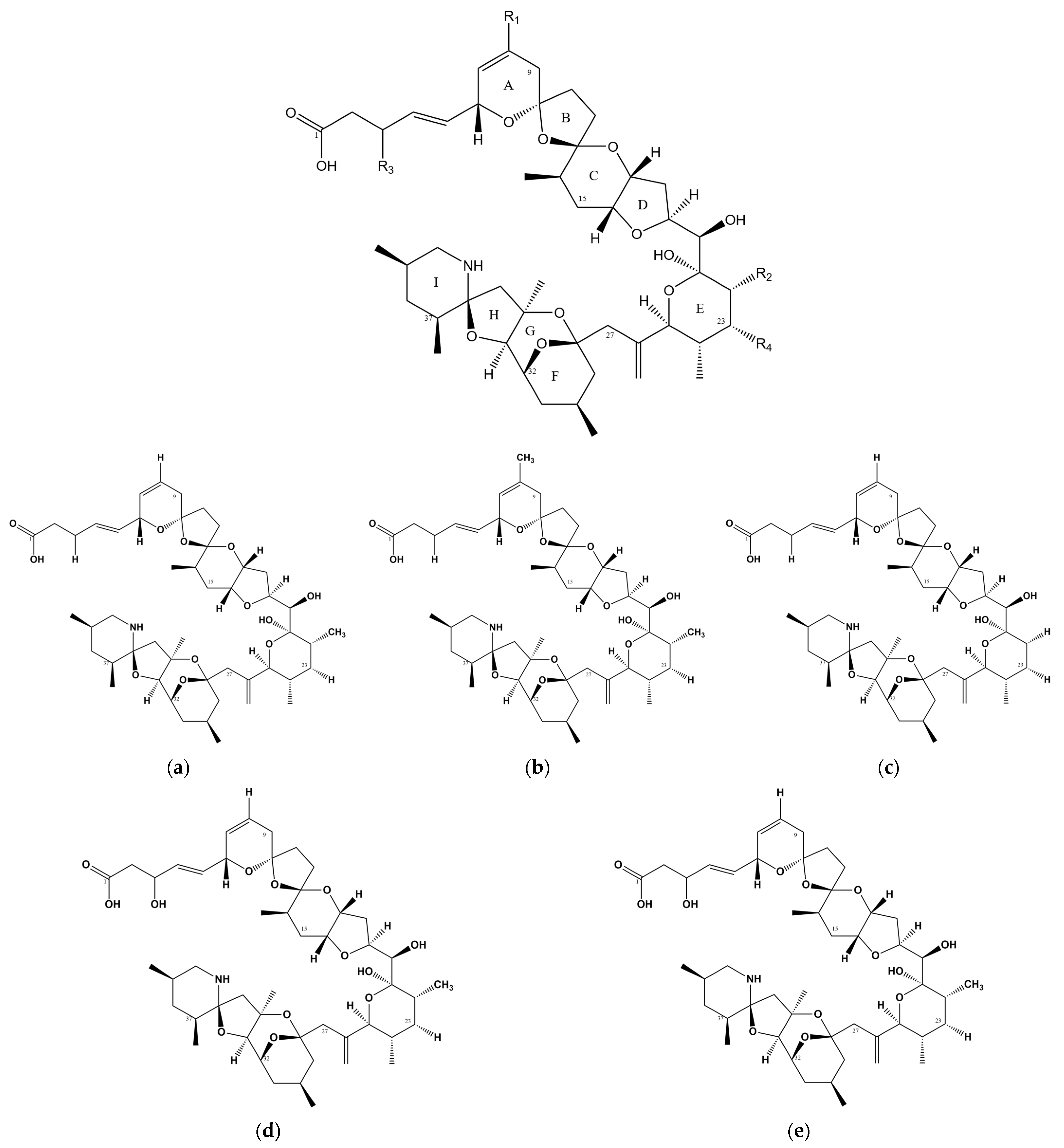
| Toxins | R1 | R2 | R3 | R4 |
|---|---|---|---|---|
| AZA-1 | H | CH3 | H | H |
| AZA-2 | CH3 | CH3 | H | H |
| AZA-3 | H | H | H | H |
| AZA-6 | CH3 | H | H | H |
| AZA-7 | H | CH3 | OH | H |
| AZA-1 | AZA-2 | AZA-3 | AZA-6 | AZA-7 |
|---|---|---|---|---|
| AZA-8 | AZA-11 | AZA-4 | AZA-9 | AZA-35 |
| AZA-14 | AZA-12 | AZA-5 | AZA-10 | AZA-54 |
| AZA-18 | AZA-16 | AZA-13 | AZA-15 | AZA-55 |
| AZA-22 | AZA-20 | AZA-17 | AZA-19 | AZA-56 |
| AZA-26 | AZA-24 | AZA-21 | AZA-23 | AZA-57 |
| AZA-30 | AZA-28 | AZA-25 | AZA-27 | AZA-58 |
| AZA-33 | AZA-32 | AZA-29 | AZA-31 | |
| AZA-34 | AZA-41 | AZA-43 | ||
| AZA-36 | AZA-42 | |||
| AZA-37 | AZA-62(AZA-11) | |||
| AZA-38 | ||||
| AZA-39 | ||||
| AZA-40 | ||||
| AZA-50 | ||||
| AZA-51 | ||||
| AZA-59 | ||||
| AZA-63(AZA-37) | ||||
| AZA-52(AZA-38) | ||||
| AZA-53(AZA-38) |
| Year | Method | LOD | LOQ | Recovery Rate | Samples |
|---|---|---|---|---|---|
| 1999 | LC-MS [62] | 50 pg | Isolated from toxic mussels collected in Ireland | ||
| 2000 | micro-LC-MS [63] | 20 ng/g | Isolated from toxic mussels collected in Ireland | ||
| 2002 | LC-MS [9] | 4 pg | 0.8 ng/mL | Isolated from toxic mussels collected in Ireland | |
| 2002 | SPE-LC-ESI-MS [64] | 5–40 pg | 0.05–1.00 μg/mL | Isolated from toxic mussels collected in Ireland | |
| 2004 | LC-ESI-MS [65] | 5 pg | 0.05–1.00 μg/mL | Isolated from toxic mussels collected in Ireland | |
| 2010 | SPE-CID-MS [66] | 0.0021 mg/g | 0.007 μg/g | From the National Research Council of Canada | |
| 2010 | SPE-LIT-MS [66] | 0.003 mg/g | 0.010 μg/g | From the National Research Council of Canada | |
| 2010 | SPE-HPLC-MS [67] | 11.00 pg/g | 75.8–82.5% | Extracted from the samples from China | |
| 2011 | LC-Orbitrap-MS [68] | 0.041–0.10 μg/L | 96–105% | From the National Research Council of Canada | |
| 2014 | LC-MS [69] | 0.12–13.6μg/kg | 0.39–45.4 μg/kg | 81.9–119.6% | Extracted from the samples from China |
| 2015 | SPE-HPLC-MS [70] | 0.013–0.085 μg/kg | 1.00 μg/kg | 99.2–102% | Extracted from the samples from China |
| 2015 | UHPLC-HR-Orbitrap-MS [71] | 0.006–0.050 ng/mL | 0.018–0.227 ng/mL | 96–114% | Isolated from toxic mussels collected in Ireland |
| 2015 | ELISA [72] | 0.45–8.6 ng/mL | 57 µg/kg | Isolated from toxic mussels collected in Ireland | |
| 2017 | MB-based direct immunoassay [73] | 63 μg/kg | 120–2875 μg/kg | From the National Research Council of Canada | |
| 2019 | ELISA [74] | 0.30–4.1 ng/mL | 37 μg/kg | From the Marine Institute, Ireland | |
| 2019 | MSPE-UPLC-MS [75] | 0.4–1.0 μg/kg | 1.0–4.0 μg/kg | 82.8–118.6% | From the National Research Council of Canada |
| 2020 | SPATT-UPLC-ESI-MS [76] | 0.001–0.05 μg/L | 0.04 μg/ml | From the National Research Council of Canada | |
| 2013 | QuEChERS-UHPLC-ESI-MS [77] | 0.10 μg/kg | 71–108% | Extracted from the samples from China | |
| 2014 | LC-HRMS [78] | 0.9–4.8 pg | 80–94% | From the National Research Council of Canada | |
| 2020 | LC-MS [79] | 0.3–0.4 μg/kg | 68–129% | From the National Research Council of Canada |
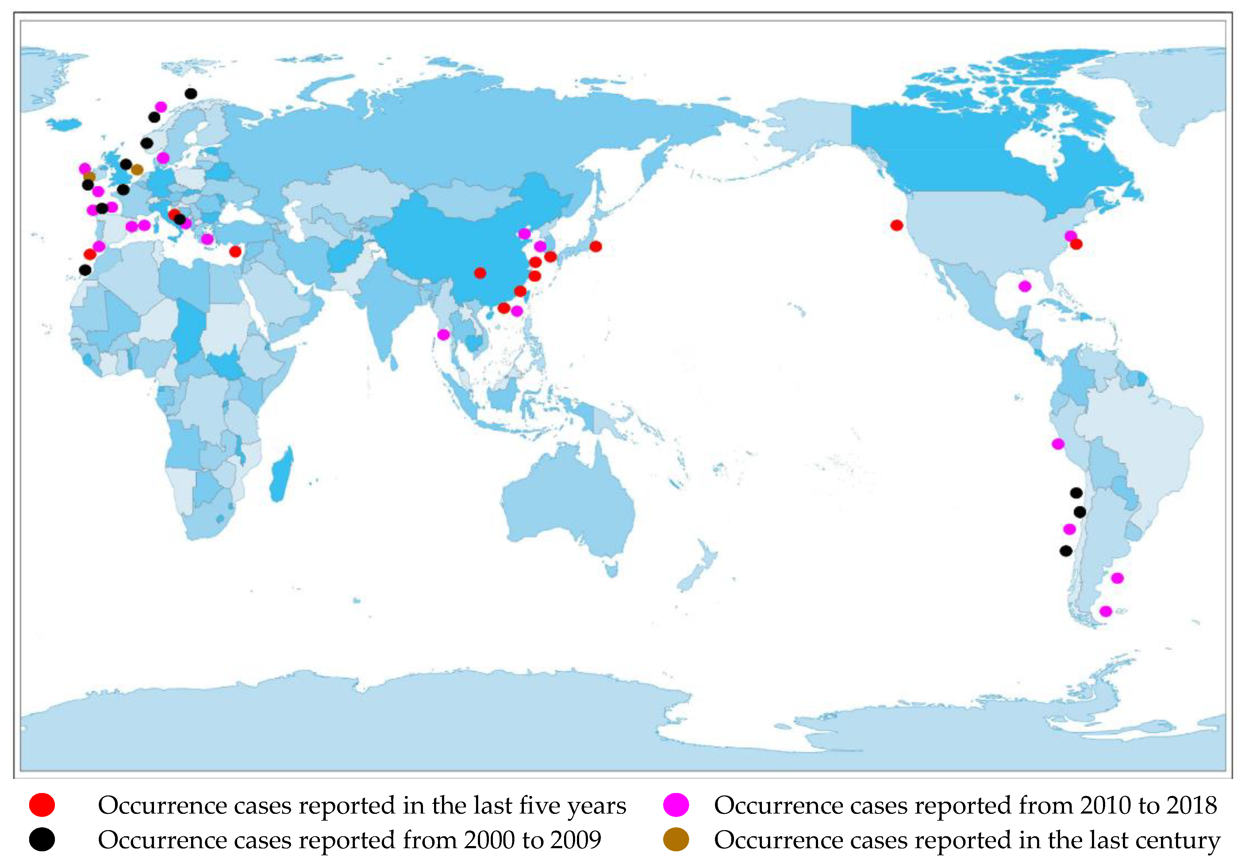
| Location of AZP | Year | Area of Production | The Types of Toxins | Results |
|---|---|---|---|---|
| Ireland | 1999 [62] | Arranmore Island | AZA-1-3 | Nearly 95% of the total AZAs. |
| England and Norway | 2002 [84] | Craster and Sognefjord | AZA-1-3 | 61%(AZA-1), 22%(AZA-2), and 17%(AZA-3) (Norway); 72% (AZA-1) and 28% (AZA-3) (UK). |
| Spain | 2007 [85] | Galica | AZA-1 | Nearly 15.46% of the total toxins. |
| Sweden and Norway | 2008 [86] | The west coast of Sweden and northwest coast of Norway | AZA-1-3 | 70.6% (AZA-1) (Sweden); 16.7% (AZA-1) (Norway). |
| France | 2008 [87] | The North Brittany coast | AZAs | 80% (AZA-1) and 20%(AZA-2). |
| Scotland | 2008 [88] | The Food Standards Agency, Scotland | AZA-1,2 | Nearly 69% of the total samples. |
| Portugal | 2008 [96] | Foz do Arelho beach | AZA-1-3 | 23.5% (AZA-1), 42.8% (AZA-2), and 33.6% (AZA-3). |
| Chile | 2010 [16] | Coquimbo Bay | AZA-1 | Below the quantification limit. |
| China | 2011 [89] | The whole coast of China | AZA-1 | Not mentioned. |
| China | 2015 [90] | The main seafood markets along the Chinese coastline | AZA-1 | 2.75% of the total samples; 27.1% of the total toxins. |
| Spain | 2016 [91] | The North Patagonian coast | AZAs | Not mentioned. |
| Spain | 2017 [92] | The Gulf of Cadiz | AZAs | 49.15% (AZA-43) and 50.85% (AZA-2). |
| The Adriatic Sea | 2018 [93] | The coast of Abruzzo and Molise regions | AZA-1-3 | 23.80% (AZA-1), 42.87% (AZA-2), and 33.33% (AZA-3). |
| Denmark | 2019 [94] | The Limfjord and the Kattegat/Belt area | AZAs | Not mentioned. |
| Spain | 2020 [95] | Galicia | AZA-1-3 | Not mentioned. |
Disclaimer/Publisher’s Note: The statements, opinions and data contained in all publications are solely those of the individual author(s) and contributor(s) and not of MDPI and/or the editor(s). MDPI and/or the editor(s) disclaim responsibility for any injury to people or property resulting from any ideas, methods, instructions or products referred to in the content. |
© 2024 by the authors. Licensee MDPI, Basel, Switzerland. This article is an open access article distributed under the terms and conditions of the Creative Commons Attribution (CC BY) license (https://creativecommons.org/licenses/by/4.0/).
Share and Cite
Yang, J.; Sun, W.; Sun, M.; Cui, Y.; Wang, L. Current Research Status of Azaspiracids. Mar. Drugs 2024, 22, 79. https://doi.org/10.3390/md22020079
Yang J, Sun W, Sun M, Cui Y, Wang L. Current Research Status of Azaspiracids. Marine Drugs. 2024; 22(2):79. https://doi.org/10.3390/md22020079
Chicago/Turabian StyleYang, Jiaping, Weiqin Sun, Mingjuan Sun, Yunyi Cui, and Lianghua Wang. 2024. "Current Research Status of Azaspiracids" Marine Drugs 22, no. 2: 79. https://doi.org/10.3390/md22020079
APA StyleYang, J., Sun, W., Sun, M., Cui, Y., & Wang, L. (2024). Current Research Status of Azaspiracids. Marine Drugs, 22(2), 79. https://doi.org/10.3390/md22020079


