Abstract
A detailed chemical study of the culture of a coral-derived fungus Penicillium chrysogenum resulted in the isolation and identification of four new aromatic heterocycles chrysoquinazolinones A–B (1–2) and chrysobenzothiazoles A–B (3–4), along with a new sorbicillinoid 4-carboxylsorbicillin (5). Chrysoquinazolinones A–B (1–2) combine a quinazolinone fragment with a bicyclo[2.2.2]octane or a pyrrolidone moiety, respectively, demonstrating the unexpected structures of marine natural products. Chrysobenzothiazoles A–B (3–4) possess a benzothiazole system and are the second isolation of this class of skeleton compounds from marine organisms. The existence of the pair of enantiomers (±3) was deduced by chiral HPLC analysis. Their structures and absolute configurations were elucidated by detailed spectroscopic analysis, comparison with the literature data, single-crystal X-ray crystallographic analysis and TDDFT-ECD calculations. Compound 5 exhibited moderate cytotoxicity against K562 and NCI-H446 cell lines, with IC50 values of 15.00 μM and 16.87 μM, respectively.
1. Introduction
Marine-derived filamentous fungi, which can produce bioactive secondary metabolites with unique structures, are recognized as valuable sources for the discovery of marine natural products []. As one of the most widespread fungal species, Penicillium chrysogenum has drawn considerable attention with medicinally valuable and structurally novel metabolites. As reported in the literature, the bioactive natural products from marine-derived P. chrysogenum are comprised of mono- and dimeric sorbicillinoids [,,], alkaloids [,,], polyketides [,,], cerebrosides [], xanthones [,], flavones [], and terpenoids [,], which have wide range of biological activities, such as antitumor [,,], antiviral [,], anti-inflammatory [] and antibacterial [,].
As described in the literature above, the types of the bioactive natural products are various and unpredictable, demonstrating the high research value of fungus P. chrysogenum. In our ongoing search to discover novel natural products, the orthogonal experiment was conducted to determine the optimal culture condition of the coral-derived fungus P. chrysogenum, collected from the Xisha Islands in the South China Sea. Then, a chemical investigation of the extracts of fermentation broth and mycelia was conducted, which resulted in the isolation and identification of five new compounds, including two quinazolinone alkaloids chrysoquinazolinones A–B (1–2), two benzothiazole heterocycles chrysobenzothiazoles A–B (3–4), and a sorbicillinoid derivative 4-carboxylsorbicillin (5). Natural quinazolinones are extensively derived from terrestrial as well as marine plants, animals, and microorganisms, which have been widely reported with various biological activities []. Dimeric sorbicillinoids containing a bicyclo[2.2.2]octane moiety usually have been isolated from the fungi of the family Penicillium [,,,,] and Trichoderma [,]. However, the unexpected combination of a quinazolinone fragment and a bicyclo[2.2.2]octane moiety in chrysoquinazolinone A (1) has never been found before, nor the conjunction of a quinazolinone fragment and a pyrrolidone moiety in chrysoquinazolinone B (2). Numerous benzothiazole derivatives with a wide spectrum of biological activity have been synthesized through various synthetic pathways, as these compounds have unique flexibility and dynamic cores [,,]. Compared to the large number of synthetic benzothiazole derivatives, fewer benzothiazole heterocyclic compounds have been found from natural sources. Chrysobenzothiazoles A–B (3–4) are the second isolation of this class of compounds from marine organisms []. Herein, we report the details for isolation, structure elucidation, and bioactivities of these isolated compounds (Figure 1).
Figure 1.
Structures of 1–5.
2. Results
Chrysoquinazolinone A (1) was obtained as colorless crystals. Its molecular formula was determined as C21H24O4N2 based on the HRESIMS data (m/z 369.1813 [M + H]+, calcd. 369.1809), indicating eleven degrees of unsaturation. The 13C NMR data (Table 1) and HSQC spectrum showed the existence of 21 carbons, including 3 methyl, 3 methylene, 7 methine (4 olefinic), and 8 quaternary carbons. The 1H NMR data (Table 1) and HSQC spectrum showed 22 non-exchangeable protons, including 4 olefinic protons at δH 8.26 (1H, d, J = 7.8 Hz, H-6′), δH 7.50 (1H, m, H-7′), δH 7.79 (1H, m, H-8′), and δH 7.71 (d, J = 8.1 Hz, H-9′), 3 methyl groups at δH 0.94 (3H, d, J = 6.8 Hz, H3-11), δH 1.13 (3H, s, H3-12), and δH 1.07 (3H, s, H3-13), 6 methylene protons at δH 2.57 (1H, d, J = 19.5 Hz, H-3a), δH 2.91 (1H, overlapped, H-3b) δH 2.19 (1H, m, H-9a), δH 1.96 (1H, m, H-9b), δH 2.96 (1H, m, H-10a), and δH 2.89 (1H, overlapped, H-10b), and 3 sp3 hybridized methine protons at δH 2.40 (1H, s, H-4), δH 1.63 (1H, m, H-7), and δH 2.12 (1H, m, H-8). In addition, the existence of an exchangeable proton and a hydroxyl group was verified by the molecular formula. The planar structure of 1 was deduced by detailed analysis of 1H–1H COSY and HMBC spectra (Figure 2). The 1H–1H COSY correlations revealed the presence of three spin systems H2-3/H-4, H3-11/H-7/H-8, and H-6′/H-7′/H-8′/H-9′. The HMBC correlations from H3-13 to C-1, C-2, C-6, and C-7, H3-12 to C-4, C-5, and C-6, H3-11 to C-1, H-3a to C-2 and C-8, and H-4 to C-7 established a bicyclo[2.2.2]octane moiety. Moreover, the characteristic carbon signals [] (156.0, 164.6, 120.6, 126.3, 126.9, 135.2, 127.4, 149.5), as well as the HMBC correlations from H-6′ to C-4′ and C-5′, H-7′, to C-5′, and H-8′ to C-10′, suggested a quinazolinone moiety. HMBC correlations from H-7 to C-9, and Ha-10 to C-9 and C-2′ indicated that the bicyclo[2.2.2]octane moiety was linked to the quinazolinone moiety via two linked methylene groups, completing the planar structure construction of 1.
Table 1.
One-dimensional and two-dimensional NMR data for chrysoquinazolinone A (1) in CDCl3.
Figure 2.
Key 1H–1H COSY, HMBC and NOESY correlations of 1.
The relative configuration of 1 was revealed by NOSEY correlations (Figure 2). The NOSEY correlation between H-7 and H-9a and the correlation between H-8 and H3-11 verified the different orientations of H-7 and H-8. In addition, the NOSEY correlation between H-3a and H3-12 and the correlation between H-3b and H-9b disclosed the relative configuration of 1 is 1R*, 4R*, 5S*, 7R*, and 8R*. Fortunately, suitable single crystals were acquired and subjected to a Bruker APEX-II CCD diffractometer with Cu Kα radiation, validating the absolute configuration of 1 was 1R, 4R, 5S, 7R, and 8R, with a Flack parameter of −0.23 (16) (Figure 3).
Figure 3.
ORTEP drawing of 1, 3, and 4 (displacement ellipsoids are drawn at the 50% probability level).
Chrysoquinazolinone B (2) was obtained as a colorless oil. The molecular formula was deduced to be C14H15O2N3 according to the HRESIMS data at m/z 258.1242 [M + H]+ (calcd. 258.1237). The 13C NMR data (Table 2) and HSQC spectrum showed 14 carbon resonances, including 1 methyl, 3 methylene, 5 methine (4 olefinic), and 5 quaternary carbons. The 1H NMR data (Table 2) showed 14 non-exchangeable protons, including 4 olefinic protons at δH 8.20 (1H, d, J = 8.0 Hz, H-6), δH 7.53 (1H, m, H-7), δH 7.82 (1H, m, H-8), and δH 7.71 (1H, d, J = 8.1 Hz, H-9), 1 methyl group at δH 1.63 (3H, d, J = 7.2 Hz, H3-12), 6 sp3 hybridized methylene protons at δH 2.47 (2H, t, J = 8.1 Hz, H2-3′), δH 2.14 (2H, m, H2-4′), and δH 3.65 (2H, m, H2-5′), as well as 1 sp3 hybridized methine proton at δH 5.13 (1H, q, J = 7.2 Hz, H-11). According to the molecular formula, one additional exchangeable proton is needed. The 1H–1H COSY and HMBC spectra (Figure 4) revealed the planar structure of 2. Three spin systems H-6/H-7/H-8/H-9, H3-12/H-11, and H-3′/H-4′/H-5′ were deduced by the 1H–1H COSY spectrum. The characteristic carbon signals (156.6, 164.3, 122.4, 127.0, 128.1, 135.8, 128.6, 150.1) showed a quinazolinone moiety, which further determined by HMBC correlations from H-6 to C-4, H-7 to C-5, H-8 to C-10, and H-9 to C-5. Additionally, the HMBC correlations from H-11 to C-2, C-2′, C-5′, and H3-12 to C-2 suggested a pyrrolidone fragment, which was linked to the quinazolinone moiety by C-11, completing the planar structure construction of 2. The absolute configuration of 2 was determined as 11R by further comparison of the experimental and the calculated ECD spectra (Figure 5).
Table 2.
One-dimensional and two-dimensional NMR data for chrysoquinazolinone B (2) in CD3OD.
Figure 4.
Key 1H–1H COSY and HMBC correlations of 2–5.
Figure 5.
Experimental and calculated ECD spectra of 2–4 (a–c).
Chrysobenzothiazole A (3) was isolated as colorless crystals. Its molecular formula was elucidated as C13H15O3NS on the basis of the HRESIMS [M + H]+ data at m/z 266.0846 (calcd. 266.0845), indicating six degrees of unsaturation. The 13C NMR data (Table 3) and HSQC spectrum showed the presence of 13 carbons, including 2 methyl, 2 methylene, 3 methine, and 6 quaternary carbons. The 1H NMR data (Table 3) and HSQC spectrum showed 13 protons, including 2 olefinic protons at δH 6.88 (1H, s, H-5) and δH 8.95 (1H, s, H-13), 2 methyl groups at δH 1.24 (3H, d, J = 7.0 Hz, H3-11) and δH 2.64 (3H, s, H3-12), 4 sp3 hybridized methylene protons at δH 2.87 (2H, m, H2-7), δH 2.02 (1H, m, H-8a), and δH 1.75 (1H, m, H-8b), together with 1 sp3 hybridized methine proton at δH 2.48 (1H, m, H-9). Moreover, two additional hydroxyl groups were needed to satisfy the molecular formula. The 1D NMR data [δC 118.9, 136.8, 146.9, 132.2, 117.5, 154.2 and 152.1; δH 6.88 (1H, s) and 8.95 (1H, s)] suggested a trisubstituted benzothiazole moiety, which was further verified by the HMBC correlations. The HMBC correlations (Figure 4) from H-13 to C-2 and C-3, H3-12 to C-3, C-4 and C-5, H-5 to C-1 indicated a methyl linking to C-4 and the connections of C-13 to C-2 via S, and C-13 to C-3 via N, which were further deduced by downfield chemical shift of C-2 (δC 136.8) and C-3 (δC 146.9). A hydroxyl group was connected to C-6 based on its downfield chemical shift (δC 154.2). Moreover, the 1H–1H COSY correlations (Figure 4) H2-7/H2-8 and H3-11/H-9, as well as the HMBC correlations from H2-7 to C-1, C-2, C-6, and C-9, H2-8 to C-9 and C-10, and H3-11 to C-8 and C-10 constructed the planar structure of 3, in which another hydroxyl group was assigned to C-10.
Table 3.
1H and 13C NMR data for chrysobenzothiazole A (3) and B (4) in CD3OD.
To verify the absolute configuration of 3, an X-ray crystallographic measurement was performed (Figure 3). The existence of enantiomers was confirmed by detailed analysis of the X-ray diffraction data without a Flack parameter (Table S4). Then, the racemate was subjected to chiral HPLC analysis. The result showed two peaks with the same UV absorption and good resolution in the HPLC chromatogram (Figure S5). The corresponding enantiomers 3a and 3b were obtained by using chiral HPLC, with [α]25D = −1.57 (c 0.03, MeOH) and [α]25D = +1.30 (c 0.03, MeOH), respectively. The absolute configuration of 3a was determined as (−)-9R-3 while the absolute configuration of 3b was determined as (+)-9S-3 through the comparison of experimental and calculated ECD spectra (Figure 5).
Chrysobenzothiazole A (4) was isolated as colorless crystals. Its molecular formula was determined as C13H15O3NS based on the HRESIMS [M + H]+ data at m/z 266.0847 (calcd. 266.0845), which was identical to 3. The characteristic 1D NMR signals (Table 3) [δC 135.8, 146.3, 137.6, 115.1, 154.2, 116.5, and 151.9; δH 6.90 (1H, s) and 8.93 (1H, s)] revealed a trisubstituted benzothiazole moiety in 4, the same as 3. The 13C NMR data showed resonances of C-2 at δC 146.3 and C-3 at δC 137.6, while the corresponding signals in 3 at δC 136.8 and δC 146.9, indicating the opposite positions of N and S. The HMBC correlations (Figure 4) from H-13 to C-2 and C-3, H3-12 to C-3, C-4 and C-5, H-6 to C-2, and H-7 to C-2, and C-6 verified a methyl linking to C-4 and a hydroxyl group linking to C-5, which was further confirmed by the downfield chemical shift of C-5 (δC 154.2). Analysis of the rest 1H–1H COSY (Figure 4) and HMBC correlations established the planar structure of 4. The differences in the planar structures between 3 and 4 are the positions of N, S, and the hydroxyl group in the benzothiazole moiety.
The ECD calculations were performed to verify the absolute configuration of 4, which was determined as 9R (Figure 5). Suitable crystals for X-ray crystallographic analysis were obtained. A detailed analysis of X-ray data suggested that the absolute configuration of 4 was 9R (Figure 3), which showed the same conclusion as the ECD calculations.
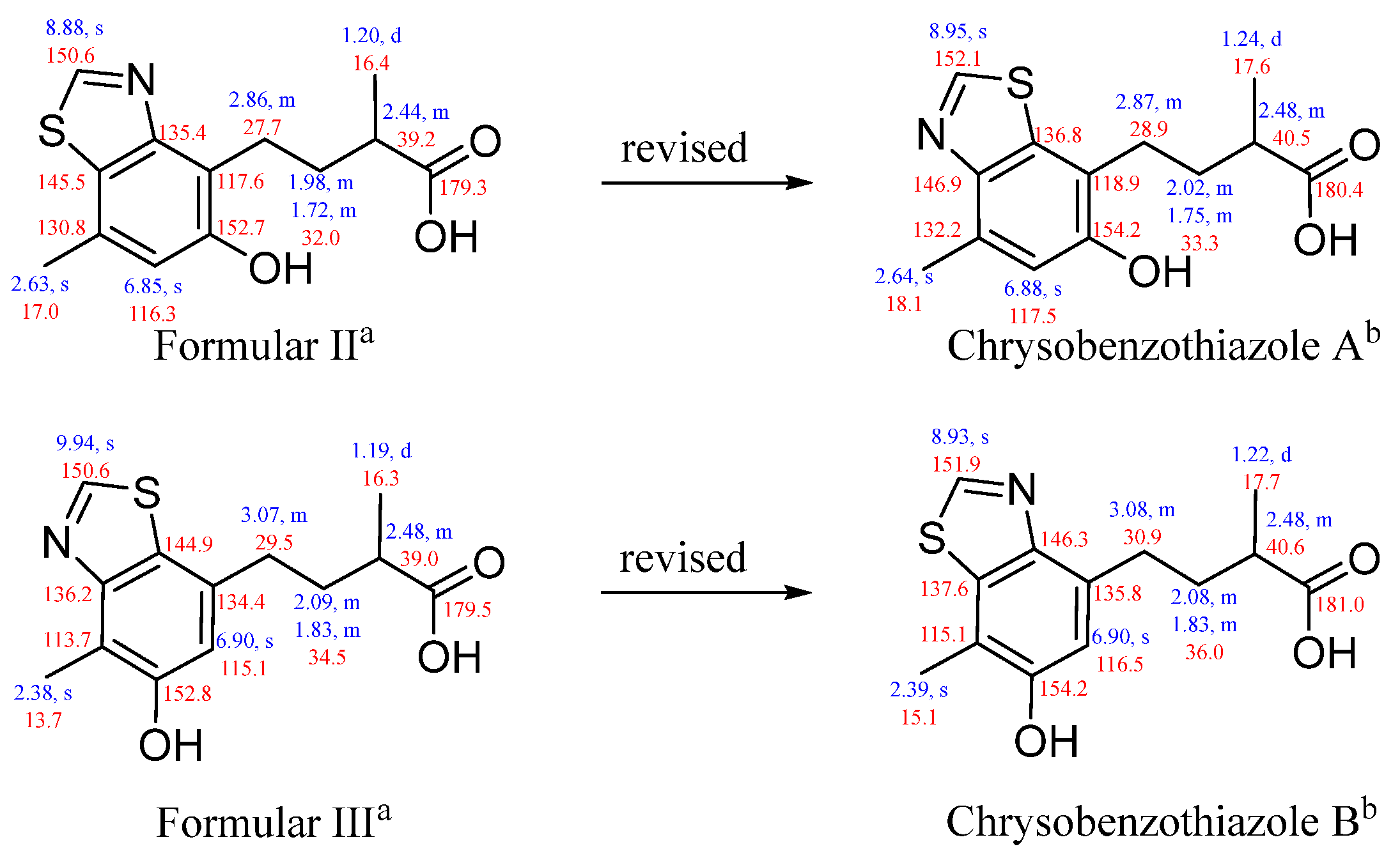
As the second isolation of this class of skeleton compounds, compounds 3–4 were compared with formulars II–III, which were isolated from a deep-sea fungus Penicillium allii-sativi. There was a puzzle about the positions of N and S in the benzothiazole moiety. The 1H and 13C NMR data for 3–4 agreed perfectly with those for II–III. As mentioned above, the positions of N and S in 3–4 were undoubtedly verified by the downfield chemical shift of C-2 and C-3, as well as the X-ray data. Therefore, the planar structures of formulars II–III should be revised (Figure 6).
Figure 6.
1H and 13C NMR data for formulars II–III and chrysobenzothiazoles A–B (a Recorded in CD3OD. b Recorded in CD3OD, 1H NMR at 500 MHz and 13C NMR at 125 Hz).
4-Carboxylsorbicillin (5) was isolated as yellow powder. Its molecular formula was elucidated as C15H16O4 based on the HRESIMS [M + H]+ data at m/z 261.1127 (calcd. 261.1121). The 13C NMR data (Table 4) and HSQC spectrum suggested 15 carbon resonances, including 3 methyl, 5 methine, and 7 quaternary (2 carbonyl) carbons. The 1H NMR data (Table 4) showed 14 protons, including 5 olefinic protons at δH 7.51 (1H, s, H-6), δH 7.00 (1H, d, J = 14.9 Hz, H-8), δH 7.53 (1H, overlapped, H-9), δH 6.38 (1H, overlapped, H-10), and δH 6.37 (1H, overlapped, H-11), 3 methyl groups at δH 1.94 (3H, d, J = 5.3 Hz, H3-12), δH 2.37 (3H, s, H3-13), and δH 2.31 (3H, s, H3-15), along with a phenolic hydroxyl group at δH 13.09 (1H, s). Moreover, one additional hydroxyl group was needed to satisfy the molecular formula. The 1H–1H COSY correlations (Figure 4) revealed the presence of two spin systems H-8/H-9/H-10 and H-11/H3-12. Evidently, the 1D NMR data [δC 119.7, 160.0, 124.8, 139.6, 123.6, 128.2; δH 7.51 (1H, s)] belonged to a penta-substituted benzene ring. The positions of substituents on the benzene ring were determined by relevant HMBC correlations (Figure 4) from H3-13 to C-4, C-5 and C-6, H3-15 to C-2, C-3, and C-4, and –OH to C-1, C-2, and C-3. In addition, a carboxyl group (δC 171.8) was assigned to C-4 on the basis of the molecular formula. The characteristic proton signals at δH 7.53, 7.00, 6.38, 6.37, and 1.94 suggested a sorbyl group, which was further verified by the HMBC correlations from H-8 to C-7 and C-10, H-9 to C-7 and C-11, and H3-12 to C-10. The sorbyl fragment was linked to C-1 according to the HMBC correlation from H-6 to C-7, completing the planar structure construction of compound 5.
Table 4.
1H and 13C NMR data for 5 in CDCl3.
The configuration of the Δ8,9 was substantiated as E according to the large coupling constants of 3JH-8,H-9 (14.9 Hz). However, the coupling constants of 3JH-10,H-11 were indeterminate because of the overlapped signals of H-10 and H-11 in the 1H NMR spectrum. As reported in the literature [], E configurations of double bonds are structural features in sorbicillinoids, therefore, the configuration of the Δ10,11 was deduced to be E. Compare with the reported compound sorbicillin [], the structure of compound 5 shows that the hydroxyl group attached to C-4 is substituted by a carboxyl group.
As discussed above, the quinazolinone, benzothiazole, and sorbicillinoid derivatives exhibited a wide range of biological activities, including cytotoxicity. Thus, cytotoxic assays of compounds 1–5 were conducted. The results showed that compound 5 exhibited moderate cytotoxic activities against K562 and NCI-H446 cell lines, with IC50 values of 15.00 μM and 16.87 μM, respectively. Unfortunately, the other compounds did not show any cytotoxic activity (Table 5).
Table 5.
Cytotoxic activities of 1–5.
3. Materials and Methods
3.1. General Experimental Procedures
The optical rotation data were obtained on a Jasco P-1020 polarimeter (Jasco, Tokyo, Japan). The UV spectra and circular dichroism data were measured on a Jasco J-815 CD spectropolarimeter (Jasco, Tokyo, Japan), and IR spectra were recorded with a Nicolet NEXUS 470 spectrophotometer (Thermo Scientific, Beijing, China) in KBr disks. The NMR spectra were measured in CDCl3 or CD3OD on an Agilent DD2-500 (1H, 500 MHz; 13C, 125 MHz; Agilent, Beijing, China). The 7.26 and 77.2 ppm resonances of CDCl3, as well as the 3.31 and 49.0 ppm resonances of CD3OD, were used as internal references for 1H and 13C NMR spectra, respectively. HRESIMS data were obtained on Micromass Q-Tof Ultima GLOBAL GAA076LC mass spectrometer (Autospec-Ultima-TOF, Waters, Shanghai, China). The crystallographic data were acquired on a Bruker APEX-II CCD diffractometer (Bruker, Beijing, China) equipped with graphite-monochromatized Cu Kα radiation []. Analytic HPLC was performed by a Shimadzu LC-20AT liquid chromatograph (Shimadzu, Shanghai, China) equipped with an LC-20A array detector, using an analytic reversed-phased column (Shimadzu, ODS, 5 μm, 150 × 4.6 mm, 1.0 mL/min) or an analytic chiral-phase column (Daicel, Shanghai, China, IC, 5 μm, 250 × 4.6 mm, 1.0 mL/min). The semi-preparative HPLC was implemented by an Agilent 1100 series liquid chromatograph (Agilent, Tokyo, Japan) equipped with a DAD G1315A detector, using a semi-preparative reversed-phased column (SilGreen ODS, Beijing, China, 5 μm, 250 × 10 mm, 2.0 mL/min). Silica gel (300–400 mesh, Qingdao Marine Chemical Factory, Qingdao, China) was used for column chromatography (CC). Sephadex LH-20 (Amersham Pharmacia Biotech AB, Uppsala, Sweden) was used for molecular exclusion chromatography. Analytical TLC plates were precoated silica gel plates (GF254).
3.2. Fungal Material
The fungal strain 19-7-ZM-4 was isolated from an unidentified soft coral (190721-07) collected from the Xisha Islands in the South China Sea and identified as Penicillium chrysogenum according to the sequencing of the ITS region (GenBank accession number PP347996.1) with 100% similarity to P. chrysogenum. The strain was deposited at the State Key Laboratory of Marine Drugs, Ocean University of China.
3.3. Fermentation, Extraction and Isolation
The fungal strain 19-7-ZM-4 was initially cultured on the panels of PDA medium at 28 °C for 3 days. The fresh mycelia were inoculated into 500 mL Erlenmeyer flasks each containing 150 mL of the seed medium, comprised of dextrose (20 g/L), potato (200 g/L), and seawater (Huiquan Bay, Yellow Sea). The flasks were grown on a rotary shaker (180 rpm) at 28 °C for 24 h. Then, 1.0 mL seed culture was transferred into each 500 mL Erlenmeyer flask containing 150 mL of liquid culture medium, composed of dextrose (20 g/L), maltose (20 g/L), yeast extract powder (5 g/L), peptone (10 g/L), corn steep liquor (10 g/L), KH2PO4 (0.5 g/L), MgSO4 (0.5 g/L), and seawater (Huiquan Bay, Yellow Sea). These flasks were cultured on rotary shakers (180 rpm) at 28 °C for 13 days.
The fermented whole broth (40 L) was filtered under reduced pressure to separate the supernatant from mycelia. The supernatant was extracted three times with EtOAc, while the mycelia were macerated and extracted three times with MeOH. All the extracts were evaporated under reduced pressure and desalinated to yield 234.6 g of residue.
The extract was subjected to silica gel vacuum liquid chromatography, using a step gradient of petroleum ether/acetone (from 50/1 to 1/1, v/v) and CH2Cl2/MeOH (from 20/1 to 0/1, v/v). Based on the TLC analysis, 8 fractions (Fr.1-Fr.8) were acquired. Fraction Fr.5 (2.41 g) was subjected to silica gel CC eluting with petroleum ether/acetone (from 50/1 to 0/1, v/v) to yield six subfractions (Fr.5-1–5-6). Subfraction Fr.5-3 (120 mg) was purified by semi-preparative HPLC (ODS, 5 μm, 250 × 10 mm; CH3OH/H2O/HCOOH, 65:35:0.035, v/v/v; 2 mL/min) to obtain compound 5 (tR = 60 min, 1.0 mg). Fraction Fr.6 (4.76 g) was subjected to silica gel CC eluting with petroleum ether/acetone (from 25/1 to 0/1, v/v) to acquire six subfractions (Fr.6-1–6-6). Subfraction Fr.6-5 (450 mg) was purified by semi-preparative HPLC (ODS, 5 μm, 250 × 10 mm; CH3OH/H2O/HCOOH, 65:35:0.035, v/v/v; 2 mL/min) to afford compounds 3 (tR = 32 min, 1.8 mg) and 4 (tR = 36 min, 1.5 mg). Then, 3 was analyzed and purified by chiral HPLC (IC, 5 μm, 250 ×10 mm; n-hexane/isopropanol, 80:20, v/v), which obtained 3a (1.2 mg) and 3b (0.3 mg). Fraction Fr.7 (5.31 g) was carried out by silica gel CC eluting with petroleum ether/acetone (from 5/1 to 0/1, v/v) to yield four subfractions (Fr.7-1–7-4). Compound 2 (tR = 36 min, 7.0 mg) was purified from subfraction Fr.7-2 (2.06g) by Sephadex LH-20 column chromatography eluting with MeOH-CH2Cl2 (1:1, v/v) and semipreparative HPLC (ODS, 5 μm, 250 × 10 mm; CH3OH/H2O/HCOOH, 45:55:0.055, v/v/v; 2 mL/min). Subfraction Fr.7-3 (940 mg) was performed by Sephadex LH-20 column chromatography eluting with MeOH-CH2Cl2 (1:1, v/v) and semipreparative HPLC (ODS, 5 μm, 250 × 10 mm; CH3OH/H2O/HCOOH, 50:50:0.05, v/v/v; 2 mL/min) to afford compound 1 (tR = 48 min, 4.8 mg).
Chrysoquinazolinone A (1): colorless crystals; [α]25D −39.07 (c 0.10, MeOH); UV (MeOH) λmax (log ε) 200.0 (1.86) nm, 224.5 (1.78) nm, 305.5 (0.28) nm; IR (KBr) νmax 3410, 2832, 1594, 1363, 776 cm−1; HRESIMS m/z 369.1813 [M + H]+ (calcd. for C21H25O4N2, 369.1809). For 1H NMR and 13C NMR data, see Table 1.
Chrysoquinazolinone B (2): colorless oil; [α]25D +2.53 (c 0.10, MeOH); UV (MeOH) λmax (log ε) 199.0 (2.23), 224.5 (2.24), 266.0 (0.67), 302.0 (0.36) nm; ECD (0.25 mg/mL, CH3OH) λmax (Δε) 218 (−13.63), 253 (1.66), 219 (−0.23) nm; IR (KBr) νmax 2832, 2359, 1595, 1363, 775 cm−1; HRESIMS at m/z 258.1242 [M + H]+ (calcd. for C14H16O2N3 258.1237). For 1H NMR and 13C NMR data, see Table 2.
Chrysobenzothiazole A (±3): colorless crystals; [α]25D −1.57 (c 0.03, MeOH) for (−)-9R-3, [α]25D +2.30 for (+)-9S-3; UV (MeOH) λmax (log ε) 207.0 (1.75), 227.0 (1.16), 243.0 (1.19), 281.5 (0.56) nm for (−)-9R-3, UV (MeOH) λmax (log ε) 207.0 (1.28), 227.0 (0.85), 243.0 (0.82), 281.5 (0.39) nm for (+)-9S-3; ECD (0.25 mg/mL, CH3OH) λmax (Δε) 190 (5.79) nm for (−)-9R-3, ECD (0.50 mg/mL, CH3OH) λmax (Δε) 190 (−9.04) nm for (+)-9S-3; IR (KBr) νmax 3406, 2834, 2360, 2341, 1598, 1364, 777, 669 cm−1; HRESIMS at m/z 266.0846 [M + H]+ (calcd. for C13H16O3NS, 266.0845). For 1H NMR and 13C NMR data, see Table 3 and Table S1.
Chrysobenzothiazole A (4): colorless crystals; [α]25D −2.70 (c 0.03, MeOH); UV (MeOH) λmax (log ε) 207.0 (2.28), 225.0 (1.58), 244.5 (1.43), 279.5 (0.76) nm; ECD (0.50 mg/mL, CH3OH) λmax (Δε) 190 (8.31), 208 (1.93) nm; IR (KBr) νmax 3405, 2833, 2360, 2342, 1594, 1363, 777, 669 cm−1; HRESIMS at m/z 266.0847 [M + H]+ (calcd. for C13H16O3NS, 266.0845). For 1H NMR and 13C NMR data, see Table 3 and Table S2.
4-Carboxylsorbicillin (5): yellow powder; UV (MeOH) λmax (log ε) 200.0 (2.24), 309.5 (2.43) nm; IR (KBr) νmax 3406, 2360, 2341, 1595, 1364, 669 cm−1; HRESIMS at m/z 261.1127 [M + H]+ (calcd. for C15H17O4, 261.1121). For 1H NMR and 13C NMR data, see Table 4.
3.4. X-Ray Crystallographic Analysis
The crystals for chrysoquinazolinone A (1), chrysobenzothiazole A (±3) and B (4) were obtained from a solution of EtOH-H2O using the vapor diffusion method at 4 °C. X-ray crystallographic data were collected on a Bruker APEX-II CCD diffractometer with Cu-Kα radiation. The crystals were kept at 293 K, 150 K, or 100 K during data collection. Crystallographic data have been deposited in the Cambridge Crystallographic Data Centre. The data can be obtained via https://www.ccdc.cam.ac.uk/ (accessed on 24 March 2022).
Crystal data for chrysoquinazolinone A (1): C21H24N2O4, M = 368.42, a = 13.1209(5) Å, b = 21.3061(7) Å, c = 13.3607(5) Å, α = 90°, β = 90.100(10)°, γ = 90°, V = 3735.0(2) Å3, T = 293(2) K, space group P21, Z = 8, μ = 0.742 mm−1, F(000) = 1568. A total of 10,888 independent reflections [Rint = 0.0559, Rsigma = 0.0456] were used for the analysis. The final R indexes [all data] gave R1 =0.0694, wR2 = 0.1755. The goodness of fit on F2 was 1.028 and the Flack parameter was −0.23(16). CCDC 2342721.
Crystal data for chrysobenzothiazole A (±3): C13H15NO3S, M =265.32, a = 8.5937(3) Å, b = 9.1721(4) Å, c = 16.9210(7) Å, α = 90°, β = 93.851(2)°, γ = 90°, V = 1330.74(9) Å3, T = 150 K, space group P21/n, Z = 4, μ = 2.174 mm−1, F(000) = 560. A total of 2724 independent reflections [Rint = 0.0506, Rsigma = 0.0512] were used for the analysis. The final R indexes [all data] gave R1 =0.0818, wR2 = 0.2000. The goodness of fit on F2 was 1.109. CCDC 2342719.
Crystal data for chrysobenzothiazole B (4): C13H15NO3S, M =265.32, a = 4.69830(10) Å, b = 15.3583(2) Å, c = 17.0647(3) Å, α = 90°, β = 90°, γ = 90°, V = 1231.35(4) Å3, T = 100 K, space group P212121, Z = 4, μ = 2.35 mm−1, F(000) = 560. A total of 2484 independent reflections [Rint = 0.0295, Rsigma = 0.0372] were used for the analysis. The final R indexes [all data] gave R1 =0.0335, wR2 = 0.0855. The goodness of fit on F2 was 1.071, and the Flack parameter was 0.020(11). CCDC 2342720.
3.5. Quantum Chemical Calculations
The conformational search of possible configurations whose energy was within 10 kJ/mol was carried out in an OPLS3e force field by MacroModel integrated into Maestro (version 11.9) (Schrödinger version 2019.1). The root mean squared distance (RMSD) cutoff of 0.5 Å and the maximum iteration of 2500 were executed to eliminate redundant conformers. Geometry optimizations and frequency calculations of stable conformers were performed at the B3LYP/6-31 G(d,p) level using density functional theory (DFT). Subsequently, a set of the lowest-energy conformers, whose Boltzmann distributions of Gibbs free energies were more than 1.0% were used for the next calculations. The ECD calculations were performed at the CAM-B3LYP/6-311G(d, p) level using the TD-DFT method with the IEFPCM solvent model for methanol in agreement with the experimental condition []. The Gaussian 09 package (version D. 01) was used for all the calculations.
3.6. Cytotoxicity Assay
The cytotoxic activity against the K562 (human leukemia) cell line was evaluated by MTT method [], and the cytotoxic activities against L-02 (normal human hepatocytes), ASPC-1 (human pancreatic cancer), MDA-MB-231 (human breast cancer), NCI-H446, and NCI-H446/EP (human small cell lung cancer) cell lines were determined by SRB method []. The difference between the two human small-cell lung cancer cell lines is that NCI-H446 is a sensitive cell line while the NCI-H446/EP cell line is resistant to cisplatin, etoposide, SN38, and doxorubicin. As a positive control, doxorubicin was used.
4. Conclusions
In summary, two quinazolinone alkaloids chrysoquinazolinones A–B (1–2), two benzothiazole heterocycles chrysobenzothiazoles A–B (3–4), together with a sorbicillinoid derivative 4-carboxylsorbicillin (5), were isolated from the culture of a coral-derived fungus Penicillium chrysogenum collected from the Xisha Islands in the South China Sea. The structures of chrysoquinazolinones A–B (1–2) are unusual as they combined a quinazolinone system with a bicyclo[2.2.2]octane or a pyrrolidone moiety, respectively. Chrysobenzothiazoles A–B (3–4) containing a benzothiazole system were isolated as natural products from marine organisms for the second time. Chiral HPLC was carried out to obtain optically pure compounds (−)-9R-3 (3a) and (+)-9S-3 (3b) and their absolute configurations were elucidated by ECD calculations and single-crystal X-ray diffraction. Moreover, the structures of formulars II–III reported in the patent were revised. Finally, biological evaluation results revealed that compound 5 exhibited moderate cytotoxicity against K562 and NCI-H446 cell lines with IC50 values of 15.00 μM and 16.87 μM, respectively. The discovery of heterocyclic compounds enriched the structural diversity of this family. However, the biosynthetic pathway of these heterocyclic compounds is still uncertain, and the heterocycles need to be further tested for their bioactivities. The discovery of these novel compounds displayed high research value of fungus P. chrysogenum and enriched chemical libraries of marine fungi.
Supplementary Materials
The following supporting information can be downloaded at: https://www.mdpi.com/article/10.3390/md22110517/s1, Tables S1 and S2: NMR data of 3–4; Tables S3–S5 and Figures S1–S4: X-ray crystallographic analysis of 1, 3, and 4; Figures S5 and S6: Chiral HPLC chromatograms for 3 and 4; Figures S7–S9 and Table S6: Stable conformers of 2–4; Tables S7–S10: The gradient of media, HPLC and TLC chromatograms, and mass of residue; Tables S11–S13: The cytotoxicity of 1–5. Figures S10–S45: The HRESIMS, UV, and NMR spectra of 1–5.
Author Contributions
Conceptualization, methodology, and funding acquisition, P.L.; supervision, C.W.; software K.L.; validation, Y.Z. (Yue Zhang); investigation and writing—original draft preparation, J.Y. and Y.Z. (Yuan Zong); writing—review and editing, P.L. All authors have read and agreed to the published version of the manuscript.
Funding
This research was funded by the National Natural Science Foundation of China (Grant No. 42276088, 81991522) and the Foundation of the State Key Laboratory of Component-based Chinese Medicine (Grant No. CBCM2024203).
Institutional Review Board Statement
Not applicable.
Data Availability Statement
Data are contained within the article or Supplementary Materials, further inquiries can be directed to the corresponding author.
Acknowledgments
Special thanks to the Center for High-Performance Computing and System Simulation (National Laboratory for Marine Science and Technology) for the support extended toward computer calculations.
Conflicts of Interest
The authors declare no conflicts of interest.
References
- Carroll, A.R.; Copp, B.R.; Grkovic, T.; Keyzers, R.A.; Prinsep, M.R. Marine natural products. Nat. Prod. Rep. 2024, 41, 162–207. [Google Scholar] [CrossRef] [PubMed]
- Bringmann, G.; Lang, G.; Gulder, T.A.M.; Tsuruta, H.; Mühlbacher, J.; Maksimenka, K.; Steffens, S.; Schaumann, K.; Stöhr, R.; Wiese, J.; et al. The first sorbicillinoid alkaloids, the antileukemic sorbicillactones A and B, from a sponge-derived Penicillium chrysogenum strain. Tetrahedron 2005, 61, 7252–7265. [Google Scholar] [CrossRef]
- Peng, J.; Zhang, X.; Du, L.; Wang, W.; Zhu, T.; Cu, Q.; Li, D. Sorbicatechols A and B, Antiviral Sorbicillinoids from the Marine-Derived Fungus Penicillium chrysogenum PJX-17. J. Nat. Prod. 2014, 77, 424–428. [Google Scholar] [CrossRef]
- Cao, M.-J.; Zhu, T.; Liu, J.-T.; Ouyang, L.; Yang, F.; Lin, H.-W. New sorbicillinoid derivatives with GLP-1R and eEF2K affinities from a sponge-derived fungus Penicillium chrysogenum 581F1. Nat. Prod. Res. 2020, 34, 2880–2886. [Google Scholar] [CrossRef]
- Zhen, X.; Gong, T.; Wen, Y.-H.; Yan, D.-J.; Chen, J.-J.; Zhu, P. A Chrysoxanthones A-C, Three New Xanthone-Chromanone Heterdimers from Sponge-Associated Penicillium chrysogenum HLS111 Treated with Histone Deacetylase Inhibitor. Mar. Drugs 2018, 16, 357. [Google Scholar] [CrossRef] [PubMed]
- Huang, S.; Chen, H.; Li, W.; Zhu, X.; Ding, W.; Li, C. Bioactive Chaetoglobosins from the Mangrove Endophytic Fungus Penicillium chrysogenum. Mar. Drugs 2016, 14, 172. [Google Scholar] [CrossRef]
- Xu, W.-F.; Mao, N.; Xue, X.-J.; Qi, Y.-X.; Wei, M.-Y.; Wang, C.-Y.; Shao, C.-L. Structures and Absolute Configurations of Diketopiperazine Alkaloids Chrysopiperazines A-C from the Gorgonian-Derived Penicillium chrysogenum Fungus. Mar. Drugs 2019, 17, 250. [Google Scholar] [CrossRef] [PubMed]
- Gao, S.-S.; Li, X.-M.; Du, F.-Y.; Li, C.-S.; Proksch, P.; Wang, B.-G. Secondary Metabolites from a Marine-Derived Endophytic Fungus Penicillium chrysogenum QEN-24S. Mar. Drugs 2011, 9, 59–70. [Google Scholar] [CrossRef]
- Guo, W.; Li, D.; Peng, J.; Zhu, T.; Gu, Q.; Li, D. Penicitols A-C and Penixanacid A from the Mangrove-Derived Penicillium chrysogenum HDN11-24. J. Nat. Prod. 2015, 78, 306–310. [Google Scholar] [CrossRef]
- Niu, S.; Xia, M.; Chen, M.; Liu, X.; Li, Z.; Xie, Y.; Shao, Z.; Zhang, G. Cytotoxic Polyketides Isolated from the Deep-Sea-Derived Fungus Penicillium chrysogenum MCCC 3A00292. Mar. Drugs 2019, 17, 686. [Google Scholar] [CrossRef]
- Peng, X.; Wang, Y.; Sun, K.; Liu, P.; Yin, X.; Zhu, W. Cerebrosides and 2-Pyridone Alkaloids from the Halotolerant Fungus Penicillium chrysogenum Grown in a Hypersaline Medium. J. Nat. Prod. 2011, 74, 1298–1302. [Google Scholar] [CrossRef] [PubMed]
- Zhao, D.-L.; Yuan, X.-L.; Du, Y.-M.; Zhang, Z.-F.; Zhang, P. Benzophenone Derivatives from an Algal-Endophytic Isolate of Penicillium chrysogenum and Their Cytotoxicity. Molecules 2018, 23, 3378. [Google Scholar] [CrossRef] [PubMed]
- Chen, S.; Wang, J.; Lin, X.; Zhao, B.; Wei, X.; Li, G.; Kaliaperumal, K.; Liao, S.; Yang, B.; Zhou, X.; et al. Chrysamides A-C, Three Dimeric Nitrophenyl trans-Epoxyamides Produced by the Deep-Sea-Derived Fungus Penicillium chrysogenum SCSIO41001. Org. Lett. 2016, 18, 3650–3653. [Google Scholar] [CrossRef] [PubMed]
- Hou, X.-M.; Wang, C.-Y.; Gu, Y.-C.; Shao, C.-L. Penimethavone A, a flavone from a gorgonian-derived fungus Penicillium chrysogenum. Nat. Prod. Res. 2016, 30, 2274–2277. [Google Scholar] [CrossRef]
- Xu, K.; Wei, X.-L.; Xue, L.; Zhang, Z.-F.; Zhang, P. Antimicrobial Meroterpenoids and Erythritol Derivatives Isolated from the Marine-Algal-Derived Endophytic Fungus Penicillium chrysogenum XNM-12. Mar. Drugs 2020, 18, 578. [Google Scholar] [CrossRef]
- Qiao, H.; Zhang, S.-H.; Dong, Y.; Yang, Y.; Xu, R.; Chen, B.; Wang, Y.; Zhu, T.-J.; Cui, C.-B.; Zhang, G.-G.; et al. Chrysomutanin and related meroterpenoids from a DES mutant of the marine-derived fungus Penicillium chrysogenum S-3-25. Nat. Prod. Res. 2022, 36, 1834–1841. [Google Scholar] [CrossRef]
- Li, H.B.; Fu, G.H.; Zhong, W.H. Natural quinazolinones: From a treasure house to promising anticancer leads. Eur. J. Med. Chem. 2023, 245, 114915. [Google Scholar] [CrossRef]
- Wang, J.J.; Li, K.L.; Luo, X.W.; Wu, Z.Y.; Gu, T.W.; Liao, S.R.; Lin, X.P.; Yang, B.; Liu, Y.H.; Fang, W.; et al. Sorbicillfurans A and B, two novel sorbicillinoid adducts from the fungus Penicillium citrinum SCSIO41402. Org. Biomol. Chem. 2019, 17, 8721–8725. [Google Scholar] [CrossRef]
- Li, D.H.; Chen, L.; Zhu, T.J.; Kurtán, T.; Mándi, A.; Zhao, Z.M.; Li, J.; Gu, Q.Q. Chloctanspirones A and B, novel chlorinated polyketides with an unprecedented skeleton, from marine sediment derived fungus Penicillium terrestre. Tetrahedron 2011, 67, 7913–7918. [Google Scholar] [CrossRef]
- Yu, J.; Han, H.; Zhang, X.; Ma, C.; Sun, C.; Che, Q.; Gu, Q.; Zhu, T.; Zhang, G.; Li, D. Discovery of Two New Sorbicillinoids by Overexpression of the Global Regulator LaeA in a Marine-Derived Fungus Penicillium dipodomyis YJ-11. Mar. Drugs 2019, 17, 446. [Google Scholar] [CrossRef]
- Guo, W.Q.; Peng, J.J.; Zhu, T.Q.; Gu, Q.Q.; Keyzers, R.A.; Li, D.H. Sorbicillamines A-E, Nitrogen-Containing Sorbicillinoids from the Deep-Sea-Derived Fungus Penicillium sp. F23-2. J. Nat. Prod. 2013, 76, 2106–2112. [Google Scholar] [CrossRef] [PubMed]
- Liu, Y.F.; Chen, T.; Sun, B.; Tan, Q.; Ouyang, H.; Wang, B.; Yu, H.J.; She, Z.G. Mono- and Dimeric Sorbicillinoid Inhibitors Targeting IL-6 and IL-1β from the Mangrove-Derived Fungus Trichoderma reesei BGRg-3. Int. J. Mol. Sci. 2023, 24, 16096. [Google Scholar] [CrossRef] [PubMed]
- Li, J.L.; Chen, T.; Yu, J.C.; Jia, H.; Chen, C.; Long, Y.H. New Sorbicillinoids from the Mangrove Endophytic Fungus Trichoderma reesei SCNU-F0042. Mar. Drugs 2023, 21, 442. [Google Scholar] [CrossRef] [PubMed]
- Aayishamma, I.; Matada, G.S.P.; Pal, R.; Ghara, A.; Aishwarya, N.; Kumaraswamy, B.; Hosamani, K.R.; Manjushree, B.V.; Haripriya, E. Benzothiazole a privileged scaffold for Cutting-Edges anticancer agents: Exploring drug design, structure-activity relationship, and docking studies. Eur. J. Med. Chem. 2024, 279, 116831. [Google Scholar]
- Yadav, R.; Meena, D.; Singh, K.; Tyagi, R.; Yadav, Y.; Sagar, R. Recent advances in the synthesis of new benzothiazole based anti-tubercular compounds. Rsc Adv. 2023, 13, 21890–21925. [Google Scholar] [CrossRef]
- Shainyan, B.A.; Zhilitskaya, L.V.; Yarosh, N.O. Synthetic Approaches to Biologically Active C-2-Substituted Benzothiazoles. Molecules 2022, 27, 2598. [Google Scholar] [CrossRef]
- Yang, X.W.; Xie, C.L.; Yan, Q.X.; Zou, Z.B.; He, Z.H. Preparation of Benzothiazole Heteroterpene Derivatives as anticancer Agents. WO2022057223, 24 March 2022. [Google Scholar]
- Tsantrizos, Y.S.; Xu, X.J.; Sauriol, F.; Hynes, R.C. Novel quinazolinones and enniatins from Fusarium lateritium Nees. Can. J. Chem. 1993, 71, 1362–1367. [Google Scholar] [CrossRef]
- Zhang, P.P.; Deng, Y.L.; Lin, X.J.; Chen, B.; Li, J.; Liu, H.J.; Chen, S.H.; Liu, L. Anti-inflammatory Mono- and Dimeric Sorbicillinoids from the Marine-Derived Fungus Trichoderma reesei 4670. J. Nat. Prod. 2019, 82, 947–957. [Google Scholar] [CrossRef]
- Ying, Y.-M.; Zhan, Z.-J.; Ding, Z.-S.; Shan, W.-G. Bioactive metabolites from Penicillium sp. P-1, a fungal endophyte in Huperzia serrata. Chem. Nat. Compd. 2011, 47, 541–544. [Google Scholar] [CrossRef]
- Zong, Y.; Jin, T.Y.; Yang, J.J.; Wang, K.Y.; Shi, X.; Zhang, Y.; Li, P.L. Lemneolemnanes A-D, Four Uncommon Sesquiterpenoids from the Soft Coral Lemnalia sp. Mar. Drugs 2024, 22, 145. [Google Scholar] [CrossRef]
- Wang, C.L.; Zhang, J.R.; Gan, Y.; Wang, M.F.; Li, X.L.; Liu, X.H.; Shi, X.; Mi, Y.; Liu, K.C.; Zhang, Y.; et al. Sarcoelegans A-H, eight undescribed cembranes with anti-inflammatory and anti-thrombotic activities from the South China Sea soft coral Sarcophyton elegans. Phytochemistry 2023, 207, 113578. [Google Scholar] [CrossRef] [PubMed]
- Mosmann, T. Rapid colorimetric assay for cellular growth and survival: Application to proliferation and cytotoxicity assays. J. Immunol. Methods 1983, 65, 55–63. [Google Scholar] [CrossRef] [PubMed]
- Skehan, P.; Storeng, R.; Scudiero, D.; Monks, A.; McMahon, J.; Vistica, D.; Warren, J.T.; Bokesch, H.; Kenney, S.; Boyd, M.R. New colorimetric cytotoxicity assay for anticancer-drug screening. J. Natl. Cancer Inst. 1990, 82, 1107–1112. [Google Scholar] [CrossRef] [PubMed]
Disclaimer/Publisher’s Note: The statements, opinions and data contained in all publications are solely those of the individual author(s) and contributor(s) and not of MDPI and/or the editor(s). MDPI and/or the editor(s) disclaim responsibility for any injury to people or property resulting from any ideas, methods, instructions or products referred to in the content. |
© 2024 by the authors. Licensee MDPI, Basel, Switzerland. This article is an open access article distributed under the terms and conditions of the Creative Commons Attribution (CC BY) license (https://creativecommons.org/licenses/by/4.0/).