Article

Terpenoids from the Soft Coral Sinularia densa Collected in the South China Sea

by
Cili Wang
1,2,3,
Jiarui Zhang
2,3,
Kai Li
2,3,
Junjie Yang
2,3,
Lei Li
4,
Sen Wang
2,3,
Hu Hou
1,2,3,* and
Pinglin Li
2,3,*
1
Key Laboratory of Marine Food Processing & Safety Control, College of Food Science and Engineering, Ocean University of China, Qingdao 266003, China
2
Key Laboratory of Marine Drugs, Chinese Ministry of Education, School of Medicine and Pharmacy, Ocean University of China, Qingdao 266003, China
3
Laboratory of Marine Drugs and Biological Products, National Laboratory for Marine Science and Technology, Qingdao 266235, China
4
Biology Institute, Qilu University of Technology (Shandong Academy of Sciences), Jinan 250103, China
*
Authors to whom correspondence should be addressed.
Mar. Drugs 2024, 22(10), 442; https://doi.org/10.3390/md22100442
Submission received: 30 August 2024 / Revised: 26 September 2024 / Accepted: 26 September 2024 / Published: 27 September 2024
(This article belongs to the Special Issue Target Identification of Marine Natural Products)

Abstract

:
The chemical investigation of the South China Sea soft coral Sinularia densa has resulted in the isolation of seven new terpenoids, including two new meroterpenoids, namely sinudenoids F–G (12), and five new cembranes, namely sinudenoids H–L (37). Their structures and absolute configurations were elucidated based on extensive analyses of spectroscopic data, single-crystal X-ray diffraction, comparison with the literature data, and quantum chemical calculations. Among them, sinudenoid F (1) and sinudenoid G (2) are rare meroterpenoids featuring a methyl benzoate core. Sinudenoid H (3) possesses a rare carbon skeleton of 8, 19-bisnorfuranocembrenolide, which is the second reported compound with this skeleton. In a bioassay, sinudenoid H (3) exhibited better anti-inflammatory activity compared to the positive control indomethacin at 20 µM in CuSO4-treated transgenic fluorescent zebrafish. Moreover, sinudenoid J (5) and sinudenoid L (7) exhibited moderate anti-thrombotic activity in arachidonic acid (AA)-induced thrombotic zebrafish at 20 µM.

Graphical Abstract

1. Introduction

Soft corals of the genus Sinularia (order Malacalcyonacea, family Sinulariidae) are one of the most widely distributed soft coral genera across the oceans. Since the first report of this genus in 1975 [1], more than 150 species of this genus have been identified, and more than 70 species have been chemically investigated [2]. As an important source of marine-derived natural products, the genus has yielded over 700 metabolites, such as diterpenes [3], norditerpenes [4], sesquiterpenes [5], steroids [6], and other types [7,8]. The majority of secondary metabolites isolated from the genus Sinularia are terpenoid derivatives [2], and have exhibited diverse biological activities, including anti-inflammatory [9], cytotoxic [10], antifouling [11], antibacterial [12], and anti-HBV activities [13]. The genus Sinularia deserves further investigation due to its chemical diversity and extensive biological activities.
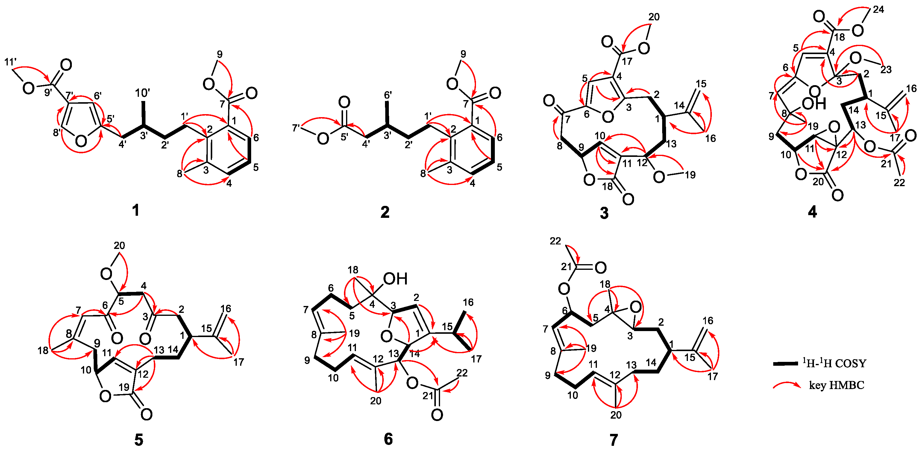
In our recent chemical investigations of the Xisha soft coral Sinularia densa, which is the first systematic investigation for this genus, five C19-Norcembranoid diterpenes with unusual scaffolds, named sinudenoids A–E, were discovered, showing the chemical diversity of this genus [14]. In order to find more bioactive metabolites, our ongoing research on the soft coral Xisha from Sinularia densa (Sinulariidae) has uncovered seven undescribed substances (17) (Figure 1). Here, we report the isolation, structure elucidation, and anti-inflammatory and anti-thrombotic effects of these isolated compounds.

2. Results

Sinudenoid F (1) was isolated as a colourless oil, and its molecular formula was determined as C20H24O5 by the HRESIMS ion peak at m/z 367.1511 [M + Na]+, implying nine degrees of unsaturation. Analysis of the 1H and 13C NMR data of 1 (Table S1) revealed the presence of 20 carbons, including seven non-protonated carbons (five olefinic and two carbonyl), six methines (one sp3 hybridized and five olefinic), three methylenes (all sp3 hybridized), and four methyls (two linked to oxygen, one to a sp3 carbon and one to an olefinic carbon).
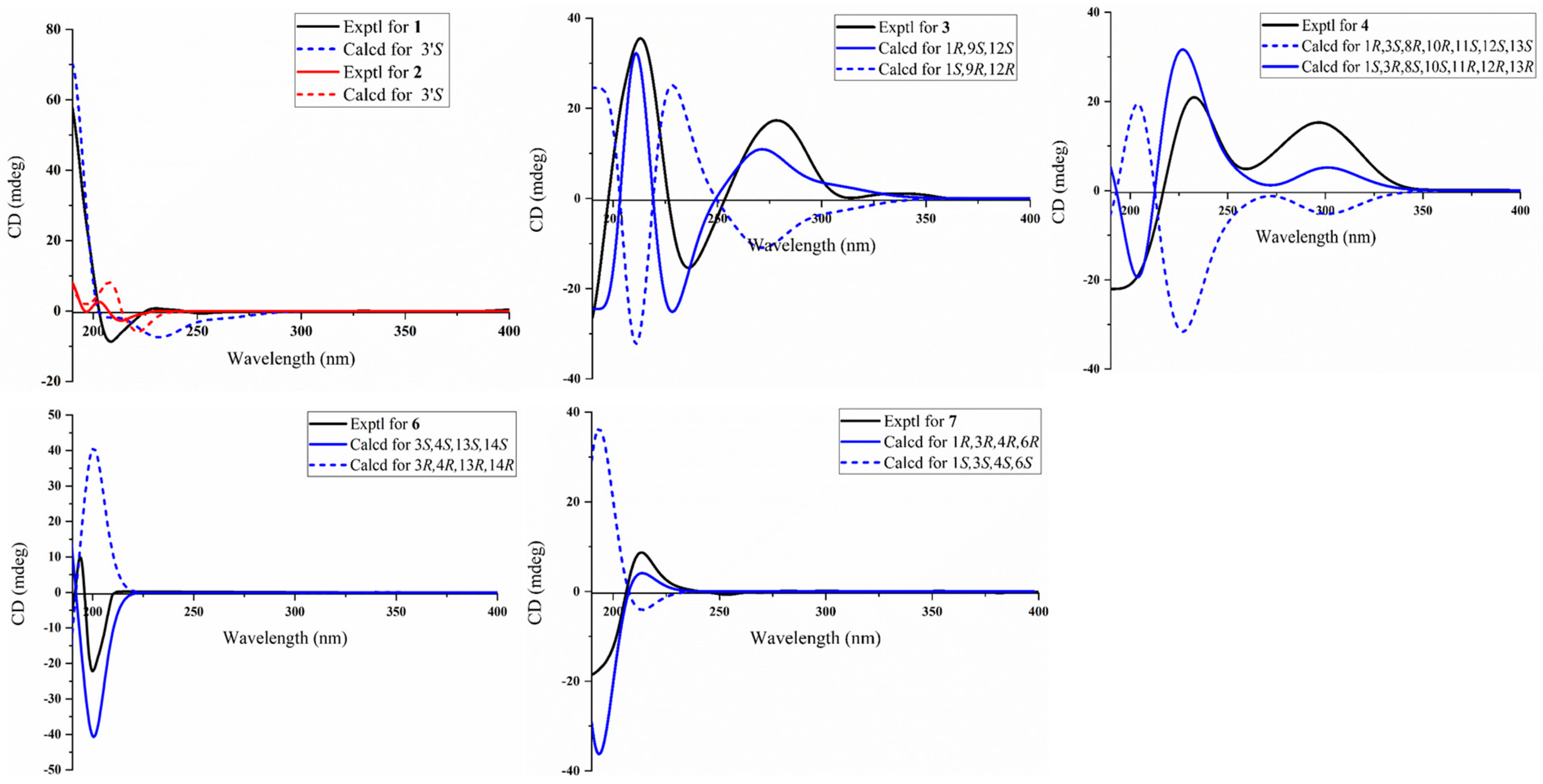
The presence of an isopentyl group was confirmed by the 1H–1H COSY correlations from H-1′ to H-4′ and H-3′ to H3-10′ (Figure 2). Subsequently, the presence of a trisubstituted benzene ring was supported by the HMBC correlations from H3-8 to C-2 (δC142.0), C-3 (δC137.4), and C-4 (δC133.9); H-5 to C-1; and H-6 to C-7, together with the 1H–1H COSY correlation from H-4 to H-6. Furthermore, the HMBC correlation from H3-9 to C-7 (δC169.0) indicated the presence of a methyl ester group at C-1 of the aromatic ring. Considering the aforementioned data, the HMBC correlations from H-1′ to C-1 and C-2 indicated the attachment of the isopentyl group to the trisubstituted benzene ring at C-2. Moreover, the presence of a furan ring located at C-4′ was inferred by the HMBC correlations from H-8′ to C-5′ and C-7′; H-6′ to C-5′ and C-7′; and H-4′ to C-5′, together with the downfield chemical shifts of C-5′ (δC157.0) and C-8′ (δC146.4). The HMBC correlation from H3-11′ to C-9′ (δC164.1) indicated the presence of a methyl ester group, which was further confirmed by its molecular formula and the requirement of degrees of unsaturation. Due to the absence of the key HMBC correlation from either H-6′ to C-9′ or H-8′ to C-9′, the connection between C-7′ and C-9′ was established through a meticulous comparison of the 1D NMR data between 1 and 3-furancarboxylic acid [15], in conjunction with the absence of a substituent at C-7′. Consequently, the planer structure of 1 was established. Finally, the absolute configuration of 1 was defined as 3′S in the TDDFT-ECD calculations (Figure 3).
Sinudenoid G (2), a colourless oil, had a molecular formula of C16H22O4, as established by the HRESIMS ion peak at m/z 301.1412 [M + Na]+, which was 66 mass units less than that of 1. The 1H and 13C NMR data of 2 (Table S2) resemble those of 1, with the exception of the disubstituted furan ring at C-4′ in 1 replaced by a methyl ester group in 2. This alteration was confirmed by the HMBC correlations (Figure 2) from H3-7′ to C-5′ (δC173.6) and H-4′ to C-5′. Finally, the absolute configuration of 2 was defined as 3′S in the TDDFT-ECD calculations (Figure 3).
Sinudenoid H (3) was isolated as a colourless oil, with a molecular formula of C20H22O7 deduced from the HRESIMS ion peak at m/z 375.1448 [M + H]+, implying ten degrees of unsaturation. The 1D NMR data of 3 (Table S3) resemble those of sarcofuranocembrenolide A, a known 8, 19-bisnorfuranocembrenolide previously isolated from the soft coral Sarcophyton sp. [16]. In fact, the structure of 3 closely resembles sarcofuranocembrenolide A, differing in the presence of a methoxy group at C-12 in 3 instead of an ethoxy group in sarcofuranocembrenolide A (Figure S1). The HMBC correlation (Figure 2) from H3-19 (δH 3.19) to C-12 confirmed these functional group disparities. In the NOESY spectrum of 3 (Figure 4), the correlation of H-12 (δH 4.08)/H-1 (δH 2.43) suggested their co-facial orientation. Due to the absence of definitive NOESY correlations, the configuration of C-9 was determined as S* using the DP4+ calculation (Supporting Information, Figures S2 and S3). Moreover, the TDDFT/ECD calculations also supported the 1R, 9S, 12S-configurations of 3 (Figure 3), instead of the 1R, 9R, 12S configurations (Figure S10).
Sinudenoid I (4) was isolated as a colourless oil. Its molecular formula was determined to be C24H30O10 from the sodium adduct ion peak at m/z 501.1729 [M + Na]+, implying ten degrees of unsaturation. Analysis of the 1D NMR data (Table S4) and HSQC spectrum of 4 suggested the presence of twenty-four carbons, including nine non-protonated carbons (three olefinic, three carbonyl, and three oxygenated), six methines (one sp3 hybridized, two olefinic, and three oxygenated), four methylenes (three sp3 hybridized and one olefinic), and five methyls (one linked to a carbonyl, one to an oxygenated sp3 carbon, two to oxygen atoms, and one to an olefinic carbon). These dates indicated that 4 was a polycyclic diterpenoid.
Upon scrutinizing the NMR data of 4, it was found that the 1D NMR data resemble that of sinumaximol C, a diterpenoid previously isolated from the soft coral Sinularia maxima [17]. In fact, the structure of 4 was truly similar to sinumaximol C (Figure S1), except for the presence of an additional acetoxy group at C-13 in compound 4 in comparison to sinumaximol C. This deduction was further proven by HMBC correlations (Figure 2) from H3-22 (δH 2.01) to C-21 (δC 170.7), and H-13 (δH 4.84) to C-21. The 1D NOE correlations (Figure 4) from H-10 to H-11, H3-19, along with the NOESY correlation between H-13 and H-11, indicated that H-10, H-11, H-13, and H-19 were positioned on the same face. Subsequently, the similar chemical shifts of carbons from C-3 to C-12 compared with sinumaximol C indicated the 8R*, 10R*, 11S*, and 12S* configurations in 4, which was same as those of sinumaximol C. In the absence of definitive NOESY correlations, the relative configurations of C-1 and C-3 were determined as 1R*, 3S* by the DP4+ calculation (Supporting Information, Figures S4 and S5). Finally, the absolute configurations of 4 were defined as 1S, 3R, 8S, 10S, 11R, 12R, and 13R using the TDDFT-ECD calculations (Figure 3).
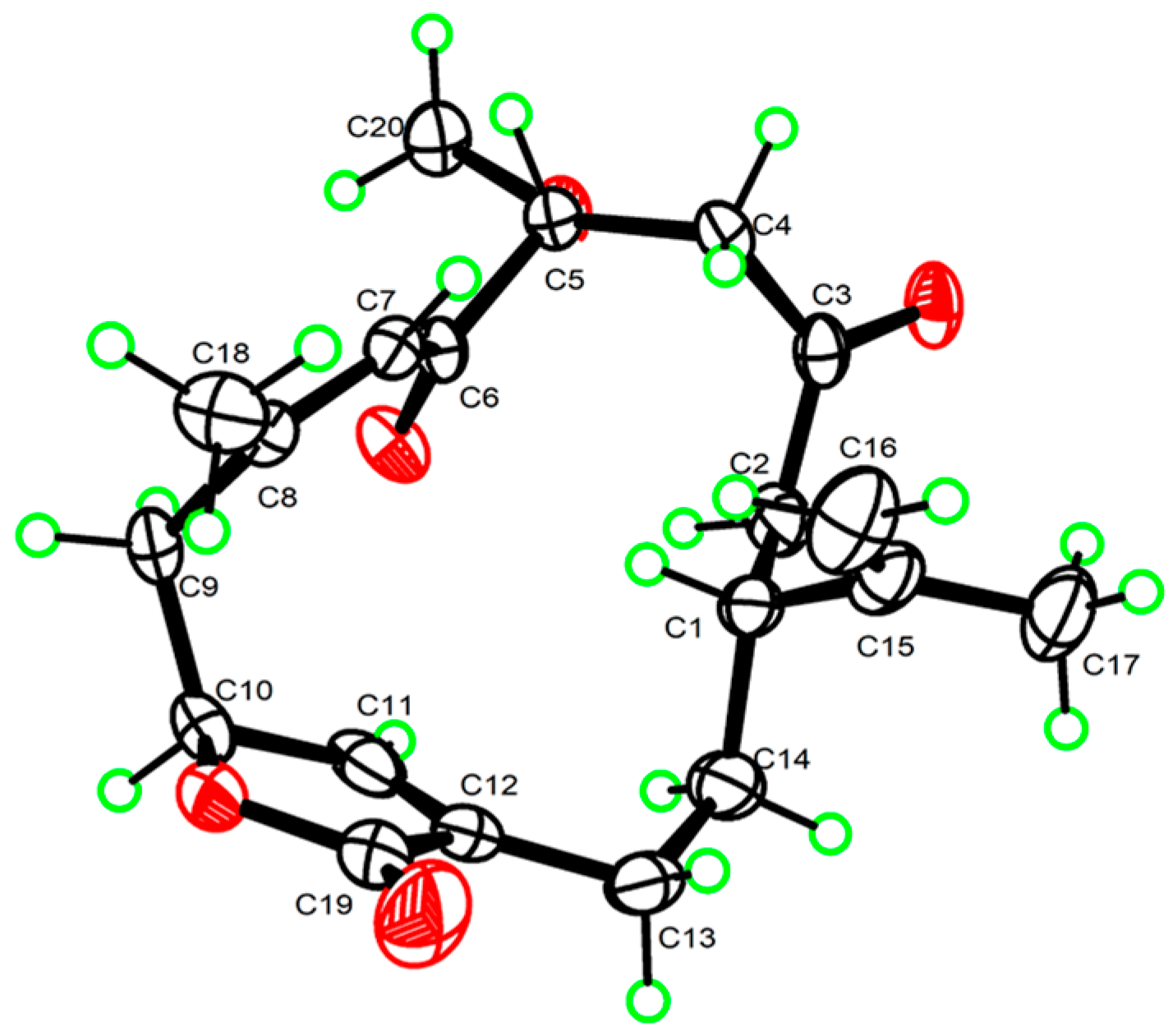
Sinudenoid J (5) was purified as a colourless crystal, with a molecular formula of C20H26O5 determined from its HRESIMS ion peak at m/z 369.1674 [M + Na]+. The 1H and 13C NMR data (Table S5) of 5 resemble those of gyrosanolide D (Figure S1), a known cembranoid previously isolated from the soft coral Sinularia gyrosa [18]. The difference in the planar structures between 5 and gyrosanolide D was that the hydroxy group at C-5 in gyrosanolide D was replaced by a methoxy group in 5. This deduction was further proven by the HMBC correlation (Figure 2) from H3-20 (δH 3.41) to C-5 (δC 82.6). Due to the lack of effective NOESY correlations, the geometry of the double bond and the configurations of chiral centres in compound 5 could not be definitively determined. However, an X-ray crystallographic experiment with the Cu Kα radiation of 5 (Figure 5) confirmed the Z-geometry of the Δ7 double bond. Subsequently, the absolute configurations of 5 were defined as 1R, 5R, and 10S, indicating that the geometry of the Δ7 double bond and the configuration at C-5 in 5 differ from those of gyrosanolide D.
Sinudenoid K (6) was isolated as a colourless oil. Its molecular formula of C22H32O4 was deduced from the sodium adduct ion peak at m/z 361.2374 [M + H]+, indicating seven degrees of unsaturation. Analysis of the 1D NMR data (Table S6) and HSQC spectrum of 6 revealed the presence of twenty-two carbons, including five non-protonated carbons (three olefinic, one carbonyl, and sp3 hybridized), seven methines (one sp3 hybridized, three olefinic, and three oxygenated), four methylenes (all sp3 hybridized), and six methyls (one linked to a carbonyl, one to an oxygenated sp3 carbon, two to sp3 carbons and two to olefinic carbons). These dates indicated that 6 possessed a cembrane nucleus.
Upon analysis the NMR data of 6, it was found that the 1D NMR data of 6 resemble that of klyflaccicembranols H, a cembranoid previously isolated from the soft coral Klyxum flaccidum [19]. In fact, the structure of 6 was truly similar to that of klyflaccicembranol H (Figure S1), except for the presence of the trisubstituted double bond at C-11, C-12, and C-20 in 6, instead of a trisubstituted epoxide ring in klyflaccicembranol H. This deduction was further proven by the HMBC correlations (Figure 2) from the H3-20 (δH 1.53) to C-11 (δC 133.3), C-12 (δC 129.2), and C-13 (δC 81.2).
In the NOESY spectrum of 6 (Figure 4), the correlations of H3-19/H-6a (δH 2.42), H-11/H3-13, and H3-20/H-10a (δH 2.29) indicated that the Δ7 and Δ11 double bonds exhibited E-geometry. The correlation of H-2/H3-16 (δH 1.16) suggested that the Δ2 double bond had a Z-geometry. Additionally, the NOESY cross-peak of H3-18/H-3 suggested that these protons were co-facial. Furthermore, the correlation from H-14 to H-13 in the 1D-NOE difference spectrum indicated that H-13 and H-14 were on the same side. In the absence of the key NOESY correlations, the relative configurations of these chiral centres were determined as 3S*, 4S*, 13S*, and 14S* using the DP4+ calculation (Supporting Information, Figures S6 and S7). Finally, the absolute configurations of 6 were defined through the TDDFT-ECD calculations (Figure 3).
Sinudenoid L (7) was isolated as a colourless oil, with a molecular formula of C22H34O3, as indicated by its HRESIMS ion peak at m/z 369.2399 [M + Na]+. The 1D NMR data of 7 (Table S7) resemble those of (7E,11E)-3,4-epoxy-7,11,15-cembratriene (Figure S1), a cembranoid previously isolated from a south Pacific soft coral [20]. The only difference was the replacement of one hydrogen atom of methylene at C-6 in the known cembranoid with an acetoxy group in 7. The differentiation was supported by the HMBC correlations (Figure 2) from H3-22 to C-21 (δC 170.1) and H-6 (δH 5.66) to C-21.
Then, the relative configurations of 7 were determined by the analysis of the NOESY spectrum (Figure 4). The NOESY correlations of H-7/H-9a (δH 1.98) and H3-20/H-10a (δH 2.25) were used to establish the E-geometry of the Δ7 and Δ11 double bonds. Additionally, the NOESY correlations of H-1/H-3 and 5a (δH 1.45)/H-3 indicated that these protons were all co-facial. Moreover, the NOESY correlations of H-5b/H3-18 indicate these protons were also co-facial. By utilizing the 13C NMR chemical shift calculations for the DP4+ calculations (Supporting Information, Figures S8 and S9), in conjunction with the NOESY correlation of H-3/H-6, the configuration of C-6 was defined as R*. Finally, the TDDFT/ECD calculations of 7 also supported the 1R, 3R, 4R, and 6R-configurations (Figure 3), instead of the 1R, 3R, 4R, and 6S-configurations (Figure S11).
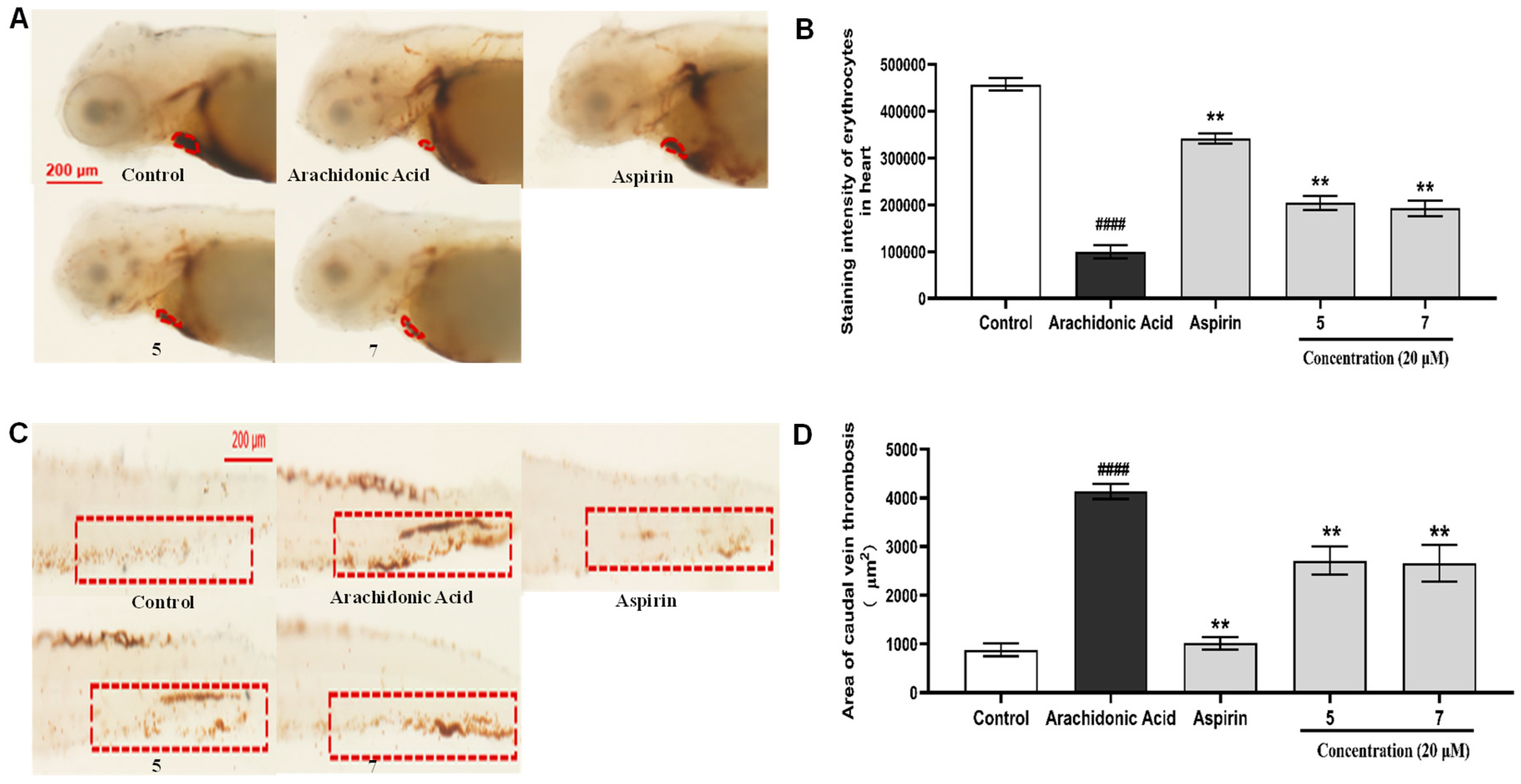
In the bioassay, these new compounds (17) were evaluated for anti-inflammatory activity in CuSO4-treated transgenic fluorescent zebrafish. The results (Figure 6) showed that 3 could reduce migration and decrease the number of macrophages surrounding the neuromasts in zebrafish, displaying better anti-inflammatory activity with an inhibition rate of 56.8% compared to the indomethacin positive control with an inhibition rate of 38.4% at 20 μM. Moreover, due to the limited availability, 5 and 7 were selectively evaluated for anti-thrombotic activity in arachidonic acid (AA)-induced thrombotic zebrafish. In arachidonic acid (AA)-induced thrombotic zebrafish, 5 and 7 enhanced the staining intensity of erythrocytes in the heart and inhibited the area of caudal vein thrombosis, demonstrating anti-thrombotic activity at 20 μM (Figure 7).

3. Materials and Methods

3.1. General Experimental Procedures

Optical rotations were measured using a Jasco P-1020 digital polarimeter. Ultraviolet (UV) spectra were measured using a Beckman DU640 spectrophotometer. CD spectra were measured using a Jasco J-810 spectropolarimeter. NMR spectra were measured using an Agilent 500 MHz and a JEOL JNMECP 600 spectrometer. The 7.26 ppm (1H) resonance of the residual CHCl3 in CDCl3 and the 77.16 ppm (13C) resonance of CDCl3 were used as internal references for 1H and 13C NMR spectra, respectively. HRESIMS spectra were measured using a Micromass Q-Tof Ultima GLOBAL GAA076LC mass spectrometer. The crystallographic data were measured on a Bruker D8 Venture diffractometer (Bruker, Beijing, China) equipped with graphite-monochromatized Cu Kα radiation. A semi-preparative HPLC was performed using a Waters 1525 pump equipped with a 2998 photodiode array detector and a YMC C18 column (YMC, 10 × 250 mm, 5 μm). Silica gel (200–300 mesh, 300–400 mesh and silica gel H) was used for the column chromatography.

3.2. Animal Material

The soft coral Sinularia densa (Sinulariidae) was collected from Xisha Island (Ganquan island) of the South China Sea in July 2018 (111°58′ E, 16°50′ N), and was frozen immediately after collection. The specimen was dispatched via express delivery and subsequently identified based on its morphology and sclerites by Prof Ping-Jyun Sung, Institute of Marine Biotechnology, National Museum of Marine Biology & Aquarium, Pingtung 944, Taiwan. The voucher specimen (No. xs-18-yg-99) has been deposited at the State Key Laboratory of Marine Drugs, Ocean University of China, People’s Republic of China.

3.3. Extraction and Isolation

A frozen specimen of Sinularia densa (3.5 kg, wet weight) was homogenized and then exhaustively extracted with CH3OH six times (for 3 days each time) at room temperature. The combined solutions were concentrated in vacuo and desalted by redissolving with CH3OH to yield a residue (83.0 g). The crude extract was subjected to silica gel vacuum column chromatography eluted with a gradient of petroleum ether/acetone (from 200:1 to 1:1, v/v) and subsequently CH2Cl2/MeOH (from 10:1 to 1:1, v/v) to obtain fourteen fractions (Fr.1–Fr.14). Each fraction was detected by TLC. Fr.2 was subjected to silica gel vacuum column chromatography (petroleum ether/acetone, from 50:1 to 1:1, v/v) to give four subfractions, Fr.2.1–Fr.2.4. Fr.2.3 was separated by semi-preparative HPLC (ODS, 5 µm, 250 × 10 mm; MeOH/H2O, 85:15, v/v; 1.5 mL/min) to afford 1 (6.3 mg, tR = 24 min). Fr.2.4 was separated by semi-preparative HPLC (ODS, 5 µm, 250 × 10 mm; MeOH/H2O, 85:15, v/v; 1.5 mL/min) to afford 2 (1.4 mg, tR = 15 min). Fr.4 was subjected to silica gel vacuum column chromatography (petroleum ether/acetone, from 40:1 to 1:1, v/v) to give four subfractions, Fr.4.1–Fr.4.4. Fr.4.3 was separated by semi-preparative HPLC (ODS, 5 µm, 250 × 10 mm; MeOH/H2O, 65:35, v/v; 1.5 mL/min) to afford 3 (1.0 mg, tR = 42 min) and 6 (2.4 mg, tR = 48 min). Fr.6 was subjected to silica gel vacuum column chromatography (petroleum ether /acetone, from 30:1 to 1:1, v/v) to give six subfractions, Fr.6.1–Fr.6.6. Fr.6.4 was separated by semi-preparative HPLC (ODS, 5 µm, 250 × 10 mm; MeOH/H2O, 45:55, v/v; 1.5 mL/min) to afford 5 (2.2 mg, tR = 48 min) and 4 (3.0 mg, tR = 30 min). Fr.6.5 was separated by semi-preparative HPLC (ODS, 5 µm, 250 × 10 mm; MeOH/H2O, 45:55, v/v; 1.5 mL/min) to afford 7 (2.3 mg, tR = 48 min).
Sinudenoid F (1): Colourless oil; [α]25D +85.2 (c 0.1, MeOH); UV (MeOH) λmax (log ε) = 200 (2.22) nm, 233 (0.36) nm; IR (KBr) νmax = 3420, 2980, 1740, 1635, 1033 cm−1; HRESIMS m/z 345.1693 [M + H]+ (calcd. for C20H25O5, 345.1697) and HRESIMS m/z 367.1511 [M + Na]+ (calcd. for C20H24O5Na, 367.1516). For 1H NMR and 13C NMR data, see Table S1.
Sinudenoid G (2): Colourless oil; [α]25D −32.1 (c 0.1, MeOH); UV (MeOH) λmax (log ε) = 204 (1.20) nm, 242 (0.40) nm; IR (KBr) νmax = 2924, 1719, 1615, 1384, 1269 cm−1; HRESIMS m/z 279.1594 [M + H]+ (calcd. for C16H23O4, 279.1591) and HRESIMS m/z 301.1412 [M + Na]+ (calcd. for C16H22O4Na, 301.1410). For 1H NMR and 13C NMR data, see Table S2.
Sinudenoid H (3): Colourless oil; [α]25D +35.2 (c 0.5, MeOH); UV (MeOH) λmax (log ε) = 193 (1.50) nm, 280 (0.69) nm; IR (KBr) νmax = 3420, 1756, 1718, 1683, 1592, 1384 cm−1; HRESIMS m/z 375.1448 [M + H]+ (calcd. for C20H23O7, 375.1438). For 1H NMR and 13C NMR data, see Table S3.
Sinudenoid I (4): Colourless oil; [α]25D −124.0 (c 1.0, MeOH); UV (MeOH) λmax (log ε) = 192 (0.84) nm, 300 (0.92) nm; IR (KBr) νmax = 2925, 1750, 1708, 1670, 1645, 1363 cm−1; HRESIMS m/z 501.1729 [M + Na]+ (calcd. for C24H30O10Na, 501.1731). For 1H NMR and 13C NMR data, see Table S4.
Sinudenoid J (5): Colourless crystal; [α]25D +67.0 (c 0.3, MeOH); UV (MeOH) λmax (log ε) = 194 (0.89) nm; IR (KBr) νmax = 1757, 1734, 1716 cm−1; HRESIMS m/z 347.1859 [M + H]+ (calcd. for C20H27O5, 347.1853). For 1H NMR and 13C NMR data, see Table S5.
Sinudenoid K (6): Colourless oil; [α]25D −29.0 (c 0.5, MeOH); UV (MeOH) λmax (log ε) = 192 (0.04) nm, 214 (0.04) nm, 240 (0.03) nm; IR (KBr) νmax = 3431, 2359, 1631, 1375 cm−1; HRESIMS m/z 361.2374 [M + H]+ (calcd. for C22H33O4, 361.2384). For 1H NMR and 13C NMR data, see Table S6.
Sinudenoid L (7): Colourless oil; [α]25D +28.3 (c 0.1, MeOH); UV (MeOH) λmax (log ε) = 195 (1.68) nm; IR (KBr) νmax = 3419, 2925, 1683, 1615, 1384 cm−1; HRESIMS m/z 369.2399 [M+Na]+ (calcd. for C22H34O3Na, 369.2400). For 1H NMR and 13C NMR data, see Table S7.

3.4. X-ray Crystallographic Analysis

Sinudenoid J (5) was obtained as a colourless crystal in an EtOH-H2O solvent system using the vapour diffusion method. Crystal data for Sinudenoid J (5): C20H26O5, M = 346.41, a = 7.4110(3) Å, b = 12.6917(5) Å, c = 19.7025(8) Å, α = 90°, β = 90°, γ = 90°, V = 1853.18 (13) Å3, T = 150 K, space group P212121, Z = 4, μ (Cu Kα) = 0.720 mm−1, 3717 independent reflections (Rint = 0.0488). The final R1 values were 0.0356 (I > 2σ(I)). The final wR2 values were 0.0869 (I > 2σ(I)). The final R1 values were 0.0370 (all data). The final wR2 values were 0.0882 (all data). The goodness of fit on F2 was 1.044. The Flack parameter was −0.02(7). The crystallographic data for 5 in this article have been deposited at the Cambridge Crystallographic Data Centre under supplementary publication number 2150265. The data can be obtained via https://www.ccdc.cam.ac.uk/ (accessed on 5 February 2022).

3.5. Bioassay

Three-day post-fertilization (dpf) healthy macrophage fluorescent transgenic zebrafish were employed as animal models to evaluate the anti-thrombotic and anti-inflammatory effects of the isolates. Healthy macrophage fluorescent transgenic zebrafish (Tg: zlyz-EGFP) were provided by the Biology Institute of Shandong Academy of Sciences (Jinan, China). Zebrafish larvae with CuSO4-treated transgenic fluorescent (Tg:zlyz-EGFP) expressing enhanced green fluorescent protein (EGFP) were used to evaluate anti-inflammatory effects. Zebrafish larvae with arachidonic acid (AA)-induced thrombus were utilized to evaluate anti-thrombosis effects. Each zebrafish larva was imaged using a fluorescence microscope (AXIO, Zom.V16), and the number of macrophages around the nerve mound were calculated using Image-Pro Plus software (version 5.1). Statistical analysis was conducted using a one-way analysis of variance with GraphPad Prism 7.00 software.

4. Conclusions

In summary, two rare meroterpenoids (12) and five new cembranes (37) were isolated from the soft coral Sinularia densa. Sinudenoid F (1) and sinudenoid G (2) are rare meroterpenoids featuring a methyl benzoate core. While the skeleton of 1 has been obtained in synthetic studies, it represents the first instance of this natural product skeleton [21]. Sinudenoid H (3) possesses a rare carbon skeleton of 8, 19-bisnorfuranocembrenolide, and it is the second reported compound of this skeleton [15]. Sinudenoid J (5) is a new furanobutenolide-derived C19-norcembranoid diterpene, whose structure was confirmed by X-ray diffraction analysis. In the biological assays, 3 showed potent anti-inflammatory activity, while 5 and 7 demonstrated moderate anti-thrombotic activity in zebrafish models. The current study shows the potential of the cembranes as marine-derived anti-inflammatory and anti-thrombotic lead molecules, and provides a basis for developing new drugs.

Supplementary Materials

The following supporting information can be downloaded at https://www.mdpi.com/article/10.3390/md22100442/s1, Tables S1–S7: NMR data of 17; Figure S1: Structures of 37 and structurally related compounds of 37; Tables S8–S11 and Figures S2–S9: The determination of the relative and absolute configurations for 3, 4, 6, and 7, respectively; Figures S10 and S11: Experimental and calculated ECD spectra of 3a and 7b, respectively; Table S12: Anti-inflammation assay of 17; Table S13: Anti-thrombotic assay of 5 and 7; Table S14 and Figures S12–S17: Computational details; Figures S18–S84: Spectra for 17.

Author Contributions

Conceptualization, H.H. and P.L.; methodology, H.H., P.L. and C.W.; software, K.L.; validation, J.Y.; investigation, L.L. and S.W.; writing—original draft preparation, C.W. and J.Z.; writing—review and editing, H.H. and P.L. All authors have read and agreed to the published version of the manuscript.

Funding

This research was funded by the National Natural Science Foundation of China (No. 42276088).

Institutional Review Board Statement

Not applicable.

Data Availability Statement

The data are contained within the article or Supplementary Materials; further inquiries can be directed to the corresponding author.

Acknowledgments

We give special thanks to the Center for High Performance Computing and System Simulation (Pilot National Laboratory for Marine Science and Technology) for the support extended toward the computer calculations. Special thanks also go to the major scientific and technological innovation project in Shandong Province (No. 2022CXGC020414) and major scientific and technological innovation project of Qingdao West Coast New Area (No. ZDKC-2022-593 01) for the support extended toward the computer calculations.

Conflicts of Interest

The authors declare no conflicts of interest.

References

  1. Tursch, B.; Braekman, J.C.; Daloze, D.; Herin, M.; Karlsson, R.; Losman, D. Chemical studies of marine invertebrates. XI. Sinulariolide, a new cembranolide diterpene from the soft coral Sinularia flexibilis. Tetrahedron 1975, 31, 129–133. [Google Scholar] [CrossRef]
  2. Yan, X.; Liu, J.; Leng, X.; Ouyang, H. Chemical Diversity and Biological Activity of Secondary Metabolites from Soft Coral Genus Sinularia since 2013. Mar. Drugs 2021, 19, 335. [Google Scholar] [CrossRef]
  3. Tseng, H.J.; Kuo, L.M.; Tsai, Y.C.; Hu, H.C.; Chen, P.J.; Chien, S.Y.; Sheu, J.H.; Sung, P.J. Sinulariaone A: A novel diterpenoid with a 13-membered carbocyclic skeleton from an octocoral Sinularia species. RSC Adv. 2023, 13, 10408–10413. [Google Scholar] [CrossRef] [PubMed]
  4. Du, Y.Q.; Yao, L.G.; Li, X.W.; Guo, Y.W. Yonarolide A, an unprecedented furanobutenolide-containing norcembranoid derivative formed by photoinduced intramolecular [2+2] cycloaddition. Chin. Chem. Lett. 2023, 34, 107512. [Google Scholar] [CrossRef]
  5. Pham, G.N.; Kang, D.Y.; Kim, M.J.; Han, S.J.; Lee, J.H.; Na, M.K. Isolation of Sesquiterpenoids and Steroids from the Soft Coral Sinularia brassica and Determination of Their Absolute Configuration. Mar. Drugs 2021, 19, 523. [Google Scholar] [CrossRef]
  6. Phan, G.H.; Tsai, Y.C.; Liu, Y.H.; Fang, L.S.; Wen, Z.H.; Hwang, T.L.; Chang, Y.C.; Sung, P.J. Sterol constituents from a cultured octocoral Sinularia sandensis (Verseveldt 1977). J. Mol. Struct. 2021, 1246, 131175. [Google Scholar] [CrossRef]
  7. Jiang, W.J.; Wang, D.D.; Wilson, B.A.P.; Voeller, D.; Bokesch, H.R.; Smith, E.A.; Lipkowitz, S.; O’Keefe, B.R.; Gustafson, K.R. Sinularamides A-G, Terpenoid-Derived Spermidine and Spermine Conjugates with Casitas B-Lineage Lymphoma Proto-Oncogene B (Cbl-b) Inhibitory Activities from a Sinularia sp. Soft Coral. J. Nat. Prod. 2021, 84, 1831–1837. [Google Scholar] [CrossRef]
  8. Yang, B.; Wei, X.Y.; Huang, J.X.; Lin, X.P.; Liu, J.; Liao, S.R.; Wang, J.F.; Zhou, X.F.; Wang, L.S.; Liu, Y.H. Sinulolides A-H, new cyclopentenone and butenolide derivatives from soft coral Sinularia sp. Mar. Drugs 2014, 12, 5316–5327. [Google Scholar] [CrossRef]
  9. Wu, M.J.; Yu, D.D.; Su, M.Z.; Wang, J.R.; Gong, L.; Zhang, Z.Y.; Wang, H.; Guo, Y.W. Discovery and photosynthesis of sinuaustones A and B, diterpenoids with a novel carbon scaffold isolated from soft coral Sinularia australiensis from Hainan. Org. Chem. Front. 2022, 9, 5921–5928. [Google Scholar] [CrossRef]
  10. Kamada, T.; Phan, C.S.; Hamada, T.; Hatai, K.; Vairappan, C.S. Cytotoxic and Antifungal Terpenoids from Bornean Soft Coral, Sinularia flexibilis. Nat. Prod. Commun. 2018, 13, 17–19. [Google Scholar] [CrossRef]
  11. Wu, Z.W.; Wang, Z.X.; Guo, Y.Q.; Tang, S.A.; Feng, D.Q. Antifouling activity of terpenoids from the corals Sinularia flexibilis and Muricella sp. against the bryozoan Bugula neritina. J. Asian Nat. Prod. Res. 2023, 25, 85–94. [Google Scholar] [CrossRef] [PubMed]
  12. Xia, Z.Y.; Sun, M.M.; Jin, Y.; Su, M.Z.; Li, S.W.; Wang, H.; Guo, Y.W. Four uncommon cycloamphilectane-type diterpenoids with antibacterial activity from the South China Sea soft coral Sinularia brassica. Phytochemistry 2024, 219, 113960. [Google Scholar] [CrossRef] [PubMed]
  13. Huang, C.; Jin, Y.; Sun, R.N.; Hu, K.Y.; Yao, L.G.; Guo, Y.W.; Yuan, Z.H.; Li, X.W. Anti-HBV Activities of Cembranoids from the South China Sea Soft Coral Sinularia pedunculata and Their Structure Activity Relationship. Chem. Biodivers. 2024, 21, e202401146. [Google Scholar] [CrossRef] [PubMed]
  14. Wang, C.L.; Jin, T.Y.; Liu, X.H.; Zhang, J.R.; Shi, X.; Wang, M.F.; Huang, R.; Zhang, Y.; Liu, K.C.; Li, G.Q. Sinudenoids A-E, C19-Norcembranoid Diterpenes with Unusual Scaffolds from the Soft Coral Sinularia densa. Org. Lett. 2022, 24, 9007–9011. [Google Scholar] [CrossRef]
  15. Tamaso, K.I.; Hatamoto, Y.; Obora, Y.; Sakaguchi, S.; Ishii, Y. Synthesis of substituted furoates from acrylates and aldehydes by Pd(oAc)2/HPMoV/CeCl3/O2 system. J. Org. Chem. 2007, 72, 8820–8823. [Google Scholar] [CrossRef]
  16. Kapojos, M.M.; Lee, J.S.; Oda, T.; Nakazawa, T.; Takahashi, O.; Ukai, K.; Mangindaan, R.E.P.; Rotinsulu, H.; Wewengkang, D.S.; Tsukamoto, S.; et al. Two unprecedented cembrene-type terpenes from an indonesian soft coral sarcophyton sp. Tetrahedron 2010, 66, 641–645. [Google Scholar] [CrossRef]
  17. Thao, N.P.; Nam, N.H.; Cuong, N.X.; Quang, T.H.; Tung, P.T.; Tai, B.H.; Bui, T.T.L.; Chae, D.; Kim, S.; Koh, Y.S.; et al. Diterpenoids from the Soft Coral Sinularia maxima and Their Inhibitory Effects on Lipopolysaccharide-Stimulated Production of Pro-inflammatory Cytokines in Bone Marrow-Derived Dendritic Cells. Chem. Pharm. Bull. 2012, 60, 1581–1589. [Google Scholar] [CrossRef]
  18. Cheng, S.Y.; Chuang, C.T.; Wen, Z.H.; Wang, S.K.; Chiou, S.F.; Hsu, C.H.; Dai, C.F.; Duh, C.Y. Bioactive norditerpenoids from the soft coral Sinularia gyrosa. Bioorg. Med. Chem. 2010, 18, 3379–3386. [Google Scholar] [CrossRef]
  19. Ahmed, A.F.; Tsai, C.R.; Huang, C.Y.; Wang, S.Y.; Sheu, J.H. Klyflaccicembranols A-I, New Cembranoids from the Soft Coral Klyxum flaccidum. Mar. Drugs 2017, 15, 23. [Google Scholar] [CrossRef]
  20. Ravi, B.N.; Faulkner, D.J. Cembranoid diterpenes from a South Pacific soft coral. J. Org. Chem. 1978, 43, 2127–2131. [Google Scholar] [CrossRef]
  21. Gros, J.; Reddy, C.M.; Aeppli, C.; Nelson, R.K.; Carmichael, C.A.; Arey, J.S. Resolving Biodegradation Patterns of Persistent Saturated Hydrocarbons in Weathered Oil Samples from the Deepwater Horizon Disaster. Environ. Sci. Technol. 2014, 48, 1628–1637. [Google Scholar] [CrossRef]
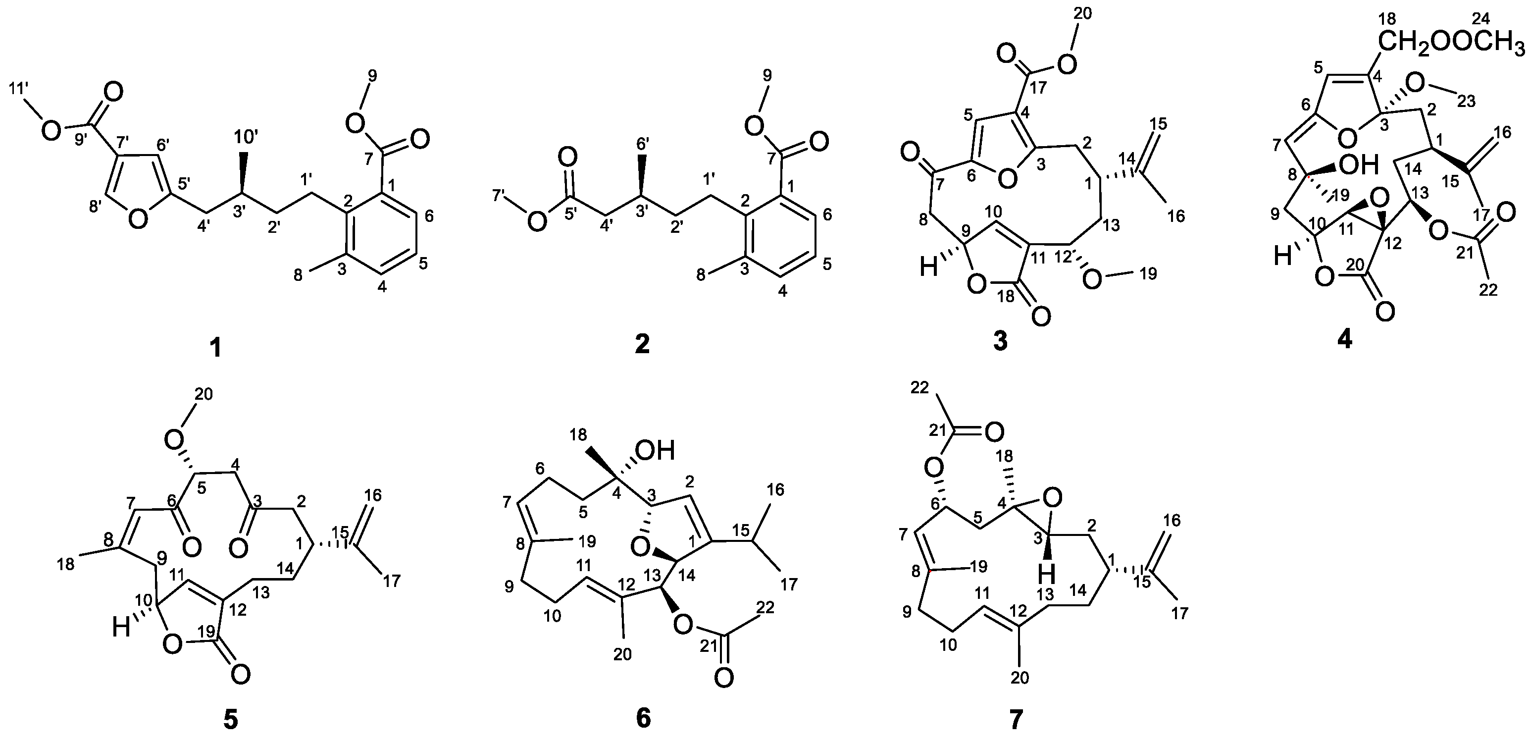
Figure 1. Structures of 17 from the soft coral Sinularia densa.
Marinedrugs 22 00442 g001
Figure 2. Selected 1H–1H COSY and HMBC correlations of 17.
Marinedrugs 22 00442 g002
Figure 3. Experimental and calculated ECD spectra of 14 and 67.
Marinedrugs 22 00442 g003
Figure 4. NOESY correlations of 34 and 67.
Marinedrugs 22 00442 g004
Figure 5. Perspective ORTEP drawings of the X-ray structures of 5 (displacement ellipsoids are drawn at the 50% probability level).
Marinedrugs 22 00442 g005
Figure 6. Anti-inflammatory assays of 17. (a) Images of inflammatory sites in CuSO4-treated transgenic fluorescent zebrafish (Tg:zlyz-EGFP) expressing enhanced green fluorescent protein (EGFP) treated with 1 through 7, using indomethacin as a positive control. (b) Quantitative analysis of macrophages in the region of inflammatory sites in zebrafish treated with 1 through 7. #### indicates that the CuSO4 model group has a very significant difference compared with the control group (p < 0.01). ** indicates that sample groups have significant differences compared with the CuSO4 model group (p < 0.01).
Marinedrugs 22 00442 g006
Figure 7. Anti-thrombotic assays of 5 and 7. (A) Images of the staining intensity of erythrocytes in the heart in arachidonic acid (AA)-induced thrombotic zebrafish, treated with either 5 or 7, using aspirin as a positive control. (B) Quantitative analysis of the staining intensity of erythrocytes in the heart in zebrafish treated with either 5 or 7. (C) Images of areas of caudal vein thrombosis in arachidonic acid (AA)-induced thrombotic zebrafish, treated with either 5 or 7, using aspirin as a positive control. (D) Quantitative analysis of the area of caudal vein thrombosis in zebrafish treated with either 5 or 7. #### indicates that the arachidonic acid model group has a very significant difference compared with the control group (p < 0.01). ** indicates that sample groups have significant differences compared with the arachidonic acid model group (p < 0.01).
Marinedrugs 22 00442 g007
Disclaimer/Publisher’s Note: The statements, opinions and data contained in all publications are solely those of the individual author(s) and contributor(s) and not of MDPI and/or the editor(s). MDPI and/or the editor(s) disclaim responsibility for any injury to people or property resulting from any ideas, methods, instructions or products referred to in the content.