Article

Dolabelladienols A–C, New Diterpenes Isolated from Brazilian Brown Alga Dictyota pfaffii

by
Alonso Pardo-Vargas
1,
Ingrid De Barcelos Oliveira
2,3,
Paulo Roberto Soares Stephens
2,3,
Claudio Cesar Cirne-Santos
2,3,
Izabel Christina Nunes De Palmer Paixão
3,
Freddy Alejandro Ramos
1,
Carlos Jiménez
4,
Jaime Rodríguez
4,
Jackson Antonio Lamounier Camargos Resende
5,
Valeria Laneuville Teixeira
6,* and
Leonardo Castellanos
1,*
1
Departamento de Química, Universidad Nacional de Colombia, Cra. 30 N° 45-03, Bogotá D.C., Colombia
2
Laboratório de Imunologia Clínica, Departamento de Imunologia, Instituto Oswaldo Cruz/FIOCRUZ, Av. Brasil 4365, Manguinhos, Pavilhão Leônidas Deane/409, Rio de Janeiro 21045-900, RJ, Brazil
3
Laboratório de Virologia Molecular e Biotecnologia Marinha, Programa de Pós-graduação em Ciências e Biotecnologia, Departamento de Biologia Celular e Molecular, Instituto de Biologia, Universidade Federal Fluminense, Niterói 24020-141, RJ, Brazil
4
Departamento de Química Fundamental, Facultade de Ciencias and Centro de Investigaciones Científicas Avanzadas (CICA) Universidade da Coruña, 15071 A Coruña, Spain
5
Laboratório de Difração de Raios X, Instituto de Química, Universidade Federal Fluminense, Niterói 24210-346, RJ, Brazil
6
Laboratório Algamar, Departmento de Biologia Marinha, Instituto de Biologia, Universidade Federal Fluminense, Niterói 24020-141, RJ, Brazil
*
Authors to whom correspondence should be addressed.
Mar. Drugs 2014, 12(7), 4247-4259; https://doi.org/10.3390/md12074247
Submission received: 31 March 2014 / Revised: 27 June 2014 / Accepted: 30 June 2014 / Published: 23 July 2014

Abstract

:
The marine brown alga Dictyota pfaffii from Atol das Rocas, in Northeast Brazil is a rich source of dolabellane diterpene, which has the potential to be used in future antiviral drugs by inhibiting reverse transcriptase (RT) of HIV-1. Reexamination of the minor diterpene constituents yielded three new dolabellane diterpenes, (1R*,2E,4R*,7S,10S*,11S*,12R*)10,18-diacetoxy-7-hydroxy-2,8(17)-dolabelladiene (1), (1R*,2E,4R*,7R*,10S*,11S*,12R*)10,18-diacetoxy-7-hydroxy-2,8(17)-dolabelladiene (2), (1R*,2E,4R*,8E,10S*,11S,12R*)10,18-diacetoxy-7-hydroxy-2,8-dolabelladiene (3), termed dolabelladienols A–C (13) respectively, in addition to the known dolabellane diterpenes (46). The elucidation of the compounds 13 was assigned by 1D and 2D NMR, MS, optical rotation and molecular modeling, along with the relative configuration of compound 4 and the absolute configuration of 5 by X-ray diffraction. The potent anti-HIV-1 activities displayed by compounds 1 and 2 (IC50 = 2.9 and 4.1 μM), which were more active than even the known dolabelladienetriol 4, and the low cytotoxic activity against MT-2 lymphocyte tumor cells indicated that these compounds are promising anti-HIV-1 agents.

1. Introduction

Human immunodeficiency virus (HIV) remains a cause of worldwide concern. According to the WHO and UNAIDS in October 2013, approximately 35.3 million people were infected with HIV. In 2012, approximately 1.6 million people died of AIDS-related causes. Since the introduction of the highly active anti-retroviral therapy (HAART), the life expectancy of HIV-infected individuals has increased, even in cases in which the virus remains active [1]. However, the discovery and development of new drugs candidates is required, as current drugs do not completely eradicate HIV from infected tissues and the long-term use of these drugs is restricted by the emergence of drug-resistant viruses, metabolic disorders and toxicity [1,2]. Of the several classes of anti-HIV compounds, inhibitors of reverse transcriptase are considered one of the most successful compounds in clinical use. Research has proven marine organisms to be excellent sources of biologically active compounds with the potential to combat HIV [1]. In particular, terpenoids isolated from marine algae [1,3,4] have been described as anti-HIV-1 inhibitors.
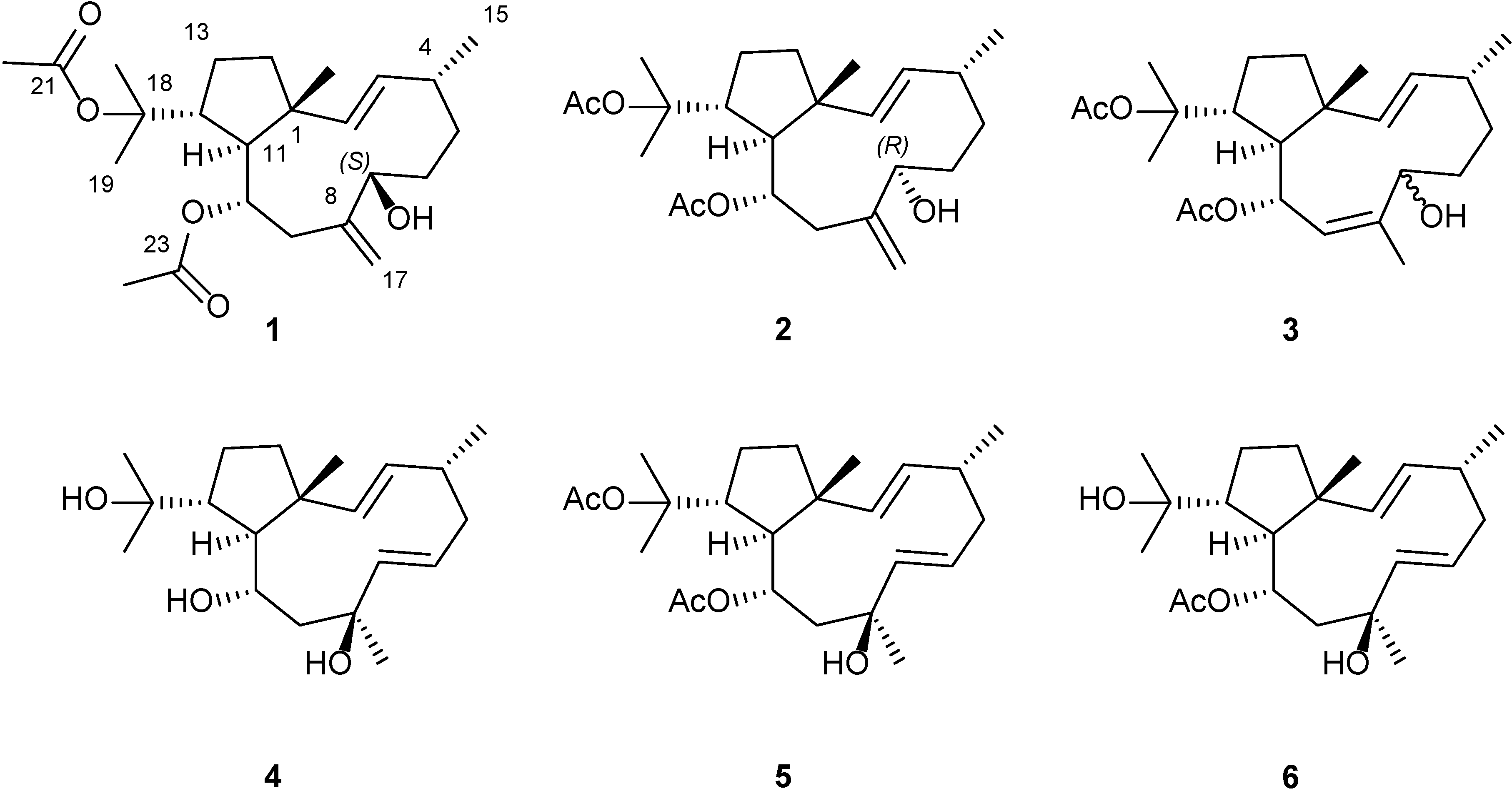
Mechanisms of action include the blocking of different steps of the HIV-1 replicative cycle as entry inhibitors [5,6], reverse transcriptase inhibitors [1,5] or protease inhibitors [1,7]. More particularly, diterpenes isolated from marine alga represent one of the most active and promising compounds in the development of new drugs to control HIV-1 during the infective process [1,8]. In our search for new anti-HIV compounds from marine sources, we have identified a dolabellane diterpene from the brown seaweed Dictyota pfaffii Schnetter (Dictyotaceae, Phaeophyceae) with strong anti-HIV-1 activity. Due to the highly relevant results obtained thus far, we further investigated the anti-HIV inhibiting capacities of the minor dolabellane diterpenes. Moreover, dolabellane diterpenes demonstrate a wide array of interesting pharmacological properties such as antibacterial [9], antiprotozoal [10], antifungal [11], anti-inflammatory [12] and antimalarial [13] activities. We found one such dolabellane diterpene, dolabelladienetriol, to be a non-competitive inhibitor of HIV-1 reverse transcriptase [2,14,15]. In this paper we report the isolation and structural determination of three new dolabellane diterpenes 13, (Figure 1), as will be demonstrated, two of these compound, 1 and 2, exhibited potent antiviral activity.
Figure 1. Structure of the new dolabellane diterpenoids dolabelladienols A–C (13), along with the known dolabelladienetriol (46) isolated from D. pfaffii.
Marinedrugs 12 04247 g001

2. Results and Discussion

Specimens of D. pfaffii were collected during scuba dives (6–9 m) at Atol das Rocas reef, Rio Grande do Norte State, the only atol in the South Atlantic, and were subsequently air-dried. In order to obtain 3.5 g of organic residue, the material was extracted exhaustively with CH2Cl2, filtered and solvent removed under vacuum at 40 °C. The resulting extract was fractionated by vacuum column chromatography (VC) and subjected to successive silica gel column chromatographies (CC) in order to afford pure compounds 16.

2.1. Structural Elucidation

Compound 1 (28 mg) was isolated as a white solid. EIMS analysis (m/z 406 [M]+) and 13C NMR spectra, which was confirmed by the observation of a [M + Na]+ peak at m/z 429.2631 in its (+)-HRESIMS suggested the molecular formula of C24H38O5 (six degrees of unsaturation) for compound 1. The 13C NMR data of 1 (Table 1) showed the presence of 24 carbon signals, which were assigned, using an APT experiment, as six methyls, six methylenes (five sp3 and one sp2), seven methines (two sp3 and five sp2) and five quaternary carbons (three sp3 and two sp2). A detailed analysis of 1D and 2D NMR spectra determined the gross structure of 1 (Supplementary Figures S1–S6). The presence of two acetate residues were easily deduced from the ester carbonyl signals at δC 170.5 and 170.1, along with two methyl groups at δH 2.04 (s, 3H) and 1.93 (s, 3H). Therefore, we inferred the diterpene structure of 1 from the 20 carbons after subtraction of the four carbons associated with two acetate groups. The presence of two oxygenated methines were deduced from NMR signals at δC 70.2 which was coupled via HSQC with the proton signal at δH 4.37 (dd, J = 8.4, 3.5 Hz) and at δC 68.6 correlated with the proton signal at δH 4.91 (ddd, J = 8.9, 4.7, 2.4 Hz) which was assigned to an hydroxymethine and an acetoxy-bearing methine, respectively. The methlylene carbon NMR signal observed at δC 113.4 which correlated according to HMBC with the olefinic methylene protons at δH 5.12 (1H, brs) and 4.99 (1H, brs), along with olefinic quaternary carbon at δC 146.2, indicated the existence of an exocyclic double bond. The existence of disubstituted double bond with an E configuration in 1 was inferred by the proton signals at δH 5.66 (dd, J = 16.4, 7.2 Hz) and 5.17 (d, J = 16.4 Hz), which correlated according to HMBC with the sp2 carbons at δC 133.6 and 137.5. The quaternary signal at δC 84.8, characteristic of a bearing acetoxy quaternary carbon, along with the methyl singlets at δH 1.60 (s, 3H), 1.42 (s, 3H) were indicative of the presence of a 2-propan-2-yl acetate group [15]. Furthermore, the NMR data of 1 (Table 1) displayed an additional two methyl groups at δH 0.97 (d, J = 6.9 Hz, 3H)/δC 22.2 and δH 0.83 (s, 3H)/δC 16.8. The above-presented NMR data closely resemble those for bicyclic dolabellane skeleton, which were established as taxonomic markers for this alga [16]. However, differences in the chemical shifts as a result of the unsaturation and oxygenation pattern indicated of a new natural product. Two main proton spin systems, outlined in Figure 2a, were established by 1H-1H COSY experiments after assignments of the direct 1H-13C correlations via HSQC analysis.
The first spin system was identified by 1H-1H COSY correlations between the olefinic proton at δH 5.17 (H-2) and the hydroxymethyne at δH 4.37, (H-7). A second was recognized by correlating the methylene protons at δH 2.43 and 2.46 (H-9) with those at δH 1.46 and 1.39 (H-14). Analysis of the HMBC spectrum granted the connectivity of these two spin-systems. The observed correlations between the methylene olefinic protons H-17a (δH 5.12, brs) and H-17b (δH 4.99, brs) and the oxygenated methine C-7 (δc 70.2) and methylene C-9 (δc 41.0) allowed for the connection of the exocyclic double bond Δ8(17) to both spin systems. On the other hand, HMBC cross-peaks between the olefinic H-2 (δH 5.17, d, J = 16.4 Hz), the methylene H2-14 (δH 1.46, m) and H-11 (δH 1.84, dd, J = 10.8, 2.4 Hz) protons with the methyl carbon C-15 (δC 16.8) confirmed the bicyclic dolabelladiene structure presented in Figure 2a. The location of the acetate group at C-10 was established through the HMBC correlations between H-10 (δH 4.91, ddd, J = 8.9, 4.7, 2.4 Hz) and H3-24 (δH 1.93, s) protons and the carbonyl signal at δC 170.1 (C-23).
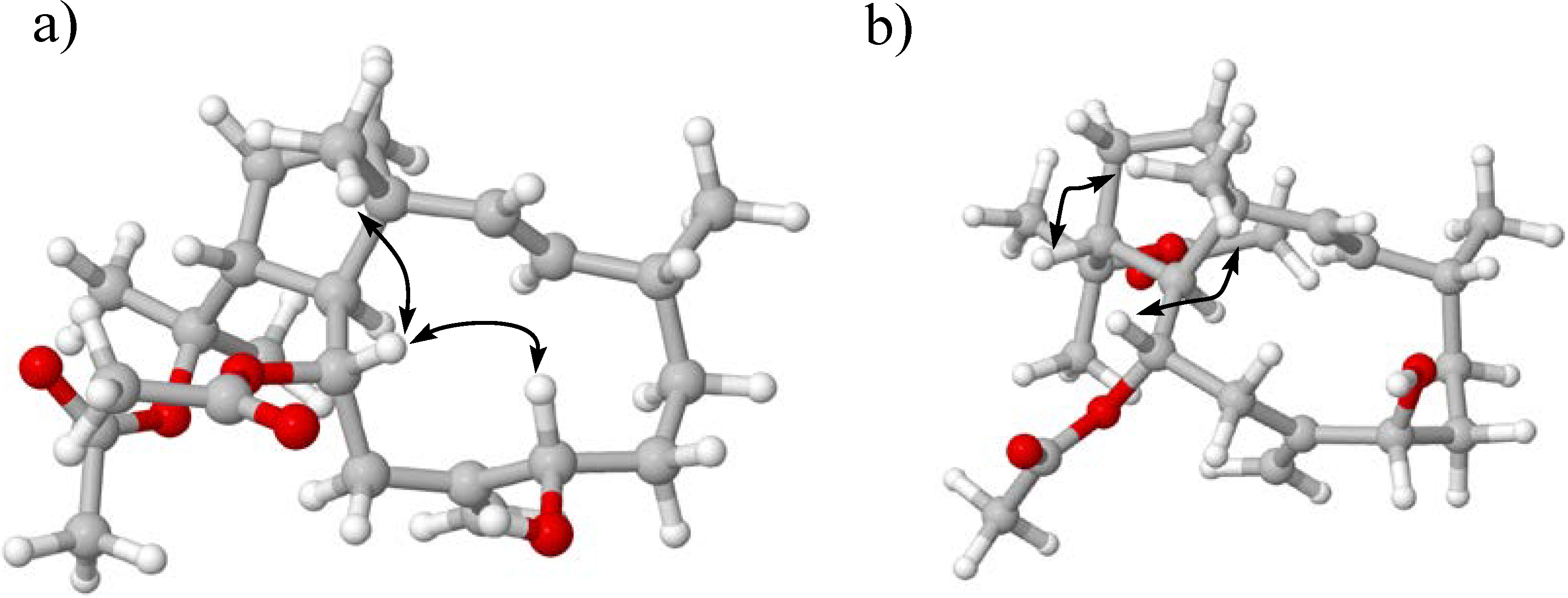
We demonstrated the assignment of the 2-propan-2-yl acetate group to position 12 by was the long range 1H-13C coupling of the methine proton H-12 at 2.95 (td, 10.8, 4.7) with methyl singlets C-18 and C-20 at δC 23.4 and 26.4 (Figure 2a). Therefore, on the basis of the above analysis, we established the planar structure of 1 as illustrated in Figure 2a. Having established the planar structure of dolabellane 1, we focused our attention on its stereochemistry. Key NOESY correlations observed between methyl protons H3-15, H-12 and H-10, and this in turn to H-7, led to the conclusion that protons H3-15, H-12, H-10 and H-7 are on the same side of the bicyclic ring (Figure 3a) and further suggested a 1R*, 7S*, 10S* and 12R* relative configuration. In order to confirm these stereochemical assignments, a conformational space of the two possible 7R*- or 7S*-epimers was explored using the GMMX stochastic conformational search procedure as implemented in the program PC-MODEL, which itself used an MMX force field.
Table 1. 13C and 1H NMR data (500 MHz in CDCl3) of dolabelladienols A–C (1–3) (δ in ppm, J in Hz).
Position123
δCδHδCδHδCδH
148.4-48.1-48.9-
2137.55.17 (d, 16.4)138.95.24 (d, 16.4)136.95.23 (dd, 16.4, 0.7)
3133.65.66 (dd, 16.4, 7.2)134.55.53 (dd, 16.4, 8.5)134.15.70 (dd, 16.4, 6.9)
434.82.20 (m)34.72.04 (m)34.92.39 (ddd, 13.9, 7.7, 3.3)
532.01.58 (m) 1.63 (m)33.21.76 (dd, 11.2, 2.3) 1.08 (dd, 11.2, 4.9)33.52.34 (m) 1.71 (m)
632.61.62 (m) 1.90 (m)33.31.85 (dd, 9.0, 4.9) 1.64 (m)30.71.45 (m) 1.66 (m)
770.24.37 (dd, 8.4, 3.5)76.73.95 (dd, 11.4, 4.9)69.05.08 (t, 6.6)
8146.2-144.8-140.1-
941.02.43 (dd, 13.2, 4.7) 2.46 (dd, 13.2, 8.9)36.52.65 (dd, 13.8, 10.1) 2.50 (br d, 13.8)125.85.31 (d, 10.4)
1068.64.91 (ddd, 8.9, 4.7, 2.4)70.44.88 (ddd, 10.1, 3.2, 2.1)67.95.58 (dd, 10.4, 2.3)
1151.11.84 (dd, 10.8, 2.4)50.61.94 (dd, 10.5, 3.2)53.31.76 (dd, 10.1, 2.1)
1244.22.95 (td, 10.8, 4.7)44.23.06 (td, 10.5, 4.9)44.32.98 (m)
1326.41.95 (m) 1.45 (m)26.51.96 (m) 1.48 (m)26.21.95 (m), 1.46 (m)
1438.91.46 (m) 1.39 (m)38.71.51 (m) 1.47 (m)39.21.49 (m) 1.39 (m)
1516.80.83 (s)18.40.83 (s)16.90.93 (s)
1622.20.97 (d 6.9 )21.11.00 (d, 6.7)19.80.96 (d, 6.9)
17113.55.12 (brs) 4.99 (brs)117.05.04 (brs) 4.89 (brs)17.01.66 (d, 1.4)
1884.8-84.8-84.9-
1923.41.42 (s)23.21.45 (s)26.01.59 (s)
2026.41.60 (s)26.61.61 (s)23.61.43 (s)
21170.5-171.5-170.2-
2221.02.04 (s)21.02.05 (s)23.01.97 (s)
23170.1-170.0-170.8-
2423.01.93 (s)22.91.92 (s)21.12.02 (s)
Figure 2. 1H-1H COSY correlations (bold lines) and key H→C HMBC correlations (arrows) for compounds (a) 12 and (b) 3.
Marinedrugs 12 04247 g002
Figure 3. Key NOE Correlations for the main conformers found for compounds 1 (a) and 2 (b).
Marinedrugs 12 04247 g003
In the case of the epimers 7S* and 7R*, 37 and 23 different conformers were observed, respectively, within an energy cutoff of 3 kcal/mol. The conformations identified for the 7S* configuration alone were enough to placed protons H-7 and H-10 in a 2 Å environment, which fits the observed NOEs for the proposed stereochemistry. The 11,1-trans-fused ring system of the dolabellane was confirmed by the carbon chemical shift of CH3-15 at δC 16.8, indicating a 1R* and 11S* configuration [10]. Finally, the carbon chemical shifts of C-4 methine at δC 34.8 and CH3-16 methyl at δC 22.2 suggested a 4R* relative configuration, which accorded with other reported compounds [15,17]. Hence, compound 1, named dolabelladienol A, was identified as (1R*,2E,4R*,7R*,10S*,11S*,12R*)-10,18-diacetoxy-7-hydroxy-2,8(17)-dolabelladiene, resulting in a new natural product.
The new dolabellane diterpene 2 (6 mg) was also isolated as a white solid. Its molecular formula of C24H38O5 was deduced from the [M + Na]+ pseudo molecular ion at m/z 429.2627 (C24H38O5Na, ∆1.6) and 13C NMR spectra (Supplementary Figures S7–S12). The close resemblance between the NMR spectra of 1 and 2 (Table 1), including 1H-1H COSY and HMBC correlations (Figure 2a), indicated that 2 shares the same planar structure with 1. However, slight differences in the NMR data of these two compounds were observed for the carbon C-7 (δc 70.2, and δc 76.7, respectively) and surrounding atoms (Table 1), which indicated that 2 corresponds to the C-7 epimer of 1.
These findings accord with the NOESY analysis of 2 which demonstrated NOE correlations between the H3-15 methyl protons and H-12 and H-10 methine protons, along with the lack of correlation between H-7 and H-10 observed in the NOESY spectrum of 1 (Figure 3b). As mentioned above, the space-conformational search in a 3.0 kcal/mol range for the epimer 7S, showed all conformers with no dipolar correlations between H-7 and the other vicinal protons. The first is located in a populated averaged of 4.9 Å away from other proximal protons, and therefore the lack of NOE correlations in the NOESY experiment supports the hypothesis that 2 should be the epimer of 1 at C-7. Thus their stereocenters were assigned as follows 1R*,10S*,11S*,12R*. In this way, dolabelladienol A (2) was identified as (1R*,2E,4R*,7S*,10S*,11S*,12R*)10,18-diacetoxy-7-hydroxy-2,8(17)-dolabelladiene, and is here reported for the first time.
Compound 3 (4 mg) was isolated as a white solid, as well, and assigned the molecular formula C24H38O5 based on LREIMS analysis (m/z 406 [M]+) and 13C NMR spectra which required six degrees of unsaturation and demonstrated that 3 is an isomer of 1 (Table 1). The NMR data of compound 3 (Supplementary Figures S13–S17) also showed spectroscopic features for a dolabellane-type diterpene, similar to those of 1 and 2, including the presence of an acetate group at C-18, an acetoxy-bearing methine at C-10 and a hydroxyl-bearing methine at C-7. By contrast, the main differences from 1 and 2 are the absence of chemical signals corresponding to the exocyclic double bond present in 1 and 2 and the presence of two endocyclic double bonds at δH 5.70 (dd J = 16.3, 6.9 Hz), 5.31 (d J = 10.4 Hz), and 5.21 (d J = 16.4 Hz). Assignments of H-C signals of 3 were conducted by HSQC experiments. The 1H-1H COSY experiment allowed for the establishment of two spins systems that were connected by HMBC correlations. In this way, the planar structure of compound 3 could be elucidated as shown in Figure 2b.
The relative stereochemistry of 3 could not be addressed because the compound decomposed. The E configuration of Δ2 double bond was established by the J = 16.4 Hz between the olefinic protons H2 and H3. Furthermore, the proton and chemical shifts were similar to the compound (1R*,2E,4R*,8Z,10S*,11S*,12R*)10,18-diacetoxydolabella-2,8-dien-6-one, reported by Lu and Faulkner in 1998, in that they share the same Z configuration at Δ8 double bond [18]. Comparison of the 1H and 13C NMR chemical shifts of compound 3 to those of 1 demonstrated that they share the same relative configuration for stereocenters at 1, 4, 10, 11 and 12. In this way, we identified and reported for the first time the compound 3 as (1R*,2E,4R*,8E,10S*,11S*,12R*)-10,18-diacetoxy-7-hydroxy-2,8-dolabelladiene. It is worth noting that compounds 1 or 2 may potentially convert to 3 during the course of isolation or purification due to slight acidity.
Spectral data (1H, 13C NMR, MS, optical rotation) of compound 4 (30 mg) are the same as those of dolabelladienetriol, which was previously isolated from the digestive glands of the sea hare Dolabella californica Stearns, 1877 [17] and obtained as semisynthetic product in 2004 [15]. It is important to note that this compound has shown strong anti-HIV and anti-HSV activity [2,19]. The dolabellanes 5 and 6 of D. pfaffii were also isolated and their spectral data proved identical with those of 10,18-diacetoxy-8-hydroxy-2,6-dolabelladiene (5) and 10-acetoxy-8,18-dihydroxy-2,6-dolabelladiene (6), which were previously described previously by us in 2004 [15]. We conducted a single crystal X-ray diffraction study of the compounds 4 and 5. In the case of compound 4, it was possible to confirm the proposed structure, but the results related to compound 5 permitted us an unambiguous description of the absolute configuration (Flack parameter = −0.03(6)). Thus, we described the configuration of compound 5 as (1R,2E,4R,6E,8S,10S,11S,12R)-8,10,18-diacetoxy-8-hydroxy-2,6-dolabelladiene.

2.2. Antiviral Activity

In order to establish the anti-HIV-1 potential of the isolated compounds, we first evaluated the safety of compound 1, 2 and 4 for use in human cells by treating MT-2 lymphocytes with various concentrations of the test compound. After 4–5 days of treatment, MT-2 viability, determined colorimetrically, was higher than 90% for concentrations up to 250 μM, resulting in a CC50 between 1345 ± 2 and 1456 ± 3.4 μM. It is important to emphasize that the compounds were not cytotoxic and were in fact even less toxic than the neviparine (325 μM ± 1.4) used as a control.
Subsequently, we evaluated the antiviral activity of natural compounds on MT-2 cells at concentrations between 6.25 and 25 μM. At 6.25 μM, compounds 1, 2 and 4 inhibited the HIV-1 virus in 83%, 69% and 52% of cases, resulting in EC50 values of 2.9 ± 0.2, 4.1 ± 0.4 and 6.16 ± 0.7 μM, respectively. There was a slight difference in activity between epimers 1 and 2, but the antiviral activity of the 2,8(17)-dolabelladienes series was twice as active as the 2,6-dolabelladienes series and exhibited a positive influence on the inhibition of HIV-1.
It is noteworthy that when compounds 1 and 2 were evaluated in the antiviral test, they proved more active than compound 4, hitherto the most active compound reported by us to have demonstrated non-competitive inhibition of HIV-1 reverse transcriptase [2,14,15], This suggests that these diterpenes could be considered potential new agents for HIV-1 therapy. Therefore, we recommend further studies on the precise mechanism of activity and on the in vivo antiviral activity.

3. Experimental Section

3.1. General Experimental Procedures

LREIMS and HRESIMS were measured on QQq-TOF mass spectrometer, Applied Biosystems QSTAR Elite. For the HRGC-MS analysis a GC7890A (Agilent Technologies, Santa Clara, CA, USA) with a DB-5MS (5% Phenyl methyl siloxane) chromatographic column (60 m × 250 μm × 0.25 μm) was coupled with an MSD-5975 (Agilent Technologies) with an electronic impact ion source (70 eV) for detecting ions between m/z 50–500 in scan mode. Injector and detector temperatures were set at 270 °C and 290 °C, respectively. We maintained the temperature program at 160 °C, and then programmed to 260 °C at a rate of 4 °C/min before finally raising the temperature at a rate of 10 °C/min to 290 °C for 15 min. Helium served as the carrier gas at a flow rate of 1 mL/min. Linear retention indices (RI) were calculated according to the Kovats method using a mixture of normal paraffin C6–C26 as external references. Nuclear magnetic resonance spectra (proton and carbon) were recorded on Varian VNMRS or Bruker 500 MHz Advance spectrometers, employing CDCl3 (99.8%, Merck, Whitehouse Station, NJ, USA) as a solvent. Multiplicities of 13C signals were obtained by DEPT. For column chromatography, we used silica gel 60. All the solvents were analytical grade. Optical rotations were measured on a Polartronic ADP440+, Bellinghan + Stanley Polarimeter (North Farm Industrial Estate, Tunbridge Wells, UK).

3.2. Sample Collection

The brown algae Dictyota pfaffii was collected during July 2009 at a depth of 6–9 m, by scuba dives at the Atol das Rocas reef, a Biological Marine Reserve in the Rio Grande do Norte State, lat. 03°51′03″S, long. 33°40′29″W, Brazil. The legal authorization for sample colleting was obtained from VLT (SISBIO/IBAMA Brazil) (number 17352). Dr. Roberto Villaça (Departamento de Biologia Marinha, Instituto de Biologia, UFF) collected and identified the seaweeds. A voucher specimen (HRJ 9117) was deposited in the herbarium of the Universidade do Estado do Rio de Janeiro (UERJ) and the seaweed was air-dried.

3.3. Extraction, Isolation and Structural Elucidation of Compounds

The air-dried material of Dictyota pfaffii (65 g) was extracted three times with 500 mL of CH2Cl2 each time for 24 h. The extracts were combined, filtered and further dried under vacuum at 40 °C. The dried crude extract (3.5 g) was subjected to vacuum column chromatography (CC) using a discontinuous gradient (Hexane/CH2Cl2/EtOAc/MeOH) in order to obtain 12 fractions (F1 to F12). Fraction F7 (CH2Cl2/EtOAc, 9:1) yields white crystals of compound 5 (750 mg). Fraction F8 was chromatographed on a silica gel CC using as eluent an isocratic mixture of CH2Cl2/EtOAc, 9:1 in order to obtain 12 fractions (F8.1 to F8.12). From these, fraction F8.3 yielded compound 2 (6 mg) and compound 3 (4 mg) after silica gel column chromatography eluted with CH2Cl2/EtOAc, 8:2 as solvent. Fraction F8.7 yielded the pure compound 6 (20 mg), and fraction F8.10 the pure compound 1 (28 mg). Finally, by crystallization in hexane, fraction F10 yielded pure compound 4 (30 mg) in the form of white needle crystals. All the isolated compounds were identified the analysis of their 1D- and 2D-NMR spectra, together with their EI-MS spectra analysis.

3.4. Conformational Searches

Conformational searches were performed using 50,000 GMMX steps as implemented in the PCmodel software (version 8.5), the MMFF94s force field and the TNCG algorithm. All local minima within 3.5 kcal/mol of the global minimum were saved and subsequently re-minimized using the FMNR algorithm within an energy cut-off of 3.0 kcal/mol. For compound 1, 15 out of 37 conformers, 56.9% of the population, exhibited NOE between H-7 and H-10 (distances of 2.11 and 2.96 Å). For compound 2, all 23 found conformers, 100% of the population, did not show NOE between H7 and H10 (distances larger than 3.8 Å).
Dolabelladienol A (1R*,2E,4R*,7S*,10S*,11S*,12R*)10,18-diacetoxy-7-hydroxy-2,8(17)-dolabelladiene (1). White solid (28 mg), Marinedrugs 12 04247 i001 +11° (c 0.002, CHCl3), RI 2593. 1H and 13C NMR data, see Table 1. EIMS m/z (relative intensity) 406 (4), 346 (3), 304 (4), 286 (100), 271 (46), 243 (61), 215 (25), 173 (34), 145 (32), 133 (30), 107 (75), 93 (80), 55 (65). HRESIMS: [M + Na]+ 429.2631 (C24H38O5Na 429.2611, ∆1.95 mmu).
Dolabelladienol B (1R*,2E,4R*,7R*,10S*,11S*,12R*)10,18-diacetoxy-7-hydroxy-2,8(17)-dolabelladiene (2). White solid, Marinedrugs 12 04247 i001 +4° (c 0.002, CHCl3), RI 2567. 1H and 13C NMR data, see Table 1. HRESIMS 835.5353 [2M + Na]+; 429.2627 [M + Na]+; EIMS m/z (relative intensity) 406 [M]+ (3), 328 [M – AcOH − H2O]+ (5), 304 (7), 286 [M − AcOH − AcOH]+ (45), 243 (43), 215 (20), 187 (28), 161 (47), 135 (65), 107 (77), 93 (100), 55 (60).
Dolabelladienol C (1R*,2E,4R*,8E,10S*,11S*,12R*)10,18-diacetoxy-7-hydroxy-2,8-dolabelladiene (3). White solid, Marinedrugs 12 04247 i001 +14° (c 0.002, CHCl3), RI 2475. 1H and 13C NMR data, see Table 1 EIMS m/z (relative intensity) 406 (4) 304 (5) 286 (38) 271 (31) 243 (32) 229 (12) 203 (23) 175 (51) 133 (52) 121 (71) 107 (75) 95 (100) 79 (58) 69 (53) 55 (63).
(1R*,2E,4R*,6E,8S*,10S*,11S*,12R*)-8,10,18-trihydroxy-2,6-dolabelladiene (4). White needles, Marinedrugs 12 04247 i001 −27° (c, 0.5, CHCl3), RI 2257, NMR, and MS data are consistent with literature values [15].
(1R,2E,4R,6E,8S,10S,11S,12R)-10,18-diacetoxy-8-hydroxy-2,6-dolabelladiene (5). White needles, Marinedrugs 12 04247 i001 −123° (c 0.002, CHCl3), RI 2475, NMR, and MS data are consistent with literature values [15].
(1R*,2E,4R*,6E,8S*,10S*,11S*,12R*)-10-acetoxy-8,18-dihydroxy-2,6-dolabelladiene (6). White solid, Marinedrugs 12 04247 i001 −70° (c, 0.5, CHCl3), RI 2314, NMR, and MS data are consistent with literature values [15].

3.5. X-Ray Diffraction Analysis of Compound 4
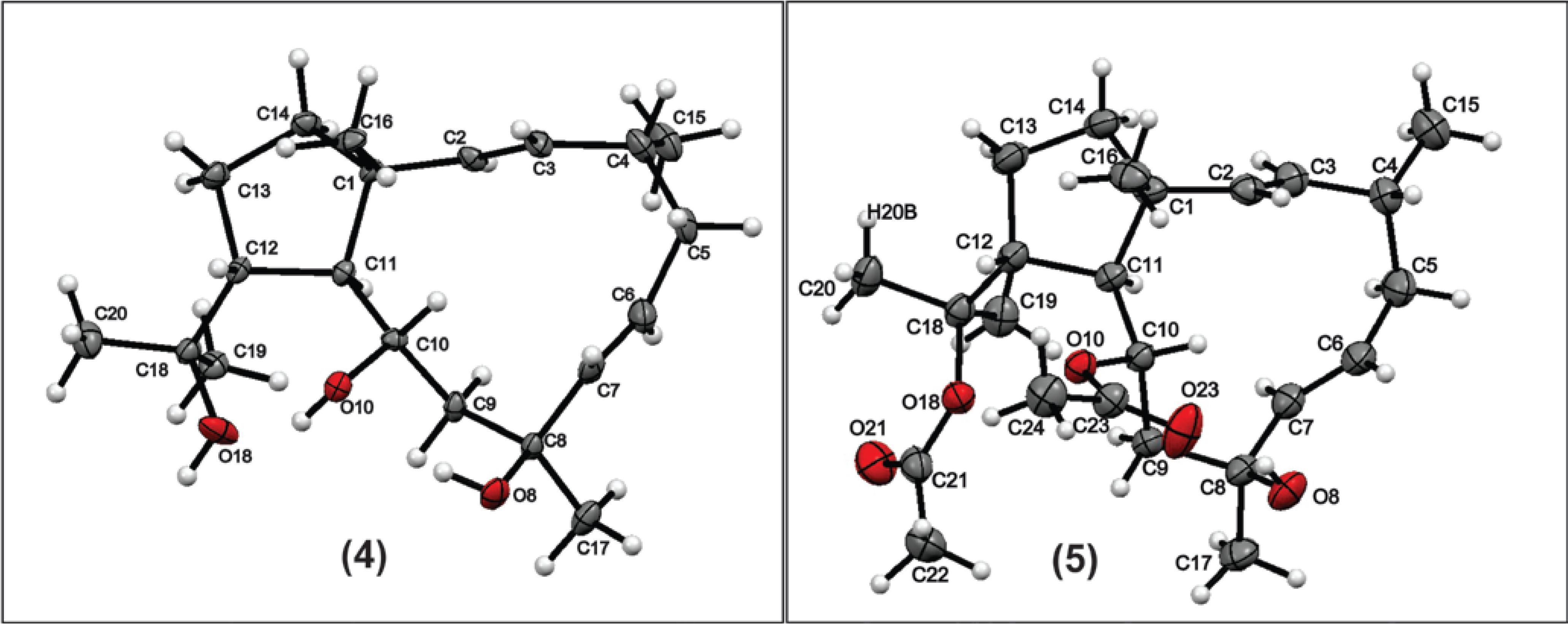
X-ray diffraction data was carried out in Bruker D8 Venture with radiation CuKα (λ = 1.5418 Å). The structure was solved by direct methods and refined by full-matrix least squares on F2 with SHELX-97 package (Figure 4) [20]. The positions of hydrogen atoms were generated geometrically and refined according to a riding model. All non-hydrogen atoms were refined anisotropically. Crystallographic data for the structures of 4 and 5 have been deposited with the Cambridge Crystallographic Data Centre database.
Figure 4. X-ray crystal structure of the compound 4 and 5.
Marinedrugs 12 04247 g004
Crystallographic data for compound 4: C20H34O3, F.W. = 322.5, colorless needle, monoclinic, space group P21, a = 9.4394(7) Å, b = 7.6155(5) Å, c = 13.4810(8) Å, β = 95.527(4)°, volume = 964.59(11) Å3, Z = 2, Dc = 1.11g/cm3, μ = 0.56 mm−1, F(000) = 356, Crystal dimensions: 0.065 mm × 0.097 mm × 0.268 mm. Independent reflections: 3415 (Rint = 0.058). The final anisotropic full-matrix least-squares refinement on F2 with 216 variables converged at R1 = 9.73%, for the observed data and wR2 = 25, 38% for all data. CCDC number: 1000466.
Crystallographic data for compound 5: C24H38O5, F.W. = 408.5, colorless prismatic, orthorhombic, space group P212121, a = 8.3069(2) Å, b = 13.6440(3) Å, c = 20.7705(5) Å, volume = 2354.11(10) Å3, Z = 4, Dc = 1.15 g/cm3, μ = 0.63 mm−1, F(000) = 888, Crystal dimensions: 0.16 mm × 0.29 mm × 0.50 mm. Independent reflections: 4221 (Rint = 0.041). The final anisotropic full-matrix least-squares refinement on F2 with 267 variables converged at R1 = 3.77%, for the observed data and wR2 = 9.87% for all data. The Flack parameter was −0.03 (6) using 1609 quotients [(I+) − (I−)]/[(I+) + (I−)] [21] CCDC number: 1005607.

3.6. Cells and Viruses

MT-2 cells were grown in RPMI 1640 media (LGC Bio, São Paulo, Brazil), which contained 10% heat-inactivated fetal bovine serum (Hyclone, Logan, UT, USA). Cell cultures were incubated at 37 °C in a humidified air atmosphere containing 5% CO2. HIV-1 isolates IIIB (X4-tropic, subtype B, donated by Eva Maria Fenyo, University of Lund, Lund, Sweden) were prepared in PHA-activated MT-2 cells and stored at −70 °C.

3.7. Cytotoxicity Assays

The cytotoxicity of the compounds in triplicates was tested in MT-2 cells (2 × 10⁶ cell/100 μL/well) grown in 96-well plates for 72 h at 37 °C with a 5% CO2 atmosphere. Cells were incubated with culture medium containing the compounds at different concentrations. After 4 to 5 days the HIV-1, cytopathic effect was determined colorimetrically by the MTT method [22]. In brief, 20 μL of MTT Sigma Chemical Co. (St. Louis, MO, USA) (5 mg/mL) were added to wells and 4 h later the optical density was measured at 540 nm. The 50% cytotoxic concentration (CC50) was determinate as the concentration capable of reducing the optical density by 50% in comparison with the control. The CC50 was calculated by linear regression analysis of the dose-response curves generate as previously described [23].

3.8. Antiviral Assays

The MT-2 cells (2 × 10⁶ cell/100 μL/well) were initially exposed to viral suspensions containing 5 to 10 ng/mL of HIV-1 p24 Ag for 2 to 3 h. The cells were treated with compounds at different concentrations. After 2–3 days at 37 °C in 5% CO2, HIV-1 cytopathic effect was determined colorimetrically by the MTT method [22]. EC50 was calculated as described [23]. The positive control was neviparine 5 μM (replication viral inhibition of 99%).

4. Conclusions

This study identified four hitherto undescribed dolabellane diterpenes from Dictyota pfaffii (14), three of which are new (13), while one (4) is here described for the first time in this species, in addition to the major known compounds 5 and 6. Biological assays demonstrated that none of the compounds were cytotoxic. Compounds 1 and 2 were evaluated in the antiviral test and seen to be more active than dolabelladienetriol 4 and nevirapine. Such results confirm that these diterpenes should be considered as potential new agents for HIV-1 therapy.

Supplementary Files

  • Supplementary File 1:

    Supplementary Information (PDF, 2074 KB)

  • Acknowledgments

    This work was financially supported by grants from Conselho Nacional de Desenvolvimento Científico e Tecnológico–CNPq (490425/2010-0), Fundação de Amparo à Pesquisa do Estado do Rio de Janeiro–FAPERJ (E-26/103.176/2011), Ministerio de Economia y Competitividad (MINECO, España) (AGL2012-12266-C02), Colciencias and DIB–Universidad Nacional de Colombia.

    Author Contributions

    APV, FR, CJ, JR, VLT, LC conducted natural products isolation and structural determination, algae identification and drafted the manuscript. IBO, PRSS, CCCS and ICNPP conducted biological (antiviral) tests. JALCR conducted X-ray diffraction analyses.

    Conflicts of Interest

    The author declares no conflict of interest.

    References

    1. Zhou, X.; Liu, J.; Yang, B.; Lin, X.; Yang, X.W.; Liu, Y. Marine natural products with anti-HIV activities in the last decade. Curr. Med. Chem. 2013, 20, 953–973. [Google Scholar]
    2. Cirne-Santos, C.C.; Souza, T.M.; Teixeira, V.L.; Fontes, C.F.; Rebello, M.A.; Castello-Branco, L.R.; Abreu, C.M.; Tanuri, A.; Frugulhetti, I.C.; Bou-Habib, D.C.; et al. The dolabellane diterpene Dolabelladienetriol is a typical noncompetitive inhibitor of HIV-1 reverse transcriptase enzyme. Antivir. Res. 2008, 77, 64–71. [Google Scholar] [CrossRef]
    3. Huang, L.; Chen, C.H. Molecular targets of anti-HIV-1 triterpenes. Curr. Drug Targets Infect. Disord. 2002, 2, 33–36. [Google Scholar]
    4. Pereira, H.S.; Leão-Ferreira, L.R.; Moussatché, N.; Teixeira, V.L.; Cavalcanti, D.N.; Costa, L.J.; Diaz, R.; Frugulhetti, I.C.P.P. Antiviral activity of diterpenes isolated from the Brazilian marine alga Dictyota menstrualis against human immunodeficiency virus type 1 (HIV-1). Antivir. Res. 2004, 64, 69–76. [Google Scholar]
    5. Aiken, C.; Chen, C.H. Betulinic acid derivatives as HIV-1 antivirals. Trends Mol. Med. 2005, 11, 31–36. [Google Scholar] [CrossRef]
    6. Dang, Z.; Qian, K.; Ho, P.; Zhu, L.; Lee, K.H.; Huang, L.; Chen, C.H. Synthesis of betulinic acid derivatives as entry inhibitors against HIV-1 and bevirimat-resistant HIV-1 variants. Bioorg. Med. Chem. Lett. 2012, 22, 5190–5194. [Google Scholar]
    7. Xu, H.X.; Zeng, F.Q.; Wan, M.; Sim, K.Y. Anti-HIV triterpene acids from Geum japonicum. J. Nat. Prod. 1996, 59, 643–645. [Google Scholar] [CrossRef]
    8. Gochfeld, D.J.; el Sayed, K.A.; Yousaf, M.; Hu, J.F.; Bartyzel, P.; Dunbar, D.C.; Wilkins, S.P.; Zjawiony, J.K.; Schinazi, R.F.; Schlueter Wirtz, S.; et al. Marine natural products as lead anti-HIV agents. Mini Rev. Med. Chem. 2003, 3, 401–424. [Google Scholar] [CrossRef]
    9. Ioannou, E.; Quesada, A.; Rahman, M.M.; Gibbons, S.; Vagias, C.; Roussis, V. Dolabellanes with antibacterial activity from the brown alga Dilophus spiralis. J. Nat. Prod. 2011, 74, 213–222. [Google Scholar]
    10. Wei, X.; Rodríguez, A.D.; Baran, P.; Raptis, R.G. Dolabellane-Type Diterpenoids with Antiprotozoan Activity from a Southwestern Caribbean Gorgonian Octocoral of the Genus Eunicea. J. Nat. Prod. 2010, 73, 925–934. [Google Scholar] [CrossRef]
    11. Konig, G.M.; Wright, A.D.; Sticher, O.; Angerhofer, C.K.; Pezzuto, J.M. Biological activities of selected marine natural products. Planta Med. 1994, 60, 532–537. [Google Scholar] [CrossRef]
    12. Yodsaoue, O.; Sonprasit, J.; Karalai, C.; Ponglimanont, C.; Tewtrakul, S.; Chantrapromma, S. Diterpenoids and triterpenoids with potential anti-inflammatory activity from the leaves of Aglaia odorata. Phytochemistry 2012, 76, 83–91. [Google Scholar] [CrossRef]
    13. Konig, G.M.; Wright, A.D.; Franzblau, S.G. Assessment of antimycobacterial activity of a series of mainly marine derived natural products. Planta Med. 2000, 66, 337–342. [Google Scholar] [CrossRef]
    14. Cirne-Santos, C.C.; Teixeira, V.L.; Castello-Branco, L.R.; Frugulhetti, I.C.; Bou-Habib, D.C. Inhibition of HIV-1 replication in human primary cells by a dolabellane diterpene isolated from the marine algae Dictyota pfaffii. Planta Med. 2006, 72, 295–299. [Google Scholar] [CrossRef]
    15. Barbosa, J.P.; Pereira, R.C.; Abrantes, J.L.; Cirne dos Santos, C.C.; Rebello, M.A.; Frugulhetti, I.C.; Texeira, V.L. In vitro antiviral diterpenes from the Brazilian brown alga Dictyota pfaffii. Planta Med. 2004, 70, 856–860. [Google Scholar] [CrossRef]
    16. Vallim, M.A.; de Paula, J.C.; Pereira, R.C.; Teixeira, V.L. The diterpenes from Dictyotacean marine brown algae in the Tropical Atlantic American region. Biochem. Syst. Ecol. 2005, 33, 1–16. [Google Scholar]
    17. Ireland, C.; Faulkner, D.J. Diterpenes from Dolabella californica. J. Org. Chem. 1977, 42, 3157–3162. [Google Scholar] [CrossRef]
    18. Lu, Q.; Faulkner, D.J. Three dolabellanes and a macrolide from the sponge Dysidea sp. from Palau. J. Nat. Prod. 1998, 61, 1096–1100. [Google Scholar] [CrossRef]
    19. Abrantes, J.L.; Barbosa, J.; Cavalcanti, D.; Pereira, R.C.; Frederico Fontes, C.L.; Teixeira, V.L.; Moreno Souza, T.L.; Paixao, I.C. The effects of the diterpenes isolated from the Brazilian brown algae Dictyota pfaffii and Dictyota menstrualis against the herpes simplex type-1 replicative cycle. Planta Med. 2010, 76, 339–344. [Google Scholar] [CrossRef]
    20. Sheldrick, G.M. A short history of SHELX. Acta Cryst. 2008, A64, 112–122. [Google Scholar] [CrossRef]
    21. Parsons, S.; Flack, H. Precise absolute-structure determination in light-atom crystals. Acta Crystallogr. A 2004, A60, s61. [Google Scholar]
    22. Scudiero, D.A.; Shoemaker, R.H.; Paull, K.D.; Monks, A.; Tierney, S.; Nofziger, T.H.; Currens, M.J.; Seniff, D.; Boyd, M.R. Evaluation of a soluble tetrazolium/formazan assay for cell growth and drug sensitivity in culture using human and other tumor cell lines. Cancer Res. 1988, 48, 4827–4833. [Google Scholar]
    23. Richman, D.D.; Johnson, V.A.; Mayers, D.M.; Shirasaka, T.; O’Brien, M.C.; Mitsuya, H. In Vitro Evaluation of Experimental Agents for Anti-HIV Activity. In Current Protocols in Immunology; John Wiley & Sons, Inc.: Hoboken, NJ, USA, 2001. [Google Scholar]

    Share and Cite

    MDPI and ACS Style

    Pardo-Vargas, A.; De Barcelos Oliveira, I.; Stephens, P.R.S.; Cirne-Santos, C.C.; De Palmer Paixão, I.C.N.; Ramos, F.A.; Jiménez, C.; Rodríguez, J.; Resende, J.A.L.C.; Teixeira, V.L.; et al. Dolabelladienols A–C, New Diterpenes Isolated from Brazilian Brown Alga Dictyota pfaffii. Mar. Drugs 2014, 12, 4247-4259. https://doi.org/10.3390/md12074247

    AMA Style

    Pardo-Vargas A, De Barcelos Oliveira I, Stephens PRS, Cirne-Santos CC, De Palmer Paixão ICN, Ramos FA, Jiménez C, Rodríguez J, Resende JALC, Teixeira VL, et al. Dolabelladienols A–C, New Diterpenes Isolated from Brazilian Brown Alga Dictyota pfaffii. Marine Drugs. 2014; 12(7):4247-4259. https://doi.org/10.3390/md12074247

    Chicago/Turabian Style

    Pardo-Vargas, Alonso, Ingrid De Barcelos Oliveira, Paulo Roberto Soares Stephens, Claudio Cesar Cirne-Santos, Izabel Christina Nunes De Palmer Paixão, Freddy Alejandro Ramos, Carlos Jiménez, Jaime Rodríguez, Jackson Antonio Lamounier Camargos Resende, Valeria Laneuville Teixeira, and et al. 2014. "Dolabelladienols A–C, New Diterpenes Isolated from Brazilian Brown Alga Dictyota pfaffii" Marine Drugs 12, no. 7: 4247-4259. https://doi.org/10.3390/md12074247

    APA Style

    Pardo-Vargas, A., De Barcelos Oliveira, I., Stephens, P. R. S., Cirne-Santos, C. C., De Palmer Paixão, I. C. N., Ramos, F. A., Jiménez, C., Rodríguez, J., Resende, J. A. L. C., Teixeira, V. L., & Castellanos, L. (2014). Dolabelladienols A–C, New Diterpenes Isolated from Brazilian Brown Alga Dictyota pfaffii. Marine Drugs, 12(7), 4247-4259. https://doi.org/10.3390/md12074247

    Article Metrics

    Back to TopTop