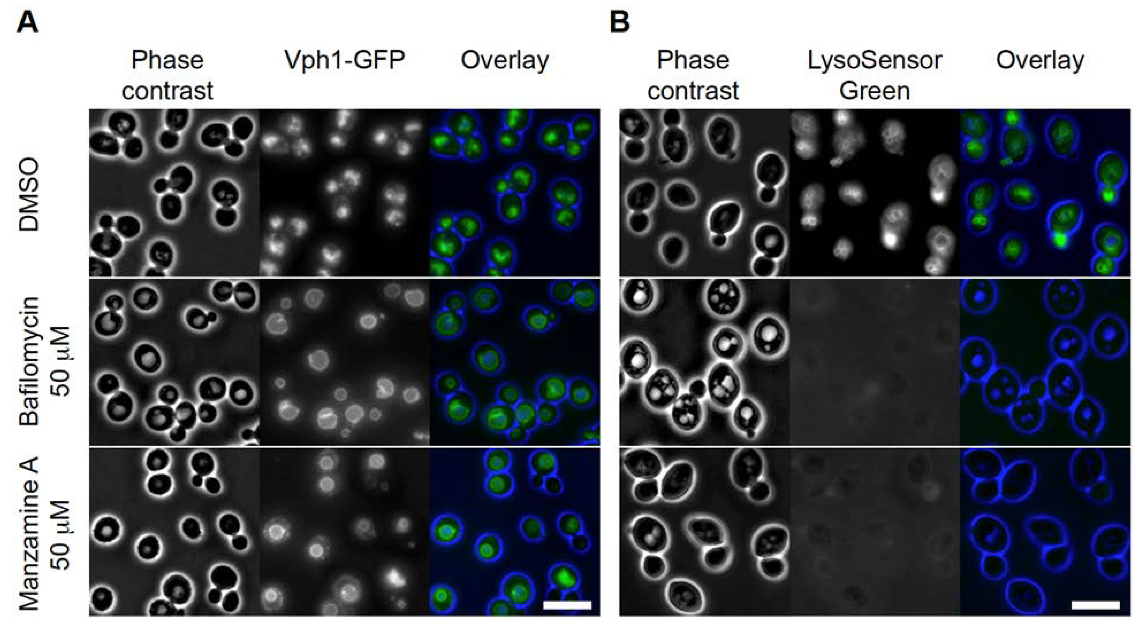
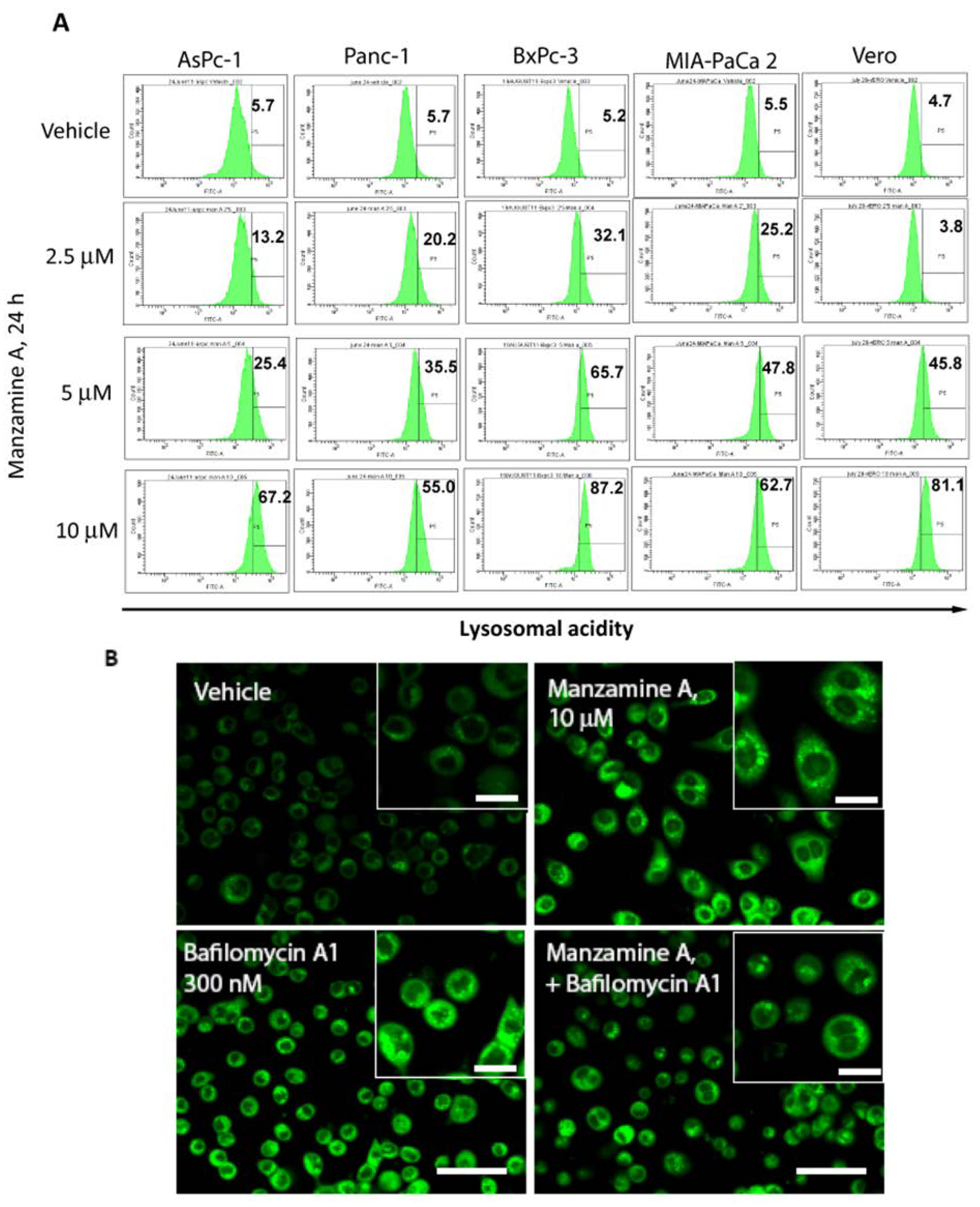
Correction: Kallifatidis, G. et al. The Marine Natural Product Manzamine A Targets Vacuolar ATPases and Inhibits Autophagy in Pancreatic Cancer Cells. Mar. Drugs 2013, 11, 3500–3516


Reference
- Kallifatidis, G.; Hoepfner, D.; Jaeg, T.; Guzmán, E.A.; Wright, A.E. The Marine Natural Product Manzamine A Targets Vacuolar ATPases and Inhibits Autophagy in Pancreatic Cancer Cells. Mar. Drugs 2013, 11, 3500–3516. [Google Scholar] [CrossRef]
© 2014 by the authors; licensee MDPI, Basel, Switzerland. This article is an open access article distributed under the terms and conditions of the Creative Commons Attribution license (http://creativecommons.org/licenses/by/3.0/).
Share and Cite
Kallifatidis, G.; Hoepfner, D.; Jaeg, T.; Guzmán, E.A.; Wright, A.E. Correction: Kallifatidis, G. et al. The Marine Natural Product Manzamine A Targets Vacuolar ATPases and Inhibits Autophagy in Pancreatic Cancer Cells. Mar. Drugs 2013, 11, 3500–3516. Mar. Drugs 2014, 12, 2305-2307. https://doi.org/10.3390/md12042305
Kallifatidis G, Hoepfner D, Jaeg T, Guzmán EA, Wright AE. Correction: Kallifatidis, G. et al. The Marine Natural Product Manzamine A Targets Vacuolar ATPases and Inhibits Autophagy in Pancreatic Cancer Cells. Mar. Drugs 2013, 11, 3500–3516. Marine Drugs. 2014; 12(4):2305-2307. https://doi.org/10.3390/md12042305
Chicago/Turabian StyleKallifatidis, Georgios, Dominic Hoepfner, Tiphaine Jaeg, Esther A. Guzmán, and Amy E. Wright. 2014. "Correction: Kallifatidis, G. et al. The Marine Natural Product Manzamine A Targets Vacuolar ATPases and Inhibits Autophagy in Pancreatic Cancer Cells. Mar. Drugs 2013, 11, 3500–3516" Marine Drugs 12, no. 4: 2305-2307. https://doi.org/10.3390/md12042305
APA StyleKallifatidis, G., Hoepfner, D., Jaeg, T., Guzmán, E. A., & Wright, A. E. (2014). Correction: Kallifatidis, G. et al. The Marine Natural Product Manzamine A Targets Vacuolar ATPases and Inhibits Autophagy in Pancreatic Cancer Cells. Mar. Drugs 2013, 11, 3500–3516. Marine Drugs, 12(4), 2305-2307. https://doi.org/10.3390/md12042305