Article

Polyketides from a Marine-Derived Fungus Xylariaceae sp.

1
Key Laboratory of Marine Bio-Resources Sustainable Utilization, Guangdong Key Laboratory of Marine Materia Medica, RNAM Center for Marine Microbiology, South China Sea Institute of Oceanology, Chinese Academy of Sciences, 164 West Xingang Road, Guangzhou 510301, Guangdong, China
2
University of Chinese Academy of Sciences, Beijing 100049, China
3
New Drug Research & Development Center of North China Pharmaceutical Group Corporation, Shijiazhuang, Hebei 052260, China
*
Author to whom correspondence should be addressed.
Mar. Drugs 2013, 11(5), 1718-1727; https://doi.org/10.3390/md11051718
Submission received: 19 March 2013 / Revised: 22 April 2013 / Accepted: 25 April 2013 / Published: 21 May 2013

Abstract

:
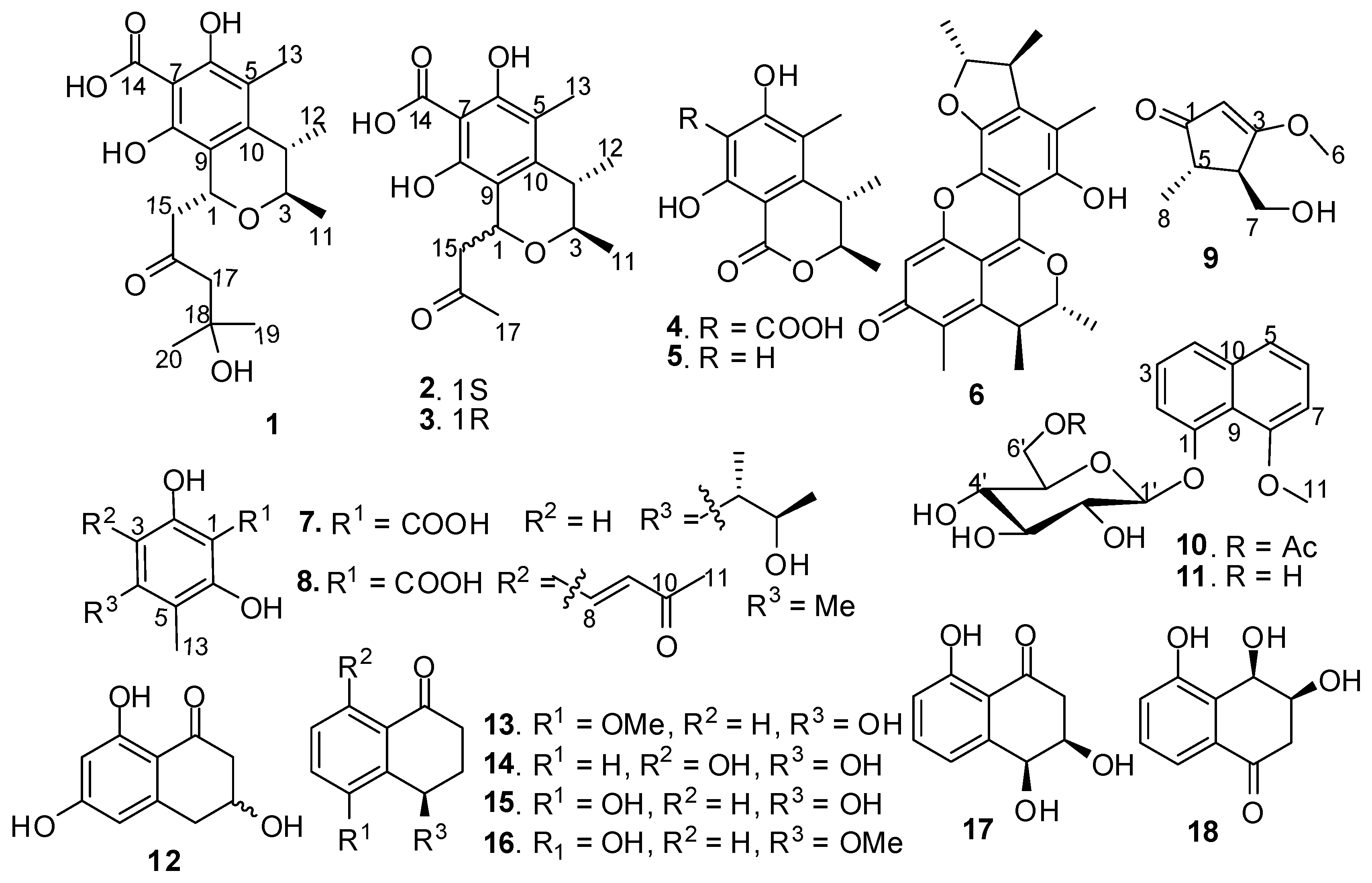
Eighteen polyketides (118) including six citrinin derivatives, two phenol derivatives, one cyclopentenone, two naphthol derivatives, and seven tetralone derivatives were isolated from the culture broth of a marine-derived fungal strain Xylariaceae sp. SCSGAF0086. Five of these compounds (1, 2, 8, 9, and 10) were new, and their structures were determined by spectroscopic methods. Compounds 4, 6, 7, and 17 showed enzyme-inhibitory activities towards several tested enzymes, and 6 and 7 showed strong antifouling activity against Bugula neritina larvae settlement. This is the first time that the antifouling and enzyme-inhibitory activities of these compounds has been reported.

Graphical Abstract

1. Introduction

Gorgonian surfaces are frequently colonized by diverse fungi. These symbiotic fungi are becoming new sources of various bioactive compounds [1,2,3,4,5]. During the course of our investigation on secondary metabolites of marine gorgonian-derived fungi, the fungal strain Xylariaceae sp. SCSGAF0086 attracted our attention. Preliminary experiment showed that the culture extract of the strain had antifouling and antibacterial activities. Further investigation on the chemical constituents of the extract led to the obtainment of 18 polyketides (118) (Figure 1) from Xylariaceae sp. SCSGAF0086. These polyketides included six citrinin derivatives, two phenol derivatives, one cyclopentenone, two naphthol derivatives, and seven tetralone derivatives, which displayed the diversity of secondary metabolites of the marine-derived fungal strain. Five of these compounds (1, 2, 8, 9, and 10) were new, and the other compounds were identified as 1-acetonyl-7-carboxyl-6,8-dihydroxy-3,4,5-trimethylisochroman (3) [6], dihydrocitrinone (4) [7], stoloniferol B (5) [7], dicitrinin A (Wakana named it penicitrinone A, 6) [8,9], phenol A acid (7) [9], 8-methoxy-1-naphthyl β-glucopyranoside (11) [10], (±)-scytalone (12) [11], (4R)-(−)-5-O-methylsclerone (13) [12], (4R)-(−)-4,8-dihydroxy-1-tetralone (14) [13,14], (4R)-(−)-regiolone (15) [15], (4R)-(−)-5-hydroxy-4-methoxy-1-tetralone (16) [16], (3R,4S)-(+)-4-hydroxy-6-deoxyscytalone (17) [17], and (3S,4R)-(+)-3,4,5-trihydroxy-1-tetralone (18) [18].
Figure 1. Structures of compounds 118.
Marinedrugs 11 01718 g001
Compounds 118 were tested for enzyme-inhibitory activities towards cathepsin B, inosine monophosphate dehydrogenase (IMPDH), protein tyrosine phosphatase 1B (PTPlB), and Src homology 2 domain-containing phosphotyrosine phosphatase (SHP2), and antifouling activity toward Bugula neritina larvae settlement. This paper describes the isolation, structure elucidation, enzyme-inhibitory and antifouling activities of these compounds.

2. Results and Discussion

Compound 1 was obtained as turquoise amorphous solid. Its molecular formula of C19H26O7 was determined by HRESIMS (m/z 389.1592 [M + Na]+). The 1H NMR spectrum exhibited five methyl signals at δH 1.01 (d, J = 6.5 Hz, Me-11), 1.15 (s, Me-19), 1.16 (s, Me-20), 1.18 (d, J = 6.5 Hz, Me-12), 1.92 (s, Me-13), and two oxymethine protons at δH 3.85 (qd, J = 2.0, 6.5 Hz, H-3), 5.07 (dd, J = 2.0, 9.5 Hz, H-1). The 13C NMR and DEPT spectra revealed the presence of 19 carbon signals including five methyls [δC 9.5 (Me-13), 18.2 (Me-11), 19.9 (Me-12), 29.4 (overlapped, Me-19 and Me-20)], two methylenes [δC 49.8 (C-15), 55.5 (C-17)], three methines [δC 35.0 (C-4), 65.3 (C-1), 71.5 (C-3)], one oxyquaternary carbon (δC 68.5, C-18), one fully substituted phenyl ring [δC 101.7 (s, C-7), 109.4 (s, C-5), 110.6 (s, C-9), 139.5 (s, C-10), 155.7 (s, C-8), 158.4 (s, C-6)], one carboxyl group (δC 175.5, C-14), and one carbonyl group (δC 209.0, C-16). These data showed close similarity to those of 3 [6], which suggested that 1 had a citrinin skeleton. Comparison of the NMR data of 1 and 3 showed that the obvious difference between them was the additional appearance of one methyl (δC 29.4), one methylene (δC 55.5), and one oxyquaternary carbon (δC 68.5) in 1, which indicated that the only difference between 1 and 3 was the side chain substituted at C-1.
This was proved by the HMBC spectrum of 1 (Figure 2). The HMBC correlations of Me-19 with C-17/C-18/C-20, Me-20 with C-17/C-18/C-19, H-17 with C-16/C-18/C-19/C-20, H-15 with C-1/C-16, and H-1 with C-9/C-15/C-16, suggested the attachment of a (CH3)2C(OH)–CH2–CO–CH2– group at C-1. The relative configuration of 1 was further determined by NOESY spectrum (Figure 2). The NOE correlation of H-3 with Me-12 suggested the α-configuration of H-3 and Me-12, while the NOE correlations of Me-11 with H-1/H-4 suggested that H-1, H-4 and Me-11 possesed β-configuration. Its relative configuration was identical to that of compound 3 according to their identical NOE data. The specific optical rotation value of 1 ([α]2D 24.8 (c 0.08, MeOH)) was also similar to that of 3 ([α]2D 59.2 (c 0.37, MeOH)), which suggested that the absolute configuration of 1 was 1R,3R,4S. So, the structure of 1 was elucidated as shown and named penicitrinol F.
Figure 2. Key HMBC and NOESY correlations of 1, 2, 8 and 9.
Marinedrugs 11 01718 g002
Compound 2 has the same molecular formula of C16H20O6 as 3, which was inferred from HRESIMS (m/z 331.1171 [M + Na]+). The 1H and 13C NMR spectroscopic data of 2 showed great similarity to those of 3 [6], except the obvious chemical shift changes of two signals [δC 68.5 (d, C-1), 73.7 (d, C-3) in 3, and correspondingly δC 65.5 (d, C-1), 71.7 (d, C-3) in 2]. Further detailed analysis of HSQC and HMBC spectra (Figure 2) suggested that 2 should have the same plane structure as 3, and it might be an isomer of 3 at C-1. This was supported by the NOESY spectrum of 2 (Figure 2). The NOE correlations of H-3 with H-1/Me-12 indicated that H-1, H-3, and Me-12 possessed α-configuration, while the NOE correlation of H-4 with Me-11 suggested that H-4 and Me-11 possessed β-configuration. By comparison with that of 3, the absolute configuration of 2 was deduced as 1R,3R,4S. Therefore, the structure of 2 was determined as shown. Because the only structural difference between 2 and penicitrinol C [19] was the additional substituent of a carboxyl group at C-7 in 2, so 2 was named 7-carboxypenicitrinol C.
Compound 8 has a molecular formula of C13H14O5 as inferred from HRESIMS (m/z 251.0666 [M + H]+). The 1H NMR spectrum showed three methyls at δH 2.00 (s, Me-13), 2.23 (s, Me-11), 2.27 (s, Me-12), two olefin protons at δH 7.02 (d, J = 16.0 Hz, H-9), 7.77 (d, J = 16.0 Hz, H-8) and two hydroxyl groups at δH 15.70 (2-OH), 16.23 (6-OH). The 13C NMR spectrum showed the presence of three methyls [δC 11.2 (Me-13), 16.6 (Me-12), 27.5 (Me-11)], six aromatic quaternary carbons [δC 101.3 (C-1), 109.8 (C-3), 112.7 (C-5), 141.1 (C-4), 162.5 (C-2), 162.6 (C-6)], one double bond [δC 125.5 (d, C-9), 138.6 (d, C-8)], one carboxyl carbon (δC 175.5, C-7), and one carbonyl carbon (δC 198.3, C-10). These NMR data showed similarity to those of 7 [9], which suggested that 8 was a fully substituted benzoic acid derivative. In the HMBC spectrum (Figure 2), the correlations of Me-11 with C-9/C-10, H-8 with C-2/C-4/C-9/C-10, and H-9 with C-3/C-10, suggested a CH3–CO–CH=CH– group attached at C-3 of the aromatic ring. A large coupling constant between H-8 and H-9 (J8,9 = 16.0 Hz) indicated E-configuration of the double bond. Furthermore, the HMBC correlations of Me-13 with C-4/C-5/C-6, and Me-12 with C-3/C-4/C-5, suggested two methyl groups attached at C-4 and C-5, respectively. Based on the above data, the structure of 8 was determined as shown and named 2, 6-dihydroxy-4,5-dimethyl-3-(3-oxo-1-butenyl)benzoic acid.
Compound 9 has a molecular formula of C8H12O3 as inferred from HRESIMS (m/z 157.0854 [M + H]+). The 1H NMR spectrum exhibited one methyl at δH 1.06 (d, J = 7.5 Hz, H-8), two methines at δH 2.32 (qd, J = 7.5, 3.0 Hz, H-5), 2.47 (ddd, J = 1.0, 3.0, 6.5 Hz, H-4), one oxymethylene at δH 3.61 (m, H-7), one oxymethyl at δH 3.80 (s, H-6) and one olefinic methine at δH 5.32 (s, H-2). The 13C NMR and DEPT spectra revealed the presence of eight carbons including one methyl (δC 15.3, Me-8), two methines [δC 42.8 (C-4), 50.9 (C-5)], one oxymethyl (δC 58.7, C-6), one oxymethylene (δC 59.5, C-7), one double bond [δC 103.4 (d, C-2), 188.8 (s, C-3)], and one carbonyl group (δC 205.9, C-1). Furthermore, the HMBC spectrum (Figure 2) showed that the correlations of H-8 with C-1/C-4/C-5, H-7 with C-3/C-4/C-5, H-6 with C-3, H-2 with C-1/C-3/C-4/C-5, and H-4 with C-3/C-7, which established the plane structure of 9. The relative configuration of 9 was further assigned by NOESY spectrum. The NOE correlation of H-7 with H-5 suggested that protons H-4 and H-5 were on the different side, which was supported by the coupling constant between H-4 and H-5 (J4,5 = 3.0 Hz). Based on the above data, the structure of 9 was determined as shown and named 4-(hydroxymethyl)-3-methoxy-5-methylcyclopent-2-enone. It is interesting that 9 has the same cyclopent-2-enone skeleton as cyclopentenone prostaglandins those are commonly present in many species of gorgonians [20].
Compound 10 has a molecular formula of C19H22O8 as inferred from HRESIMS (m/z 401.1209 [M + Na]+). The 1H NMR spectrum exhibited signals for one methyl at δH 2.17 (s), seven sugar protons at δH 3.59–4.81 [3.59 (1H, m, H-2′), 3.71 (1H, m, H-4′), 3.72 (1H, m, H-5′), 3.82 (1H, t, J = 9.5, H-3′), 4.44 (1H, dd, J = 1.5, 12.5 Hz, H-6′a), 4.62 (1H, dd, J = 4.5, 12.5 Hz, H-6′b), 4.81 (1H, d, J = 7.0 Hz, H-1′)], one oxymethyl at δH 4.00 (s, Me-11), and six protons of aromatic ABC and A′B′C′ systems at δH 6.96 (1H, d, J = 8.0 Hz, H-2), 7.36 (1H, br t, J = 8.0 Hz, H-3), 7.38 (1H, br t, J = 8.0 Hz, H-6), 7.41 (1H, dd, J = 2.5, 8.0 Hz, H-4), 7.48 (1H, d, J = 8.0 Hz, H-7), 7.58 (1H, dd, J = 2.5, 8.0 Hz, H-5). The 13C NMR and DEPT spectra revealed the presence of 19 carbon signals including one methyl (δC 20.9, Me-13), one oxymethyl (δC 57.2, Me-11), a sugar moiety [δC 63.3 (t, C-6′), 69.9 (d, C-4′), 74.0 (d, C-3′), 74.5 (d, C-5′), 75.8 (d, C-2′), 105.1 (d, C-1′)], ten aromatic carbons [δC 108.5 (d, C-2), 114.7 (d, C-7), 118.4 (s, C-9), 122.1 (d, C-4), 124.2 (d, C-5), 126.4 (d, C-6), 126.5 (d, C-3), 136.2 (s, C-10), 155.0 (s, C-1), 155.6 (s, C-8)], and one carboxyl group [δC 171.6 (COOCH3)]. These NMR data showed remarkable similarity to those of 11 [10], and the only difference between 10 and 11 was the additional presence of one acetyl group in 10, which suggested that 10 had the same structural skeleton as 11 and the sugar moiety was glucose [10,21]. In the HMBC spectrum, the correlations of H-6′ with C-12, and Me-13 with C-12 suggested the acetylation of 6′-OH of the glucose moiety. The large coupling constant of 7.0 Hz for the anomeric proton of the glucose moiety (δH 4.81) suggested that the glucose was β-glucose [10,21,22]. Therefore, the structure of 10 was determined as shown and named 8-methoxy-1-naphthyl 6′-O-acetyl-β-glucopyranoside.
Compounds 118 were tested for their inhibitory activities towards enzymes of cathepsin B, IMPDH, PTPlB and SHP2. The results displayed that 4 and 7 had inhibitory activities towards cathepsin B with IC50 values of 28.5 ± 1.7, 20.4 ± 1.9 μM, respectively, 6 showed inhibitory activities towards SHP2 and IMPDH with IC50 values of 58.4 ± 4.0 and 98.7 ± 6.2 μM, respectively, and 17 showed inhibitory activities towards SHP2, PTPlB, and IMPDH with IC50 values of 4.1 ± 0.6, 13.9 ± 1.8, and 41.2 ± 3.6 μM, respectively, while other compounds showed very weak or no activity towards all tested enzymes. The positive controls used in cathepsin B and IMPDH enzyme-inhibitory assays were leupeptin and mycophenolic acid, with IC50 values of 0.03 and 0.4 μM, respectively. Similarly, the ursolic acid used as positive controls in PTPlB and SHP2 enzyme-inhibitory assays showed an identical IC50 value of 2.8 μM. Cathepsin B and SHP2 are targets for screening anti-tumor agents, and IMPDH and PTPlB are targets for screening immunosuppressive and anti-diabetic agents, respectively. Previous studies reported that 17 was a phytotoxic substance and an important melanin biosynthetic intermediate in fungi [23]. Moreover, 17 was found to be a SHP2 and PTPlB inhibitor. This is the first time that the enzyme-inhibitory activities of these compounds has been reported.
In addition, antifouling activities of compounds 118 were tested against larvae settlement of B. neritina larva. The results showed that 6 and 7 had strong antifouling activity against B. neritina larvae settlement with EC50 values of 1.76 ± 0.8 and 14.35 ± 1.72 μg/mL, and LC50/EC50 values of >56, and >15, respectively, while other compounds showed weak or no activity. Usually, the standard requirement of an efficacy EC50 level for natural antifoulant is 25 μg/mL, and an antifouling compound with LC50/EC50 >15 is often considered as a non-toxic antifouling compound [24]. The results indicated that 6 and 7 were potential natural antifouling candidates. This is the first time that the antifouling activities of these compounds has been reported.

3. Experimental Section

3.1. General Experimental Procedures

Optical rotations were measured with a PL341 spectropolarimeter. UV spectra were measured with a Shimadzu UV-2501PC spectrophotometer in MeOH solution. IR spectra were measured with a FT-IR NICOLET spectrophotometer. 1H, 13C NMR and 2D NMR spectra were recorded on a Bruker AV-500 MHz NMR spectrometer with TMS as reference. MS spectroscopic data were obtained on a LCQDECA XP HPLC/MSn spectrometer for ESIMS. High-resolution electrospray-ionization (HRESIMS) was performed on a UPLC/Q-TOF Micro MS spectrometer under 70 eV. Semi-preparative HPLC was performed on a Shimadzu LC-20A preparative liquid chromatography with an YMC-Pack ODS column, 250 × 10 mm i.d., S-5 μm. Sephadex LH-20 (GE Healthcare) was used for CC. Silica gel (200–300 mesh) for CC and GF254 for TLC were obtained from the Qindao Marine Chemical Factory, Qindao, China.

3.2. Fungal Material

The fungal strain SCSGAF0086 (GenBank access number JN851017) was isolated from the South China Sea gorgonian corals Melitodes squamata, and identified as Xylariaceae sp. SCSGAF0086 by a molecular biological protocol calling for DNA amplification and ITS region sequence comparison with GenBank database, sharing a similarity of 89% with Xylariaceae sp., which was deposited in RNAM center, South China Sea Institute of Oceanology, Chinese Academy of Sciences.

3.3. Fermentation and Extraction

The strain was inoculated in PDA liquid medium (containing 20 g/L glucose, 200 g/L potato and 30 g/L sea salt) in 500 mL shake flask loading 120 mL as seed culture and incubated on a rotary shaker (200 rpm) at 28 °C for 3 days. Then, 20 L broth inoculated with seed culture as scale-up cultivation were carried out under static condition at 28 °C for 50 days in 120 of 500-mL conical flasks containing the liquid medium (150 mL/flask) composed of glucose (10 g/L), maltose (20 g/L), mannitol (20 g/L), monosodium glutamate (10 g/L), KH2PO4 (0.5 g/L), MgSO4·7H2O (0.3 g/L), corn steep liquor (0.5 g/L), yeast extract (3 g/L), and seawater (adjusted to pH 7.0 with 1 mol/L hydrochloric acid before sterilization).
After incubation, the 20 L fermentation broth was filtered through cheesecloth to separate the broth supernatant and mycelia, respectively. For fermentation broth, the supernatant was extracted with ethyl acetate while mycelia was extracted with 80% acetone; the acetone extract was evaporated under reduced pressure to afford an aqueous solution, and extracted with ethyl acetate; The extracts of filtrate and mycelium were concentrated in vacuo to give a crude residue.

3.4. Isolation and Purification

The residue (10.5 g) was subjected to a reversed phase RP-18 CC eluting with MeOH-H2O solvent system at the ratios of 100:0, 80:20, 65:35, 45:55, 25:75, 0:100 (v/v) to yield 11 fractions. Fraction 3 (234 mg) was subjected to repeated Sephadex LH-20 CC eluting with MeOH to obtain 18 (2.3 mg). Fraction 4 (640 mg) was fractionated by Sephadex LH-20 CC eluting with MeOH to offer six subfractions (4-1–4-6), and then subfraction 4-1 was purified by semi-preparative reversed-phase (SP-RP) HPLC (YMC-Pack, ODS S-5 μ 250 × 10 mm i.d., 3 mL/min) eluting with MeOH-H2O (20:80) to give 9 (1.2 mg), further purification of subfraction 4-2 was achieved by preparative TLC with a mobile phase of CHCl3-MeOH (8:2) to give 12 (5.3 mg), 15 (3.3 mg), and 17 (20.3 mg), subfraction 4-4 was purified by a silica gel CC eluting with CHCl3-MeOH at the ratios of 95:5, 90:10, 80:20, 70:30 (v:v), to offer 7 (4.6 mg), and subfraction 4-6 was purified by SP-RP-HPLC with MeOH-H2O-TFA (30:70:0.03) to obtain 4 (8.9 mg). Fraction 5 (260 mg) was separated by Sephadex LH-20 CC to afford seven subfractions. Subfraction 5-5 was purified by SP-RP-HPLC eluting with MeOH-H2O (30:70) to obtain 13 (8.2 mg) and 14 (6.9 mg). Subfraction 5-6 was subjected to Sephadex LH-20 CC to give a mixture of isomers 2 and 3, and the mixture was further purified by SP-RP-HPLC, eluting with MeOH-H2O-TFA (47:53:0.03) to afford 2 (2.8 mg, tR = 16 min) and 3 (4.8 mg, tR = 20 min). Subfraction 5-7 was purified by repeated Sephadex LH-20 CC to obtain 8 (2.9 mg). Fraction 6 (100 mg) was subjected to repeated Sephadex LH-20 CC eluting with MeOH to obtain 1 (3.5 mg), and 16 (7.4 mg). Fraction 7 (460 mg) was fractionated by Sephadex LH-20 CC, then further purified by SP-RP-HPLC using MeOH-H2O (45:55) to give 5 (2.3 mg) and 11 (4.3 mg). Fraction 8 (565 mg) was fractionated by repeated Sephadex LH-20 CC eluting with MeOH to give 10 (2.9 mg). Fraction 9 (410 mg) was subjected to Sephadex LH-20 CC, then purified by SP-RP-HPLC eluting with MeOH-H2O (70:30) to afford 6 (2.3 mg).
Penicitrinol F (1): Turquoise amorphous solid; [α]2D 24.8 (c 0.08, MeOH); UV (MeOH) λmax (log ε): 383(2.15) nm; IR (KBr) νmax: 3417, 2975, 2932, 1686, 1626, 1595, 1497, 1427, 1407, 1382, 1208 cm−1; 1H-NMR (500 MHz, DMSO-d6) δH: 15.18 (1H, br s, 8-OH), 14.67 (1H, br s, 6-OH), 5.07 (1H, dd, J = 2.0, 9.5 Hz, H-1), 4.47 (1H, br s, 18-OH), 3.85 (1H, qd, J = 2.0, 6.5 Hz, H-3), 3.23 (1H, dd, J = 2.0, 15.3 Hz, CH2-15), 2.63 (1H, dd, J = 9.5, 15.3 Hz, CH2-15), 2.56 (2H, s, CH2-17), 2.53 (1H, qd, J = 2.0, 6.5 Hz, H-4), 1.92 (3H, s, Me-13), 1.18 (3H, d, J = 6.5 Hz, Me-12), 1.16 (3H, s, Me-20), 1.15 (3H, s, Me-19), 1.01 (3H, d, J = 6.5 Hz, Me-11); 13C-NMR (125 MHz, DMSO-d6) δC: 209.0 (C, C-16), 175.5 (C, C-14), 158.4 (C, C-6), 155.7 (C, C-8), 139.5 (C, C-10), 110.6 (C, C-9), 109.4 (C, C-5), 101.7 (C, C-7), 71.5 (CH, C-3), 68.5 (C, C-18), 65.3 (CH, C-1), 55.5 (CH2, C-17), 49.8 (CH2, C-15), 35.0 (CH, C-4), 29.4 (CH3, overlapped, C-19 and C-20), 19.9 (CH3, C-12), 18.2 (CH3, C-11), 9.5 (CH3, C-13); HRESIMS m/z 389.1592 [M + Na]+ (calcd. for C19H26NaO7, 389.1576).
7-Carboxypenicitrinol C (2): Yellow amorphous solid; [α]2D −31.7 (c 0.42, MeOH); UV (MeOH) λmax (log ε): 215(3.15), 252(2.65), 320(2.19) nm; IR (KBr) νmax: 3427, 2973, 2931, 2832, 2718, 1698, 1591, 1418, 1364, 1264 cm−1; 1H-NMR (500 MHz, DMSO-d6) δH: 14.91 (1H, br s, 8-OH), 14.41 (1H, br s, 6-OH), 5.08 (1H, dd, J = 3.0, 9.5 Hz, H-1), 3.87 (1H, qd, J = 1.0, 6.5 Hz, H-3), 3.44 (1H, dd, J = 3.0, 14.7 Hz, H-15a), 2.55 (1H, qd, J = 1.0, 7.0 Hz, H-4), 2.48 (1H, dd, J = 9.5, 14.7 Hz, H-15b), 2.12 (3H, s, Me-17), 1.93 (3H, s, Me-13), 1.19 (3H, d, J = 7.0 Hz, Me-12), 1.03 (3H, d, J = 6.5 Hz, Me-11); 13C-NMR (125 MHz, DMSO-d6) δC: 207.4 (C, C-16), 175.8 (C, C-14), 158.4 (C, C-6), 155.6 (C, C-8), 140.0 (C, C-10), 110.7 (C, C-9), 109.8 (C, C-5), 101.4 (C, C-7), 71.7 (CH, C-3), 65.5 (CH, C-1), 49.1 (CH2, C-15), 35.1 (CH, C-4), 30.0 (CH3, C-17), 20.0 (CH3, C-11), 18.2 (CH3, C-12), 9.5 (CH3, C-13); HRESIMS m/z 331.1171 [M + Na]+ (calcd. for C16H20NaO6, 331.1158).
2,6-Dihydroxy-4,5-dimethyl-3-(3-oxo-1-butenyl) benzoic acid (8): Yellow amorphous solid; UV (MeOH) λmax (log ε): 214(2.50), 249(2.43), 372(2.37) nm; IR (KBr) νmax: 3434, 2922, 1581, 1406, 1364, 1256, 1497, 1228, 1155 cm−1; 1H-NMR (500 MHz, DMSO-d6) δH: 16.23 (1H, s, 2-OH), 15.70 (1H, s, 6-OH), 7.77 (1H, d, J = 16.0 Hz, H-8), 7.02 (1H, d, J = 16.0 Hz, H-9), 2.27 (3H, s, Me-12), 2.23 (3H, s, Me-11), 2.00 (3H, s, Me-13); 13C-NMR (125 MHz, DMSO-d6) δC: 198.3 (C, C-10), 175.5 (C, C-7), 162.6 (C, C-6), 162.5 (C, C-2), 141.1 (C, C-4), 138.6 (CH, C-8), 125.5 (CH, C-9), 112.7 (C, C-5), 109.8 (C, C-3), 101.3 (C, C-1), 27.5 (CH3, C-11), 16.6 (CH3, C-12), 11.2 (CH3, C-13); HRESIMS m/z 251.0666 [M + H]+ (calcd. for C13H15O5, 251.0642).
4-(Hydroxymethyl)-3-methoxy-5-methylcyclopent-2-enone (9): White amorphous solid; [α]2D −4.2 (c 0.12, MeOH); UV (MeOH) λmax (log ε) 237 (2.93) nm; IR (KBr) νmax: 3389, 2962, 2934, 2877, 1678, 1590, 1515, 1455, 1439, 1383, 1360, 1255, 1195 cm−1; 1H-NMR (500 MHz, DMSO-d6) δH: 5.32 (1H, s, H-2), 4.77 (1H, t, J = 5.0 Hz, 7-OH), 3.80 (3H, s, OCH3), 3.61 (2H, m, CH2-7), 2.47 (1H, ddd, J = 1.0, 3.0, 6.5 Hz, H-4), 2.32 (1H, qd, J = 7.5, 3.0 Hz, H-5), 1.06 (3H, d, J = 7.5 Hz, Me-8), 13C-NMR (125 MHz, DMSO-d6) δC: 205.9 (C, C-1), 188.8 (C, C-3), 103.4 (CH, C-2), 59.5 (CH2, C-7), 58.7 (CH3, C-6), 50.9 (CH, C-5), 42.8 (CH, C-4), 15.3 (CH3, C-8); HRESIMS m/z 157.0854 [M + H]+ (calcd. for C8H13O3, 157.0859).
8-Methoxy-1-naphthyl 6′-O-acetyl-β-glucopyranoside (10): Yellow amorphous solid; [α]2D −16.6 (c 0.10, MeOH); UV (MeOH) λmax (log ε) 225(3.47), 284(2.69) nm; IR (KBr) νmax: 3405, 2933, 1723, 1660, 1641, 1455, 1381, 1268, 1074 cm−1; 1H-NMR (500 MHz, CDCl3) δH: 7.58 (1H, dd, J = 2.5, 8.0 Hz, H-5), 7.48 (1H, d, J = 8.0 Hz, H-7), 7.41 (1H, dd, J = 2.5, 8.0 Hz, H-4), 7.38 (1H, br t, J = 8.0 Hz, H-6), 7.36 (1H, br t, J = 8.0 Hz, H-3), 6.96 (1H, d, J = 8.0 Hz, H-2), 4.81 (1H, d, J = 7.0 Hz, H-1′), 4.62 (1H, dd, J = 4.5, 12.5 Hz, H-6′b), 4.44 (1H, dd, J = 1.5, 12.5 Hz, H-6′a), 4.00 (3H, s, Me-11), 3.82 (1H, t, J = 9.5, H-3′), 3.72 (1H, m, H-5′), 3.71 (1H, m, H-4′), 3.59 (1H, m, H-2′), 2.17 (3H, s, Me-13); 13C-NMR (125 MHz, CDCl3) δC: 171.6 (C, COOCH3), 155.6 (C, C-8), 155.0 (C, C-1), 136.2 (C, C-10), 126.5 (CH, C-3), 126.4 (CH, C-6), 124.2 (CH, C-5), 122.1 (CH, C-4), 118.4 (C, C-9), 114.7 (CH, C-7), 108.5 (CH, C-2), 105.1 (CH, C-1′), 75.8 (CH, C-2′), 74.5 (CH, C-5′), 74.0 (CH, C-3′), 69.9 (CH, C-4′), 63.3 (CH2, C-6′), 57.2 (CH3, C-11), 20.9 (CH3, C-13); HRESIMS m/z 401.1209 [M + Na]+ (calcd. for C19H22NaO8, 401.1212).

3.5. Enzyme-Inhibitory Activity Assays

Enzyme-inhibitory activity assays were tested according to literatures reported [25,26,27,28].

3.6. Larval Settlement Assays

Larval culture and larval settlement assays matched the method reported in reference [29].

4. Conclusions

In conclusion, our investigation on the chemical constituents of the extract from gorgonian-derived fungal strain Xylariaceae sp. SCSGAF0086 led to the obtainment of 18 polyketides (118). Among these compounds, 1, 2, 8, 9, and 10 were new 4, 6, 7, and 17 showed enzyme-inhibitory activities towards several tested enzymes, and 6 and 7 showed strong antifouling activity against B. neritina larvae settlement. This is the first time that the antifouling and enzyme-inhibitory activities of these compounds has been reported.

Acknowledgments

The authors are grateful to 973 Program of China (2010CB833803), 863 Program of China (2012AA092104), National Marine Public Welfare Research Project of China (grant 201305017), and National Natural Science Foundation of China (40931160435 and 40976090) for financial support.

Conflict of Interest

The authors declare no conflict of interest.

References

  1. Du, L.; Zhu, T.; Fang, Y.; Liu, H.; Gu, Q.; Zhu, W. Aspergiolide A, a novel anthraquinone derivative with naphtho 1,2,3-de chromene-2,7-dione skeleton isolated from a marine-derived fungus Aspergillus glaucus. Tetrahedron 2007, 63, 1085–1088. [Google Scholar] [CrossRef]
  2. Ishino, M.; Kiyomichi, N.; Takatori, K.; Sugita, T.; Shiro, M.; Kinoshita, K.; Takahashi, K.; Koyama, K. Phomactin I, 13-epi-Phomactin I, and Phomactin J, three novel diterpenes from a marine-derived fungus. Tetrahedron 2010, 66, 2594–2597. [Google Scholar] [CrossRef]
  3. Shao, C.L.; Wang, C.Y.; Gu, Y.C.; Wei, M.Y.; Pan, J.H.; Deng, D.S.; She, Z.G.; Lin, Y.C. Penicinoline, a new pyrrolyl 4-quinolinone alkaloid with an unprecedented ring system from an endophytic fungus Penicillium sp. Bioorg. Med. Chem. Lett. 2010, 20, 3284–3286. [Google Scholar] [CrossRef]
  4. Shao, C.L.; Wu, H.X.; Wang, C.Y.; Liu, Q.A.; Xu, Y.; Wei, M.Y.; Qian, P.Y.; Gu, Y.C.; Zheng, C.J.; She, Z.G.; et al. Potent antifouling resorcylic acid lactones from the gorgonian-derived fungus Cochliobolus lunatus. J. Nat. Prod. 2011, 74, 629–633. [Google Scholar] [CrossRef]
  5. Wei, M.Y.; Wang, C.Y.; Liu, Q.A.; Shao, C.L.; She, Z.G.; Lin, Y.C. Five sesquiterpenoids from a marine-derived fungus Aspergillus sp. isolated from a gorgonian Dichotella gemmacea. Mar. Drugs 2010, 8, 941–949. [Google Scholar]
  6. Xu, L.; Xue, J.; Xu, H.; Liu, X.; Ma, W.; Wei, X. Three new isochromans from the mycelial culture of a Cylindrocarpon fungus. Heterocycles 2006, 68, 1955–1959. [Google Scholar] [CrossRef]
  7. Xin, Z.H.; Li, T.; Zhu, T.J.; Wang, W.L.; Du, L.; Fang, Y.C.; Gu, Q.Q.; Zhu, W.M. Isocoumarin derivatives from the sea squirt-derived fungus Penicillium stoloniferum QY2-10 and the halotolerant fungus Penicillium notatum B-52. Arch. Pharm. Res. 2007, 30, 816–819. [Google Scholar] [CrossRef]
  8. Wakana, D.; Hosoe, T.; Itabashi, T.; Okada, K.; de Campos Takaki, G.M.; Yaguchi, T.; Fukushima, K.; Kawai, K.-I. New citrinin derivatives isolated from Penicillium citrinum. J. Nat. Med. 2006, 60, 279–284. [Google Scholar]
  9. Clark, B.R.; Capon, R.J.; Lacey, E.; Tennant, S.; Gill, J.H. Citrinin revisited: From monomers to dimers and beyond. Org. Biomol. Chem. 2006, 4, 1520–1528. [Google Scholar] [CrossRef]
  10. Cameron, D.W.; Craik, J.C.A. Colouring matters of the aphididae. Part XXXVI. The configuration of the glucoside linkage in protoaphins. J. Chem. Soc. C 1968. [Google Scholar] [CrossRef]
  11. Sankawa, U.; Shimada, H.; Sato, T.; Kinoshita, T.; Yamasaki, K. Biosynthesis of scytalone. Chem. Pharm. Bull. 1981, 29, 3536–3542. [Google Scholar] [CrossRef]
  12. Yamamoto, K.; Hatano, H.; Arai, M.; Shiomi, K.; Tomoda, H.; Omura, S. Structure elucidation of new monordens produced by Humicola sp FO-2942. J. Antibiot. 2003, 56, 533–538. [Google Scholar] [CrossRef]
  13. Inacio, M.L.; Silva, G.H.; Teles, H.L.; Trevisan, H.C.; Cavalheiro, A.J.; Bolzani, V.D.S.; Young, M.C.M.; Pfenning, L.H.; Araujo, A.R. Antifungal metabolites from Colletotrichum gloeosporioides, an endophytic fungus in Cryptocarya mandioccana Nees (Lauraceae). Biochem. Syst. Ecol. 2006, 34, 822–824. [Google Scholar] [CrossRef]
  14. Kokubun, T.; Veitch, N.C.; Bridge, P.D.; Simmonds, M.S.J. Dihydroisocoumarins and a tetralone from Cytospora eucalypticola. Phytochemistry 2003, 62, 779–782. [Google Scholar] [CrossRef]
  15. Cimmino, A.; Villegas-Fernandez, A.M.; Andolfi, A.; Melck, D.; Rubiales, D.; Evidente, A. Botrytone, a new naphthalenone pentaketide produced by Botrytis fabae, the causal agent of chocolate spot disease on Vicia faba. J. Agric. Food Chem. 2011, 59, 9201–9206. [Google Scholar] [CrossRef]
  16. Machida, K.; Matsuoka, E.; Kasahara, T.; Kikuchi, M. Studies on the constituents of Juglans species. I. Structural determination of (4S)- and (4R)-4-hydroxy-alpha-tetralone derivatives from the fruit of Juglans mandshurica MAXIM. var. sieboldiana MAKINO. Chem. Pharm. Bull. 2005, 53, 934–937. [Google Scholar]
  17. Krohn, K.; Biele, C.; Drogies, K.H.; Steingrover, K.; Aust, H.J.; Draeger, S.; Schulz, B. Fusidilactones, a new group of polycyclic lactones from an endophyte, Fusidium sp. Eur. J. Org. Chem. 2002, 14, 2331–2336. [Google Scholar]
  18. Yan, S.; Li, S.; Wu, W.; Zhao, F.; Bao, L.; Ding, R.; Gao, H.; Wen, H.A.; Song, F.; Liu, H.W. Terpenoid and phenolic metabolites from the fungus Xylaria sp associated with Termite Nests. Chem. Biodivers. 2011, 8, 1689–1700. [Google Scholar] [CrossRef]
  19. Lu, Z.Y.; Lin, Z.J.; Wang, W.L.; Du, L.; Zhu, T.J.; Fang, Y.C.; Gu, Q.Q.; Zhu, W.M. Citrinin dimers from the halotolerant fungus Penicillium citrinum B-57. J. Nat. Prod. 2008, 71, 543–546. [Google Scholar] [CrossRef]
  20. Gerwick, W.H.; Nagle, D.G.; Proteau, P.J. Oxylipins from marine-invertebrates. Top. Curr. Chem. 1993, 167, 117–180. [Google Scholar] [CrossRef]
  21. Orsini, F.; Pelizzoni, F.; Verotta, L.; Aburjai, T.; Rogers, C.B. Isolation, synthesis, and antiplatelet aggregation activity of resveratrol 3-O-beta-d-glucopyranoside and related compounds. J. Nat. Prod. 1997, 60, 1082–1087. [Google Scholar] [CrossRef]
  22. Pittayakhajonwut, P.; Suvannakad, R.; Thienhirun, S.; Prabpai, S.; Kongsaeree, P.; Tanticharoen, M. An anti-herpes simplex virus-type 1 agent from Xylaria mellisii (BCC 1005). Tetrahedron Lett. 2005, 46, 1341–1344. [Google Scholar] [CrossRef]
  23. Jimenez-Teja, D.; Daoubi, M.; Collado, I.G.; Hernandez-Galan, R. Lipase-catalyzed resolution of 5-acetoxy-1,2-dihydroxy-1,2,3,4-tetrahydronaphthalene. Application to the synthesis of (+)-(3R,4S)-cis-4-hydroxy-6-deoxyscytalone, a metabolite isolated from Colletotrichum acutatum. Tetrahedron 2009, 65, 3392–3396. [Google Scholar] [CrossRef]
  24. Qian, P.Y.; Xu, Y.; Fusetani, N. Natural products as antifouling compounds: Recent progress and future perspectives. Biofouling 2010, 26, 223–234. [Google Scholar] [CrossRef]
  25. Halangk, W.; Lerch, M.M.; Brandt-Nedelev, B.; Roth, W.; Ruthenbuerger, M.; Reinheckel, T.; Domschke, W.; Lippert, H.; Peters, C.; Deussing, J. Role of cathepsin B in intracellular trypsinogen activation and the onset of acute pancreatitis. J. Clin. Invest. 2000, 106, 773–781. [Google Scholar] [CrossRef]
  26. Nimmesgern, E.; Black, J.; Futer, O.; Fulghum, J.R.; Chambers, S.P.; Brummel, C.L.; Raybuck, S.A.; Sintchak, M.D. Biochemical analysis of the modular enzyme inosine 5′-monophosphate dehydrogenase. Protein Expr. Purif. 1999, 17, 282–289. [Google Scholar] [CrossRef]
  27. Montalibet, J.; Skorey, K.I.; Kennedy, B.P. Protein tyrosine phosphatase: Enzymatic assays. Methods 2005, 35, 2–8. [Google Scholar] [CrossRef]
  28. Nakajima, K.; Powers, J.C.; Ashe, B.M.; Zimmerman, M. Mapping the extendedsubstrate binding-site of cathepsing and human-leukocyte elastase—Studies with peptide-substrates related to the alpha-1-protease inhibitor reactive site. J. Biol. Chem. 1979, 254, 4027–4032. [Google Scholar]
  29. Qi, S.H.; Xu, Y.; Xiong, H.R.; Qian, P.Y.; Zhang, S. Antifouling and antibacterial compounds from a marine fungus Cladosporium sp F14. World J. Microbiol. Biotechnol. 2009, 25, 399–406. [Google Scholar] [CrossRef]

Supplementary Files

  • Supplementary File 1:

    Supporting Information (PDF, 5483 KB)

  • Share and Cite

    MDPI and ACS Style

    Nong, X.-H.; Zheng, Z.-H.; Zhang, X.-Y.; Lu, X.-H.; Qi, S.-H. Polyketides from a Marine-Derived Fungus Xylariaceae sp. Mar. Drugs 2013, 11, 1718-1727. https://doi.org/10.3390/md11051718

    AMA Style

    Nong X-H, Zheng Z-H, Zhang X-Y, Lu X-H, Qi S-H. Polyketides from a Marine-Derived Fungus Xylariaceae sp. Marine Drugs. 2013; 11(5):1718-1727. https://doi.org/10.3390/md11051718

    Chicago/Turabian Style

    Nong, Xu-Hua, Zhi-Hui Zheng, Xiao-Yong Zhang, Xin-Hua Lu, and Shu-Hua Qi. 2013. "Polyketides from a Marine-Derived Fungus Xylariaceae sp." Marine Drugs 11, no. 5: 1718-1727. https://doi.org/10.3390/md11051718

    Article Metrics

    Back to TopTop