Article

Long COVID Neuropsychological Deficits after Severe, Moderate, or Mild Infection

by
Philippe Voruz
1,2,3,
Gilles Allali
2,3,
Lamyae Benzakour
3,4,
Anthony Nuber-Champier
1,2,
Marine Thomasson
1,2,
Isabele Jacot de Alcântara
1,2,
Jordan Pierce
1,
Patrice H. Lalive
2,3,
Karl-Olof Lövblad
3,5,
Olivia Braillard
6,
Matteo Coen
3,7,
Jacques Serratrice
3,7,
Jérôme Pugin
3,8,
Radek Ptak
3,9,
Idris Guessous
3,6,
Basile N. Landis
3,10,
Frédéric Assal
2,3,† and
Julie A. Péron
1,2,*,†
1
Clinical and Experimental Neuropsychology Laboratory, Faculty of Psychology, University of Geneva, 1205 Geneva, Switzerland
2
Neurology Department, Geneva University Hospitals, 1205 Geneva, Switzerland
3
Faculty of Medicine, University of Geneva, 1205 Geneva, Switzerland
4
Psychiatry Department, Geneva University Hospitals, 1205 Geneva, Switzerland
5
Diagnostic and Interventional Neuroradiology Department, Geneva University Hospitals, 1205 Geneva, Switzerland
6
Division and Department of Primary Care, Geneva University Hospitals, 1205 Geneva, Switzerland
7
Internal Medicine Department, Geneva University Hospitals, 1205 Geneva, Switzerland
8
Intensive Care Department, Geneva University Hospitals, 1205 Geneva, Switzerland
9
Neurorehabilitation Department, Geneva University Hospital, 1205 Geneva, Switzerland
10
Rhinology-Olfactology Unit, Otorhinolaryngology Department, Geneva University Hospitals, 1205 Geneva, Switzerland
*
Author to whom correspondence should be addressed.
These authors contributed equally to this work.
Clin. Transl. Neurosci. 2022, 6(2), 9; https://doi.org/10.3390/ctn6020009
Submission received: 10 January 2022 / Revised: 14 March 2022 / Accepted: 21 March 2022 / Published: 29 March 2022

Abstract

:
There is growing awareness that severe acute respiratory syndrome coronavirus 2 (SARS-CoV-2) infection, even in its mild or moderate respiratory forms, can include long-term neuropsychological deficits. Standardized neuropsychological, psychiatric, neurological, and olfactory tests were administered to 45 patients 236.51 ± 22.54 days after hospital discharge following severe, moderate, or mild respiratory severity from SARS-CoV-2 infection (severe = intensive care unit hospitalization, moderate = conventional hospitalization, mild = no hospitalization). Deficits were found in all domains of cognition, and the prevalence of psychiatric symptoms was relatively high in the three groups. The severe infection group performed more poorly on long-term episodic memory tests and exhibited greater anosognosia than did the other two groups. Those with moderate infection had poorer emotion recognition, which was positively correlated with persistent olfactory dysfunction. Individuals with mild infection were more stressed, anxious, and depressed. The data support the hypothesis that the virus targets the central nervous system (notably the limbic system) and the notion that there are different neuropsychological phenotypes.

1. Introduction

The presence of long-term neuropsychological deficits following severe acute respiratory syndrome coronavirus 2 (SARS-CoV-2) infection is strongly suspected, even in its mild or moderate forms. This is based on four main arguments.
First, longitudinal studies of SARS-CoV and the Middle East respiratory syndrome, which share many pathogenetic similarities with SARS-CoV-2, have demonstrated the presence of sleep disorders, frequent recall of traumatic memories, emotional lability, impaired concentration, fatigue, and impaired memory in more than 15% of affected patients 1 month to 3.5 years following infection [1].
Second, neurological and cognitive symptoms observed in 38.6% of patients in the acute phase [2] are hypothesized to have pathophysiological causes similar to those responsible for short- and long-term cognitive impairment in other pathologies. Neuropsychological studies among patients with neuro-immunological diseases such as HIV [3], multiple sclerosis [4], and encephalitis [5] have reported specific long-term deficits in cognitive functions (e.g., memory, executive, or emotional processes) with a neuro-infectious and neuro-immunological pathogenesis. Furthermore, increased prevalence of stroke has been reported in patients with COVID-19 [6,7], leading to additional short- and long-term neurological and cognitive deficits, depending on the location of the lesion, as described, for example, by Oxley, Mocco et al., [8], who examined five patients under 50 years of age with large-vessel stroke.
Third, sudden-onset anosmia is a symptom that has been described frequently by patients following infection with SARS-CoV-2, regardless of the severity of their respiratory symptoms [9,10]. Researchers have identified sustentacular cells as the potential entry point into the olfactory epithelium [11]. Unlike olfactory neurons, these cells carry angiotensin-converting enzyme 2 receptors [12]. However, the extent to which the olfactory epithelium is affected remains unclear, and so it is currently impossible to predict which patients with COVID-19 will develop long-term olfactory disorders [13]. It is not known if and how olfactory neurons are affected by the disruption of sustentacular cell function. Additionally, it is unclear whether the SARS-CoV-2 infection is confined solely to the olfactory epithelium [14] or whether it follows a neuro-invasive pathway via the cribriform plate. On the basis of other neuro-olfactory pathologies, entry through the nose-brain barrier has been suggested as likely and probably underestimated [15,16]. Some authors suggest that the olfactory bulb is damaged following COVID-19 infection [17,18]. Interestingly, an 18F-fluorodeoxyglucose positron emission tomography (18F-FDG PET) study among patients with SARS-CoV-2 and anosmia highlighted hypometabolism specifically in the neural substrates of the olfactory circuit, which could indicate an attack on the central nervous system (CNS) (pre/postcentral gyrus, thalamus/hypothalamus, cerebellum, and brainstem) via the olfactory pathway [19,20].
Fourth, to our knowledge, only two studies have so far explored the short-term impact (10 40 days post-hospital discharge) of SARS-CoV-2 infection on cognition by using a validated and standardized methodology with face-to-face interviews [21,22]. These authors reported short-term disruption of memory, attention, and executive functions. Unfortunately, they did not explore the impact of the severity of the respiratory symptoms. Hampshire, Trender et al., [23], on the other hand, did consider the influence of severity in their study, but found only a trend toward significance and used online tests that had not been psychometrically validated. Zhou, Lu [24] showed short-term executive dysfunctions but used computerized tasks. Woo, Malsy et al., [25] also addressed the short-term (20–105 days post-infection) impact of SARS-CoV-2 in patients with mild or moderate disease by administering the Modified Telephone Interview for Cognitive Status, a screening battery that was initially developed for the early detection of dementia. They reported memory and attentional deficits in patients in comparison to matched controls. These approaches had several potential methodological issues, such as the use of an online survey relying on participants’ unverified self-reports [23] and the failure to collect information about patients’ clinical history or medical antecedents [21,25], which may have induced interindividual variability in the results. Moreover, no study has investigated the long-term effects of infection on the instrumental domains (including visuospatial processing, ideomotor praxis, and language) or emotion recognition. Finally, to our knowledge, the impact of psychiatric factors on the cognitive functioning of patients with SARS-CoV-2 has not been studied thus far. Epidemiological studies have highlighted the impact of the pandemic and related health measures, such as lockdown, on mental health [26,27,28], reporting increased anxiety and depressive symptoms [29] in the general population. Being infected by SARS-CoV-2 also has a major affective impact [21]. Long-term psychiatric consequences of COVID-19 described so far include anxiety, depressive symptoms, insomnia, and posttraumatic stress disorder (PTSD) [30], especially among patients with a history of psychiatric illness or who required intensive care. All these symptoms may arise from a neurobiological disturbance and the ensuing neuroinflammation process [31].
In this context, the present study had three main objectives: (i) to investigate whether SARS-CoV-2 causes long-term (6–9 months after the acute phase) neuropsychological deficits, identify the nature of the affected cognitive and psychiatric domains, and determine their impact on quality of life; (ii) to explore whether cognitive and psychiatric symptoms are a function of the severity of the respiratory symptoms in the acute phase and whether patients who present with moderate or even mild forms also exhibit cognitive dysfunctions and/or psychiatric symptoms; and (iii) to look for correlations between long-term neuropsychological deficits and psychiatric symptoms resulting from a neurobiological disturbance caused by SARS-CoV-2 and/or a personal stressful experience in the context of the global health crisis, as well as between these deficits and olfactory functions. To this end, patients underwent a comprehensive neuropsychological assessment that probed multiple cognitive domains, emotion recognition, psychiatric symptoms, and olfaction. They were divided into three groups according to the respiratory severity of the disease in the acute phase: severe (intensive care with respiratory assistance), moderate (hospitalized without respiratory assistance), or mild (not hospitalized).
Corresponding to our three objectives, we developed three hypotheses. First, we hypothesized that SARS-CoV-2 causes long-term neuropsychological deficits that continue to affect patients’ functioning and quality of life 6–9 months post-infection. We expected to observe cognitive deficits in memory, executive function, and logical reasoning [21], as well as the emergence of psychiatric disorders such as anxiety, depressive symptoms, insomnia, and PTSD [32,33]. Second, we hypothesized that the presence of neuropsychological deficits is positively correlated with disease severity in the acute phase [23]. Third, although ours was an exploratory study, we hypothesized that pandemic- and disease-related psychiatric symptoms explain a significant proportion (but not all) of the variance observed for neuropsychological measures [34]. From the studies by Soudry, Lemogne et al., [35] and Guedj, Campion et al., [20], we also predicted that a long-term reduction in olfactory performance would correlate positively with any impaired performance on memory and emotion recognition, owing to common neuronal substrates.

2. Materials and Methods
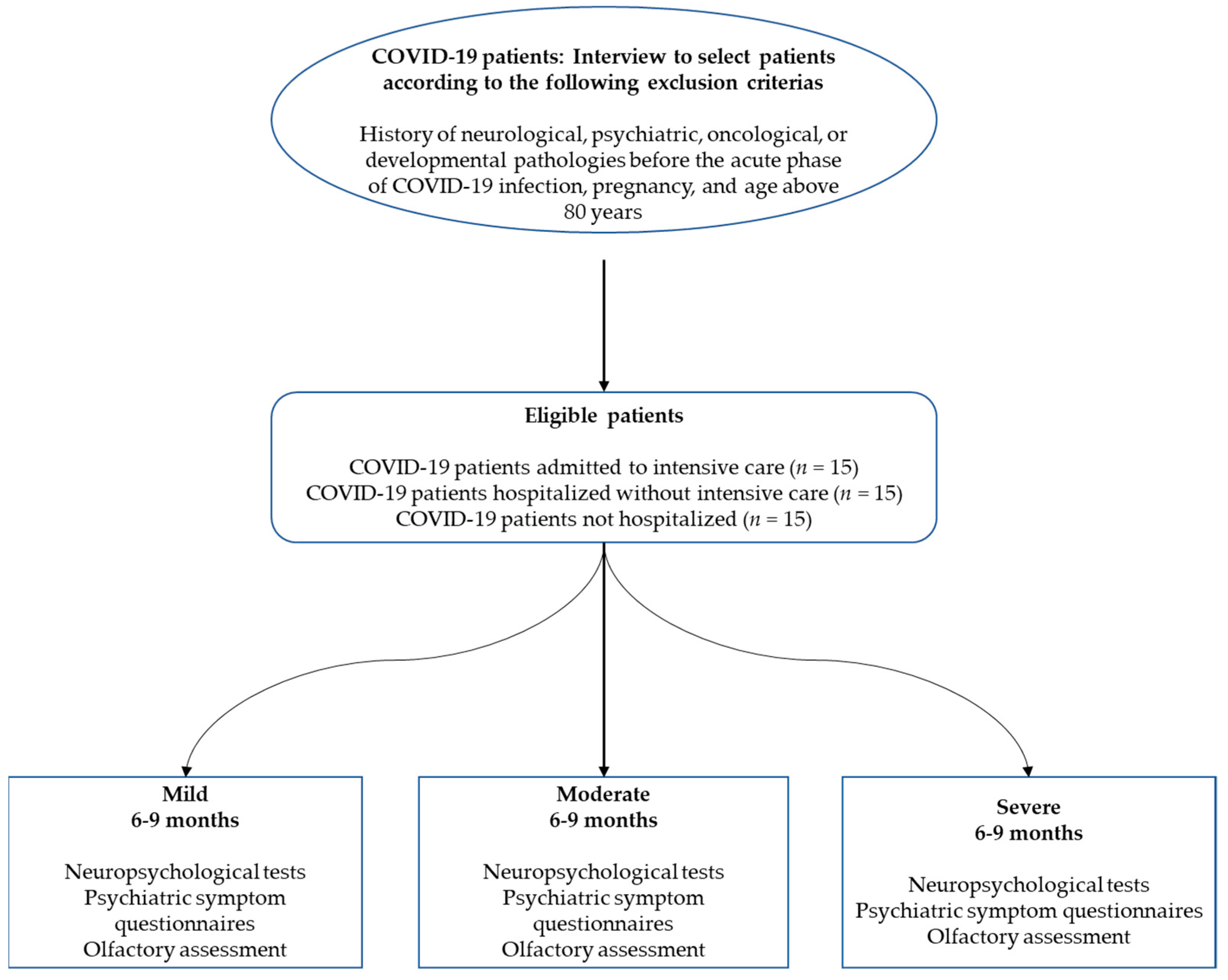
2.1. Participants

Three groups of patients who had been infected with SARS-CoV-2 were included in the study (see Table 1): 15 patients who had been admitted to intensive care during the acute phase of the infection (severe), 15 patients who had been hospitalized but did not require intensive care (moderate), and 15 patients who had tested positive but had not been hospitalized. All the patients had had their infection confirmed by positive polymerase chain reaction (PCR) results from a nasopharyngeal swab and/or by positive serological results. On average, the moderate patients had been hospitalized for 9.27 days (±9.52) and the severe patients for 37.40 days (±30.50). In comparison with other studies on SARS-CoV-2, the mean duration of hospitalization for the moderate group was somewhat longer, but this was driven by a single patient. The median number of days for this group was 7, which is comparable to that observed in other studies in Switzerland [36].
The required number of participants in each group was determined by a power analysis involving the comparison of two means: N = 2 × σ 2   ( z α 2 + z β )   ( X ¯ 1 X ¯ 2 ) 2 . This analysis was based on the literature that evaluated the short-term neuropsychological effects of SARS-CoV-2 on mild patients [25]. To achieve the desired statistical power (1 − β) of 90% and risk of Type I error (α) of 0.05, results indicated that for a one-sided hypothesis, 13 participants were needed in each group. As we planned to perform nonparametric analyses, we had to increase the sample size by 15% [37], resulting in 15 participants per group.
The three groups were comparable for median age (mild = 57 years, moderate = 55 years, severe = 59 years), sociocultural level (educational level), language (all were French speaking and Swiss citizens or residents of the French part of Switzerland), and clinical variables. Given the risk factors associated with the severe form of SARS-CoV-2, there were significantly higher proportions of men (severe = 86.66%, moderate = 40%, mild = 46.66%) and patients with diabetes. Participants were recruited via admission lists provided by the treating doctors at Geneva University Hospitals: LB and OB. For each patient, we carried out a medical file review, followed by a telephone call inviting the patient to take part in the study if all the eligibility criteria were met. Exclusion criteria were a history of neurological issues, psychiatric disorders (two of the included participants had had an episode of depression more than 10 years before their SARS-CoV-2 infection), cancer (to exclude possible chemotherapy- and radiotherapy-related cognitive impairment [38]), and neuro-developmental pathologies; pregnancy; and age above 80 years.

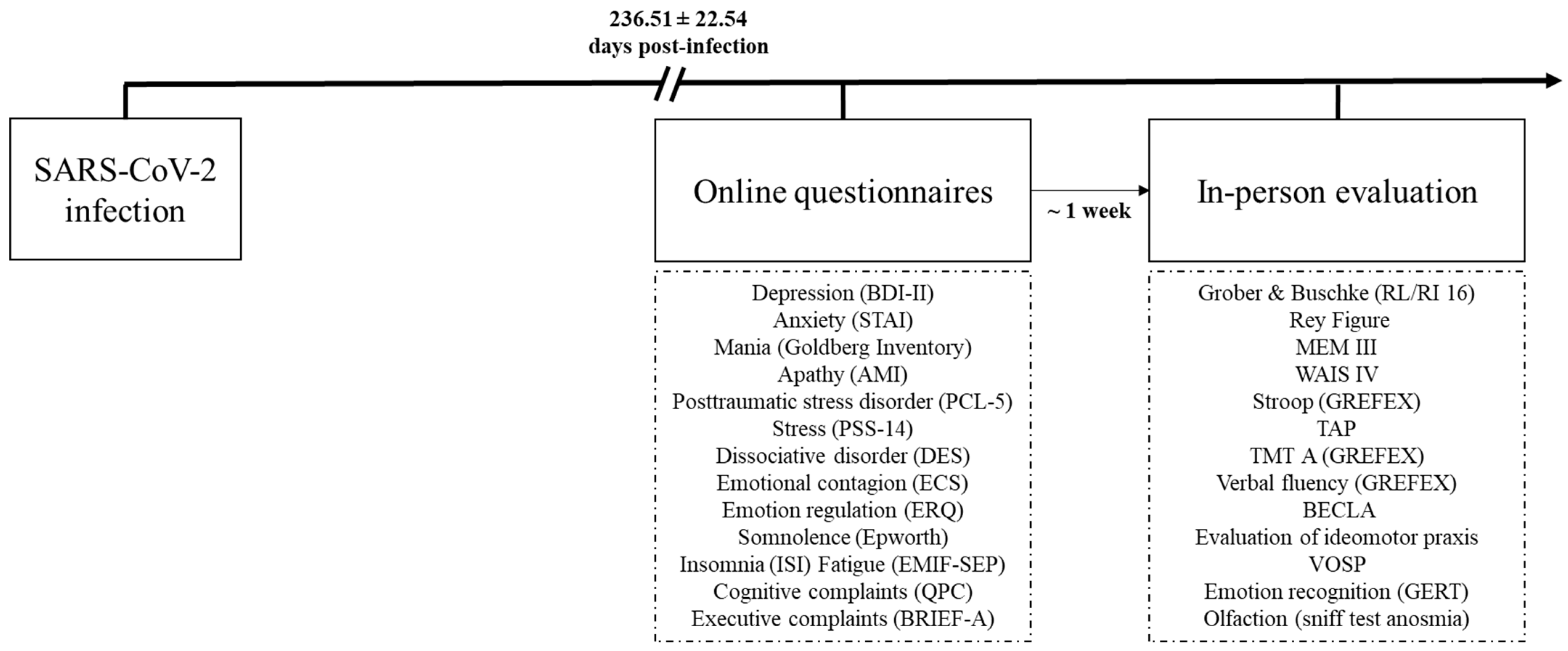
2.2. General Procedure and Ethics

A flowchart displaying the successive stages of the study according to the eligibility criteria for each experimental group is provided in Figure 1.
After being given a complete description of the study, participants provided their written informed consent. The study was conducted in accordance with the Declaration of Helsinki, and the study protocol was approved by the cantonal ethics committee of Geneva (CER-02186).

2.3. Neuropsychological Assessment

A comprehensive neuropsychological battery (based only on tests of norms validated in a French-speaking population) was administered in French to participants 6–9 months after their positive PCR test result (236.51 ± 22.54 days). This battery included a series of tests and questionnaires that assessed most of the domains of cognition, emotion recognition, fatigue, and quality of life (see below in the next paragraph). The tests were administered by clinical psychologists (mean duration: approximately 180 min), and the questionnaires were administered online via Qualtrics software (Qualtrics, Provo, UT) (mean duration: approximately 60 min). Details about which test/questionnaire was provided online or in person are provided in Figure 2.
Executive functions. Several tasks were administered to evaluate three executive functions (i.e., inhibition, shifting, and updating), in accordance with Miyake, Friedman [39]: the Stroop task, the Trail Making Test, and categorical and lexical verbal fluency from the GREFEX battery [40]. Verbal working memory and visuospatial working memory were assessed by using the backward digit span [41] and backward Corsi tests [42]. We also administered computer-based tasks designed to gauge focused attention, divided attention, phasic alertness, working memory, and incompatibility, using version 2.1 of the Test for Attentional Performance [43].
Memory systems. The short-term memory system was assessed with forward digit spans [41] and the Corsi test [42]. Verbal episodic memory was assessed with the 16-item Grober and Buschke free/cued recall (RL/RI 16) paradigm [44], as it distinguishes between the cognitive subprocesses of encoding, storage, and recall [45]. Visual episodic memory was assessed with the delayed recall of the Rey–Osterrieth Complex Figure test [46].
Instrumental function. Language was assessed with the BECLA battery [47], ideomotor praxis with a short validated battery [48], visuoconstructive abilities with the Rey–Osterrieth Complex Figure test [46], and visuoperceptual functions with four subtests from the Visual Object and Space Perception battery [49] that measured object perception (fragmented letters, object decision) and spatial perception (localization of numbers, analysis of cubes).
Logical reasoning. This was assessed by using the Puzzle and Matrices subtests of the Wechsler Adult Intelligence Scale–Fourth Edition [50].
Emotion. Multimodal emotion recognition was assessed with the Geneva Emotion Recognition Test (GERT) [51]. In this emotion recognition task, participants watched 42 video clips, in which 10 actors displayed 14 different emotions (pride, fun, joy, pleasure, relief, interest, anger, irritation, fear, anxiety, disgust, despair, sadness, surprise) while expressing nonverbal content. After each clip, participants were asked to choose one emotion from the list of 14 that best described the emotion played by the actor.
Anosognosia and cognitive complaints. We administered the Cognitive Complaints Questionnaire (QPC) [52] and the Behavior Rating Inventory of Executive Function–Adult Version (BRIEF-5) [53]. To quantify anosognosia, we calculated a self-appraisal discrepancy (SAD) score for each memory and executive domain evaluated by the QPC and BRIEF-5 [54,55,56]. In addition, the BRIEF-A not only measures the validity of the patients’ responses but also the presence of any non-credible symptoms [57,58]. First, we calculated standardized scores for the cognitive complaints, dividing the raw scores of the self-report questionnaires into four categories: 0 = normal behavior, 1 = limited influence on daily life, 2 = noticeable influence on daily life, and 3 = substantial influence on daily life. Then, we subtracted each standardized score yielded by one of these self-administered questionnaires of cognitive complaints from the standardized score for the relevant function. For example, if a patient reported no memory disorders (QPC score = 3) but performed very poorly on Grober and Buschke’s (RL/RI 16) delayed free recall test (score = 0), he or she would exhibit anosognosia for memory dysfunction: 0 (standardized score on episodic memory test)–3 (score on self-questionnaire of memory complaints) = −3. SAD scores could therefore range from −3 to 3, and any score below 0 indicated anosognosia.

2.4. Other Clinical Outcomes

We collected patients’ sociodemographic data and medical history. Psychiatric data (including current fatigue, insomnia, and somnolence) and data on olfactory abilities and quality of life at the time of the interview were also collected. Finally, a neurological assessment of CNS and peripheral nervous system functions and walking was carried out by two certified neurologists (FA and GA).
Sociodemographic and clinical data. In addition to age, collected during the inclusion interview, we recorded patients’ gender, handedness, and education level. To complement information about previous neurological, psychiatric, and developmental conditions and cancer collected during the inclusion interview, we asked patients about previous cardiovascular disease, respiratory disorders, immunosuppression status, sleep apnea syndrome, diabetes, and smoking. Participants were asked to describe the symptoms they had experienced, both during the acute phase of the infection and currently (6–9 months post-infection), and the number of days they had spent in hospital, where relevant.
Psychiatric data. Depression was assessed with the Beck Depression Inventory–Second edition (BDI-II) [59], anxiety with the State–Trait Anxiety Inventory (STAI-S and STAI-T) [60], apathy and its distinct subtypes with the Apathy Motivation Index (AMI) [61], PTSD with the Posttraumatic Stress Disorder Checklist for DSM-5 [62], manic symptoms with the Goldberg Mania Inventory [63], dissociative symptoms in the patient’s daily life with the Dissociative Experience Scale [64], current stress perception with the Perceived Stress Scale–14 items (PSS-14) [65], cognitive reappraisal of an emotional episode and expressive emotional suppression capacities with the Emotion Regulation Questionnaire [66], and susceptibility to others’ emotions with the Emotional Contagion Scale [67]. Finally, fatigue was assessed with the French version of the Fatigue Impact Scale [68], potential sleeping disorders with the Insomnia Severity Index [69], and symptoms of sleepiness in daily life with the Epworth Sleepiness Scale [70].
Olfaction. Olfactory performance was measured with the Sniffin’ Sticks test battery. This test consists of commercially available pens with 16 common odors, which were each presented for 2 s in front of both nostrils. For each odor, patients had to choose between four descriptors in a multiple-choice task. Participants’ scores ranged from 0 to 16. Based on the work by Kobal, Klimek [71], we set three thresholds: Patients with an identification score of 0–7 were considered anosmic, 8–12 hyposmic, and 13–16 normosmic.
Quality of life. We administered the SF-36 [72], which distinguishes between the physical and mental aspects of quality of life.

2.5. Statistical Analyses

2.5.1. Prevalence of Neuropsychological Deficits and Psychiatric Symptoms (Objective 1)

For each neuropsychological test, we first compared patients’ performances with normative data for the validated neuropsychological tools. As the standardization depended on the distribution of the normative data collected from the reference sample (t- and z-scores, percentiles, or standard scores), the comparative tests were adjusted according to the guidelines provided by the authors of the validation study for each test (each personal score was transformed as either t-scores, z-scores, percentiles, or standard scores). Second, we normalized the data according to the guidelines of the Swiss Association of Neuropsychology [73,74]. When the test norms were in percentiles, the following standardization was performed: far below the norm (<2nd percentile), substandard (2nd and 5th percentiles), borderline or below the normal limit (6th and15th percentiles), normal (≥16th percentile). When the test norms were in z-score, the following standardization was performed: far below the norm (<−2 z-score), substandard (−1.99 and 1.60 z-score), borderline or below the normal limit (−1.59 and −1.01 z-score), normal (≥−1.00 z-score). When the test norms were in t-score, the following standardization was performed: far below the norm (<30 t-score), substandard (30.01 and 33.60 t-score), borderline or below the normal limit (33.61 and 39.99 t-score), normal (≥40 t-score). This standardization allowed us to quantify the prevalence of each type of disorder, while controlling for variables such as age, education level, and gender. To consider the possible effect of fatigue and increase the robustness of the results, we used only those performances that were far below the norm or below the norm to calculate the prevalence of neuropsychological deficits. Results that were just below the norm were, therefore, not considered in the prevalence table.

2.5.2. Neuropsychological Deficits as a Function of Disease Severity (Objective 2)

For each neuropsychological, psychiatric, or quality-of-life measure, we compared the three groups (severe = intensive care unit [ICU] hospitalization; moderate = conventional hospitalization; mild = no hospitalization) in terms of raw data. Given the distribution of samples, we used nonparametric Kruskal–Wallis tests. For significant (p < 0.050) measures, Mann–Whitney tests were performed for the 2 × 2 comparisons with FDR correction for each domain (neuropsychological tests; psychiatric questionnaires; quality of life questionnaire).

2.5.3. Relationships between Neuropsychological Deficits, Psychiatric Symptoms, and Other Secondary Variables (Objective 3)

Because of the lack of knowledge about possible predictors of neuropsychological deficits following a SARS-CoV-2 infection, for each neuropsychological variable of interest, we performed a forward stepwise multiple regression, with the objective of determining which predictor improves the model the most. The analyses were performed on the raw cognitive data with the significant sociodemographic variables, sniff test results, and psychiatric measures to quantify relationships between these variables and the neuropsychological functions. To avoid an effect on the variance (test error), we chose not to perform all possible models but only the one that included all predictors for each neuropsychological variable of interest.
In parallel, and to elucidate the underlying structure of the cognitive data, we performed a principal component analysis (PCA) on the raw test and questionnaire scores assessing cognition and emotion recognition. The list of variables included in the PCA is available in Supplementary Index 3. We extracted the first three components with the highest eigenvalues. We then reran forward stepwise multiple regressions for each cognitive component, with the same variables of interest as those described above.

3. Results

3.1. Neuropsychological, Psychiatric, and Olfactory Profiles 6–9 Months Post-Infection

The first aim of this study was to assess the prevalence of neuropsychological impairments and psychiatric symptoms 6–9 months after SARS-CoV-2 infection. We compared patients’ performances with available normative data to identify the number of impaired scores per patient, group, and test. The cumulative prevalence of cognitive impairments in each group 236.51 ± 22.54 days after infection is set out in Table 2 (neuropsychological tests involved in the cumulative percentages can be found in Supplementary Index 1), the prevalence for each neuropsychological test can be found in Supplementary Index 1, and the prevalence of psychiatric symptoms in Table 3.
Cognition. Cognitive deficits common to all three groups were observed in the following domains: long-term episodic memory in both the verbal and visual modalities, executive functions (e.g., inhibition and mental flexibility, and both categorical and literal verbal fluency), sustained and divided attention, and language (semantic matching and naming). All three groups exhibited anosognosia for executive dysfunction (see Table 2).
Psychiatric disorders. All three groups displayed anxiety, mania, the social component of apathy, stress, PTSD, and dissociative disorders. All three groups also reported insomnia, fatigue, and pathological somnolence (see Table 3). The only psychiatric variable for which the prevalence score stood out for severe patients was emotional apathy, as measured with AMI (see Table 3).
Olfaction. Hyposmia (not counting the patients with an anosmic score) was displayed by 33.33% of the mild group, 73.33% of the moderate group, and 46.66% of the severe group. There was no anosmia in the mild and moderate groups, but 13.33% of the severe group were anosmic (see Table 3).
Symptom validity. The measurement of symptom validity, congruence, but also the measurement of non-credible symptoms by the BRIEF-A, showed good to excellent results for all participants, validating the results of the neuropsychological tests and the psychiatric symptoms questionnaires.
Table 2. Cumulated prevalence deficits for each cognitive function among patients with mild, moderate, or severe COVID-19 6–9 months post-infection.
Mild
(No Hospitalization)
n = 15
Moderate
(Conventional Hospitalization)
n = 15
Severe
(ICU Hospitalization)
n = 15
FunctionPrevalence under P5
(mean%)
Prevalence under P16
(mean%)
Prevalence under P5
(mean%)
Prevalence under P16
(mean%)
Prevalence under P5
(mean%)
Prevalence under P16
(mean%)
Memory
Verbal episodic memory (/5)1.346.676.6713.268.0017.33
Visuospatial episodic memory (/4)1.673.3415.0020.001.675.00
Verbal short-term memory (/1)0.006.676.6733.336.676.67
Visuospatial short-term memory (/1)0.006.670.000.000.000.00
Executive functions
Inhibition (/3)15.5622.2211.9023.8113.3328.89
Verbal Working memory (/1)6.676.670.0013.330.000.00
Visuospatial working memory (/3)6.6713.344.4511.116.8213.33
Mental flexibility (/6)3.337.7814.4418.8911.1118.89
Verbal fluency (/2)6.6726.6713.3353.336.6726.67
Incompatibility (/4)0.0013.335.0021.676.6710.00
Interhemispheric transfer (/2)10.0020.006.6730.006.6726.67
Attentional functions
Phasic alertness (/5)10.6718.670.0020.001.3314.67
Sustained attention (/2)10.0016.6726.9334.627.6911.54
Divided attention (/4)8.3421.676.6716.678.3321.67
Instrumental functions
Language (/5)4.009.345.338.006.6716.00
Ideomotor praxis (/3)4.44-0.00-2.22-
Object perception (/2)10.00-20.00-0.00-
Spatial perception (/2)0.00-13.33-3.33-
Logical reasoning (/2)3.3310.0010.0013.330.006.67
Anosognosia for memory0.00-40.00-40.00-
Anosognosia-Executive functions-Inhibition20.00-26.67-53.33-
Anosognosia-Executive functions-Flexibility20.00-33.33-33.33-
Anosognosia-Executive functions-Working memory6.67-6.67-0.00-
Cognitive complaints 6.67-13.33-0-
Note. Performances that were far below the norm or below the norm were used to calculate the prevalence of neuropsychological deficits (<P5). Performances that were just below the norm were considered, in addition to performances below and far below the norm in the prevalence table.
Table 3. Psychiatric symptoms and olfaction in patients with mild, moderate, or severe COVID-19 6–9 months post-infection.
Psychiatric SymptomsMild
(No Hospitalization)
n = 15
Moderate
(Conventional Hospitalization)
n = 15
Severe
(ICU Hospitalization)
n = 15
Kruskal–Wallis
p Value
Depression (BDI-II)
(prevalence)
Minor = 46.67%Minor = 66.67%Minor = 80%0.009
Mild = 20%Mild = 20%Mild = 20%
Moderate = 33.33%Moderate = 13.33%Moderate = 0%
Severe = 0%Severe = 0%Severe = 0%
State anxiety (STAI-state)
(prevalence)
Very low = 26.67%Very low = 60%Very low = 86.67%0.002
Low = 33.33%Low = 6.67%Low = 6.67%
Moderate = 13.33%Moderate = 20%Moderate = 6.67%
High = 26.67%High = 13.33%High = 0%
Very high = 0%Very high = 0%Very high = 0%
Trait anxiety (STAI-trait)
(prevalence)
Very low = 46.67%Very low = 73.33%Very low = 60%0.100
Low = 26.67%Low = 0%Low = 33.33%
Moderate = 13.33%Moderate = 20%Moderate = 6.67%
High = 13.33%High = 6.67%High = 0%
Very high = 0%Very high = 0%Very high = 0%
Mania (Goldberg Inventory)
(prevalence)
Probably absent = 26.67%Probably absent = 13.33%Probably absent = 20%0.909
Hypomania = 26.67%Hypomania = 26.67%Hypomania = 46.67%
Close to mania = 20%Close to mania = 20%Close to mania = 6.67%
Moderate = 26.67%Moderate = 40%Moderate = 26.67%
Ordinary to severe = 0%Ordinary to severe = 0%Ordinary to severe = 0%
Severe = 0%Severe = 0%Severe = 0%
Apathy (AMI-total)
(prevalence)
Absent = 86.67%Absent = 93.33%Absent = 73.33%0.602
Moderate = 13.33%Moderate = 6.67%Moderate = 26.67%
High = 0%High = 0%High = 0%
Behavioral apathy (AMI-behavioral)
(prevalence)
Absent = 100%Absent = 100%Absent = 93.33%0.211
Moderate = 0%Moderate = 0%Moderate = 6.67%
High = 0%High = 0%High = 0%
Social apathy (AMI-social)
(prevalence)
Absent = 93.33%Absent = 86.67%Absent = 73.33%0.940
Moderate = 6.67%Moderate = 6.67%Moderate = 26.67%
High = 0%High = 6.67%High = 0%
Emotional apathy (AMI-emotional)
(prevalence)
Absent = 73.33%Absent = 60%Absent = 40%0.029
Moderate = 26.67%Moderate = 40%Moderate = 33.33%
High = 0%High = 0%High = 26.67%
Posttraumatic stress disorder (PCL-5)
(prevalence)
Absent = 86.67%Absent = 86.67%Absent = 93.33%0.054
Present = 13.33%Present = 13.33%Present = 6.67%
Stress (PSS-14)
Mean (±SD)
26.13 (±9.53)19.6 (±7.47)14.93 (±9.42)0.023
Dissociative disorder (DES)
Mean (±SD)
7.68 (±11.89)10.45 (±9.23)3.98 (±3.03)0.140
Emotional contagion (ECS)
Mean (±SD)
41.40 (±7.20)44.6 (±4.22)36.13 (±8.38)0.002
Emotion regulation (ERQ)
Mean (±SD)
41.6 (±7.39)44.2 (±8.33)39.80 (±11.77)0.416
Somnolence (Epworth)
(prevalence)
Pathological = 40.00%Pathological = 53.33%Pathological = 26.67%0.036
Insomnia (ISI)
(prevalence)
Absent = 20%Absent = 46.67%Absent = 60%0.040
Mild = 40%Mild = 40%Mild = 26.67%
Moderate = 40%Moderate = 13.33%Moderate = 13.33%
Severe = 0%Severe = 0%Severe = 0%
Fatigue (EMIF-SEP)
(prevalence)
Present = 13.33%Present = 20%Present = 6.67%0.088
Sniff test (anosmia)
Mean (±SD)
13.07 (±1.44)11.53 (±2.13)11.47 (±2.90)0.067
Abbreviations: AMI-behavioral: Apathy Motivation Index–behavioral score [61]; AMI-emotional: Apathy Motivation Index–emotional score [61]; AMI-social: Apathy Motivation Index–social score [61]; AMI-total: Apathy Motivation Index–total score [61]; BDI-II: Beck Depression Inventory–Second edition [59]; DES: Dissociative Experience Scale [64]; ECS: Emotional Contagion Scale [67]; EMIF-SEP: Fatigue Impact Scale, French adaptation [68]; ERQ: Emotion Regulation Questionnaire [66]; Epworth: Epworth Sleepiness Scale [70]; Goldberg Inventory: Goldberg Mania Inventory [64]; ICU: intensive care unit; ISI: Insomnia Severity Index [69]; PCL-5: Posttraumatic Stress Disorder Checklist for DSM-5 [62]; PSS-14: Perceived Stress Scale–14 items [65]; STAI-trait: State–Trait Anxiety Inventory [60]; STAI-state: State–Trait Anxiety Inventory [60].

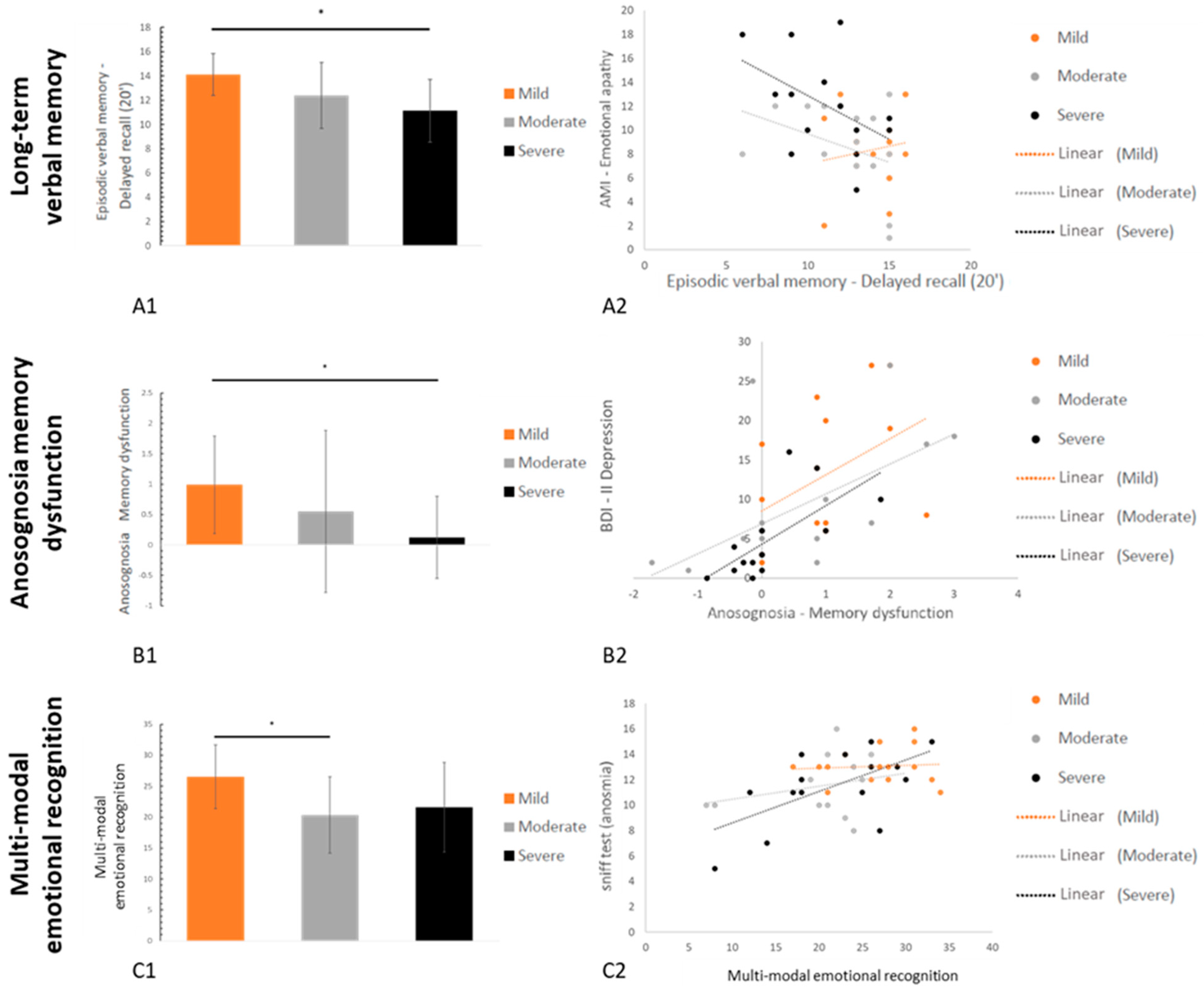
3.2. Neuropsychological and Psychiatric Symptoms as a Function of Disease Severity

The second aim was to determine whether cognitive deficits and psychiatric symptoms are a function of the severity of the respiratory symptoms in patients in the acute phase (severe = ICU hospitalization; moderate = conventional hospitalization; mild = no hospitalization). To this end, we compared the three groups for neuropsychological, psychiatric, and other clinical data (Kruskal–Wallis statistics and p values reported in Table 2, Table 3 and Table 4; FDR-corrected Mann–Whitney statistics and p values reported below).

3.2.1. Neuropsychological Data

The three groups differed significantly in: (i) long-term episodic memory in both the verbal (Grober and Buschke (RL/RI 16) delayed free recall, H = 10.75, p = 0.005, r = 0.21) and visual (Rey Figure delayed free recall, H = 6.15, p = 0.046, r = 0.10) modalities, (ii) multimodal emotion recognition (GERT; H = 7.55, p = 0.023, r = 0.13), and (iii) cognitive complaints (QPC; H = 6.38, p = 0.041, r = 0.10) and anosognosia for memory dysfunction (SAD; H = 7.84, p = 0.020, r = 0.14). The other effects were not significant (p > 0.05 for all comparisons) (see Figure 3).
Episodic memory. For Grober and Buschke delayed free recall, the mild patients scored significantly higher than the severe patients (z = 3.04, p = 0.002, r = 0.55), but the other two pairwise comparisons were not significant after FDR correction (moderate vs. severe: z = −1.47, p = 0.141, r = −0.27; mild vs. moderate: z = 2.00, p = 0.046, r = 0.37). Pairwise comparisons were not significant for visual episodic memory (mild vs. moderate: z = 2.26, p = 0.023, r = 0.41; mild vs. severe: z = 0.48, p = 0.61, r = 0.09; moderate vs. severe: z = 1.89, p = 0.059, r = 0.35).
Emotion recognition. Mild patients scored significantly higher than moderate patients (z = 2.61, p = 0.009, r = 0.48), but neither the difference between mild and severe patients (z = 1.97, p = 0.048, r = 0.36) nor the difference between moderate and severe patients (z = 0.49, p = 0.620, r = 0.08) reached significance after FDR correction.
Cognitive complaints and anosognosia. Mild patients had more cognitive complaints than severe patients did (z = −2.55, p = 0.010, r = −0.47), but there were no differences between either the mild and moderate patients (z = −1.31, p = 0.191, r = −0.24) or the moderate and severe patients (z = −0.93, p = 0.351, r = −0.17). By contrast, severe patients exhibited more anosognosia for memory dysfunction than mild patients did (z = 2.97, p = 0.003, r = 0.54), whereas there were no differences between either the mild and moderate patients (z = 1.41, p = 0.158, r = 0.26) or the moderate and severe patients (z = −0.76, p = 0.443, r = −0.14).
All means and standard deviations for neuropsychological data are available in Supplementary Index 2.

3.2.2. Psychiatric Data

The three groups differed significantly on depression (H = 9.40, p = 0.009, r = 0.18), state anxiety (H = 12.93, p = 0.002, r = 0.26), emotional apathy (H = 7.10, p = 0.029, r = 0.12), stress (H = 7.55, p = 0.023, r = 0.13), and emotional contagion (H = 9.73, p = 0.002, r = 0.18). The other effects were not significant (p > 0.05 for all comparisons). Pairwise comparisons for each of these group differences are described below.
Depression, stress, and state anxiety. The mild patients were more depressed, stressed, and anxious than the severe patients (BDI-II: z = −2.99, p = 0.003, r = −0.55; PSS-14: z = −2.55, p = 0.010, r = −0.47; STAI-S: z = −3.57, p < 0.001, r = −0.65), whereas there were no differences between either the severe and moderate patients (BDI-II: z = −1.38, p = 0.165, r = −0.25; PSS-14: z = −1.08, p = 0.281, r = −0.20; STAI-S: z = −1.76, p = 0.078, r = −0.32) or the mild and moderate patients (BDI II: z = −1.66, p = 0.097, r = −0.30; PSS-14: z = −1.08, p = 0.281, r = −0.20; STAI-S: z = −1.72, p = 0.085, r = −0.31).
Apathy. For the AMI emotional subscore, pairwise comparisons failed to reach significance after FDR correction (severe vs. mild: z = 2.32, p = 0.020, r = 0.42; severe vs. moderate: z = 2.20, p = 0.028, r = 0.40; mild vs. moderate: z = 0.08, p = 0.933, r = 0.01).
Emotional contagion. For the ECS scale, severe patients reported higher levels of emotional contagion as compared to moderate patients (z = −3.03, p = 0.017). The other pairwise comparisons failed to reach significance after FDR correction (severe vs. mild: z = −1.89, p = 0.059, r = −0.35; moderate vs. mild: z = 1.18, p = 0.237, r = 0.22).

3.2.3. Fatigue and Quality of Life

Finally, the three groups differed in insomnia (H = 6.66, p = 0.036, r = 0.11), fatigue (H = 6.45, p = 0.040, r = 0.11), vitality (H = 6.50, p = 0.039, r = 0.11), and emotional well-being (H = 9.18, p = 0.010, r = 0.17). The other effects were not significant (p > 0.05 for all comparisons).
The mild patients reported more fatigue than the severe patients did (z = −2.57, p = 0.010, r = −0.47), whereas there were no differences between either the mild and moderate patients (z = −0.71, p = 0.481, r = −0.13) or the moderate and severe patients (z = −1.52, p = 0.130, r = −0.28). For insomnia, mild reported more symptoms as compared to moderate patients (z = −1.99, p = 0.046, r = −0.36), whereas the other pairwise comparisons did not reach significance after FDR correction (mild vs. severe: z = −2.28, p = 0.023, r = −0.42; moderate vs. severe: z = −0.71, p = 0.481, r = −0.13).
Conversely, severe patients reported more vitality, emotional well-being, and social function than mild patients did (vitality: z = 2.65, p = 0.008, r = 0.48; well-being: z = 2.97, p = 0.003, r = 0.54; social function: z = 2.99, p = 0.002, r = 0.55), as well as higher well-being as compared to moderate patients (z = 2.01, p = 0.044, r = 0.37). Pairwise comparisons between severe and moderate patients did not reach significance after FDR correction (vitality: z = 1.06, p = 0.290, r = 0.19; social function: z = 1.66, p = 0.097, r = 0.30) nor between mild and moderate patients (well-being: z = 0.68, p = 0.494; vitality: z = 1.12, p = 0.263, r = 0.12; social function: z = 1.06, p = 0.290, r = 0.19) (see Table 4).
All means and standard deviations for psychiatric data are available in Supplementary Index 3.

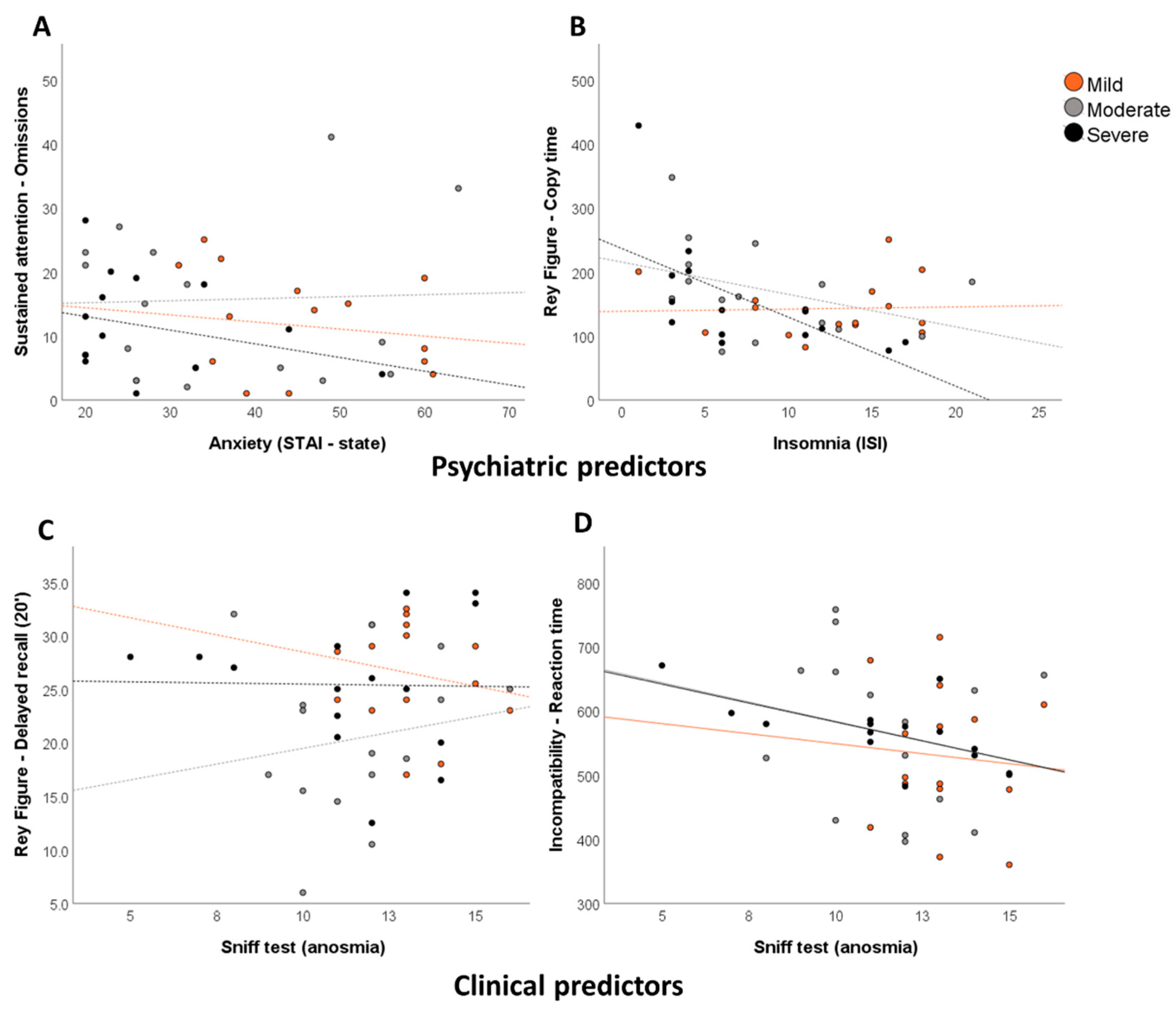
3.3. Relationships between Neuropsychological Deficits, Psychiatric Symptoms, and Other Secondary Variables

The third aim was to examine whether the presence of long-term neuropsychological deficits was correlated with psychiatric symptoms and/or other clinically relevant variables (see Figure 4).
The results of the multiple regression performed on each cognitive variable are set out in Table 5. Interestingly, apathy, depression, anxiety, emotion regulation, emotion contagion, stress, PTSD, dissociative disorders, anosmia, and diabetes all proved to be variables of interest for explaining the neuropsychological sequelae. Therefore, both psychiatric and nonpsychiatric data correlated with neuropsychological deficits across the three groups. There were at least three patterns of results, depending on the neuropsychological domain: (i) patterns in which neuropsychological sequelae did not correlate with any psychiatric variables, but did with other clinical variables, such as visuospatial long-term episodic memory (delayed recall of Rey–Osterrieth Complex Figure); (ii) patterns in which neuropsychological sequelae correlated with both psychiatric and clinical variables, such as the object and action naming task scores (language); and (iii) patterns in which neuropsychological sequelae correlated with only psychiatric variables, such as categorical verbal fluency. There was also a fourth possible pattern in which the neuropsychological sequelae correlated neither with psychiatric variables nor with clinical ones, such as the score on the object decision task (object perception).
To reduce the dimensionality of the data set, we computed a PCA (Supplementary Index 4). We selected the first three orthogonal components accounting for 43.67% of the total variance. The first component, accounting for 26.75% of the total variance, was difficult to interpret in terms of underlying cognitive processes, as it included language (semantic word and image matching), executive functions (mental flexibility), verbal episodic memory, and emotion recognition. Interestingly, these happened to be precisely the variables on which the three groups differed significantly (see section “Neuropsychological and Psychiatric Symptoms as a Function of Disease Severity”). We therefore labeled this component respiratory disease severity. The second component (9.79% of total variance) was labeled attention and anosognosia, as it included alertness, divided attention, and anosognosia for executive dysfunction. The third component (7.15% of total variance) was labeled instrumental functions, as it included language, visual perception, and ideomotor praxis.
For the respiratory disease severity component, the best fit was achieved with emotional apathy (R2 = 0.28, p = 0.007), stress (R2 = 0.19, p = 0.013), and anosmia (R2 = 0.11, p = 0.03). For the attention and anosognosia component, the multiple regression was not significant (p > 0.1). For the instrumental functions component, the best fit was achieved with anosmia (R2 = 0.23, p = 0.04), mania (R2 = 0.23, p = 0.006), and social apathy (R2 = 0.17, p = 0.004).
Table 5. Multiple regression results for each of the neuropsychological variables.
RegressorR2p Value
Memory Functions
Verbal episodic memoryGrober & Buschke (RL/RI 16)-Immediate recallnsnsns
Grober & Buschke (RL/RI 16)-Delayed free recallAMI–Emotional apathy0.450.006
Epworth-Sleepiness0.200.022
ERQ–Emotion regulation0.130.034
ECS–Emotional contagion0.080.034
Grober & Buschke (RL/RI 16)-Delayed total recallAMI–Emotional apathy 0.340.022
Epworth-Sleepiness0.220.031
AMI–Social apathy0.150.034
Visuospatial episodic memoryRey Figure-Copy timeISI-Insomnia0.460.005
ERQ–Emotion regulation0.180.035
Rey Figure-Scorensnsns
Rey Figure-Immediate recall (3’)nsnsns
Rey Figure-Delayed recall (20’)ISI–Insomnia0.300.034
Days of hospitalization0.220.039
Sniff test (anosmia)0.210.013
Verbal short-term memoryMEM III-Spansnsnsns
Visuospatial short-term memoryWAIS IV-SpansDES–Dissociation 0.300.035
Executive functions
InhibitionStroop (GREFEX)-Interference-Timensnsns
Stroop (GREFEX)-Interference-ErrorsAMI–Total apathy0.370.015
ERQ–Emotion regulation0.210.030
Stroop (GREFEX)-Interference/Naming-Score ns ns ns
Working memoryMEM III–Verbal working memoryAMI–Behavioral apathy0.330.026
WAIS IV-Visuospatial working memory STAI-T Anxiety0.390.013
Diabetes0.250.014
STAI-S Anxiety0.130.03
TAP-Working memory item omissionsAMI–Emotional apathy0.300.035
TAP-Working memory false alarmsDiabetes0.470.005
Mania–Goldberg Inventory0.160.044
Mental flexibilityTMT A (GREFEX)-Timensnsns
TMT A (GREFEX)-Errorsnsnsns
TMT B (GREFEX)-Timensnsns
TMT B (GREFEX)-ErrorsAMI–Total apathy0.410.010
ERQ–Emotion regulation0.140.024
Gender0.130.025
TMT B (GREFEX)-PerseverationsSTAI-T Anxiety0.550.002
BDI-II-Depression0.160.026
ISI–Insomnia0.20<0.001
TMT B-A (GREFEX)-Scorensnsns
Verbal fluency (GREFEX)-Literal (2’)nsnsns
Verbal fluency (GREFEX)-Categorical fluency (2’)DES–Dissociation0.280.047
IncompatibilityTAP-Compatibility-Reaction timeAMI–Social apathy0.500.003
TAP-Compatibility-False alarmsnsnsns
TAP-Incompatibility-Reaction TimeSniff test (anosmia)0.280.043
DES–Dissociation0.250.026
ERQ–Emotion regulation0.130.028
Epworth-Sleepiness0.100.017
TAP-Incompatibility-False alarmsSniff test (anosmia)0.270.045
TAP-Incompatibility-Visual field scoreDays of hospitalization0.390.013
Diabetes0.230.007
STAI-T Anxiety0.080.041
PCL-5 Posttraumatic stress disorder0.060.041
TAP-Incompatibility task-Hands scoreAMI–Social apathy0.440.008
TAP-Incompatibility task-Visual fields * Hands scoreISI–Insomnia0.330.025
STAI-T Anxiety0.240.025
BDI-II-Depression0.180.019
Attentional functions
Phasic alertnessTAP-Without warning sound-Reaction timeGender0.350.019
TAP-Without warning sound-SD of reaction timeAMI–Social apathy0.64<0.001
Diabetes0.110.041
Sniff test (anosmia)0.100.023
TAP-With warning sound-Reaction timeDES–Dissociation0.300.033
TAP-With warning sound-SD of reaction timensnsns
TAP-Alertness indexGender 0.280.041
Sustained attentionTAP-Items OmissionsSTAI-Trait Anxiety0.460.005
TAP-False alarmnsnsns
Divided attentionTAP-Audio condition-Reaction timeDES–Dissociation0.410.010
AMI–Behavioral apathy0.270.008
TAP-Visual condition-Reaction timeDays of hospitalization 0.380.014
Sniff test (anosmia)0.270.009
ISI-Insomnia0.23<0.001
AMI–Emotional apathy0.050.016
TAP-Total omissions ns ns ns
TAP-Total false alarmsAMI–Emotional apathy0.320.029
Epworth-Sleepiness0.280.015
AMI–Social apathy0.160.023
Instrumental functions
LanguageBECLA-Semantic image matchingnsnsns
BECLA-Semantic word matchingnsnsns
BECLA-Object and action image namingECS–Emotional contagion0.380.014
STAI-State Anxiety0.290.007
AMI–Emotional apathy0.100.014
ISI-Insomnia0.080.012
BECLA-Word repetitionNVNVNV
BECLA-Nonword repetitionnsnsns
Ideomotor praxisSymbolic gesturesERQ–Emotion regulation0.350.019
AMI–Behavioral apathy0.330.004
Action pantomimesBDI-II-Depression0.510.003
Meaningless gesturesAMI–Total apathy0.300.033
AMI–Social apathy0.180.035
Object perceptionVOSP-Fragmented lettersAMI–Total apathy0.260.029
VOSP-Object decisionnsnsns
Spatial perceptionVOSP-Number localizationnsnsns
VOSP-Cubic countingMania–Goldberg Inventory0.270.047
PSS-14-Stress0.240.034
Logical reasoningWAIS IV-PuzzleDiabetes0.340.021
WAIS IV-MatrixDES–Dissociation0.280.041
Gender0.400.002
Emotion recognitionGERTERQ–Emotion regulation0.330.023
AMI–Emotional apathy0.280.011
AMI–Behavioral apathy0.160.004
Sniff test (anosmia)0.060.008
AMI–Social apathy0.020.027
AnosognosiaMemory dysfunctionsBDI-II-Depression0.62<0.001
ISI-Insomnia0.120.038
AMI–Behavioral apathy0.090.040
Epworth-Sleepiness0.050.033
Executive functions-InhibitionAMI–Total apathy0.400.011
Executive functions-FlexibilityAMI–Behavioral score0.280.043
Executive functions–Working memoryEpworth-Sleepiness0.380.015
Sniff test (anosmia)0.250.015
Abbreviations: AMI-behavioral: Apathy Motivation Index–behavioral score [61]; AMI-emotional: Apathy Motivation Index–emotional score [61]; AMI-social: Apathy Motivation Index–social score [61]; AMI-total: Apathy Motivation Index–total score [61]; BDI-II: Beck Depression Inventory–Second Edition [59]; BECLA: Batterie d’Evaluation Cognitive du Langage [47]; DES: Dissociative Experience Scale [64]; ECS: Emotional Contagion Scale [67]; ERQ: Emotion Regulation Questionnaire [66]; GERT: Geneva Emotion Recognition Test [51]; Goldberg-Inventory: Goldberg Mania Inventory [63]; GREFEX: Groupe de Réflexion sur l’Evalutation des Fonctions Exécutives [40]; MEM III: Wechsler Memory Scale–Third Edition [41]; NV: no variance; ns: not significant; PCL-5: Posttraumatic Stress Disorder Checklist for DSM-5 [62]; PSS-14: Perceived Stress Scale–14 items [65]; Rey Figure: Rey–Osterrieth Complex Figure test [46]; RL/RI 16: free/cued recall 16 items (RL/RI 16) [44]; SD: standard deviation; STAI-S: State–Trait Anxiety Inventory [60] STAI-T: State–Trait Anxiety Inventory [60]; TAP: Test for Attentional Performance, Version 2.1 [43]; TMT: Trail Making Test; VOSP: Visual Object and Space Perception battery [49]; WAIS IV: Wechsler Adult Intelligence Scale–Fourth Edition [50].

4. Discussion

Growing evidence suggests that SARS-CoV-2 can cause brain damage in the long term, with a potential impact on cognition even in its mild and moderate forms [75]. Nonetheless, to date, the occurrence and nature of such sequelae, the evolution and the duration of symptoms, the impact of respiratory disease severity in the acute phase, and the relationship between these impairments and psychiatric disorders triggered or exacerbated by the pandemic are unknown and have not been studied in detail within a single sample of patients. In addition, areas such as instrumental functions (ideomotor praxis, visual perception, or language), cognitive complaints, anosognosia, and emotion recognition following SARS-CoV-2 have yet to be explored. Finally, the relevant medical events have not been controlled in studies published thus far. In the present study, we used a robust, psychometrically validated methodology and a stringent approach to the normative data of neuropsychological tests (excluding borderline scores from the interpretation, while data is disponible in Supplementary Index 1). We included patients with no history of cancer or neurological and developmental disorders, and no active psychiatric disorders before SARS-CoV-2 infection, and divided them into mild, moderate, and severe groups, according to the respiratory severity of the disease during its acute phase.
The present study, therefore, improves our understanding of what we can call neurological long COVID, highlighting three main patterns of results. First, a potentially important prevalence of patients across the three groups (severe = ICU hospitalization; moderate = conventional hospitalization; mild = no hospitalization) performed below the normality threshold in all domains of cognition (except ideomotor praxis) 6–9 months after infection with SARS-CoV-2. The prevalence of psychiatric symptoms, regardless of disease severity during the acute phase, was also high, and individuals in all three groups exhibited depressive symptoms, anxiety, mania, apathy, stress, PTSD, and dissociative disorders, as well as reporting insomnia, fatigue, and pathological somnolence. Regarding olfaction, 33.33% of the mild group, 73.33% of the moderate group, and 46.66% of the severe group were still hyposmic 6–9 months following infection; 13.33% of the severe group were still anosmic. Second, despite the presence of common cognitive deficits across the three groups, some domains of cognition and mood were differentially impacted by the severity of respiratory disease during the acute phase: the severe group performed more poorly than the mild group did on long-term episodic memory and also exhibited more anosognosia for memory dysfunction. The mild group was more depressed, stressed, and anxious and reported more cognitive complaints. Finally, the moderate group recognized multimodal emotions less well than the mild group did. All of this had a substantial impact on patients’ quality of life. Third, as predicted, neuropsychological deficits correlated with psychiatric disorders such as depressive symptoms, stress, and mania, but not all of the variance was explained by psychiatric symptoms or transdiagnostic syndrome [76]. Instead, a large proportion of the variance was explained by other clinical variables. For instance, the long-term episodic memory deficits displayed by the severe group were positively correlated with emotional apathy, their anosognosia for memory dysfunction was correlated with depression, and their diminished emotion recognition, shared by the moderate group, was positively correlated with hyposmia and/or anosmia.
This study had several limitations that need to be acknowledged and addressed before we can draw any inferences from our results. The first drawback was a possible recruitment bias. By enrolling volunteers, we may have selected the most severe cases in the mild group (who were interested in the study because of their cognitive complaints), and we may not have recruited the most cognitively affected in the severe group, because they were too disabled to join the study. Second, we had greater proportions of men and individuals with diabetes in the severe group. These factors may have had an influence on the cognitive deficits observed in this group, as diabetes is known to impact cognition [77] and gender affects depression [78], with a greater prevalence in women [30]. That said, although the proportion of women was higher for both the mild and moderate groups, the mean depression scores by gender in the mild (women: 13.50 ± 9.10; men: 12.57 ± 8.52) and moderate (women: 6.11 ± 5.25; men: 13.33 ± 11.25) groups did not indicate a greater proportion of women with depressive symptoms. Moreover, our sample does not exactly reflect the population of COVID-19 survivors because of our methodological choice to match mild and moderate to severe groups. Indeed, it is known that young people have a higher probability to have mild/moderate disease without hospitalization. Thus, our sample of mild patients does not perfectly represent the majority of mild cases, which could pose difficulties in generalizing the results. The question whether younger people experience the same neuropsychological consequences remains open. Another issue for the mild group is the fact that it did not contain a single person over the age of 65, which is known to be a cut-off age for increased prevalence of mild cognitive impairment. This imbalance could have potentially influenced the results of the groups on cognitive tests. Third, stroke is more prevalent in patients after a severe SARS-CoV-2 infection [6,7] and may have gone unseen during the acute phase. In our study, no patient had any central neurological deficit including major stroke, but minor stroke cannot be ruled out. Two patients in the severe group reported mild signs of peripheral neuropathy, which may have been due to their diabetes and not a direct consequence of the SARS-CoV-2 infection, and one patient in the severe group had an unstable gait. Fourth, the absence of a control group prevented us from observing a possible general effect of the pandemic and the resulting public health measures on mental health. When recruiting patients for this study (between October 2020 and January 2021), we chose not to carry out face-to-face neuropsychological assessments on control patients, for an ethical reason. The infection rates were high, and the vaccines were not marketed. Therefore, we did not want to take the risk of increasing the number of nosocomial infections. In this study, the analyses of prevalence were based on standardized normative data, allowing us to run comparisons with the normal population. The tests were chosen carefully for their psychometric validity, with adequate sensitivity and specificity. Notably, in a recent study, the multimodal emotion recognition task (GERT) was administered to 469 participants during the pandemic [79], but the authors failed to find a reduction in performances compared with those in validation studies [51], reinforcing the hypothesis that our results reflected a specific effect of the infection and not just the public health context. Fifth, we did not psychometrically measure participants’ motivation to complete the tasks. Nevertheless, the assessments were performed by clinical psychologists who, during the anamnesis and testing, checked the performance of the tasks, the impact of fatigue, and the motivation of the participants. Any participant who did not show willingness to complete the tasks or to be part of the cohort was excluded and his or her results were not considered. In addition, the BRIEF-A was able to measure not only the validity of the patients’ responses but also the presence of any non-credible symptoms [57,58]. The results for the participants were all within the norm with good to excellent response validity. Sixth and last, the number of participants was relatively small, which prevented us from considering more covariates. Nevertheless, the power analysis, based on a previous study of the neurocognitive effects of SARS-CoV-2, did allow us to estimate the necessary sample size.
Our results demonstrate first that cognitive deficits can be observed 6–9 months after a SARS-CoV-2 infection, regardless of the severity of the disease in the acute phase. These results corroborate previous observations for the executive, attentional, and memory domains and go one step further, with exhaustive neuropsychological and psychiatric assessments demonstrating impairments in other previously unexplored cognitive and psychiatric domains. Impairments were evident not only in the severe patient group but also in the moderate and mild groups. These deficits had an impact on quality of life, notably in the mild patients, as evidenced by our results. These findings could be of great importance in understanding the long-term damage and consequences of SARS-CoV-2 infection for cognition and mental health. The potentially high prevalence of certain cognitive and psychiatric disorders, regardless of the severity of the disease in the acute phase, suggests that long-term patient management following SARS-CoV-2 infection may need to be adapted. Notably, the etiology of these disorders needs to be established in order to provide people who are experiencing these long-term sequelae with the best possible care. One potential explanation for these effects, based on observational studies of the psychiatric impact of the pandemic in the general population [26], is that these cognitive deficits result from a stressful or traumatic context induced by the pandemic context or by hospitalization. In this case, specific interventions in certain psychiatric variables could considerably reduce their long-term impact on cognition and improve daily functioning. Another hypothesis for the higher level of cognitive complaints in the mild group could be explained by the fact that the patients in the severe group survived despite having been admitted to ICU. While prolonged hospitalization could undoubtedly be traumatic, the survival itself could have biased this group towards a more optimistic outlook and a lower tendency to notice/report lingering cognitive complaints. Nevertheless, the present results do not exclude the hypothesis of direct damage to brain networks by SARS-CoV-2 and its neurotropism, as well as indirect neurobiological effects, which could lead to both psychiatric and neurological disorders. COVID-19 may induce CNS disturbance, and four main pathogenic mechanisms may act in combination: (i) direct viral encephalitis, (ii) systemic inflammation, (iii) peripheral organ dysfunction (liver, kidney, lung), and (iv) cerebrovascular changes [80]. At this stage, it is difficult to determine whether the cognitive deficits can be regarded as a marker of brain damage and/or should be linked to psychiatric variables that may themselves result directly from infection with SARS-CoV-2, or else be triggered by the stressful nature of the general pandemic and the individual experience of the disease.
Second, this study highlighted the presence of differential cognitive and psychiatric profiles at 6–9 months post-infection as a function of the respiratory severity of the SARS-CoV-2 infection in the acute phase. This suggests the existence of different clinical phenotypes. In the identification/discrimination of these phenotypes, different cognitive variables seem to be of interest, starting with cognitive complaints and anosognosia. Although the severe patients exhibited anosognosia for their memory dysfunction and greater long-term verbal memory impairment than the mild patients did, the latter had more cognitive complaints. This fits well with the observations of Almeria, Cejudo et al., [21] who found that the patients with the most serious cognitive complaints did not have significantly more neuropsychological impairments. In this sense, the tendency of the severe patients to report greater well-being in quality-of-life assessments, together with the lack of awareness of their cognitive difficulties, may be a clinical characteristic to bear in mind when interviewing this type of patient. The present results in the domain of emotion recognition and episodic memory are also highly relevant to the current debate on the neurotropism of SARS-CoV-2. One of the main hypotheses regarding the pathways of direct attack of the CNS assumes olfactory transmucosal invasion by the virus [18]. This hypothesis appears to be supported by our results. It is worth noting that episodic memory and emotion recognition were identified in a PCA as variables that explained most of the variance of our data, and this first component was significantly correlated with hyposmia/anosmia, in addition to stress and emotional apathy. Interestingly, a recent 18F-FDG PET study demonstrated hypometabolism at about 8 weeks post-infection in brain regions common to emotion and olfaction in patients with SARS-CoV-2 [19]. Moreover, the literature suggests that the viral load was probably greater in our severe group [81,82], which may have contributed to stronger effects on olfaction and emotion recognition. Recently, evidence on humans and rodents have demonstrated neurophysiological relationships through cycle-by-cycle influences between respiratory rhythms involving olfactory regions with cortical (e.g., hippocampal) and sub-cortical regions, involved in emotional and memory processing [83]. Those observations may reinforce our results and suggest a specific link between these functions in healthy humans, potentially disrupted by SARS-CoV-2 infection. Finally, this pathway could also partially explain the psychiatric results via disruption of the limbic network, including subcortical regions [84], by SARS-CoV-2.
Our third level of analysis enabled us to go further in characterizing the hypothesized clinical phenotypes. Quantified results pointed to the presence of at least three profiles (patient clusters), corroborating the clinical impressions we had when interviewing and assessing the patients for this study. Patients with the first (neurological) profile were typically aged about 55 years, mostly men, of average educational level, and a small proportion of them had a history of diabetes, cardiovascular disease, or sleep apnea syndrome. At the cognitive level, these patients displayed long-term memory, executive, and language disorders. They had more severe anosognosia for their memory difficulties. Nearly all of them reported sleep disorders and emotional apathy. Patients with the second (psychiatric) profile were aged about 45–50 years, and there were equal numbers of men and women. No significant medical antecedents were noted, and most of them had had mild or moderate respiratory disease. At the cognitive level, they displayed executive and attentional dysfunctions, which could influence other cognitive domains (e.g., memory recall strategies). At the psychiatric level, they had high scores for depressive symptoms, anxiety, insomnia, and stress, and they more sporadically exhibited PTSD and dissociative disorders. Our results also indicated the presence of a third (mixed) profile combining the symptoms and clinical characteristics of the two previously described profiles.

5. Conclusions

This study demonstrates the presence of long-term neuropsychological sequelae following SARS-CoV-2 infection, regardless of the severity of the respiratory disease in the acute phase. Some of the cognitive deficits could be explained by psychiatric variables, emphasizing the importance of considering a broad range of psychiatric symptoms. However, not all neuropsychological sequelae could be explained by these variables. The presence of correlations between olfaction, emotion recognition, and episodic memory, which share common functional and anatomical substrates, reinforces the hypothesis that the virus targets the CNS (notably the limbic system). Finally, the data support the notion of different clinical phenotypes, paving the way for clinical guidelines and recommendations for the management of long-term neurological impairment following SARS-CoV-2 infection.

Supplementary Materials

The following supporting information can be downloaded at: https://www.mdpi.com/article/10.3390/ctn6020009/s1, Supplementary Index 1: Prevalence of cognitive deficits (under P5 and under P16) for each neuropsychological score; Supplementary Index 2: Cognitive deficits among patients with mild, moderate, or severe COVID-19 6–9 months post-infection; Supplementary Index 3: Psychiatric symptoms and olfaction in patients with mild, moderate, or severe COVID-19 6–9 months post-infection; Supplementary Index 4: Raw scores (cognitive tests; psychiatric questionnaires included in the principal component analysis).

Author Contributions

Conceptualization, P.V., F.A. and J.A.P.; methodology, P.V., F.A. and J.A.P.; formal analysis, P.V. and J.A.P.; investigation, P.V., G.A., I.J.d.A., A.N.-C., M.T. and F.A.; resources, P.V., G.A., L.B., F.A., J.A.P. and O.B.; data curation, P.V. and J.A.P.; writing—original draft preparation, P.V.; writing—review and editing, P.V., G.A., L.B., A.N.-C., M.T., I.J.d.A., J.P. (Jordan Pierce), P.H.L., K.-O.L., O.B., M.C., J.S., J.P. (Jérôme Pugin), R.P., I.G., B.N.L., F.A. and J.A.P.; supervision, F.A. and J.A.P.; project administration, F.A. and J.A.P.; funding acquisition, F.A. and J.A.P. All authors have read and agreed to the published version of the manuscript.

Funding

This research was supported by a grant from the Swiss National Science Foundation (SNSF) within the framework of the COVID-19 National Research Program (NRP 78; grant no. 407840_198438, RNP 78) to J.A.P. (PI) and F.A. (Co-PI). The funders had no role in data collection, discussion of content, preparation of the manuscript, or decision to publish. We would like to thank the patients for contributing their time to this study.

Institutional Review Board Statement

The study was conducted in accordance with the Declaration of Helsinki, and approved by the cantonal ethics committee of Geneva (CER-02186) for studies involving humans.

Informed Consent Statement

Informed consent was obtained from all subjects involved in the study.

Data Availability Statement

Data available under request.

Conflicts of Interest

The authors report no conflict of interest.

References

  1. Rogers, J.P.; Chesney, E.; Oliver, D.; Pollak, T.A.; McGuire, P.; Fusar-Poli, P.; Zandi, M.S.; Lewis, G.; David, A.S. Psychiatric and neuropsychiatric presentations associated with severe coronavirus infections: A systematic review and meta-analysis with comparison to the COVID-19 pandemic. Lancet Psychiatry 2020, 7, 611–627. [Google Scholar] [CrossRef]
  2. Mao, L.; Jin, H.; Wang, M.; Hu, Y.; Chen, S.; He, Q.; Chang, J.; Hong, C.; Zhou, Y.; Wang, D. Neurologic manifestations of hospitalized patients with coronavirus disease 2019 in Wuhan, China. JAMA Neurol. 2020, 77, 683–690. [Google Scholar] [CrossRef] [PubMed] [Green Version]
  3. Wendelken, L.A.; Valcour, V. Impact of HIV and aging on neuropsychological function. J. Neurovirol. 2012, 18, 256–263. [Google Scholar] [CrossRef] [PubMed] [Green Version]
  4. Piras, M.R.; Magnano, I.; Canu, E.D.G.; Paulus, K.S.; Satta, W.M.; Soddu, A.; Conti, M.; Achene, A.; Solinas, G.; Aiello, I. Longitudinal study of cognitive dysfunction in multiple sclerosis: Neuropsychological, neuroradiological, and neurophysiological findings. J. Neurol. Neurosurg. Psychiatry 2003, 74, 878–885. [Google Scholar] [CrossRef] [PubMed] [Green Version]
  5. van Sonderen, A.; Thijs, R.D.; Coenders, E.C.; Jiskoot, L.C.; Sanchez, E.; De Bruijn, M.A.; van Coevorden-Hameete, M.H.; Wirtz, P.W.; Schreurs, M.W.; Smitt, P.A.S. Anti-LGI1 encephalitis: Clinical syndrome and long-term follow-up. Neurology 2016, 87, 1449–1456. [Google Scholar] [CrossRef]
  6. Merkler, A.E.; Parikh, N.S.; Mir, S.; Gupta, A.; Kamel, H.; Lin, E.; Lantos, J.; Schenck, E.J.; Goyal, P.; Bruce, S.S. Risk of ischemic stroke in patients with coronavirus disease 2019 (COVID-19) vs patients with influenza. JAMA Neurol. 2020, 77, 1366–1372. [Google Scholar] [CrossRef]
  7. Nannoni, S.; de Groot, R.; Bell, S.; Markus, H.S. Stroke in COVID-19: A systematic review and meta-analysis. Int. J. Stroke 2020, 16, 137–149. [Google Scholar] [CrossRef]
  8. Oxley, T.J.; Mocco, J.; Majidi, S.; Kellner, C.P.; Shoirah, H.; Singh, I.P.; De Leacy, R.A.; Shigematsu, T.; Ladner, T.R.; Yaeger, K.A. Large-vessel stroke as a presenting feature of COVID-19 in the young. N. Engl. J. Med. 2020, 382, e60. [Google Scholar] [CrossRef]
  9. De Maria, A.; Varese, P.; Dentone, C.; Barisione, E.; Bassetti, M. High prevalence of olfactory and taste disorder during SARS-CoV-2 infection in outpatients. J. Med. Virol. 2020, 92, 2310–2311. [Google Scholar] [CrossRef]
  10. Lee, J.-M.; Lee, S.J. Olfactory and gustatory dysfunction in a COVID-19 patient with ankylosing spondylitis treated with etanercept: Case report. J. Korean Med. Sci. 2020, 35, e201. [Google Scholar] [CrossRef]
  11. Brann, D.H.; Tsukahara, T.; Weinreb, C.; Lipovsek, M.; Van den Berge, K.; Gong, B.; Chance, R.; Macaulay, I.C.; Chou, H.-J.; Fletcher, R.B. Non-neuronal expression of SARS-CoV-2 entry genes in the olfactory system suggests mechanisms underlying COVID-19-associated anosmia. Sci. Adv. 2020, 6, eabc5801. [Google Scholar] [CrossRef] [PubMed]
  12. Fodoulian, L.; Tuberosa, J.; Rossier, D.; Boillat, M.; Kan, C.; Pauli, V.; Egervari, K.; Lobrinus, J.A.; Landis, B.N.; Carleton, A. SARS-CoV-2 receptors and entry genes are expressed in the human olfactory neuroepithelium and brain. Iscience 2020, 23, 101839. [Google Scholar] [CrossRef] [PubMed]
  13. Butowt, R.; Bilinska, K. SARS-CoV-2: Olfaction, brain infection, and the urgent need for clinical samples allowing earlier virus detection. ACS Chem. Neurosci. 2020, 11, 1200–1203. [Google Scholar] [CrossRef] [PubMed] [Green Version]
  14. Vaira, L.A.; Hopkins, C.; Sandison, A.; Manca, A.; Machouchas, N.; Turilli, D.; Lechien, J.; Barillari, M.; Salzano, G.; Cossu, A. Olfactory epithelium histopathological findings in long-term coronavirus disease 2019 related anosmia. J. Laryngol. Otol. 2020, 134, 1123–1127. [Google Scholar] [CrossRef] [PubMed]
  15. Doty, R.L. The olfactory vector hypothesis of neurodegenerative disease: Is it viable? Ann. Neurol. Off. J. Am. Neurol. Assoc. Child Neurol. Soc. 2008, 63, 7–15. [Google Scholar] [CrossRef] [PubMed]
  16. Landis, B.N.; Vodicka, J.; Hummel, T. Olfactory dysfunction following herpetic meningoencephalitis. J. Neurol. 2010, 257, 439–443. [Google Scholar] [CrossRef]
  17. Kandemirli, S.G.; Altundag, A.; Yildirim, D.; Sanli, D.E.T.; Saatci, O. Olfactory Bulb MRI and paranasal sinus CT findings in persistent COVID-19 anosmia. Acad. Radiol. 2021, 28, 28–35. [Google Scholar] [CrossRef]
  18. Meinhardt, J.; Radke, J.; Dittmayer, C.; Franz, J.; Thomas, C.; Mothes, R.; Laue, M.; Schneider, J.; Brünink, S.; Greuel, S. Olfactory transmucosal SARS-CoV-2 invasion as a port of central nervous system entry in individuals with COVID-19. Nat. Neurosci. 2021, 24, 168–175. [Google Scholar] [CrossRef]
  19. Guedj, E.; Million, M.; Dudouet, P.; Tissot-Dupont, H.; Bregeon, F.; Cammilleri, S.; Raoult, D. 18F-FDG brain PET hypometabolism in post-SARS-CoV-2 infection: Substrate for persistent/delayed disorders? Eur. J. Nucl. Med. Mol. Imaging 2020, 48, 592–595. [Google Scholar] [CrossRef]
  20. Guedj, E.; Campion, J.; Dudouet, P.; Kaphan, E.; Bregeon, F.; Tissot-Dupont, H.; Guis, S.; Barthelemy, F.; Habert, P.; Ceccaldi, M. 18F-FDG brain PET hypometabolism in patients with long COVID. Eur. J. Nucl. Med. Mol. Imaging 2021, 48, 2823–2833. [Google Scholar] [CrossRef]
  21. Almeria, M.; Cejudo, J.; Sotoca, J.; Deus, J.; Krupinski, J. Cognitive profile following COVID-19 infection: Clinical predictors leading to neuropsychological impairment. Brain Behav. Immun.-Health 2020, 9, 100163. [Google Scholar] [CrossRef] [PubMed]
  22. Jaywant, A.; Vanderlind, W.M.; Alexopoulos, G.S.; Fridman, C.B.; Perlis, R.H.; Gunning, F.M. Frequency and profile of objective cognitive deficits in hospitalized patients recovering from COVID-19. Neuropsychopharmacology 2021, 46, 2235–2240. [Google Scholar] [CrossRef] [PubMed]
  23. Hampshire, A.; Trender, W.; Chamberlain, S.R.; Jolly, A.E.; Grant, J.E.; Patrick, F.; Mazibuko, N.; Williams, S.C.R.; Barnby, J.M.; Hellyer, P.; et al. Cognitive deficits in people who have recovered from COVID-19. EClinicalMedicine 2021, 39, 101044. [Google Scholar] [CrossRef]
  24. Zhou, H.; Lu, S.; Chen, J.; Wei, N.; Wang, D.; Lyu, H.; Shi, C.; Hu, S. The landscape of cognitive function in recovered COVID-19 patients. J. Psychiatr. Res. 2020, 129, 98–102. [Google Scholar] [CrossRef] [PubMed]
  25. Woo, M.S.; Malsy, J.; Pöttgen, J.; Seddiq Zai, S.; Ufer, F.; Hadjilaou, A.; Schmiedel, S.; Addo, M.M.; Gerloff, C.; Heesen, C. Frequent neurocognitive deficits after recovery from mild COVID-19. Brain Commun. 2020, 2, fcaa205. [Google Scholar] [CrossRef] [PubMed]
  26. Bäuerle, A.; Teufel, M.; Musche, V.; Weismüller, B.; Kohler, H.; Hetkamp, M.; Dörrie, N.; Schweda, A.; Skoda, E.-M. Increased generalized anxiety, depression and distress during the COVID-19 pandemic: A cross-sectional study in Germany. J. Public Health 2020, 42, 672–678. [Google Scholar] [CrossRef] [PubMed]
  27. Röhr, S.; Müller, F.; Jung, F.; Apfelbacher, C.; Seidler, A.; Riedel-Heller, S.G. Psychosocial impact of quarantine measures during serious coronavirus outbreaks: A rapid review. Psychiatr. Prax. 2020, 47, 179–189. [Google Scholar]
  28. Salari, N.; Hosseinian-Far, A.; Jalali, R.; Vaisi-Raygani, A.; Rasoulpoor, S.; Mohammadi, M.; Rasoulpoor, S.; Khaledi-Paveh, B. Prevalence of stress, anxiety, depression among the general population during the COVID-19 pandemic: A systematic review and meta-analysis. Glob. Health 2020, 16, 28. [Google Scholar] [CrossRef]
  29. Wang, C.; Pan, R.; Wan, X.; Tan, Y.; Xu, L.; Ho, C.S.; Ho, R.C. Immediate psychological responses and associated factors during the initial stage of the 2019 coronavirus disease (COVID-19) epidemic among the general population in China. Int. J. Environ. Res. Public Health 2020, 17, 1729. [Google Scholar] [CrossRef] [Green Version]
  30. Mazza, M.G.; De Lorenzo, R.; Conte, C.; Poletti, S.; Vai, B.; Bollettini, I.; Melloni, E.M.T.; Furlan, R.; Ciceri, F.; Rovere-Querini, P. Anxiety and depression in COVID-19 survivors: Role of inflammatory and clinical predictors. Brain Behav. Immun. 2020, 89, 594–600. [Google Scholar] [CrossRef]
  31. Kohler, O.; Krogh, J.; Mors, O.; Eriksen Benros, M. Inflammation in depression and the potential for anti-inflammatory treatment. Curr. Neuropharmacol. 2016, 14, 732–742. [Google Scholar] [CrossRef] [PubMed] [Green Version]
  32. Varatharaj, A.; Thomas, N.; Ellul, M.A.; Davies, N.W.; Pollak, T.A.; Tenorio, E.L.; Sultan, M.; Easton, A.; Breen, G.; Zandi, M. Neurological and neuropsychiatric complications of COVID-19 in 153 patients: A UK-wide surveillance study. Lancet Psychiatry 2020, 7, 875–882. [Google Scholar] [CrossRef]
  33. Troyer, E.A.; Kohn, J.N.; Hong, S. Are we facing a crashing wave of neuropsychiatric sequelae of COVID-19? Neuropsychiatric symptoms and potential immunologic mechanisms. Brain Behav. Immun. 2020, 87, 34–39. [Google Scholar] [CrossRef] [PubMed]
  34. Baig, A.M.; Khaleeq, A.; Ali, U.; Syeda, H. Evidence of the COVID-19 virus targeting the CNS: Tissue distribution, host–virus interaction, and proposed neurotropic mechanisms. ACS Chem. Neurosci. 2020, 11, 995–998. [Google Scholar] [CrossRef] [Green Version]
  35. Soudry, Y.; Lemogne, C.; Malinvaud, D.; Consoli, S.-M.; Bonfils, P. Olfactory system and emotion: Common substrates. Eur. Ann. Otorhinolaryngol. Head Neck Dis. 2011, 128, 18–23. [Google Scholar] [CrossRef] [Green Version]
  36. Regina, J.; Papadimitriou-Olivgeris, M.; Burger, R.; Le Pogam, M.-A.; Niemi, T.; Filippidis, P.; Tschopp, J.; Desgranges, F.; Viala, B.; Kampouri, E. Epidemiology, risk factors and clinical course of SARS-CoV-2 infected patients in a Swiss university hospital: An observational retrospective study. PLoS ONE 2020, 15, e0240781. [Google Scholar] [CrossRef]
  37. Lehmann, E.L. Parametric versus nonparametrics: Two alternative methodologies. In Selected Works of EL Lehmann; Springer: Berlin/Heidelberg, Germany, 2012; pp. 437–445. [Google Scholar]
  38. Cascella, M.; Di Napoli, R.; Carbone, D.; Cuomo, G.F.; Bimonte, S.; Muzio, M.R. Chemotherapy-related cognitive impairment: Mechanisms, clinical features and research perspectives. Recenti Progress. Med. 2018, 109, 523–530. [Google Scholar]
  39. Miyake, A.; Friedman, N.P.; Emerson, M.J.; Witzki, A.H.; Howerter, A.; Wager, T.D. The unity and diversity of executive functions and their contributions to complex “frontal lobe” tasks: A latent variable analysis. Cogn. Psychol. 2000, 41, 49–100. [Google Scholar] [CrossRef] [Green Version]
  40. Roussel, M.; Godefroy, O. La batterie GREFEX: Données normatives. In Fonctions Exécutives et Pathologies Neurologiques et Psychiatriques, 1st ed.; de Boeck Superieur: Ottignies, Belgium, 2008; pp. 231–252. [Google Scholar]
  41. Drozdick, L.W.; Raiford, S.E.; Wahlstrom, D.; Weiss, L.G. The Wechsler Adult Intelligence Scale—Fourth Edition and the Wechsler Memory Scale—Fourth Edition. In Contemporary Intellectual Assessment: Theories, Tests, and Issues; Flanagan, D.P., McDonough, E.M., Eds.; The Guilford Press: New York, NY, USA, 2018. [Google Scholar]
  42. Kessels, R.P.; Van Zandvoort, M.J.; Postma, A.; Kappelle, L.J.; De Haan, E.H. The Corsi block-tapping task: Standardization and normative data. Appl. Neuropsychol. 2000, 7, 252–258. [Google Scholar] [CrossRef]
  43. Zimmermann, P.; Fimm, B. Test for Attentional Performance (TAP), Version 2.1, Operating Manual; PsyTest: Herzogenrath, Germany, 2007. [Google Scholar]
  44. Grober, E.; Buschke, H. Genuine memory deficits in dementia. Dev. Neuropsychol. 1987, 3, 13–36. [Google Scholar] [CrossRef]
  45. Van der Linden, M.; Coyette, F.; Poitrenaud, J.; Kalafat, M.; Calacis, F.; Wyns, C.; Adam, S. L’épreuve de rappel libre/rappel indicé à 16 items (RL/RI-16). In L’évaluation des Troubles de la Mémoire: Présentation de Quatre Tests de Mémoire Épisodique (Avec Leur Étalonnage); Van der Liden, M., Adam, S., Agniel, A., Baisset Mouly, C., et al., Eds.; Solal: Marseille, France, 2004. [Google Scholar]
  46. Meyers, J.E.; Meyers, K.R. Rey Complex Figure Test and Recognition Trial Professional Manual; Psychological Assessment Resources: Lutz, FL, USA, 1995. [Google Scholar]
  47. Macoir, J.; Gauthier, C.; Jean, C.; Potvin, O. BECLA, a new assessment battery for acquired deficits of language: Normative data from Quebec-French healthy younger and older adults. J. Neurol. Sci. 2016, 361, 220–228. [Google Scholar] [CrossRef] [PubMed]
  48. Mahieux-Laurent, F.; Fabre, C.; Galbrun, E.; Dubrulle, A.; Moroni, C.; Sud, G.; Groupe de réflexion sur les praxies du CMRR Île-de-France Sud. Validation d’une batterie brève d’évaluation des praxies gestuelles pour consultation Mémoire. Évaluation chez 419 témoins, 127 patients atteints de troubles cognitifs légers et 320 patients atteints d’une démence. Rev. Neurol. 2009, 165, 560–567. [Google Scholar] [CrossRef] [PubMed]
  49. Warrington, E.K.; James, M. The Visual Object and Space Perception Battery; Thames Valley Test Company: Bury St. Edmunds, UK, 1991. [Google Scholar]
  50. Wechsler, D. Wechsler Adult Intelligence Scale (WAIS–IV), 4th ed.; NCS Pearson: San Antonio, TX, USA, 2008; Volume 22, p. 1. [Google Scholar]
  51. Schlegel, K.; Grandjean, D.; Scherer, K.R. Introducing the Geneva emotion recognition test: An example of Rasch-based test development. Psychol. Assess. 2014, 26, 666. [Google Scholar] [CrossRef] [PubMed] [Green Version]
  52. Thomas-Antérion, C.; Ribas, C.; Honoré-Masson, S.; Million, J.; Laurent, B. Evaluation de la plainte cognitive de patients Alzheimer, de sujets MCI, anxiodépressifs et de témoins avec le QPC (Questionnaire de Plainte Cognitive). Neurol. Psychiatr. Gériatrie 2004, 4, 30–34. [Google Scholar] [CrossRef]
  53. Roth, R.M.; Gioia, G.A.; Isquith, P.K. BRIEF-A: Behavior Rating Inventory of Executive Function—Adult Version; Mapi Research Trust: Lyin, France, 2005. [Google Scholar]
  54. Leicht, H.; Berwig, M.; Gertz, H.-J. Anosognosia in Alzheimer’s disease: The role of impairment levels in assessment of insight across domains. J. Int. Neuropsychol. Soc. 2010, 16, 463. [Google Scholar] [CrossRef]
  55. Rosen, H.J.; Alcantar, O.; Rothlind, J.; Sturm, V.; Kramer, J.H.; Weiner, M.; Miller, B.L. Neuroanatomical correlates of cognitive self-appraisal in neurodegenerative disease. Neuroimage 2010, 49, 3358–3364. [Google Scholar] [CrossRef] [Green Version]
  56. Tondelli, M.; Barbarulo, A.M.; Vinceti, G.; Vincenzi, C.; Chiari, A.; Nichelli, P.F.; Zamboni, G. Neural correlates of Anosognosia in Alzheimer’s disease and mild cognitive impairment: A multi-method assessment. Front. Behav. Neurosci. 2018, 12, 100. [Google Scholar] [CrossRef] [Green Version]
  57. Harrison, A.G.; Beal, A.L.; Armstrong, I.T. Predictive value of performance validity testing and symptom validity testing in psychoeducational assessment. Appl. Neuropsychol. 2021; Online ahead of print. [Google Scholar]
  58. Abeare, K.; Razvi, P.; Sirianni, C.D.; Giromini, L.; Holcomb, M.; Cutler, L.; Kuzmenka, P.; Erdodi, L.A. Introducing alternative validity cutoffs to improve the detection of non-credible symptom report on the BRIEF. Psychol. Inj. Law 2021, 14, 2–16. [Google Scholar] [CrossRef]
  59. Beck, A.T.; Steer, R.A.; Brown, G.K. Manual for the Beck Depression Inventory-II; Psychological Corporation: San Antonio, TX, USA, 1996; Volume 1, p. 82. [Google Scholar]
  60. Spielberger, C.D.; Gorsuch, R.L.; Lushene, R.; Vagg, P.R.; Jacobs, G.A. Manuel de L’inventaire D’anxiété État-Trait Forme Y (STAI-Y); Editions du Centre de Psychologie Appliquée: Paris, France, 1993. [Google Scholar]
  61. Ang, Y.-S.; Lockwood, P.; Apps, M.A.; Muhammed, K.; Husain, M. Distinct subtypes of apathy revealed by the apathy motivation index. PLoS ONE 2017, 12, e0169938. [Google Scholar]
  62. Ashbaugh, A.R.; Houle-Johnson, S.; Herbert, C.; El-Hage, W.; Brunet, A. Psychometric validation of the English and French versions of the Posttraumatic Stress Disorder Checklist for DSM-5 (PCL-5). PLoS ONE 2016, 11, e0161645. [Google Scholar] [CrossRef]
  63. Goldberg, I.K. Questions & Answers about Depression and Its Treatment: A Consultation with a Leading Psychiatrist; Charles Press Pub: Prague, Czech Republic, 1993. [Google Scholar]
  64. Carlson, E.B.; Putnam, F.W. Dissociative experiences scale. J. Nerv. Ment. Dis. 1986, 4, 174. [Google Scholar]
  65. Lesage, F.-X.; Berjot, S.; Deschamps, F. Psychometric properties of the French versions of the Perceived Stress Scale. Int. J. Occup. Med. Environ. Health 2012, 25, 178–184. [Google Scholar] [CrossRef] [PubMed]
  66. Gross, J.; John, O. Emotion regulation questionnaire. NeuroImage 2003, 48, 9. [Google Scholar]
  67. Doherty, R.W. The emotional contagion scale: A measure of individual differences. J. Nonverbal Behav. 1997, 21, 131–154. [Google Scholar] [CrossRef]
  68. Debouverie, M.; Pittion-Vouyovitch, S.; Louis, S.; Guillemin, F. Validity of a French version of the fatigue impact scale in multiple sclerosis. Mult. Scler. J. 2007, 13, 1026–1032. [Google Scholar] [CrossRef]
  69. Morin, C.M. Insomnia: Psychological Assessment and Management; Guilford Press: New York, NY, USA, 1993. [Google Scholar]
  70. Johns, M.W. A new method for measuring daytime sleepiness: The Epworth sleepiness scale. Sleep 1991, 14, 540–545. [Google Scholar] [CrossRef] [Green Version]
  71. Kobal, G.; Klimek, L.; Wolfensberger, M.; Gudziol, H.; Temmel, A.; Owen, C.; Seeber, H.; Pauli, E.; Hummel, T. Multicenter investigation of 1036 subjects using a standardized method for the assessment of olfactory function combining tests of odor identification, odor discrimination, and olfactory thresholds. Eur. Arch. Oto-Rhino-Laryngol. 2000, 257, 205–211. [Google Scholar] [CrossRef]
  72. Bousquet, J.; Bullinger, M.; Fayol, C.; Marquis, P.; Valentin, B.; Burtin, B. Assessment of quality of life in patients with perennial allergic rhinitis with the French version of the SF-36 Health Status Questionnaire. J. Allergy Clin. Immunol. 1994, 94, 182–188. [Google Scholar] [CrossRef]
  73. Frei, A.; Balzer, C.; Gysi, F.; Leros, J.; Plohmann, A.; Steiger, G. Kriterien zur Bestimmung des Schweregrades einer neuropsychologischen Störung sowie Zuordnungen zur Funktions-und Arbeitsfähigkeit. Z. Für Neuropsychol. 2016, 27, 107–119. [Google Scholar] [CrossRef]
  74. Heaton, R.K.; Grant, I.; Matthews, C.G. Comprehensive Norms for an Expanded Halstead-Reitan Battery: Demographic Corrections, Research Findings, and Clinical Applications; Psychological Assessment Resources: Lutz, FL, USA, 1991. [Google Scholar]
  75. Qin, Y.; Wu, J.; Chen, T.; Li, J.; Zhang, G.; Wu, D.; Zhou, Y.; Zheng, N.; Cai, A.; Ning, Q. Long-term microstructure and cerebral blood flow changes in patients recovered from COVID-19 without neurological manifestations. J. Clin. Investig. 2021, 131, e147329. [Google Scholar] [CrossRef]
  76. Husain, M.; Roiser, J.P. Neuroscience of apathy and anhedonia: A transdiagnostic approach. Nat. Rev. Neurosci. 2018, 19, 470–484. [Google Scholar] [CrossRef] [PubMed]
  77. McCrimmon, R.J.; Ryan, C.M.; Frier, B.M. Diabetes and cognitive dysfunction. Lancet 2012, 379, 2291–2299. [Google Scholar] [CrossRef]
  78. Spagnolo, P.A.; Manson, J.E.; Joffe, H. Sex and Gender Differences in Health: What the COVID-19 Pandemic Can Teach Us; American College of Physicians: Philadelphia, PA, USA, 2020. [Google Scholar]
  79. Schlegel, K.; von Gugelberg, H.M.; Makowski, L.M.; Gubler, D.A.; Troche, S.J. Emotion Recognition Ability as a Predictor of Well-Being During the COVID-19 Pandemic. Soc. Psychol. Personal. Sci. 2021, 12, 1380–1391. [Google Scholar] [CrossRef]
  80. Iadecola, C.; Anrather, J.; Kamel, H. Effects of COVID-19 on the nervous system. Cell 2020, 183, 16–27.e1. [Google Scholar] [CrossRef]
  81. Fajnzylber, J.; Regan, J.; Coxen, K.; Corry, H.; Wong, C.; Rosenthal, A.; Worrall, D.; Giguel, F.; Piechocka-Trocha, A.; Atyeo, C. SARS-CoV-2 viral load is associated with increased disease severity and mortality. Nat. Commun. 2020, 11, 1–9. [Google Scholar] [CrossRef]
  82. Magleby, R.; Westblade, L.F.; Trzebucki, A.; Simon, M.S.; Rajan, M.; Park, J.; Goyal, P.; Safford, M.M.; Satlin, M.J. Impact of SARS-CoV-2 viral load on risk of intubation and mortality among hospitalized patients with coronavirus disease 2019. Clin. Infect. Dis. 2020, 73, e4197–e4205. [Google Scholar] [CrossRef]
  83. Heck, D.H.; Kozma, R.; Kay, L.M. The rhythm of memory: How breathing shapes memory function. J. Neurophysiol. 2019, 122, 563–571. [Google Scholar] [CrossRef]
  84. Lane, R.D. Neural substrates of implicit and explicit emotional processes: A unifying framework for psychosomatic medicine. Psychosom. Med. 2008, 70, 214–231. [Google Scholar] [CrossRef] [Green Version]
Figure 1. Flowchart of the study.
Ctn 06 00009 g001
Figure 2. Testing procedure.
Ctn 06 00009 g002
Figure 3. Mean ratings (and standard deviations) for all three groups (severe [ICU hospitalization] in black, moderate [conventional hospitalization] in gray, and mild [no hospitalization] in orange) on tasks evaluating verbal episodic memory (A1), anosognosia for memory dysfunction (B1), and multimodal emotion recognition (C1), as well as their respective predictors (A2,B2,C2). Note. (A2) The greater the emotional apathy, the poorer the performance on verbal memory (except for the mild group). (B2) The lower the depression, the greater the anosognosia for memory dysfunction. (C2) The poorer the olfactory recognition, the poorer the emotion recognition.
Ctn 06 00009 g003
Figure 4. Psychiatric and clinical predictors of neuropsychological performances. Note. (A): The lesser the anxiety, the fewer omissions during sustained attention task (except for the moderate group). (B): The lower the insomnia symptoms, the lower the time for Rey Figure copy (except for mild). (C): The poorer the olfactory recognition, the poorer the emotion visual episodic memory for moderate patients, while the higher olfactory recognition, the poorer the visual episodic memory for mild patients. (D): The poorer the olfactory recognition, the higher the reaction time for incompatibility task.
Ctn 06 00009 g004
Table 1. Sociodemographic data and relevant medical antecedents.
Mild (No Hospitalization)
n = 15
Moderate (Conventional Hospitalization)
n = 15
Severe (ICU Hospitalization)
n = 15
p Value b
Age in years (mean ± SD)
(range)
53.33 (±8.93)
(39–65)
55.87 (±11.45)
(38–74)
61.80 (±10.42)
(44–78)
ns
Education level in years [level 1 to 3] a (mean ± SD)2.67 (±0.49)2.53 (±0.74)2.40 (±0.63)ns
Gender (F/M)8/79/62/130.021
Number of day’s post-discharge (mean ± SD) and (range)247.33 (±19.61)
(216–282)
226.53 (±24.85)
(195–273)
236.00 (±20.30)
(195–270)
ns
Days of hospitalization (mean ± SD)-9.27 (±9.52)37.40 (±30.50)-
Diabetes0/152/155/150.041
Smoking2/150/151/15ns
History of respiratory disorders3/153/155/15ns
History of cardiovascular disorders3/152/154/15ns
History of neurological disorders0/150/150/15-
History of psychiatric disorders1/150/151/15ns
History of cancer0/150/150/15-
History of severe immunosuppression0/150/150/15-
History of developmental disorders0/150/150/15-
Chronic renal failure0/150/152/15ns
Sleep apnea syndrome1/151/153/15 ns
Abbreviations: F: female; ICU: intensive care unit; M: male; ns: not significant; SD: standard deviation. a Level 1 is equivalent to the compulsory Swiss scholarship (<11 years of study); level 2 is equivalent to a vocational diploma (11–12 years of study); level 3 is equivalent to Matura level and higher education (>12 years of study) b statistical analyses carried out. Chi-square for dichotomized variables and Kruskal–Wallis U-test, as well as Mann–Whitney U-test for continuous non-parametric data.
Table 4. Quality of life of patients with mild, moderate, or severe COVID-19 6–9 months post-infection.
Quality-of-Life Domains (SF-36) +Mild
(No Hospitalization)
(n = 15)
Mean (±SD)
Moderate
(Conventional Hospitalization)
(n = 15)
Mean (±SD)
Severe
(ICU Hospitalization)
(n = 15)
Mean (±SD)
Kruskal–Wallis
p Value
Overall health62.67 (±16.89)59.33 (±27.31)66.00 (±24.14)0.808
Physical function80.00 (±17.22)82.33 (±19.44)77.33 (±24.41)0.806
Physical role58.33 (±30.86)53.33 (±43.16)71.67 (±36.43)0.353
Emotional role64.45 (±36.67)73.34 (±36.08)80.00 (±37.38)0.314
Social function57.50 (±23.05)66.67 (±31.93)85.00 (±18.42) **0.011
Physical pain57.83 (±20.81)72.00 (±29.40)71.83 (±25.61)0.153
Emotional well-being58.13 (±17.75)61.33 (±24.96)79.2 (±17.90) **’’0.010
Vitality score38.66 (±16.20)49.00 (±27.14)56.00 (±14.17) **0.039
Health modification30.00 (±16.90)35.00 (±24.64)43.33 (±17.59)0.143
Abbreviation: ICU: intensive care unit. + The higher the score, the better the quality of life. ** FDR corrected (mild vs. severe: p < 0.0167) when compared to mild patients. ’’ FDR corrected (moderate vs. severe: p < 0.05) when compared to moderate patients.
Publisher’s Note: MDPI stays neutral with regard to jurisdictional claims in published maps and institutional affiliations.

