Article

Development and Evaluation of Six Novel Recombinant GRA Proteins in Serodiagnosis of Human Toxoplasmosis

by
Karolina Sołowińska
and
Lucyna Holec-Gąsior
*
Department of Biotechnology and Microbiology, Faculty of Chemistry, Gdańsk University of Technology, 11/12 Narutowicza Str., 80-233 Gdańsk, Poland
*
Author to whom correspondence should be addressed.
Curr. Issues Mol. Biol. 2025, 47(11), 879; https://doi.org/10.3390/cimb47110879
Submission received: 30 September 2025 / Revised: 20 October 2025 / Accepted: 22 October 2025 / Published: 23 October 2025
(This article belongs to the Section Molecular Microbiology)

Abstract

Toxoplasma gondii is a globally distributed protozoan parasite, and reliable serodiagnosis is essential for effective management of toxoplasmosis. Conventional assays rely on tachyzoite lysate antigen (TLA), which suffers from limited standardization and reproducibility. In this study, immunodominant fragments of six dense granule proteins—GRA29, GRA35, GRA36, GRA45, GRA54, and GRA64—were expressed in Escherichia coli, purified, and evaluated as candidate antigens in IgG ELISAs using human sera. This study represents the first assessment of their diagnostic utility. Initial screening identified GRA29, GRA45, and GRA54 as promising candidates, with AUC values of 0.9983, 0.8507, and 0.9323, respectively, while GRA35-, GRA36-, and GRA64-based ELISA showed poor discrimination between seropositive and seronegative samples. Extended evaluation of GRA29-based assay with a larger serum panel (n = 286) confirmed excellent diagnostic performance, yielding an AUC of 0.9942 and higher sensitivity, specificity, positive predictive value (PPV), and negative predictive value (NPV) compared with TLA-ELISA. A comparative analysis revealed that GRA29 produced stronger reactivity in positive sera and lower background in negatives. These findings highlight GRA29 as a promising recombinant antigen for the serodiagnosis of human toxoplasmosis and a potential standardized alternative to TLA.

1. Introduction

Toxoplasma gondii is an obligate intracellular protozoan parasite and the causative agent of toxoplasmosis. Its broad host range, together with multiple routes of transmission, contributes to its global distribution in both humans and animals [1]. Members of the family Felidae serve as definitive hosts, while all other warm-blooded animals act as intermediate hosts [2]. Infection can occur via ingestion of sporulated oocysts from contaminated soil, water, or food, through consumption of undercooked meat containing tissue cysts, ingestion of unpasteurized milk or by vertical transplacental transmission; less commonly, it may follow blood transfusion or organ transplantation [3].
While T. gondii infection is prevalent, most immunocompetent individuals remain asymptomatic or develop only mild, self-limiting, flu-like symptoms. In contrast, toxoplasmosis represents a major health risk for immunocompromised patients, neonates, and congenitally infected fetuses. In individuals with impaired immunity, such as those with HIV/AIDS or undergoing immunosuppressive therapy, latent infections may reactivate and cause life-threatening complications [4]. Congenital toxoplasmosis occurs when primary infection is acquired during pregnancy, with severity influenced by gestational age and parasite strain virulence [5], and may lead to severe outcomes such as miscarriage, hydrocephalus, intracranial calcifications, chorioretinitis, or long-term neurological and ocular sequelae [6,7]. Beyond these established risks, an increasing body of evidence also links chronic infection to psychiatric and behavioral changes in humans, suggesting that its clinical impact may be broader than previously recognized [4].
Given the wide spectrum of clinical manifestations and the potential for severe outcomes, accurate diagnosis of toxoplasmosis is critical for disease management and prevention of complications. Laboratory diagnosis relies on detection of parasite-specific antibodies or DNA [8]. Molecular methods are mainly used in immunocompromised patients [9] and in prenatal cases, where PCR on amniotic fluid can confirm fetal infection [10]. In contrast, serological assays remain the routine diagnostic approach worldwide [11].
Despite their widespread use, serological assays face limitations in standardization and reproducibility, largely due to their reliance on tachyzoite lysate antigen (TLA), a protein mixture prone to batch variability. This heterogenicity affects sensitivity, specificity, and comparability between laboratories. Additional challenges include antibody cross-reactivity and differences in protocols and interpretation, which contribute to inconsistent results [8,12]. These drawbacks highlight the need for diagnostic tools that are reliable, standardized, and able to discriminate between infection stages. Recombinant antigens offer a promising alternative, providing consistent production, improved biosafety, and the ability to target immunodominant epitopes for enhanced accuracy [13].
Among the recombinant candidates under investigation, excretory–secretory antigens (ESA) have long been recognized for their strong immunogenicity and central role in host–parasite interactions [14,15]. Within this group, dense granule proteins (GRAs) are attractive as diagnostic candidates, as they are secreted in abundance during infection and serve as major components of both tachyzoite vacuoles and bradyzoite cyst walls [16]. Following invasion, the parasite establishes a parasitophorous vacuole (PV) that separates it from the host cytoplasm, and GRAs are secreted to remodel both the vacuolar lumen and the parasitophorous vacuole membrane (PVM). Within the vacuole, GRAs contribute to the formation of specialized structures, including the intravacuolar network (IVN) that facilitates nutrient acquisition and communication, and, during bradyzoite differentiation, the cyst wall that ensures parasite persistence during chronic infection. Other GRAs integrate into the PVM or are exported into the host cytoplasm and nucleus, where they modulate signaling and transcriptional pathways to subvert immune responses [17,18]. Collectively, these functions support parasite survival, replication, and dissemination, while simultaneously bringing GRAs into frequent contact with host defenses. Over the years, several GRA antigens have been explored both as tools to improve the serodiagnosis of T. gondii infection [11] and as promising targets for vaccine development [19,20].
This study set out to evaluate six dense granule proteins (GRA29, GRA35, GRA36, GRA45, GRA54, and GRA64) as recombinant antigens for the serodiagnosis of toxoplasmosis. To our knowledge, this represents the first assessment of their diagnostic utility. These proteins were expressed in Escherichia coli and their immunoreactivity with human sera was assessed using enzyme-linked immunosorbent assays (ELISAs). Our results highlight GRA29 as a promising recombinant antigen with superior diagnostic performance compared to TLA.

2. Materials and Methods

2.1. Serum Samples

Human serum samples were obtained from the collection of the Department of Biotechnology and Microbiology, Gdańsk University of Technology, Poland, and originated from routine toxoplasmosis screening performed in Poland. All sera were anonymized prior to analysis, and no identifying information about the donors was available to the researchers. All procedures performed in this study were conducted in accordance with the ethical principles of the 1964 Declaration of Helsinki and its subsequent amendments, or comparable ethical standards.
Samples were categorized as either seropositive or seronegative for T. gondii on the basis of results obtained with commercial assays: VIDAS TOXO IgM, VIDAS TOXO IgG II, and VIDAS TOXO IgG Avidity tests (bioMérieux, Marcy l’Etoile, France). These assays were used to determine the presence of specific anti-T. gondii IgM and IgG antibodies and to evaluate IgG avidity, allowing classification of sera into negative, acute, or chronic infection groups.
For the preliminary evaluation of antigen immunoreactivity, ELISA assays based on each antigen were performed with the same 24 seropositive (IgG+, IgM+/) and 24 seronegative (IgG, IgM) serum samples.
For the extended analysis, a total of 286 human serum samples were analyzed by ELISA, and each assessed antigen was tested using this same panel of sera. These included 136 seronegative samples (IgG, IgM), 150 seropositive sera comprising 33 samples from patients with acute infection (IgG+, IgM+/, low IgG avidity), and 117 samples from individuals with chronic infection (IgG+, IgM, high IgG avidity). The chronic group was further subdivided according to IgG concentration: >300 IU/mL, 100–300 IU/mL, and <100 IU/mL [21].

2.2. Construction of Recombinant Plasmids

Coding sequences corresponding to selected antigenic regions of GRA29425–865 (GenBank: EPR64817.1), GRA35142–378 (GenBank: EPR62264.1), GRA36210–452 (GenBank: EPR59512.1), GRA4533–394 (GenBank: EPR58836.1), GRA54501–800 (GenBank: EPR61484.1), and GRA6420–263 (GenBank: EPT30204.1) were amplified by PCR using T. gondii (RH strain) cDNA as the template. Amplification reactions were carried out with the CloneAmp HiFi PCR Premix (Takara Bio USA, Inc., San Jose, CA, USA) according to the manufacturer’s instructions. The specific primer pairs used are listed in Table 1.
Purified PCR products were subsequently inserted into a pUET1 vector [22] using the In-Fusion® HD Cloning Kit (Takara Bio USA, Inc., San Jose, CA, USA). The expression vector was linearized by PCR with primers pUET1.FOR (5′-acccagatctgggctgtcc-3′) and pUET1.REV (5′-aattcgagctccgtcgacaag-3′). All constructs were designed in-frame with the N- and C-terminal His6-tag coding regions, enabling affinity purification. Correct insertion of the DNA fragments was confirmed by DNA sequencing (Genomed S.A., Warsaw, Poland).

2.3. Expression and Purification of Recombinant Proteins

Recombinant plasmids pUET1/GRA29, pUET1/GRA35, pUET1/GRA36, pUET1/GRA45, pUET1/GRA54, and pUET1/GRA64 were transformed into E. coli strains BL21(DE3)pLysS, BL21(DE3)placI, and Rosetta(DE3)pLysS. Overnight cultures were inoculated into LB broth supplemented with 100 μg/mL ampicillin and 34 μg/mL chloramphenicol and grown with shaking to an OD600 of 0.4. Protein expression was induced with isopropyl β-D-1-thiogalactopyranoside (IPTG) at a final concentration of 1 mM, and cells were further incubated with vigorous shaking for up to 18 h. Culture conditions, including the expression strain, post-induction temperature, and post-induction incubation time, were optimized individually for each recombinant protein.
The expected molecular weights and theoretical isoelectric points (pI) of each protein were calculated using the Compute pI/Mw tool available at ExPASy (https://web.expasy.org/compute_pi, accessed on 19 October 2025).
Cells were harvested by centrifugation and disrupted by sonication. Following clarification by centrifugation, recombinant proteins were purified from the supernatant in a single-step immobilized metal affinity chromatography (IMAC) procedure using Ni2+-IDA Sepharose resin (Novagen, Madison, WI, USA) according to the manufacturer’s instructions. The obtained protein preparations were analyzed on 12% SDS–PAGE gels stained with Coomassie Brilliant Blue. Protein concentrations were determined by the Bradford assay with bovine serum albumin (BSA) as a standard (Bio-Rad, Hercules, CA, USA).

2.4. Preparation of T. gondii Tachyzoite Lysate Antigen (TLA)

Toxoplasma lysate antigen was prepared as described previously [23]. Parasites were resuspended in distilled water and subjected to repeated freeze–thaw cycles. The suspension was clarified by sequential centrifugation, and the resulting supernatant was stored at −70 °C until use. Protein concentration in the TLA preparation was determined using the Bradford assay with BSA as a reference standard (Bio-Rad, Hercules, CA, USA).

2.5. IgG ELISA

Ninety six well microtiter plates (MaxiSorp, Thermo Fisher Scientific, Inc., Waltham, MA, USA) were coated overnight at 4 °C with either 2.5 µg/mL of recombinant GRAs or 1 µg/mL of TLA diluted in 0.05 M carbonate–bicarbonate buffer (pH 9.6). Plates were washed three times (50 mM Tris; 0.88% NaCl; 0.1% Tween 20, pH 7.4) and blocked with 1% (w/v) BSA in phosphate-buffered saline containing 0.05% Tween-20 (PBST) for 1 h at 37 °C. After another three washes, human serum samples were applied at a dilution of 1:100 in blocking buffer and incubated for 1 h at 37 °C. After washing, bound IgG was detected with HRP-conjugated goat anti-human IgG antibodies (1:16,000; Jackson ImmunoResearch, Ely, UK), followed by incubation for 1 h at 37 °C. Colour was developed using o-phenylenediamine dihydrochloride (OPD) substrate (SIGMAFAST™ OPD, Sigma, Saint Louis, MO, USA) for 30 min in the dark at 37 °C, and the reaction was stopped with 2 M H2SO4. Absorbance was measured at 492 nm using a Multiskan FC microplate reader (Thermo Fisher Scientific, Inc., Waltham, MA, USA). Each serum sample was tested in duplicate wells, and the mean absorbance value was used for analysis.
To exclude the possibility of background signal or cross-reactivity with bacterial components, an additional ELISA was performed using purified E. coli lysate as the coating antigen. All sera tested yielded low reactivity, with a mean absorbance value of 0.164 ± 0.044.

2.6. Statistical Analysis

All statistical analyses were conducted using GraphPad Prism version 10.6.0 (GraphPad Software, Boston, MA, USA).
For the preliminary evaluation, OD492 values obtained for ELISAs based on each recombinant antigen and TLA were first tested for normality using the Shapiro–Wilk test. As distributions did not meet assumptions of normality across all groups, comparisons of OD values between seropositive and seronegative sera were performed using the non-parametric Mann–Whitney U test. Diagnostic performance of each antigen was assessed by receiver operating characteristic (ROC) curve analysis. The area under the curve (AUC) with 95% confidence intervals (CI) was calculated, and optimal cut-off values were determined using the Euclidean distance criterion [24]. At these thresholds, sensitivity and specificity (with 95% CI) were established, and Fisher’s exact test was applied to evaluate the association between ELISA results and the reference classification.
For the extended evaluation of TLA- and GRA29-based ELISAs, ROC analyses were repeated, with AUC, sensitivity, specificity, positive predictive value (PPV) and negative predictive value (NPV) calculated, all with 95% CI. At the optimal cut-offs, Fisher’s exact test was again used to test the association between ELISA classifications and the reference standard. Paired OD values obtained with GRA29- and TLA-coated plates on the same serum samples were also compared. Normality of paired differences was tested with the Shapiro–Wilk test, and as the data deviated from normality, the Wilcoxon signed-rank test was applied. A p value < 0.05 was considered statistically significant.

3. Results

3.1. Construction of Recombinant Plasmids, Expression and Purification of Recombinant Proteins

The recombinant plasmids (pUET1/GRA29, pUET1/GRA35, pUET1/GRA36, pUET1/GRA45, pUET1/GRA54, and pUET1/GRA64) were successfully constructed. Plasmid maps are shown in Figures S1–S6 (Supplementary Materials).
All six recombinant GRA proteins were successfully expressed in E. coli following IPTG induction, with optimal expression conditions for each summarized in Table 2. The expected molecular weights and theoretical pI of each protein are summarized in Table 3.
Recombinant proteins were purified, resulting in single major bands corresponding to the expected molecular weights, at sufficient electrophoretic purity for immunoassay application, as confirmed by SDS–PAGE analysis (shown in Figure S7 of Supplementary Materials). Yields per liter of bacterial culture are presented in Table 4.

3.2. IgG ELISA

3.2.1. Preliminary Evaluation of Diagnostic Performance

To assess the diagnostic potential of the six recombinant GRA proteins, an initial ELISA screening was performed using a panel of 48 human serum samples (24 seropositive and 24 seronegative), with TLA included as a reference antigen. Scatter plot analysis of absorbance values illustrates the differences in diagnostic performance (Figure 1). GRA29-, GRA45-, and GRA54-ELISA produced a clear separation between seropositive and seronegative sera, with statistically significant differences in OD distributions (p < 0.0001). GRA35-, GRA36-, and GRA64-based immunoassays, however, showed substantial overlap between groups, with no significant differences detected (ns).
ROC curve analysis (Figure 2) further revealed marked differences in diagnostic performance among the tested proteins. GRA29-, GRA45-, and GRA54-based assays showed strong discriminatory capacity between seropositive and seronegative sera, with AUC values of 0.9983, 0.8507, and 0.9323, respectively. Fisher’s exact test confirmed statistically significant associations with the reference classification (p < 0.0001). In contrast, GRA35-, GRA36-, and GRA64-ELISA displayed poor diagnostic performance, with AUC values ranging from 0.5260 to 0.5503 and no significant association with the reference classification (Fisher’s exact test, ns). A summary of AUC values, optimal cut-offs, sensitivities, specificities, and Fisher’s exact test results is provided in Table 5.
Although ELISAs based on GRA29, GRA45, and GRA54 demonstrated promising diagnostic capacity, only GRA29 was selected for extended evaluation. ELISA based on this antigen achieved near-perfect classification of sera, with an AUC, sensitivity, and specificity comparable to or exceeding that of TLA-based assay. GRA45-ELISA showed overall weaker diagnostic accuracy, whereas GRA54-ELISA displayed high AUC and sensitivity but reduced specificity, resulting in an increased rate of false positives relative to TLA-ELISA. Thus, while GRA45- and GRA54-based assays are diagnostically relevant, they do not provide a feasible alternative to TLA-based detection, in contrast to GRA29-ELISA, which showed superior overall performance.

3.2.2. Extended Evaluation of GRA29-Based IgG ELISA

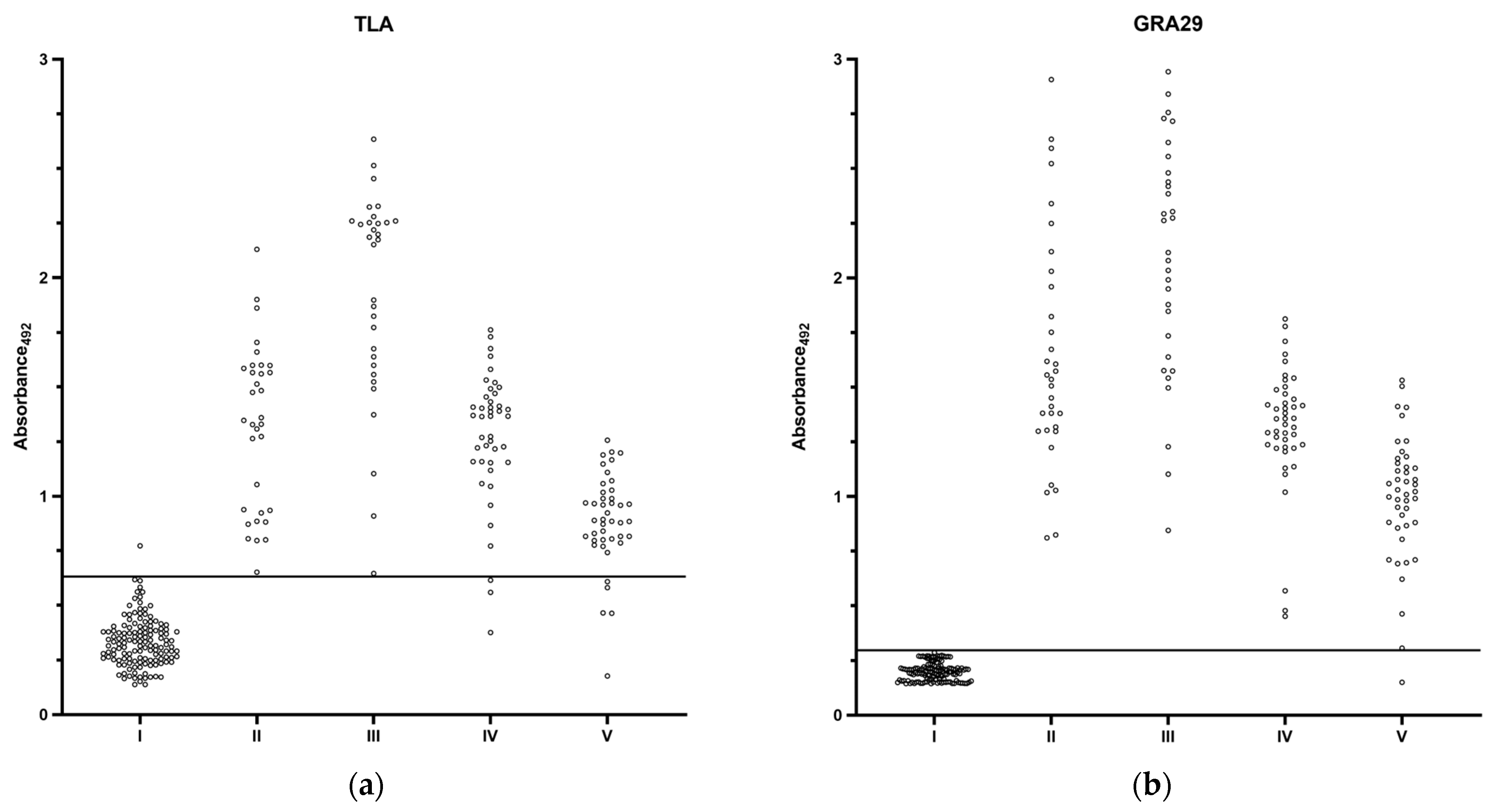
To fully determine the reactivity of the selected recombinant antigen, GRA29 and TLA were used in an indirect IgG ELISA on a total of 286 human serum samples categorized into the following five groups: I: seronegative; II: acute infection; III: chronic infection, high IgG; IV: chronic infection, medium IgG; V: chronic infection, low IgG. The distribution of OD values for the different serum groups is shown in Figure 3. The GRA29-based ELISA misclassified only sera from the low-IgG chronic group, whereas the TLA-based assay showed false negatives not only in this group but also among medium-IgG chronic samples. In addition, acute and high-IgG chronic samples produced OD values close to the cut-off in the TLA assay, while the GRA29 assay consistently yielded strong positive signals in these groups. Paired comparisons of OD values revealed that GRA29 produced significantly higher reactivity in positive sera (Wilcoxon signed-rank test, median difference = 0.1005, p < 0.0001) while maintaining significantly lower background reactivity in negative sera (median difference = −0.1090, p < 0.0001).
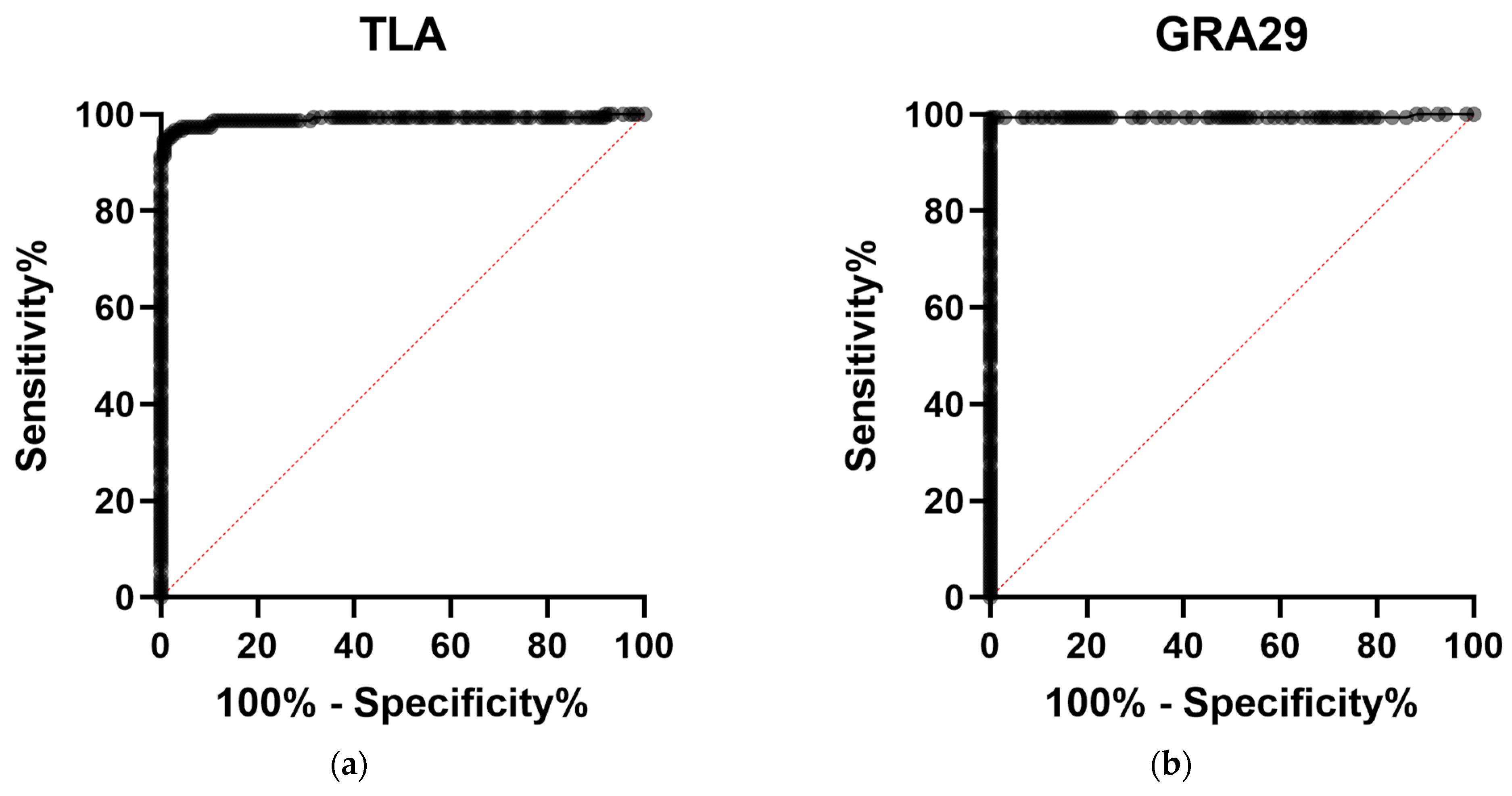
ROC curve analysis demonstrated excellent diagnostic performance for both assays, with an AUC of 0.9894 (95% CI: 0.9765–1.000) for TLA-ELISA and 0.9942 (95% CI: 0.9828–1.000) for GRA29-ELISA (Figure 4, Table 6).
At the optimal cut-off values, GRA29-ELISA exhibited superior discrimination between seropositive and seronegative samples, with higher sensitivity, specificity, PPV, and NPV compared with TLA-based assay. These parameters are summarized in Table 6. Fisher’s exact test further demonstrated that classifications obtained with both TLA- and GRA29-based assays were significantly associated with the reference standard (p < 0.0001), further validating that both tests are diagnostically meaningful.

4. Discussion

Efforts to improve the serodiagnosis of toxoplasmosis have long focused on identifying recombinant antigens that could replace TLA, which provides broad antigenic coverage, however its heterogeneity poses challenges for assay reproducibility and standardization. Recombinant proteins offer a solution by enabling consistent production and targeted selection of specific epitopes and/or immunodominant fragments. Among these, dense granule proteins have been widely studied. Several GRAs, including GRA1 [25,26,27], GRA2 [27,28,29,30], GRA4 [27,31], GRA5 [23], GRA6 [25,32,33,34], GRA7 [27,28,31,34,35], GRA8 [27] and GRA14 [34] have demonstrated diagnostic potential for detecting anti-T. gondii IgG. Notably, several studies have shown that combinations of recombinant antigens achieve higher sensitivity and specificity than single antigens alone [23,25,26,27,28,31], likely due to broader epitope coverage, though at the expense of increased production complexity and cost. As an emerging alternative, chimeric antigens that combine immunodominant fragments of different proteins into a single fusion construct are being explored to enhance diagnostic accuracy while reducing assay complexity [36,37,38].
Despite these advances, no single recombinant antigen has yet replaced TLA in routine serological testing. Nevertheless, their established diagnostic potential suggests that unexplored members of this protein family may provide additional value. To address this, we selected six previously untested GRAs (GRA29, GRA35, GRA36, GRA45, GRA54, and GRA64) for evaluation of their utility as recombinant serodiagnostic antigens.
These dense granule proteins have varying roles in Toxoplasma biology. GRA29 is secreted into the PV, where it forms filamentous structures distinct from the intravacuolar network and previously described actin filaments, and it relocalizes during stage conversion to bradyzoites [39]. It has also been shown to be non-essential for parasite survival in vivo [40]. GRA35 has been linked to inflammasome activation. Together with GRA42 and GRA43, it functions as a key factor required for activation of the NLRP1 inflammasome and induction of pyroptosis in Lewis rat macrophages [41,42]. GRA35 has also been investigated for its protective potential in experimental vaccination studies in mouse models. Immunization with recombinant protein cocktails including GRA35 [43], as well as DNA vaccines encoding GRA35 in combination with GRA42 and GRA43 [44], elicited humoral and cellular immune responses. GRA36 is less well characterized; while it shares >40% sequence similarity with GRA35, gene knockout studies indicate that it does not share the same inflammasome-activating function [41].
Other candidates included GRA45, a chaperone-like protein essential for the correct localization of GRAs into the PVM and for the export of dense granule effectors into host cells [45,46,47]. Parasites lacking GRA45 show impaired survival under IFNγ-mediated pressure and reduced virulence in mice [47,48]. On the other hand, little is known about the precise biological role of GRA54, it has been identified as a MYR1-associated protein; however, disruption of GRA54 did not produce detectable effects on effector translocation across the PVM [45]. Finally, GRA64 is a transmembrane protein localized to the PVM and partly exposed to the host cytoplasm during tachyzoite and bradyzoite stages. Although implicated in vesicular trafficking, its disruption does not affect virulence or cyst formation in mice [49]. While genetic disruption of some of these proteins does not alter parasite virulence in mice, this does not exclude the possibility that they contribute to host immune recognition.
To the best of our knowledge, only GRA29 and GRA35 have previously been produced in recombinant form, and none of the six tested antigens have been assessed for diagnostic performance. Thus, establishing their expression in E. coli, purification, and evaluation in IgG-based ELISAs with human sera represents a novel contribution of this study.
The vast majority of studies evaluating recombinant T. gondii antigens have relied on prokaryotic expression systems [50]. In the present study, the bacterial expression strategy yielded protein preparations with production levels ranging from 18.1 to 164.8 mg/L of culture. Although few studies report detailed production efficiencies, the yields obtained here are comparable to, and in some cases exceed those previously described for recombinant GRA proteins. For instance, Golkar et al. reported a yield of 12 mg/L for GRA2 [30], while another study achieved 28 mg/L for the same antigen [29]. GRA5 and GRA8 have been produced with reported yields of 15 mg/L [23] and 68 mg/L [51], respectively. In comparison, the production levels obtained in the present study, particularly for GRA35, GRA36, and GRA45 (exceeding 160, 130, and 120 mg/L of bacterial culture, respectively), show the efficiency of the developed expression systems. Such high yields are advantageous not only for research purposes but also for the potential development of standardized diagnostic assays, where scalability and cost-effectiveness of antigen production are important.
The evaluation of six recombinant GRA proteins revealed marked differences in their diagnostic potential. Among the tested candidates, GRA29, GRA45, and GRA54 demonstrated the highest discriminatory ability, with AUC values ranging from 0.85 to 0.99 and highly significant associations with the reference classification. These findings were further supported by clear separation of seropositive and seronegative sera and statistically significant differences in their absorbance values. In contrast, GRA35, GRA36, and GRA64 showed poor performance, with AUC values close to random classification and no significant differences between groups, indicating limited applicability for serological detection of T. gondii infection.
Further inspection of the data underscores the unique diagnostic value of GRA29. This antigen combined high sensitivity (95.83%) with perfect specificity (100%), outperforming TLA. GRA45, while diagnostically relevant, displayed lower sensitivity (79.17%) and specificity (87.50%), while GRA54, despite achieving high specificity (95.83%) and a good AUC (0.9323), showed a higher rate of false-negative classifications compared with TLA. These limitations reduce their feasibility as standalone diagnostic tools. Therefore, GRA29 was chosen for extended analysis on a larger pool of human sera.
The extended comparison of TLA- and GRA29-IgG ELISAs shows that although both assays perform very well, the GRA29-based consistently outperforms the TLA-based ELISA, offering higher AUC values, sensitivity, specificity, PPV, and NPV at the optimal cut-off, stronger reactivity in positive sera, and lower background in negative sera. Moreover, scatter plot analyses confirmed that the GRA29-based ELISA provided more robust performance across serum groups, with fewer misclassifications and borderline results. Thus, GRA29 represents a promising recombinant antigen for the serodiagnosis of toxoplasmosis and may serve as a standardized alternative to TLA.
Previous studies assessing recombinant GRA antigens have generally shown that, while they can elicit strong antibody responses, their overall discriminatory power between seropositive and seronegative individuals remains limited. Lecordier et al. [25] reported that a GRA1-based IgG ELISA achieved 68% sensitivity, while Luo et al. [28] found that GRA2 reached 85% sensitivity and 85% specificity. In contrast, Aubert et al. [27] excluded GRA1, GRA2, and GRA4 at the preliminary stage of reactivity assessment due to insufficient performance. Consistent with this, Nigro et al. [31] reported low diagnostic accuracy for GRA4, with sensitivity of 58.3% in acute and only 18.2% in chronic infection. GRA5 [23] and GRA14 [34] have also been evaluated, with ELISAs based on these antigens correctly classifying 70.9% and 40.7% of seropositive samples, respectively. Among the GRAs tested, GRA7 has emerged as one of the most promising, with several studies reporting high sensitivity and specificity across infection stages. However, its diagnostic performance has not been consistent. Ybanez et al. [34] showed 100% sensitivity and specificity, while Arab-Mazar et al. [35] reported values of 92% and 94%, respectively. Aubert et al. [27] found that GRA7-based ELISA correctly classified 83.40% of seropositive samples, and Luo et al. [28] observed 83.3% sensitivity and 90% specificity. In contrast, Nigro et al. [31] reported more modest performance, with 75% sensitivity in acute and only 36.3% in chronic cases.
Many GRAs have demonstrated higher sensitivity in acute infection but markedly reduced reactivity in chronic cases, positioning them as potential markers of infection stage. For example, ELISAs based on GRA2 and GRA6 achieved sensitivities above 90% for acute sera, yet performance dropped substantially in chronic infection, in some cases below 40% [30,32,33,52]. Similarly, GRA8-based assay correctly classified 70.04% of all seropositive samples but showed only 37.14% sensitivity in chronic infection compared with 98.87% in acute cases [27].
In the presented findings GRA29 exhibited strong reactivity with IgG antibodies in both acute and chronic sera, resulting in near-perfect discrimination between seropositive and seronegative individuals. While this broad reactivity limits its ability to distinguish infection stage, it positions GRA29 as a robust antigen for universal serological screening, where it could be applied as a primary diagnostic marker.
This study has certain limitations that should be considered when interpreting the findings. The sample size of tested human sera, while adequate for preliminary evaluation, was relatively limited, and future studies should examine much larger cohorts to increase statistical power and improve the generalizability of the results. Furthermore, this work focused solely on the detection of anti-T. gondii IgG antibodies. Additional studies should assess the diagnostic potential of these antigens in IgM-based assays and in IgG avidity testing, which are particularly relevant for distinguishing acute from chronic infections. Future research could also investigate whether GRA29 reliably recognized anti-T. gondii IgG in other intermediate hosts, broadening its applicability to both clinical and veterinary settings. Addressing these limitations will fully establish the diagnostic value of the investigated proteins.

5. Conclusions

The objective of this study was to biotechnologically produce and assess the diagnostic performance of six recombinant dense granule proteins—GRA29, GRA35, GRA36, GRA45, GRA54, and GRA64—for the serological detection of T. gondii infection. All proteins were successfully expressed in E. coli, purified, and evaluated by IgG-based ELISA using human sera.
Among the tested candidates, GRA29, GRA45, and GRA54 exhibited the highest discriminatory power, with AUC values ranging from 0.8507 to 0.9983. These proteins showed strong correlations with reference classifications, and statistically significant differences in absorbance values between seropositive and seronegative sera confirmed their diagnostic relevance. In contrast, GRA35, GRA36, and GRA64 demonstrated low diagnostic utility, with AUC values approaching random classification and no significant separation between positive and negative samples.
Overall, the results indicate that while most of the tested recombinant GRA proteins show limited potential for routine diagnostic application, GRA29 in particular displays strong reactivity, supporting its potential as a standardized recombinant antigen for reliable serodiagnosis of human toxoplasmosis.

Supplementary Materials

The following supporting information can be downloaded at https://www.mdpi.com/article/10.3390/cimb47110879/s1.

Author Contributions

Conceptualization, K.S. and L.H.-G.; methodology, K.S. and L.H.-G.; software, K.S.; validation, K.S. and L.H.-G.; formal analysis, K.S. and L.H.-G.; investigation, K.S. and L.H.-G.; resources, L.H.-G.; data curation, K.S.; writing—original draft preparation, K.S.; writing—review and editing, K.S. and L.H.-G.; supervision, L.H.-G. All authors have read and agreed to the published version of the manuscript.

Funding

This research received no external funding.

Institutional Review Board Statement

The present study analyzes previously collected, fully anonymized human serum samples. No new samples were collected and no interaction or intervention with human participants took place, ethics committee review was not required for the present study.

Informed Consent Statement

The present study analyzes previously collected, fully anonymized human serum samples. No new samples were collected and no interaction or intervention with human participants took place. Informed Consent Statement was not required for the present study.

Data Availability Statement

The original contributions presented in this study are included in the article/Supplementary Materials. Further inquiries can be directed to the corresponding author.

Conflicts of Interest

The authors declare no conflicts of interest.

Abbreviations

The following abbreviations are used in this manuscript:
TLATachyzoite lysate antigen
ESAExcretory–secretory antigens
GRADense granule proteins
PVParasitophorous vacuole
PVMParasitophorous vacuole membrane
IVNIntravacuolar network
ELISAEnzyme-linked immunosorbent assay
ODOptical density
PPVPositive predictive value
NPVNegative predictive value
AUCArea under the curve
ROCReceiver operating characteristic

References

  1. Montoya, J.G.; Liesenfeld, O. Toxoplasmosis. Lancet 2004, 363, 1965–1976. [Google Scholar] [CrossRef] [PubMed]
  2. Warschkau, D.; Seeber, F. Advances towards the Complete in Vitro Life Cycle of Toxoplasma gondii. Fac. Rev. 2023, 12, 1. [Google Scholar] [CrossRef]
  3. Florence, R.-G.; Marie-Laure, D. Epidemiology of and Diagnostic Strategies for Toxoplasmosis. Clin. Microbiol. Rev. 2012, 25, 264–296. [Google Scholar] [CrossRef]
  4. Akins, G.K.H.; Furtado, J.M.; Smith, J.R. Diseases Caused by and Behaviors Associated with Toxoplasma gondii Infection. Pathogens 2024, 13, 968. [Google Scholar] [CrossRef]
  5. Cerisola, A.; Francia, M.; Gesuele, J.P. Congenital Toxoplasmosis. Semin. Pediatr. Neurol. 2025, 54, 101203. [Google Scholar] [CrossRef] [PubMed]
  6. Bollani, L.; Auriti, C.; Achille, C.; Garofoli, F.; De Rose, D.U.; Meroni, V.; Salvatori, G.; Tzialla, C. Congenital Toxoplasmosis: The State of the Art. Front. Pediatr. 2022, 10, 894573. [Google Scholar] [CrossRef]
  7. Garweg, J.G.; Kieffer, F.; Mandelbrot, L.; Peyron, F.; Wallon, M. Long-Term Outcomes in Children with Congenital Toxoplasmosis-A Systematic Review. Pathogens 2022, 11, 1187. [Google Scholar] [CrossRef]
  8. Kim, M.-J.; Park, S.J.; Park, H. Trend in Serological and Molecular Diagnostic Methods for Toxoplasma gondii Infection. Eur. J. Med. Res. 2024, 29, 520. [Google Scholar] [CrossRef]
  9. Dupont, D.; Fricker-Hidalgo, H.; Brenier-Pinchart, M.-P.; Garnaud, C.; Wallon, M.; Pelloux, H. Serology for Toxoplasma in Immunocompromised Patients: Still Useful? Trends Parasitol. 2021, 37, 205–213. [Google Scholar] [CrossRef]
  10. de Oliveira Azevedo, C.T.; do Brasil, P.E.A.A.; Guida, L.; Lopes Moreira, M.E. Performance of Polymerase Chain Reaction Analysis of the Amniotic Fluid of Pregnant Women for Diagnosis of Congenital Toxoplasmosis: A Systematic Review and Meta-Analysis. PLoS ONE 2016, 11, e0149938. [Google Scholar] [CrossRef] [PubMed]
  11. Ybañez, R.H.D.; Ybañez, A.P.; Nishikawa, Y. Review on the Current Trends of Toxoplasmosis Serodiagnosis in Humans. Front. Cell. Infect. Microbiol. 2020, 10, 204. [Google Scholar] [CrossRef] [PubMed]
  12. Liu, Q.; Wang, Z.-D.; Huang, S.-Y.; Zhu, X.-Q. Diagnosis of Toxoplasmosis and Typing of Toxoplasma gondii. Parasit. Vectors 2015, 8, 292. [Google Scholar] [CrossRef] [PubMed]
  13. Holec-Gasior, L. Toxoplasma gondii Recombinant Antigens as Tools for Serodiagnosis of Human Toxoplasmosis: Current Status of Studies. Clin. Vaccine Immunol. 2013, 20, 1343–1351. [Google Scholar] [CrossRef] [PubMed]
  14. Darcy, F.; Deslee, D.; Santoro, F.; Charif, H.; Auriault, C.; Decoster, A.; Duquesne, V.; Capron, A. Induction of a Protective Antibody-Dependent Response against Toxoplasmosis by in Vitro Excreted/Secreted Antigens from Tachyzoites of Toxoplasma gondii. Parasite Immunol. 1988, 10, 553–567. [Google Scholar] [CrossRef]
  15. Cesbron-Delauw, M.F.; Capron, A. Excreted/Secreted Antigens of Toxoplasma gondii—Their Origin and Role in the Host-Parasite Interaction. Res. Immunol. 1993, 144, 41–44. [Google Scholar] [CrossRef]
  16. Rezaei, F.; Sharif, M.; Sarvi, S.; Hejazi, S.H.; Aghayan, S.; Pagheh, A.S.; Dodangeh, S.; Daryani, A. A Systematic Review on the Role of GRA Proteins of Toxoplasma gondii in Host Immunization. J. Microbiol. Methods 2019, 165, 105696. [Google Scholar] [CrossRef]
  17. Schwab, J.C.; Beckers, C.J.; Joiner, K.A. The Parasitophorous Vacuole Membrane Surrounding Intracellular Toxoplasma gondii Functions as a Molecular Sieve. Proc. Natl. Acad. Sci. USA 1994, 91, 509–513. [Google Scholar] [CrossRef]
  18. Griffith, M.B.; Pearce, C.S.; Heaslip, A.T. Dense Granule Biogenesis, Secretion, and Function in Toxoplasma gondii. J. Eukaryot. Microbiol. 2022, 69, e12904. [Google Scholar] [CrossRef]
  19. Zhang, Y.; Li, D.; Lu, S.; Zheng, B. Toxoplasmosis Vaccines: What We Have and Where to Go? npj Vaccines 2022, 7, 131. [Google Scholar] [CrossRef]
  20. Mamaghani, A.J.; Fathollahi, A.; Arab-Mazar, Z.; Kohansal, K.; Fathollahi, M.; Spotin, A.; Bashiri, H.; Bozorgomid, A. Toxoplasma gondii Vaccine Candidates: A Concise Review. Ir. J. Med. Sci. 2023, 192, 231–261. [Google Scholar] [CrossRef]
  21. Holec, L.; Gasior, A.; Brillowska-Dabrowska, A.; Kur, J. Toxoplasma gondii: Enzyme-Linked Immunosorbent Assay Using Different Fragments of Recombinant Microneme Protein 1 (MIC1) for Detection of Immunoglobulin G Antibodies. Exp. Parasitol. 2008, 119, 1–6. [Google Scholar] [CrossRef]
  22. Dabrowski, S.; Kur, J. Cloning, Overexpression, and Purification of the Recombinant His-Tagged SSB Protein of Escherichia coli and Use in Polymerase Chain Reaction Amplification. Protein Expr. Purif. 1999, 16, 96–102. [Google Scholar] [CrossRef]
  23. Holec-Gasior, L.; Kur, J. Toxoplasma gondii: Recombinant GRA5 Antigen for Detection of Immunoglobulin G Antibodies Using Enzyme-Linked Immunosorbent Assay. Exp. Parasitol. 2010, 124, 272–278. [Google Scholar] [CrossRef]
  24. Rota, M.; Antolini, L. Finding the Optimal Cut-Point for Gaussian and Gamma Distributed Biomarkers. Comput. Stat. Data Anal. 2014, 69, 1–14. [Google Scholar] [CrossRef]
  25. Lecordier, L.; Fourmaux, M.P.; Mercier, C.; Dehecq, E.; Masy, E.; Cesbron-Delauw, M.F. Enzyme-Linked Immunosorbent Assays Using the Recombinant Dense Granule Antigens GRA6 and GRA1 of Toxoplasma gondii for Detection of Immunoglobulin G Antibodies. Clin. Diagn. Lab. Immunol. 2000, 7, 607–611. [Google Scholar] [CrossRef] [PubMed]
  26. Pietkiewicz, H.; Hiszczyńska-Sawicka, E.; Kur, J.; Petersen, E.; Nielsen, H.V.; Stankiewicz, M.; Andrzejewska, I.; Myjak, P. Usefulness of Toxoplasma gondii-Specific Recombinant Antigens in Serodiagnosis of Human Toxoplasmosis. J. Clin. Microbiol. 2004, 42, 1779–1781. [Google Scholar] [CrossRef]
  27. Aubert, D.; Maine, G.T.; Villena, I.; Hunt, J.C.; Howard, L.; Sheu, M.; Brojanac, S.; Chovan, L.E.; Nowlan, S.F.; Pinon, J.M. Recombinant Antigens to Detect Toxoplasma gondii-Specific Immunoglobulin G and Immunoglobulin M in Human Sera by Enzyme Immunoassay. J. Clin. Microbiol. 2000, 38, 1144–1150. [Google Scholar] [CrossRef] [PubMed]
  28. Luo, J.; Wan, J.; Tang, Z.; Shen, S. Identification of Novel Antigens for Serum IgG Diagnosis of Human Toxoplasmosis. Exp. Parasitol. 2019, 204, 107722. [Google Scholar] [CrossRef]
  29. Holec-Gasior, L.; Kur, J.; Hiszczyńska-Sawicka, E. GRA2 and ROP1 Recombinant Antigens as Potential Markers for Detection of Toxoplasma gondii-Specific Immunoglobulin G in Humans with Acute Toxoplasmosis. Clin. Vaccine Immunol. 2009, 16, 510–514. [Google Scholar] [CrossRef]
  30. Golkar, M.; Rafati, S.; Abdel-Latif, M.S.; Brenier-Pinchart, M.-P.; Fricker-Hidalgo, H.; Sima, B.K.; Babaie, J.; Pelloux, H.; Cesbron-Delauw, M.-F.; Mercier, C. The Dense Granule Protein GRA2, a New Marker for the Serodiagnosis of Acute Toxoplasma Infection: Comparison of Sera Collected in Both France and Iran from Pregnant Women. Diagn. Microbiol. Infect. Dis. 2007, 58, 419–426. [Google Scholar] [CrossRef]
  31. Nigro, M.; Gutierrez, A.; Hoffer, A.M.; Clemente, M.; Kaufer, F.; Carral, L.; Martin, V.; Guarnera, E.A.; Angel, S.O. Evaluation of Toxoplasma gondii Recombinant Proteins for the Diagnosis of Recently Acquired Toxoplasmosis by an Immunoglobulin G Analysis. Diagn. Microbiol. Infect. Dis. 2003, 47, 609–613. [Google Scholar] [CrossRef]
  32. Golkar, M.; Azadmanesh, K.; Khalili, G.; Khoshkholgh-Sima, B.; Babaie, J.; Mercier, C.; Brenier-Pinchart, M.-P.; Fricker-Hidalgo, H.; Pelloux, H.; Cesbron-Delauw, M.-F. Serodiagnosis of Recently Acquired Toxoplasma gondii Infection in Pregnant Women Using Enzyme-Linked Immunosorbent Assays with a Recombinant Dense Granule GRA6 Protein. Diagn. Microbiol. Infect. Dis. 2008, 61, 31–39. [Google Scholar] [CrossRef]
  33. Redlich, A.; Müller, W.A. Serodiagnosis of Acute Toxoplasmosis Using a Recombinant Form of the Dense Granule Antigen GRA6 in an Enzyme-Linked Immunosorbent Assay. Parasitol. Res. 1998, 84, 700–706. [Google Scholar] [CrossRef]
  34. Ybañez, R.H.; Nishikawa, Y. Comparative Performance of Recombinant GRA6, GRA7, and GRA14 for the Serodetection of T. gondii Infection and Analysis of IgG Subclasses in Human Sera from the Philippines. Pathogens 2022, 11, 277. [Google Scholar] [CrossRef]
  35. Arab-Mazar, Z.; Fallahi, S.; Koochaki, A.; Haghighi, A.; Seyyed Tabaei, S.J. Immunodiagnosis and Molecular Validation of Toxoplasma gondii-Recombinant Dense Granular (GRA) 7 Protein for the Detection of Toxoplasmosis in Patients with Cancer. Microbiol. Res. 2016, 183, 53–59. [Google Scholar] [CrossRef]
  36. Beghetto, E.; Spadoni, A.; Bruno, L.; Buffolano, W.; Gargano, N. Chimeric Antigens of Toxoplasma gondii: Toward Standardization of Toxoplasmosis Serodiagnosis Using Recombinant Products. J. Clin. Microbiol. 2006, 44, 2133–2140. [Google Scholar] [CrossRef] [PubMed]
  37. Ferra, B.T.; Holec-Gąsior, L.; Gatkowska, J.; Dziadek, B.; Dzitko, K.; Grąźlewska, W.; Lautenbach, D. The First Study on the Usefulness of Recombinant Tetravalent Chimeric Proteins Containing Fragments of SAG2, GRA1, ROP1 and AMA1 Antigens in the Detection of Specific Anti-Toxoplasma gondii Antibodies in Mouse and Human Sera. PLoS ONE 2019, 14, e0217866. [Google Scholar] [CrossRef] [PubMed]
  38. Ferra, B.; Holec-Gąsior, L.; Kur, J. A New Toxoplasma gondii Chimeric Antigen Containing Fragments of SAG2, GRA1, and ROP1 Proteins—Impact of Immunodominant Sequences Size on Its Diagnostic Usefulness. Parasitol. Res. 2015, 114, 3291–3299. [Google Scholar] [CrossRef] [PubMed]
  39. Young, J.C.; Broncel, M.; Teague, H.; Russell, M.R.G.; McGovern, O.L.; Renshaw, M.; Frith, D.; Snijders, A.P.; Collinson, L.; Carruthers, V.B.; et al. Phosphorylation of Toxoplasma gondii Secreted Proteins during Acute and Chronic Stages of Infection. mSphere 2020, 5, e00792-20. [Google Scholar] [CrossRef]
  40. Young, J.; Dominicus, C.; Wagener, J.; Butterworth, S.; Ye, X.; Kelly, G.; Ordan, M.; Saunders, B.; Instrell, R.; Howell, M.; et al. A CRISPR Platform for Targeted in Vivo Screens Identifies Toxoplasma gondii Virulence Factors in Mice. Nat. Commun. 2019, 10, 3963. [Google Scholar] [CrossRef] [PubMed]
  41. Wang, Y.; Cirelli, K.M.; Barros, P.D.C.; Sangaré, L.O.; Butty, V.; Hassan, M.A.; Pesavento, P.; Mete, A.; Saeij, J.P.J. Three Toxoplasma gondii Dense Granule Proteins Are Required for Induction of Lewis Rat Macrophage Pyroptosis. MBio 2019, 10, e02388-18. [Google Scholar] [CrossRef] [PubMed]
  42. Wang, Y.; Hollingsworth, L.R.; Sangaré, L.O.; Paredes-Santos, T.C.; Krishnamurthy, S.; Penn, B.H.; Wu, H.; Saeij, J.P.J. Host E3 Ubiquitin Ligase ITCH Mediates Toxoplasma gondii Effector GRA35-Triggered NLRP1 Inflammasome Activation and Cell-Autonomous Immunity. MBio 2024, 15, e0330223. [Google Scholar] [CrossRef]
  43. Li, D.; Han, M.; Cao, Y.; Du, J.; An, R. Protective Effect against Toxoplasmosis in BALB/C Mice Vaccinated with Recombinant Toxoplasma gondii CDPK3, GRA35, and ROP46 Protein Cocktail Vaccine. Vaccine 2024, 42, 1342–1351. [Google Scholar] [CrossRef]
  44. Shi, Y.; Li, J.; Yang, W.; Chen, J. Protective Immunity Induced by DNA Vaccine Containing TgGRA35, TgGRA42, and TgGRA43 against Toxoplasma gondii Infection in Kunming Mice. Front. Cell. Infect. Microbiol. 2023, 13, 1236130. [Google Scholar] [CrossRef]
  45. Cygan, A.M.; Theisen, T.C.; Mendoza, A.G.; Marino, N.D.; Panas, M.W.; Boothroyd, J.C. Coimmunoprecipitation with MYR1 Identifies Three Additional Proteins within the Toxoplasma gondii Parasitophorous Vacuole Required for Translocation of Dense Granule Effectors into Host Cells. mSphere 2020, 5, e00858-19. [Google Scholar] [CrossRef]
  46. Coffey, M.J.; Dagley, L.F.; Seizova, S.; Kapp, E.A.; Infusini, G.; Roos, D.S.; Boddey, J.A.; Webb, A.I.; Tonkin, C.J. Aspartyl Protease 5 Matures Dense Granule Proteins That Reside at the Host-Parasite Interface in Toxoplasma gondii. MBio 2018, 9, e01796-18. [Google Scholar] [CrossRef]
  47. Wang, Y.; Sangaré, L.O.; Paredes-Santos, T.C.; Hassan, M.A.; Krishnamurthy, S.; Furuta, A.M.; Markus, B.M.; Lourido, S.; Saeij, J.P.J. Genome-Wide Screens Identify Toxoplasma gondii Determinants of Parasite Fitness in IFNγ-Activated Murine Macrophages. Nat. Commun. 2020, 11, 5258. [Google Scholar] [CrossRef]
  48. Tachibana, Y.; Hashizaki, E.; Sasai, M.; Yamamoto, M. Host Genetics Highlights IFN-γ-Dependent Toxoplasma Genes Encoding Secreted and Non-Secreted Virulence Factors in In Vivo CRISPR Screens. Cell Rep. 2023, 42, 112592. [Google Scholar] [CrossRef]
  49. Mayoral, J.; Guevara, R.B.; Rivera-Cuevas, Y.; Tu, V.; Tomita, T.; Romano, J.D.; Gunther-Cummins, L.; Sidoli, S.; Coppens, I.; Carruthers, V.B.; et al. Dense Granule Protein GRA64 Interacts with Host Cell ESCRT Proteins during Toxoplasma gondii Infection. MBio 2022, 13, e01442-22. [Google Scholar] [CrossRef] [PubMed]
  50. Sołowińska, K.; Holec-Gąsior, L. Single Cell Expression Systems for the Production of Recombinant Proteins for Immunodiagnosis and Immunoprophylaxis of Toxoplasmosis. Microorganisms 2024, 12, 1731. [Google Scholar] [CrossRef] [PubMed]
  51. Babaie, J.; Miri, M.; Sadeghiani, G.; Zare, M.; Khalili, G.; Golkar, M. Expression and Single-Step Purification of GRA8 Antigen of Toxoplasma gondii in Escherichia Coli. Avicenna J. Med. Biotechnol. 2011, 3, 67–77. [Google Scholar] [PubMed]
  52. Murray, A.; Mercier, C.; Decoster, A.; Lecordier, L.; Capron, A.; Cesbron-Delauw, M.F. Multiple B-Cell Epitopes in a Recombinant GRA2 Secreted Antigen of Toxoplasma gondii. Appl. Parasitol. 1993, 34, 235–244. [Google Scholar] [PubMed]
Figure 1. Scatter plots of ELISA absorbance values (OD492) for (a) GRA29-, (b) GRA35-, (c) GRA36-, (d) GRA45-, (e) GRA54-, (f) GRA64-, and (g) TLA-coated plates, obtained using seropositive (n = 24) and seronegative (n = 28) human serum samples. Horizontal lines indicate cut-off values determined by ROC curve analysis. Statistical comparisons were performed using the Mann–Whitney U test; ns, not significant; **** p < 0.0001.
Cimb 47 00879 g001
Figure 2. Receiver operating characteristic (ROC) curves of (a) GRA29-, (b) GRA35-, (c) GRA36-, (d) GRA45-, (e) GRA54-, (f) GRA64-, and (g) TLA-based IgG ELISAs. The diagonal red dotted line represents the line of no discrimination (AUC = 0.5).
Cimb 47 00879 g002
Figure 3. Scatter plots of ELISA absorbance values (OD492) for (a) TLA-coated plates; (b) and GRA29-coated plates across serum groups. I: seronegative (IgG, IgM; n = 136); II: acute infection (IgG+, IgM+/, low IgG avidity; n = 33); III: chronic infection, high IgG (IgG+, IgM, high IgG avidity, IgG > 300 IU/mL; n = 31); IV: chronic infection, medium IgG (IgG+, IgM, high IgG avidity, IgG 100–300 IU/mL; n = 43); V: chronic infection, low IgG (IgG+, IgM, high IgG avidity, IgG < 100 IU/mL; n = 43). The horizontal line represents the assay-specific cut-off determined by ROC analysis.
Cimb 47 00879 g003
Figure 4. Receiver operating characteristic (ROC) curves of ELISA assays for detection of T. gondii-specific IgG. (a) TLA-based ELISA; (b) GRA29-based ELISA. The diagonal red dotted line indicates the line of no discrimination (AUC = 0.5).
Cimb 47 00879 g004
Table 1. Primers for the amplification of genes coding the selected immunodominant protein fragments. Lowercase letters correspond to the nucleotides homologous to the linearized vector, whereas uppercase letters are nucleotides homologous to the target gene.
Primer NamePrimer SequenceAmplicon Size [bp]
gra29FORagcccagatctgggtCAAATCTTGGCTCTGCTTC1354
gra29REVgacggagctcgaattACGTGTCCCTCTTCCC
gra35FORagcccagatctgggtGCGAAGCGTCAATATGAG742
gra35REVgacggagctcgaattAGTCTGTTTCGGCTCCG
gra36FORagcccagatctgggtTATCGGCGCTATCGTGAT760
gra36REVgacggagctcgaattCGTACGCTGTGCCC
gra45FORgacggagctcgaattCGTACGCTGTGCCC1117
gra45REVgacggagctcgaattCGTACGCTGTGCCC
gra54FORagcccagatctgggtGGTGCAACACGAGGAC931
gra54REVgacggagctcgaattTTGTGCCCTCACACCAG
gra64FORgacggagctcgaattCGTCTTAAACCCCTTCAAGC763
gra64REVagcccagatctgggtGCAAACTACGATTTCTTTGTTCTT
Table 2. Summary of optimal expression conditions for GRA29, GRA35, GRA36, GRA45, GRA54, and GRA64, including E. coli expression strain, post-induction temperature and post-induction incubation time.
Protein NameE. coli Expression StrainPost-Induction
Temperature [°C]
Post-Induction
Incubation Time [h]
GRA29BL21(DE3)pLacI3718
GRA35BL21(DE3)pLacI2518
GRA36Rosetta(DE3)pLysS2518
GRA45BL21(DE3)pLysS306
GRA54BL21(DE3)pLacI2518
GRA64BL21(DE3)pLacI3718
Table 3. Predicted molecular weights and theoretical isoelectric points (pI) of recombinant GRA29, GRA35, GRA36, GRA45, GRA54, and GRA64, calculated from their amino acid sequences using the ExPASy Compute pI/Mw tool.
Protein NameMolecular Weight [kDa]Isoelectric Point
GRA2952.08.03
GRA3533.76.14
GRA3634.56.43
GRA4547.75.17
GRA5440.19.64
GRA6432.57.95
Table 4. Protein yields obtained per liter of bacterial culture.
Protein NameYield [mg]
GRA2928.3
GRA35164.8
GRA36138.0
GRA45123.0
GRA5418.1
GRA6438.0
Table 5. Diagnostic performance of recombinant GRA proteins and TLA in ELISA-based detection of T. gondii-specific IgG antibodies.
AntigenAUC 1
(95% CI)
Cut-OffSensitivity [%] (95% CI)Specificity [%] (95% CI)Fisher’s Exact
p-Value
GRA290.9983
(0.9925–1.000)
0.374095.83
(79.76–99.79)
100.00
(86.20–100.00)
<0.0001
GRA350.5269
(0.3600–0.6938)
1.19312.50
(4.34–31.00)
91.67
(74.15–98.52)
ns 2
GRA360.5260
(0.3544–0.6977)
0.994579.17
(59.53–90.76)
41.67
(24.47–61.17)
ns
GRA450.8507
(0.3544–0.6977)
0.441579.17
(59.53–90.76)
87.50
(69.00–95.66)
<0.0001
GRA540.9323
(0.8464–1.000)
0.409070.83
(50.83–85.09)
95.83
(79.76–99.79)
<0.0001
GRA640.5503
(0.3848–0.7159)
0.839041.67
(24.47–61.17)
75.00
(55.10–88.00)
ns
TLA0.9948
(0.9822–1.000)
0.635595.83
(79.76–99.79)
95.83
(79.76–99.79)
<0.0001
1 AUC—area under the curve; 2 ns—not significant.
Table 6. Diagnostic performance of TLA- and GRA29-based IgG ELISAs.
AntigenAUC 1
(95% CI)
Cut-OffSensitivity [%] (95% CI)Specificity [%] (95% CI)PPV 2 [%]NPV 3 [%]
TLA0.9894
(0.9765–1.000)
0.632099.30
(96.15–99.96)
94.41
(89.35–97.14)
94.67
(89.83–97.27)
99.26
(95.95–99.96)
GRA290.9942
(0.9828–1.000)
0.2965100.00
(97.49–100.00)
99.27
(95.98–99.96)
99.33
(96.32–99.97)
100.00
(97.25–100.00)
1 AUC—area under the curve; 2 PPV—positive predictive value; 3 NPV—negative predictive value.
Disclaimer/Publisher’s Note: The statements, opinions and data contained in all publications are solely those of the individual author(s) and contributor(s) and not of MDPI and/or the editor(s). MDPI and/or the editor(s) disclaim responsibility for any injury to people or property resulting from any ideas, methods, instructions or products referred to in the content.