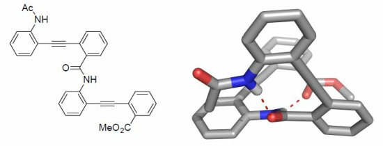
Methyl 2-[(2-{2-[(2-acetamidophenyl)ethynyl]benzamido} phenyl)ethynyl]benzoate

Abstract
:
Introduction
Experimental Section
General Information
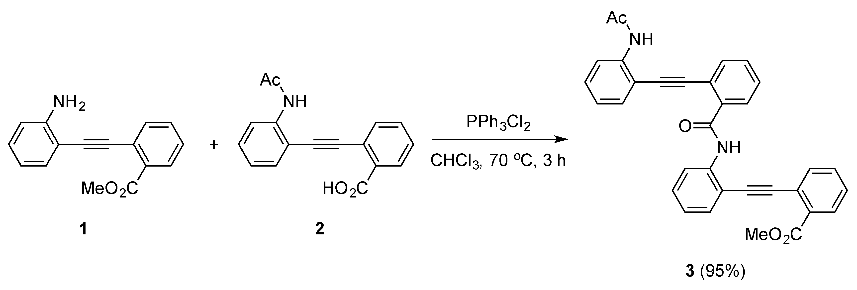
Synthesis of Methyl 2-[(2-{2-[(2-acetamidophenyl)ethynyl]benzamido}phenyl)ethynyl]benzoate (3)
Supplementary materials
Supplementary File 1Supplementary File 2Supplementary File 3Acknowledgments
Author Contributions
Conflicts of Interest
References and Notes
- Horne, W.S.; Gellman, S.H. Foldamers with heterogeneous backbones. Acc. Chem. Res. 2008, 41, 1399–1408. [Google Scholar] [CrossRef] [PubMed]
- Fischer, L.; Guichard, G. Folding and self-assembly of aromatic and aliphatic urea oligomers: Towards connecting structure and function. Org. Biomol. Chem. 2010, 8, 3101–3117. [Google Scholar] [CrossRef] [PubMed]
- Saraogi, I.; Hamilton, A.D. Recent advances in the development of aryl-based foldamers. Chem. Soc. Rev. 2009, 38, 1726–1743. [Google Scholar] [CrossRef] [PubMed]
- Yamazaki, N.; Demizu, Y.; Sato, Y.; Doi, M.; Kurihara, M. Helical foldamer containing a combination of cyclopentane-1,2-diamine and 2,2-dimethylmalonic acid. J. Org. Chem. 2013, 78, 9991–9994. [Google Scholar] [CrossRef] [PubMed]
- Demizu, Y.; Yamashita, H.; Yamazaki, N.; Sato, Y.; Doi, M.; Tanaka, M.; Kurihara, M. Oligopeptides with equal amounts of L- and D-amino acids may prefer a helix screw sense. J. Org. Chem. 2013, 78, 9991–9994. [Google Scholar] [CrossRef] [PubMed]
- Kemp, D.S.; Li, Z.Q. 2-Amino-2′-carboxydiphenylacetylenes as β-turn mimetics. Synthesis and conformational properties. Tetrahedron Lett. 1995, 36, 4175–4178. [Google Scholar] [CrossRef]
- Kemp, D.S.; Li, Z.Q. A short β-sheet containing proline nucleated by a 2,2′-substituted tolan β-turn mimetic. Tetrahedron Lett. 1995, 36, 4179–4180. [Google Scholar] [CrossRef]
- Lingard, H.; Han, J.T.; Thompson, A.L.; Leung, I.K.H.; Scott, R.T.W.; Thompson, S.; Hamilton, A.D. Diphenylacetylene-Linked Peptide Strands Induce Bidirectional β-Sheet Formation. Angew. Chem. Int. Ed. 2014, 53, 3650–3653. [Google Scholar] [CrossRef] [PubMed]
- Yang, X.W.; Yuan, L.H.; Yamamoto, K.; Brown, A.L.; Feng, W.; Furukawa, M.; Zeng, X.C.; Gong, B. Backbone-rigidfied oligo(m-phenylene ethynylenes). J. Am. Chem. Soc. 2004, 126, 3148–3162. [Google Scholar] [CrossRef] [PubMed]
- Fu, H.L.; Liu, Y.; Zeng, H.Q. Shape-persistent H-bonded macrocyclic aromatic pentamers. Chem. Commun. 2013, 49, 4127–4144. [Google Scholar] [CrossRef] [PubMed]
- Offermann, D.A.; McKendrick, J.E.; Sejberg, J.J.P.; Mo, B.; Holdom, M.D.; Helm, B.A.; Leatherbarrow, R.J.; Beavil, A.J.; Sutton, B.J.; Spivey, A.C. Synthesis and incorporation into cyclic peptides of tolan amino acids and their hydrogenated congeners: construction of an array of A-B-loop mimetics of the cε3 domain of human IgE. J. Org. Chem. 2012, 77, 3197–3214. [Google Scholar] [CrossRef] [PubMed]
- Compound 2 was prepared by N-acetylation of compound 1 and the subsequent alkaline hydrolysis of the methyl ester.
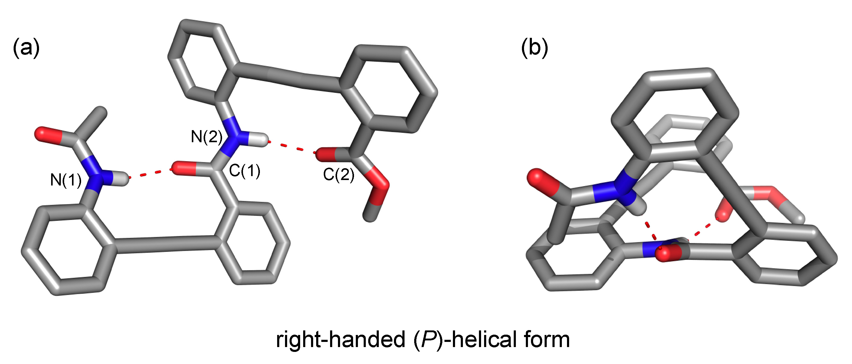
- CCDC-784138 (3) contains the supplementary crystallographic data for this paper. These data can be obtained free of charge at www.ccdc.cam.ac.uk/conts/retrieving.html (or from the Cambridge Crystallographic Data Centre, 12, Union Road, Cambridge CB2 1EZ, UK; fax: (+44) 1223-336-033; or deposit@ccdc.cam.ac.uk).
- Crystal data for 3: C33H24N2O4; Mr = 512.54; Monoclinic; Cc, a = 18.3920, b = 9.0316, c = 15.1378 Å; α = 90, β = 114.054, γ = 90°; V = 2513.2 Å3; Z = 4; Dcalc = 1.355 g/cm3; μ (MoKα) = 0.90 cm−1; No. of observations (I > 2σ(I)) = 4457; No. of variables = 352; R1 = 0.0658, and Rw = 0.1753.

© 2015 by the authors; licensee MDPI, Basel, Switzerland. This article is an open access article distributed under the terms and conditions of the Creative Commons Attribution license (http://creativecommons.org/licenses/by/4.0/).
Share and Cite
Demizu, Y.; Misawa, T.; Yamagata, N.; Doi, M.; Kurihara, M. Methyl 2-[(2-{2-[(2-acetamidophenyl)ethynyl]benzamido} phenyl)ethynyl]benzoate. Molbank 2015, 2015, M854. https://doi.org/10.3390/M854
Demizu Y, Misawa T, Yamagata N, Doi M, Kurihara M. Methyl 2-[(2-{2-[(2-acetamidophenyl)ethynyl]benzamido} phenyl)ethynyl]benzoate. Molbank. 2015; 2015(2):M854. https://doi.org/10.3390/M854
Chicago/Turabian StyleDemizu, Yosuke, Takashi Misawa, Nanako Yamagata, Mitsunobu Doi, and Masaaki Kurihara. 2015. "Methyl 2-[(2-{2-[(2-acetamidophenyl)ethynyl]benzamido} phenyl)ethynyl]benzoate" Molbank 2015, no. 2: M854. https://doi.org/10.3390/M854
APA StyleDemizu, Y., Misawa, T., Yamagata, N., Doi, M., & Kurihara, M. (2015). Methyl 2-[(2-{2-[(2-acetamidophenyl)ethynyl]benzamido} phenyl)ethynyl]benzoate. Molbank, 2015(2), M854. https://doi.org/10.3390/M854