Article

Thyroid Disorder in Obese Children and Adolescents: A Cross-Sectional Study in a Tertiary Care Hospital in Bangladesh

by
Farzana Sharmin
1,
Anika Tasneem Chowdhury
2,3,4,
Mosharop Hossian
4,5,*,
Shaima Rafiquzzaman
3,
Dhiraj C. Biswas
6,
Fatema Hashem Rupa
7 and
Suraiya Begum
8
1
100 Bedded District Hospital, Narsingdi, Dhaka 1600, Bangladesh
2
International Centre for Diarrhoeal Disease Research, Bangladesh (ICDDR,B), 68, Shaheed Tajuddin Ahmed Avenue, Mohakhali, Dhaka 1212, Bangladesh
3
Public Health Department, North South University, Dhaka 1229, Bangladesh
4
Public Health Promotion and Development Society (PPDS), Dhaka 1205, Bangladesh
5
School of Health and Rehabilitation Sciences, University of Queensland, Brisbane 4072, Australia
6
300 Bed Hospital, Khanpur, Narayanganj 1361, Bangladesh
7
Directorate General of Health Services, Dhaka 1212, Bangladesh
8
Department of Paediatrics, Bangabandhu Sheikh Mujib Medical University, Dhaka 1000, Bangladesh
*
Author to whom correspondence should be addressed.
Future 2025, 3(4), 18; https://doi.org/10.3390/future3040018
Submission received: 30 April 2025 / Revised: 27 July 2025 / Accepted: 17 September 2025 / Published: 25 September 2025

Abstract

Background: Childhood obesity is becoming an increasingly pressing issue on a global scale. This study aimed to explore the relationship between thyroid hormone levels and body mass index (BMI) in obese children and adolescents, an area with limited research, particularly in Bangladesh. Methods: This cross-sectional study was undertaken in Bangabandhu Sheikh Mujib Medical University, Bangladesh, from August 2018 to January 2020. We included 105 participants aged 10–18 years, divided into obese (n = 69) and normal-weight (n = 36) groups based on the CDC BMI percentiles. We conducted chi-square tests, Pearson correlation, and linear regression analyses. Results: Obese participants exhibited significantly higher mean levels of TSH (4.40 ± 3.20 µIU/mL vs. 2.26 ± 0.97 µIU/mL, p-value 0.0002) and FT3 (3.52 ± 0.71 pg/mL vs. 3.02 ± 0.48 pg/mL, p-value < 0.001) and lower FT4 levels (1.23 ± 0.21 ng/dL vs. 1.38 ± 0.30 ng/dL, p-value 0.0002) compared to normal-weight participants. We observed a positive correlation between BMI and TSH (p-value 0.002) and FT3 (p-value < 0.001), and a negative correlation between BMI and FT4 (p-value 0.003). Most of the obese children were euthyroid (71.01%), with 27.54% showing subclinical hypothyroidism and 1.45% showing overt hypothyroidism. Multivariable linear regression analysis revealed that with a one unit increase in BMI, FT3 increased by 0.032 ± 0.011 pg/mL (p-value 0.004), FT4 decreased by 0.010 ± 0.004 (p-value 0.017 ng/dL, and TSH increased by 0.104 ± 0.044 µIU/mL (p-value 0.020). Conclusions: The significant association between BMI and thyroid hormone levels underscores the necessity for routine thyroid function monitoring in obese paediatric populations. The early detection and management of thyroid dysfunction may enhance health and well-being outcomes in obese children and adolescents.

1. Introduction

Childhood obesity is becoming an increasingly pressing issue on a global scale with its prevalence rising dramatically in recent decades [1]. The global prevalence of obesity among children was increased from 2% in 1990 to 8% in 2022 [2]. Although the prevalence in the Southeast Asian region remains among the lowest by global comparison, childhood obesity has been on the rise in the last 10–15 years [3]. Around 38.4 million children between the ages of 5–19 were overweight in 2020 [3]. In Bangladesh, a rise in obesity among children and adolescents has been observed from 5.8% in 2011 to 9.2% in 2022 [4]. Additionally, the prevalence of the morbid obesity among this population rose from 0.9% to 1.8% during the same period [4].
Recently, the growing focus on thyroid function in obese pediatric patients have led to numerous studies exploring their connection [1,5,6]. Thyroid hormones (FT3 and FT4) play a significant role in metabolic processes, energy expenditure, and appetite regulation; an increase in thyroid hormone levels increases basal metabolic rate, thermogenesis, and lipolysis, which collectively help reduce body weight. Conversely, a reduction in these hormone levels over time can have the opposite effect in weight gain [7,8,9,10,11,12,13]. Iodine deficiency is a leading global cause of thyroid dysfunction in children. However, according to the most recent national micronutrient survey, iodine intake in Bangladesh is currently adequate among children and women of reproductive age, as indicated by median urinary iodine concentration. Therefore, iodine deficiency is unlikely to confound the observed patterns of thyroid dysfunction in this population. Studies have shown a positive relationship between BMI and TSH, as well as between BMI and FT3, while a negative association with FT4 has been observed [14,15]. The pathophysiology of obesity and its association with thyroid hormone dysregulation are well established in the literature [8]. Adipose tissues release cytokines and adipokines that influence thyroid function. In excess adipose tissue, leptin (an adipokine) is secreted and stimulates the hypothalamic-pituitary-thyroid axis, potentially leading to increased TRH, TSH, and FT3 levels [16]. Elevated TSH levels may also be attributed to reduced negative feedback between peripheral thyroid hormones and TSH in obese patients, resulting from a decreased number of T3 receptors and pituitary resistance to FT3 [7]. This leads to heightened TSH and FT3 levels [7].
Studies by H. Stichel et al. and Robabeh G et al. supported this by reporting elevated TSH and FT3 levels and a decrease in the FT4 levels among overweight and obese children compared to those with normal weight [1,14].
Obesity can result in hormonal and metabolic disorders which resolve after body weight normalization [7]. This is proven by some studies where abnormal thyroid function has shown subsequent improvement or resolution of subclinical hypothyroidism following weight loss by diet modification or bariatric surgery, suggesting that pharmacological therapy may not be required in all cases [17,18].
Few studies in Bangladesh have focused on thyroid hormone dysfunction and obesity among the paediatric population, and a study conducted by S Mahbuba et al. investigating the pattern of endocrine disorders in children revealed that 25% of the subjects presented with thyroid disorders and 25.9% had simple obesity [19]. The association between obesity and thyroid hormone levels on the adult population of Bangladesh have already researched but few studies have been surveyed this association in pediatric population [9,20]. Therefore, this cross-sectional study of obese paediatric patients was conducted in a tertiary care hospital in Bangladesh to determine the prevalence of thyroid dysfunction among obese children in Bangladesh and compare it with normal-weight peers.

2. Methods

2.1. Study Design and Participants

This prospective cross-sectional study was undertaken in the Department of Paediatrics, Endocrinology and Metabolism Division, Bangabandhu Sheikh Mujib Medical University (BSMMU) from August 2018 to January 2020. The study population were allocated into two groups in accordance with the age and sex-specific BMI criteria provided by the Centers for Disease Control and Prevention (CDC) [21]. Group A (obese group) comprised obese children, whose BMI was ≥95th centile, while Group B (normal weight group) comprised children with a normal BMI, ranging from the 5th to less than the 85th centile [21]. The sample size was determined using the formula n = z 2 p q d 2 . Based on the findings of Ozer et al. (2015), who reported a 25.7% prevalence of thyroid disorders among obese adolescents, this figure was used as the estimated prevalence (p) for the calculation [22]. A total of 72 children in the obese group met all inclusion criteria (Supplementary Figure S1). Additionally, 36 children with normal weight were included, considering logistical and economic constraints. Of the 108 participants initially enrolled, 72 were obese; however, 3 children from the obese group were excluded due to the presence of thyroid autoantibodies. As a result, the final sample comprised 69 obese and 36 normal-weight children.

2.2. Procedures

All anthropometric measurements were conducted by physicians who received training and supervision in standardized measurement procedures, following WHO guidelines, to ensure consistency and accuracy throughout the data collection period. Physicians of the BSMMU obtained a detailed history of the study participants and performed physical examinations, including measurements of anthropometric indices, using a structured questionnaire. The measurement of weight was obtained using an electronic-weighing scale (Tanita, Japan) and recorded to the nearest 100 g. The weight of the study participants was measured barefoot and in light clothing. Measurements of standing height were obtained using a stadiometer, with readings taken close to 0.1 cm. Body mass index was determined by weight-to-height ratio, where weight was expressed in kg and height in meters.
Blood samples were taken from patients in the study, and serum TSH, FT3, and FT4 were measured in obese and non-obese adolescents using a chemiluminescent immunoassay on a Siemens ADIVA Centaur machine (Siemens Healthineers Headquarters Siemens Healthcare GmbH, Henkestr. 127 91052 Erlangen, Germany). When there was an elevated level, Anti TPO and Anti TG were measured by chemiluminescent immunoassay using Immulite (Siemens Healthcare Diagnostics Inc. Specialty Lab Solutions, 511 Benedict Avenue, Tarrytown, NY 10591-5005, USA). Thyroid antibody-positive (any one of thyroid antibodies) cases were excluded from the study. These investigations were performed at the Department of Microbiology, Bangabandhu Sheikh Mujib Medical University. TSH value 0.70–5.70 µIU/mL, FT3 value 1.40–4.20 pg/mL, and FT4 value 0.80–1.80 ng/dL were considered normal (21). All thyroid hormone (TSH, FT3, FT4) assays were conducted in a single accredited laboratory using standardised chemiluminescent immunoassay protocols. Internal quality control samples and calibration procedures were performed with each batch in accordance with manufacturer instructions to ensure accuracy and reproducibility.
Thyroid status was categorized as euthyroid if normal TSH and FT4 levels, subclinical hypothyroid if high TSH, normal FT4 levels and overt hypothyroid if high TSH, low FT4 levels.
Statistical analyses were conducted using STATA version 15.0 (StataCorp. 2017. Stata Statistical Software: Release 15, College Station, TX: StataCorp LLC). For continuous variables, data are presented as mean ± standard deviation (SD); for categorical variables, as frequency and percentage. Group comparisons for continuous variables (e.g., BMI, TSH, FT3, FT4) were performed using independent sample t-tests, as the distribution of key variables was consistent with approximate normality based on descriptive statistics and sample size. Categorical variables were compared using the chi-square test. Associations between continuous variables were examined using Pearson correlation coefficients. Linear regression analysis, adjusting for potential confounders, was used to assess the relationship between BMI and thyroid hormone levels. Statistical significance was defined as a two-sided p-value < 0.05.

2.3. Ethical Consideration

Acquisition of written consent from the guardians and assent from the adolescents took place prior to data collection.

3. Results

The study population comprised 105 individuals aged 10 to 18 years. In Table 1 the background characteristics of the study participants can be observed. Around 66% of 10–13 years, 65% of 14–18 years, 69% of males, 63% of females, 56% of rural residents and 74% of urban residents were obese. No statistically significant differences were observed in the background characteristics between normal weight and obesity.
Table 2 presents anthropometric measurements of the study participants and their parents and thyroid hormone levels of the children and adolescents. The mean values of weight (Obese group: 62.29 ± 14.42 kg vs. Normal weight group: 37.80 ± 7.29 kg; p-value < 0.001), BMI (Obese group: 27.92 ± 3.72 kg/m2 vs. Normal weight group: 18.13 ± 3.72 kg/m2; p-value < 0.001), waist circumference (Obese group: 91.71 ± 9.41 cm vs. Normal weight group: 71.21 ± 7.05 cm; p-value < 0.001) and mother’s BMI (Obese group: 26.65 ± 4.04 kg/m2 vs. Normal weight group: 23.90 ± 3.44 kg/m2; p-value < 0.001) were statistically significantly different between two groups. TSH and FT3 levels were higher in the obese group than the normal weight group (4.40 ± 3.20 μIU/mL vs. 2.26 ± 0.97 μIU/mL, p value 0.0002 and 3.52 ± 0.71 142 pg/mL vs. 3.02 ± 0.48 pg/mL, p value < 0.001, respectively). But FT4 levels were lower in the obese group than the normal weight group (1.23 ± 0.21 ng/dL vs. 1.38 ± 0.30 ng/dL, p value 0.0002).
Table 3 shows the thyroid function status categories of obese children. Euthyroid, subclinical hypothyroid, and overt hypothyroid states were observed in 71.01%, 27.54%, and 1.45% of the obese children, respectively.
Figure 1 demonstrates the correlation of TSH, BMI, FT3, and FT4 values of the study participants through a scatter plot. Scatter plots revealed significant relationships between thyroid hormone levels and BMI in children and adolescents. Each panel in Figure 1 displays the relationship between a unique pair of variables (BMI, TSH, FT3, FT4), with colour-coding and clear labelling provided to facilitate interpretation of all relevant associations in the study population. There was a positive association between BMI and TSH (p-value 0.002). TSH was positively associated with FT3 (p-value < 0.001) and negatively associated with FT4 (p-value 0.003). The BMI was positively associated with FT3 (p = 0.000) and negatively associated with FT4 (p = 0.010). Additionally, FT3 were negatively associated with FT4 (p-value 0.001). These results highlight the interconnected role of thyroid hormones and BMI in children and adolescents.
Table 4 shows the association between BMI and thyroid hormone status among study participants. With one unit increase in BMI, FT3 increased by 0.032 ± 0.011 pg/mL (p-value 0.004), FT4 decreased by 0.010 ± 0.004 (p-value 0.017) ng/dL and TSH increased by 0.104 ± 0.044 µIU/mL (p-value 0.020).

4. Discussion

Our investigation focused on the evaluation of thyroid function in children and adolescents with obesity at a tertiary healthcare facility in Bangladesh. A significant association was observed between TSH and BMI, implying that alterations of body weight were associated with thyroid dysfunction. This suggests that excess energy intake in obese children leads to elevated TSH and FT3 levels [23]. The results of the present study align with the result of Kumar et al. and Ali et al., who reported elevated TSH levels in 30% and 35% of the obese children, respectively [24,25]. Different studies have reported varying frequencies of high TSH levels. Elevated TSH levels in obese children (8.3%) compared to normal-weight children (5.8%) revealed by YM et al. [26]. Several mechanisms are related to alterations in thyroid function in patients with obesity. The primary factors contributing to elevated TSH levels in obesity are influenced by leptin, mitochondrial dysfunction, thyroid hormone resistance, adaptive processes, and inflammatory conditions associated with obesity [16,27].
Our study found that 13.04% of obese children had high FT3 levels which is congruent to the study findings of Marras et al., where 18% of obese children and adolescents had high FT3 levels [28]. Complicated mechanisms, such as thyroid hormone resistance and adaptation processes, are responsible for high FT3 levels [16]. Reduced T3 receptors in the pituitary gland of obese children lead to impaired negative feedback mechanisms [29]. Variations in FT4 levels were observed among obese children. High FT4 levels were found in studies by Marras et al. and Ghergherehchi and Hazhir [14,28]. In our study, normal FT4 levels were observed in the majority of the obese children which is similar to the findings of studies conducted by Stichel et al. and Ekenci et al. [1,30]. Low FT4 level was found in studies carried out by An YM et al. and Xu et al. [26,31]. In obesity, leptin enhances the activity of deiodinases, leading to high conversion of FT4 to FT3 [16].
In this study, high TSH (28.99%), high FT3 (13.04%), and low FT4 (1.45%) levels were associated with obesity. The current study found that TSH was positively correlated with BMI which was similar to previously published studies, such as Grandone et al., Marras et al., Ekenci et al. and Ali et al., but in contrast to Kumar et al., Aeberili et al. [15,24,25,28,30,32]. TSH had positive significant correlation with FT3 which corresponded to findings of Ali et al., and in the present study, TSH had a significant negative association with FT4 which agreed with Ali et al. [25]. In the current study FT3 also had a significant positive association with BMI which was similar to the results of Grandone et al. and Marras et al. [15,28]. This widespread variability in the results has been attributed in the literature to differences in ethnicity, dietary patterns (including iodine intake), genetic background, and environmental exposures among study populations. For example, ethnic variation may influence both the prevalence of obesity and the physiological response of the thyroid axis. Dietary practices, particularly differences in iodine consumption and nutritional status, as well as underlying genetic predispositions, have been proposed as contributors to discrepancies in thyroid function observed across diverse groups of children and adolescents. As primary data on these factors were not collected in the present study, direct analysis could not be performed. Nevertheless, these variables have been acknowledged as important sources of heterogeneity in the international literature, and further research in the Bangladeshi context is recommended to address these complexities.
The significant positive association observed between FT3 and BMI suggests adaptation of thyroid function to increased body mass. This finding aligns with those of previous studies that reported similar correlations [16]. The elevation in FT3 levels with BMI may be related to enhanced peripheral conversion of T4 to T3, particularly in adipose tissue where deiodinase enzymes are active [23]. This is supported by studies indicating that adiposity can increase deiodinase activity, thereby promoting the conversion process to meet the higher metabolic demands associated with greater body mass [24]. This adaptation could be a physiological mechanism aimed at maintaining energy homeostasis in individuals with higher BMI.
Conversely, the significant negative association between BMI and FT4 levels can be considered as part of a regulatory feedback mechanism in thyroid hormone metabolism. As BMI increased, FT4 levels decreased, possibly due to enhanced conversion to FT3, suggesting a shift in hormone dynamics to support metabolic needs. This relationship might also involve the dilution of hormones in a larger blood volume, which is a common occurrence in individuals with higher body mass. The literature on thyroid function in obesity often highlights such dynamic shifts, indicating a complex interplay between hormone levels and body weight [26,27].
The finding that TSH levels increase with higher BMI points to potential subclinical hypothyroidism or a compensatory mechanism by the thyroid gland to address insufficient thyroid hormone production relative to body needs [28]. Elevated TSH levels could drive increased hormone production, overcoming inefficiencies in hormone synthesis or secretion associated with obesity [29]. This is consistent with previous research suggesting that increased adiposity can be associated with TSH levels, possibly through mechanisms involving leptin and other adipokines that interact with thyroid function [5,33].
The significant relationship between maternal BMI and child TSH in children suggests that environmental and possibly genetic factors linked to the mother’s metabolic state could be associated with thyroid function in the offspring. The mechanism might involve prenatal influences where maternal hormones, including thyroid hormones and factors such as leptin, may affect the development of thyroid regulation in children. Studies have noted that maternal health, including weight and metabolic status, can have long-lasting effects on the endocrine profiles of children, indicating transgenerational transmission of metabolic traits [14]. The cross-sectional design is a significant limitation of our study. As with all cross-sectional analyses, our data describe statistical associations between obesity and thyroid dysfunction, but do not permit any inference of causality or the direction of these associations. Because both exposure (BMI) and outcome (thyroid hormone levels) were measured at a single point in time, it is not possible to determine whether obesity precedes thyroid dysfunction or vice versa. Therefore, our findings must be interpreted as evidence of association only, not causation. In addition, we did not account for all potential confounding factors, including diet, physical activity, and genetic predisposition. In particular, it should be noted that important confounding variables, such as diet, physical activity, detailed socioeconomic status, and other familial or environmental influences, were not measured, which may have introduced residual confounding and affected the observed associations. Along with these, the single-center design and modest sample size further limit the generalizability of our findings. Additionally, we relied primarily on BMI to define obesity. Although BMI is a practical and widely used index in paediatric epidemiological research, it does not distinguish between fat mass and lean body mass, and may misclassify muscular children as overweight or obese. To partially address this limitation, we also assessed waist circumference as a measure of central adiposity. However, more direct measures of body fat, such as skinfold thickness, dual-energy X-ray absorptiometry, or bioelectrical impedance analysis, would provide a more accurate assessment of adiposity and are recommended for future studies. Furthermore, while we have updated the reference list to include the recent international literature where possible, relevant studies from Bangladesh and the wider region remain scarce. Consequently, some older references have been retained to ensure appropriate scientific context and reflect foundational evidence. This underscores the ongoing evidence gap in local populations and highlights the importance of the present study. Future research should prioritise prospective cohort and intervention studies that examine the temporal sequence between obesity and thyroid dysfunction, as well as the effects of dietary and lifestyle modifications on thyroid status. Inclusion of more comprehensive measures of adiposity and careful control of confounding factors are also recommended. For clinical practice, our findings support the recommendation that routine thyroid function screening be considered in the management of obese children and adolescents. Early identification and management of thyroid dysfunction may offer potential health benefits for this vulnerable population.

5. Conclusions

Thyroid dysfunction, particularly subclinical hypothyroidism, is prevalent in obese children. The significant association between BMI and thyroid hormone levels underscores the necessity for routine thyroid function monitoring in all obese children, both at initial presentation and at regular intervals during follow-up. Early detection and management of thyroid dysfunction may enhance health and well-being outcomes in paediatric obesity. Larger multicenter prospective studies are recommended to assess these associations and to inform evidence-based screening intervals and management strategies.

Supplementary Materials

The following supporting information can be downloaded at: https://www.mdpi.com/article/10.3390/future3040018/s1, Figure S1: Schematic Diagram Illustrating the Selection Process for Study Participants.

Author Contributions

Conceptualization: F.S., A.T.C., S.B.; methodology: F.S., A.T.C., S.B.; software: F.S., M.H., S.B.; validation: S.R., D.C.B., S.B.; formal analysis: A.T.C., M.H.; investigation: D.C.B.; resources: FS., S.B.; data curation: F.S.; F.H.R.; writing—original draft preparation: F.S., A.T.C.; writing—review and editing: F.S., A.T.C., M.H., S.R., D.C.B., F.H.R., S.B.; visualization: M.H.; supervision: S.B.; project administration: S.B.; funding acquisition: Not applicable. All authors have read and agreed to the published version of the manuscript.

Funding

This research received no external funding.

Institutional Review Board Statement

The study was conducted in accordance with the Declaration of Helsinki, and approved by the Institutional Review Board (or Ethics Committee) of Bangabandhu Sheikh Mujib Medical University (BSMMU) (BSMMU/2018/2653).

Informed Consent Statement

Informed consent was obtained from all subjects involved in the study. Written informed consent has been obtained from the patient(s) to publish this paper.

Data Availability Statement

Data will be made available upon considerable application.

Conflicts of Interest

The authors declare no conflicts of interest.

References

  1. Stichel, H.; Allemand, D.; Grüters, A. Thyroid function and obesity in children and adolescents. Horm. Res. Paediatr. 2000, 54, 14–19. [Google Scholar] [CrossRef]
  2. The World Health Organization. Obesity and Overweight; The World Health Organization: Geneva, Switzerland, 2025.
  3. World Health Organization. The Double Burden of Malnutrition: Priority Actions on Ending Childhood Obesity; World Health Organization: Geneva, Switzerland; Regional Office for South-East Asia: New Delhi, India, 2020.
  4. World Health Organization. Prevalence of Overweight Among Children and Adolescents; World Health Organization: Geneva, Switzerland, 2016.
  5. Nannipieri, M.; Cecchetti, F.; Anselmino, M.; Camastra, S.; Niccolini, P.; Lamacchia, M.; Rossi, M.; Iervasi, G.; Ferrannini, E. Expression of thyrotropin and thyroid hormone receptors in adipose tissue of patients with morbid obesity and/or type 2 diabetes: Effects of weight loss. Int. J. Obes. 2009, 33, 1001–1006. [Google Scholar] [CrossRef]
  6. Kitahara, C.M.; Platz, E.A.; Ladenson, P.W.; Mondul, A.M.; Menke, A.; de González, A.B. Body fatness and markers of thyroid function among US men and women. PLoS ONE 2012, 7, e34979. [Google Scholar] [CrossRef]
  7. Witkowska-Sędek, E.; Kucharska, A.; Rumińska, M.; Pyrżak, B. Thyroid dysfunction in obese and overweight children. Endokrynol. Pol. 2017, 68, 54–60. [Google Scholar] [CrossRef]
  8. Natah, T.M.; Wtwt, M.A.-A.; Hadi, M.A.; Farhood, H.F.; Al-Saadi, A.H. Thyroid metabolic hormones and its correlation with bmi and lipid profile in healthy people. Thyroid 2013, 18, 47–56. [Google Scholar]
  9. Muscogiuri, G.; Sorice, G.P.; Mezza, T.; Prioletta, A.; Lassandro, A.P.; Pirronti, T.; Della Casa, S.; Pontecorvi, A.; Giaccari, A. High-normal tsh values in obesity: Is it insulin resistance or adipose tissue’s guilt? Obesity 2013, 21, 101–106. [Google Scholar] [CrossRef]
  10. Pasquali, R.; Vicennati, V. Obesity and hormonal abnormalities. In International Textbook of Obesity; Björntorp, P., Ed.; John Wiley & Sons, Ltd.: Hoboken, NJ, USA, 2001; pp. 225–239. [Google Scholar]
  11. Björntorp, P. Endocrine abnormalities of obesity. Metabolism 1995, 44, 21–23. [Google Scholar] [CrossRef] [PubMed]
  12. Fox, C.S.; Pencina, M.J.; D’agostino, R.B.; Murabito, J.M.; Seely, E.W.; Pearce, E.N.; Vasan, R.S. Relations of thyroid function to body weight: Cross-sectional and longitudinal observations in a community-based sample. Arch. Intern. Med. 2008, 168, 587–592. [Google Scholar] [CrossRef] [PubMed]
  13. Staníková, D.; Krajčovičová, L.; Lobotková, D.; Vitariušová, E.; Tichá, Ľ.; Pribilincová, Z.; Ukropcová, B.; Ukropec, J.; Staník, J. Thyroid hormone levels and BMI-SDS changes in adolescents with obesity. Front. Endocrinol. 2023, 14, 1304970. [Google Scholar] [CrossRef]
  14. Ghergherehchi, R.; Hazhir, N. Thyroid hormonal status among children with obesity. Ther. Adv. Endocrinol. Metab. 2015, 6, 51–55. [Google Scholar] [CrossRef]
  15. Grandone, A.; Santoro, N.; Coppola, F.; Calabrò, P.; Perrone, L.; del Giudice, E.M. Thyroid function derangement and childhood obesity: An Italian experience. BMC Endocr. Disord. 2010, 10, 8. [Google Scholar] [CrossRef] [PubMed]
  16. Walczak, K.; Sieminska, L. Obesity and thyroid axis. Int. J. Environ. Res. Public Health 2021, 18, 9434. [Google Scholar] [CrossRef]
  17. Laurberg, P.; Knudsen, N.; Andersen, S.; Carlé, A.; Pedersen, I.B.; Karmisholt, J. Thyroid function and obesity. Eur. Thyroid. J. 2012, 1, 159–167. [Google Scholar] [CrossRef] [PubMed]
  18. Chikunguwo, S.; Brethauer, S.; Nirujogi, V.; Pitt, T.; Udomsawaengsup, S.; Chand, B.; Schauer, P. Influence of obesity and surgical weight loss on thyroid hormone levels. Surg. Obes. Relat. Dis. 2007, 3, 631–635. [Google Scholar] [CrossRef]
  19. Mahbuba, S.; Rahat, F.; Iman, K.; Sultana, A.; Parvin, R.; Afroze, S.; Ghosh, N.K. Disease Profile of Children with Endocrine Disorders: Experience in Dr. MR Khan Shishu Hospital, Dhaka, Bangladesh. J. Dr. MR Khan Shishu Child. Hosp. 2022, 3, 9–14. [Google Scholar]
  20. Åsvold, B.O.; Bjøro, T.; Vatten, L.J. Association of serum TSH with high body mass differs between smokers and never-smokers. J. Clin. Endocrinol. Metab. 2009, 94, 5023–5027. [Google Scholar] [CrossRef]
  21. Centers for Disease Control and Prevention. BMI; Centers for Disease Control and Prevention: Atlanta, GA, USA, 2024.
  22. Özer, S.; Bütün, İ.; Sönmezgöz, E.; Yılmaz, R.; Demir, O. Relationships among thyroid hormones and obesity severity, metabolic syndrome and its components in Turkish children with obesity. Nutr. Hosp. 2015, 32, 645–651. [Google Scholar]
  23. Longhi, S.; Radetti, G. Thyroid function and obesity. J. Clin. Res. Pediatr. Endocrinol. 2013, 5, 40. [Google Scholar] [PubMed]
  24. Hari Kumar, K.; Verma, A.; Muthukrishnan, J.; Modi, K. Obesity and thyrotropinemia. Indian J. Pediatr. 2009, 76, 933–935. [Google Scholar] [CrossRef]
  25. Ali, B.A.-M.; Mahrous, D.M.; Ahmed, D.M. Thyroid function status in obese children. J. Diabetes Metab. 2016, 7, 665. [Google Scholar]
  26. An, Y.M.; Moon, S.J.; Kim, S.K.; Suh, Y.J.; Lee, J.E. Thyroid function in obese Korean children and adolescents: Korea National Health and Nutrition Examination Survey 2013–2015. Ann. Pediatr. Endocrinol. Metab. 2018, 23, 141. [Google Scholar] [CrossRef]
  27. Pacifico, L.; Anania, C.; Ferraro, F.; Andreoli, G.M.; Chiesa, C. Thyroid function in childhood obesity and metabolic comorbidity. Clin. Chim. Acta 2012, 413, 396–405. [Google Scholar] [CrossRef] [PubMed]
  28. Marras, V.; Casini, M.R.; Pilia, S.; Carta, D.; Civolani, P.; Porcu, M.; Uccheddu, A.P.; Loche, S. Thyroid function in obese children and adolescents. Horm. Res. Paediatr. 2010, 73, 193–197. [Google Scholar] [CrossRef]
  29. Burman, K.D.; Latham, K.R.; Djuh, Y.-Y.; Smallridge, R.C.; Tseng, Y.-C.L.; Lukes, Y.G.; Maunder, R.; Wartofsky, L. Solubilized nuclear thyroid hormone receptors in circulating human mononuclear cells. J. Clin. Endocrinol. Metab. 1980, 51, 106–116. [Google Scholar] [CrossRef] [PubMed]
  30. Ekinci, F.; Uzuner, A.; Demet, M.C.; Turgut, B.; Tosun, N. Thyroid hormones and BMI in obese children: One year follow up results. Marmara Med. J. 2015, 28, 129–134. [Google Scholar] [CrossRef]
  31. Xu, R.; Huang, F.; Zhang, S.; Lv, Y.; Liu, Q. Thyroid function, body mass index, and metabolic risk markers in euthyroid adults: A cohort study. BMC Endocr. Disord. 2019, 19, 129. [Google Scholar] [CrossRef]
  32. Aeberli, I.; Jung, A.; Murer, S.B.; Wildhaber, J.; Wildhaber-Brooks, J.; Knöpfli, B.H.; Zimmermann, M.B. During rapid weight loss in obese children, reductions in TSH predict improvements in insulin sensitivity independent of changes in body weight or fat. J. Clin. Endocrinol. Metab. 2010, 95, 5412–5418. [Google Scholar] [CrossRef]
  33. Shimura, H.; Miyazaki, A.; Haraguchi, K.; Endo, T.; Onaya, T. Analysis of differentiation-induced expression mechanisms of thyrotropin receptor gene in adipocytes. Mol. Endocrinol. 1998, 12, 1473–1486. [Google Scholar] [CrossRef]
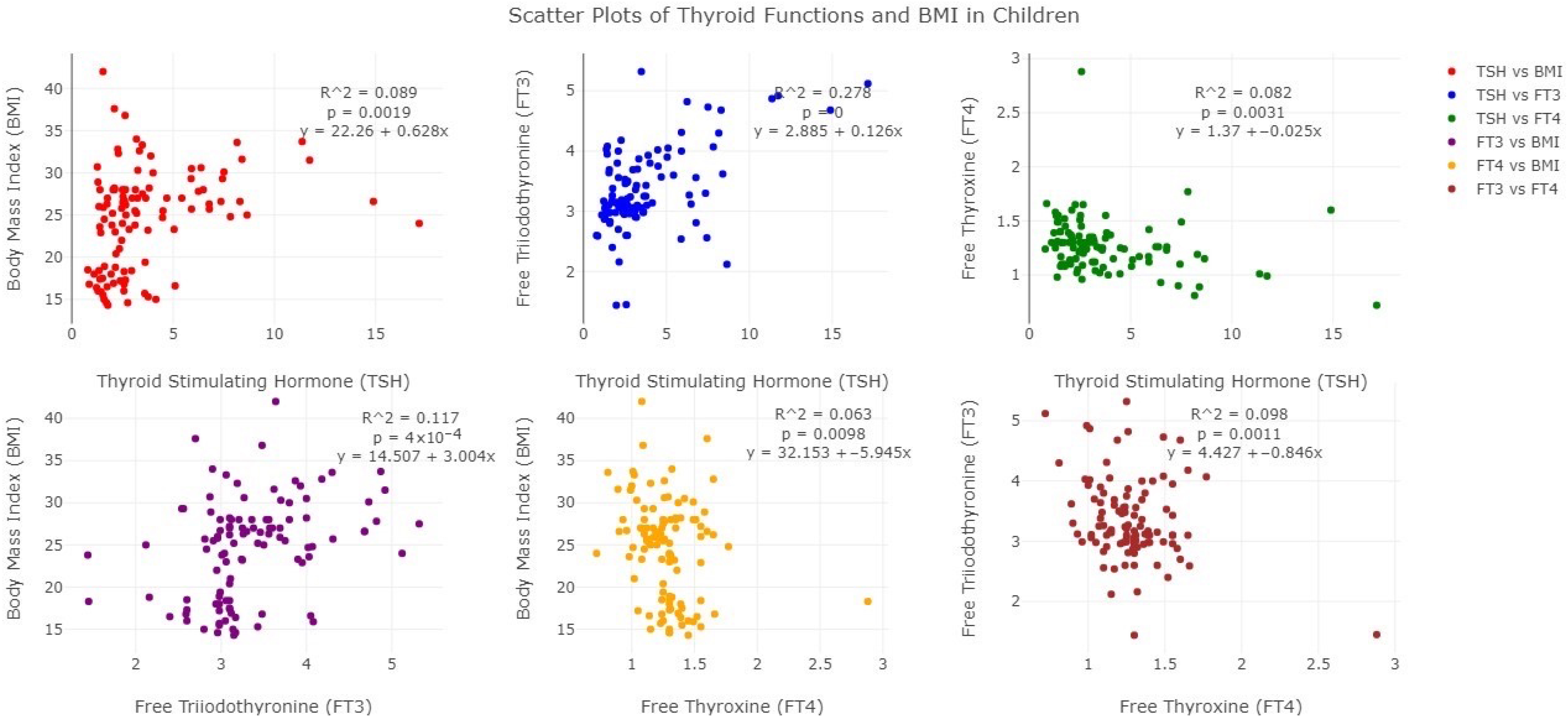
Figure 1. Pairwise Scatter Plots Showing the Relationships Between Body Mass Index (BMI) and Thyroid Hormone Levels (TSH, FT3, FT4) in Children and Adolescents.
Future 03 00018 g001
Table 1. Background characteristics of the study participants.
NormalObesep-Value
n%n%
Age (years)
10–132734.185265.820.967
14–18934.621765.38
Sex
Male1631.373568.630.541
Female2037.043462.96
Residence
Rural2043.482656.520.059
Urban 1525.864374.14
Table 2. Anthropometric measurements of the study participants and their parents and thyroid hormone levels of the children.
Normal
(Mean ± SD)
Obese
(Mean ± SD)
p-Value
Weight (kg)37.80 (±7.29)62.29 (±14.42)<0.001
Height (cm)147.68 (±9.30)148.80 (±10.26)0.5862
BMI18.13 (±3.72)27.92 (±3.72) <0.001
Waist circumference (cm)71.21 (±7.05)91.71 (±9.41)<0.001
Father’s BMI24.59 (±2.64)25.71 (±2.94)0.0573
Mother’s BMI23.90 (±3.44)26.65 (±4.04)0.0008
FT3 (pg/mL)3.02 (±0.48)3.52 (±0.71)0.0003
FT4 (ng/dL)1.38 (±0.30)1.23 (±0.21)0.0031
TSH (µIU/mL)2.26 (±0.97)4.40 (±3.20)0.0002
Table 3. Category of thyroid function status categories of obese children.
State of Thyroid Hormone StatusNumber (n = 69)Percentage (%)
Euthyroid4971.01
Subclinical hypothyroid1927.54
Overt hypothyroid011.45
Table 4. Association between BMI and thyroid hormone status among study participants.
FT3 FT4TSH
Coefficient ± SEp-ValueCoefficient ± SEp-ValueCoefficient ± SEp-Value
BMI0.032 ± 0.0110.004−0.010 ± 0.0040.0170.104 ± 0.0440.020
Age (ref. 10–13 years)
14–18 years−0.176 ± 0.1420.218 −0.066 ± 0.0560.243−0.551 ± 0.5780.342
Sex (ref. Male)
Female0.032 ± 0.1260.799−0.021 ± 0.0500.6720.842 ± 0.5130.104
Residence (ref. Rural)
Urban−0.053 ± 0.1260.6750.047 ± 0.4900.3400.661 ± 0.5110.199
Mother’s BMI−0.035 ± 0.0240.154−0.003 ± 0.0100.7340.197 ± 0.0990.048
Father’s BMI0.027 ± 0.0330.414−0.014 ± 0.0130.2860.076 ± 0.1360.577
Disclaimer/Publisher’s Note: The statements, opinions and data contained in all publications are solely those of the individual author(s) and contributor(s) and not of MDPI and/or the editor(s). MDPI and/or the editor(s) disclaim responsibility for any injury to people or property resulting from any ideas, methods, instructions or products referred to in the content.