Article

Assessment of a Natural Phytobiotic Mixture as Feed Additive for Broiler Chicken: Studies on Animal Performance, Gut Health, and Antioxidant Status After Experimental Infection with Eimeria spp.

1
Department of Animal Science, University of Thessaly, 41335 Larissa, Greece
2
Laboratory of Nutrition, School of Veterinary Medicine, Aristotle University of Thessaloniki, 54124 Thessaloniki, Greece
3
Unit of Avian Medicine, Clinic of Farm Animals, School of Veterinary Medicine, Aristotle University of Thessaloniki, 54124 Thessaloniki, Greece
4
Laboratory of Parasitology and Parasitic Diseases, School of Veterinary Medicine, Aristotle University of Thessaloniki, 54124 Thessaloniki, Greece
5
Laboratory of Pathology, School of Veterinary Medicine, Aristotle University of Thessaloniki, 54124 Thessaloniki, Greece
6
PatentCo, 24211 Misicevo, Serbia
7
Department of Pathobiology and Population Sciences, The Royal Veterinary College, Hatfield AL9 7TA, UK
*
Authors to whom correspondence should be addressed.
Submission received: 29 October 2024 / Revised: 12 December 2024 / Accepted: 17 December 2024 / Published: 2 January 2025

Abstract

:
This study evaluated the effectiveness of phytobiotic supplementation in managing coccidiosis in broiler chickens, a disease that impacts productivity. Three hundred sixty Ross-308 one-day-old chicks were assigned to five treatment groups: control negative (CN), phyto negative (PN), control infected (CI), phyto infected (PI), and salinomycin infected (SI). All diets were fed for the entire experiment duration. CN and CI groups were given a common diet, with CN remaining uninfected and CI exposed to Eimeria spp., while PN and PI groups received a phytobiotic supplement containing essential oils, saponins, and tannins (with PI challenged by Eimeria spp.), and SI received salinomycin post-infection. All infected groups were challenged on day 14 with Eimeria acervulina, E. maxima, and E. tenella. By day 21, PN had the highest body weight (744.9 g). Body weight gain (BWG) was highest in CN and PN from days 14–35, while CI consistently showed the lowest BWG. PI demonstrated significantly lower oocyst counts than CI, outperforming even SI by day 7, and showed milder intestinal lesions. A high anticoccidial index (ACI) of 188.45 was observed in PI, close to uninfected groups, while SI achieved a partially effective ACI of 136.91. Overall, PN and PI exhibited improved performance and intestinal health, highlighting the potential of phytobiotics in coccidiosis management for broilers.

1. Introduction

Avian coccidiosis poses a significant challenge to the poultry industry, with various species of the Eimeria protozoa causing substantial economic losses worldwide. This parasitic disease particularly affects broiler chickens, leading to reduced growth rates, impaired feed efficiency, and higher mortality rates [1]. The complex life cycle of Eimeria involves multiplication within the host’s intestinal cells, resulting in damage to the gut lining and clinical symptoms such as diarrhea, dehydration, and, in severe cases, death [2,3].
Despite the severity of avian coccidiosis, the ubiquity of Eimeria and its development of resistance to traditional anticoccidial drugs complicate disease management further [4]. Addressing this challenge requires a comprehensive strategy that goes beyond mere treatment, necessitating a proactive approach that includes preventative measures, elevated hygiene standards, and exploration of innovative alternatives [5].
In this context, phytobiotics emerge as a promising frontier. Derived from various plant sources, phytobiotics encompass a rich array of bioactive compounds with notable anti-parasitic properties [6]. These natural substances, including essential oils, tannins, flavonoids, and saponins, exhibit antimicrobial and immunomodulatory effects [7]. When integrated into poultry diets, phytobiotics contribute not only as growth promoters but also as defenders against coccidial infections. Their diverse modes of action, such as bolstering the immune system and creating an unfavorable environment for parasites, position them as valuable tools in mitigating the impact of coccidiosis [6].
Studies have demonstrated that specific phytobiotic compounds can interfere with the life cycle of Eimeria, inhibiting their development and replication, leading to positive effects on poultry performance, especially in the presence of coccidial infection [8,9,10]. Notably, phytobiotic compounds such as carvacrol, thymol, p-cymene, and γ-terpinene present promising solutions for mitigating avian coccidiosis.
Carvacrol, a monoterpene found in oregano (Origanum vulgare) and thyme (Thymus vulgaris), stands out for its potent antimicrobial and antioxidant properties [11]. Research indicates that carvacrol disrupts the life cycle of Eimeria, inhibiting oocyst development, interfering with sporulation, and disrupting the parasites’ ability to invade and replicate within host cells [12,13]. Moreover, its antimicrobial effects extend to the gut environment, creating less favorable conditions for Eimeria survival while promoting a balanced gut microbiota [13].
Similarly, thymol, renowned for its antimicrobial and antifungal properties, exhibits potential efficacy against various parasites, including protozoa and helminths [14]. Thymol has demonstrated specific anti-Eimeria effects in vitro, while its encapsulated form has shown superior efficacy in vivo by mitigating disease-related challenges, including weight loss, feed malabsorption, and mortality. Encapsulated forms of thymol exhibited heightened efficacy in alleviating coccidiosis symptoms compared to free thymol [15].
Additionally, constituents of essential oils, namely p-cymene and γ-terpinene, contribute both fragrance and potential health-related properties [16]. Their impacts vary based on factors like plant source and overall oil composition. Notably, p-cymene and γ-terpinene inhibit acetylcholinesterase activity, with the latter displaying robust capabilities as a lipid peroxidation inhibitor, highlighting its role in modulating oxidative stress [17].
Finally, saponins and tannins are bioactive compounds found in various plants. Studies have shown that moderate levels of dietary saponins and tannins can improve growth performance in broilers by enhancing nutrient utilization and promoting gut health [18,19]. However, high levels of these compounds may have negative effects on palatability and nutrient absorption, leading to reduced feed intake (FI) and growth performance [20].
Given these pressing concerns, it becomes crucial to evaluate the effectiveness of phytobiotics in alleviating the adverse effects of coccidiosis on poultry health and performance. This study, specifically delving into the protective role of essential oil compounds, exemplified by carvacrol and thymol, primarily sourced from oregano and thyme, aims to investigate the viability of incorporating carvacrol as a dietary supplement for broiler chickens challenged with Eimeria maxima, tenella, and acervulina—known instigators of caecal coccidiosis. Through an expanded exploration, our objective is to offer a more nuanced and comprehensive understanding of the potential benefits and efficacy associated with the incorporation of carvacrol and other phytobiotic compounds in poultry management practices.

2. Materials and Methods

2.1. Ethics and Procedures

The study strictly adhered to Greek legislation pertaining to experimental animals. These standards were strictly followed in all elements of the handling of animals, euthanasia, experimental methods, and biosecurity precautions. The trial protocol obtained approval from the Research Committee of Aristotle University, Thessaloniki, Greece, with registration number 420364 (1913)–11/07/2023 at the designated site, EL-54-BIOexp-03. The research transpired in specialized experimental facilities dedicated to ensuring the well-being and proper care of laboratory animals. Additionally, the study conformed to both institutional and national guidelines governing the ethical and responsible use of laboratory animals, upholding rigorous standards throughout the research process.

2.2. Experimental Design

A total of 360 Ross-308 chicks, one day old, were randomly distributed into five groups with 6 replicates, each containing 12 birds. The chicks were accommodated in individual floor pens, equipped with infrared lamps for temperature control. The temperature in the pens was gradually reduced from 36 °C on the first day to 24 °C on the 21st day and maintained at this level thereafter. The lighting schedule initially provided continuous light for 24 h per day until the 2nd day, after which it was reduced to 23 h per day. The birds’ health was diligently overseen by a veterinarian. The experiment was conducted in a specially designed facility at the Unit of Avian Medicine, Faculty of Veterinary Medicine, School of Health Sciences, Aristotle University of Thessaloniki. Temperature and humidity were consistently monitored using a record system. Additionally, the birds received vaccinations for Newcastle disease (ND), infectious bronchitis (IB), and infectious bursal disease (IBD) on the first day at the hatchery, PINDOS APSI SA, Ioannina Greece, number Ε 553.

2.3. Dietary Treatments

The dietary treatments for the study consisted of five groups. The groups CN and CI were fed a control diet, formulated with maize and soybean meal according to the recommendations of the breeder company. It is important to note that the control diet did not contain any coccidiostats or antibiotics. CN served as the non-infected control group, while group CI was deliberately infected with coccidia. Groups PN and PI received supplementation of a phytobiotic mixture feed, with group PN not being infected and group PI being infected. The phytobiotic mixture consisted of essential oils, saponins, and tannins, and was included in the feed at a concentration of 500 mg/kg. Group SI, which was also challenged with Eimeria spp., received a basal diet supplemented with salinomycin at a concentration of 60 mg/kg feed. Salinomycin and the phytobiotic mixture were provided to their respective groups for the entire duration of the experiment. Each experimental group received its designated diet from day 1 to day 35 of age, with feed and drinking water made available ad libitum.

2.4. Characterization of the Basal Diet and the Phytobiotic Mixture

The quality control of the phytobiotic mixture and the basal diet was conducted using two replicates per sample, with the Folin–Ciocalteu method applied three times for each replicate (Table 1). Specifically, extraction was performed by combining 1 g of sample with 50 mL of methanol, followed by incubation at 60 °C for 30 min in an ultrasound bath. The mixture was then centrifuged at 10,000× g for 10 min. For total phenolic content (TPC) analysis, 0.4 mL of the extract was reacted with 0.5 mL of Folin–Ciocalteu reagent, followed by the addition of 1.5 mL of 20% w/v sodium carbonate after 3 min. The reaction mixture was diluted with water to a final volume of 10 mL and allowed to develop for 1 h before measurement at 750 nm. The results were expressed as gallic acid equivalents (mg GAE/g feed) [21].

2.5. Eimeria Challenge

At 14 days of age, the chickens were subjected to a challenge with Eimeria oocysts. Each bird received a 2 mL suspension of sporulated oocysts, consisting of 3.5 × 10⁴ Eimeria acervulina, 7.0 × 10³ Eimeria maxima, and 5.0 × 10³ Eimeria tenella, directly into the crop through an oral gavage using a plastic tube. The negative control group did not receive any inoculation. The number of coccidian oocysts per 1 mL was determined prior to oral inoculation using standard enumeration techniques. The Eimeria oocysts used in the study were generously provided by the Department of Pathobiology and Population Sciences at the Royal Veterinary College, University of London in Hatfield, United Kingdom.

2.6. Performance Parameters

Individual chick weights were recorded upon placement in dedicated pens and subsequently noted at seven days post-placement and every week thereafter until euthanization. To ensure precision in measurements, chicks underwent a four-hour fasting period before each weighing session. Detailed records of FI were maintained for each subgroup. The feed conversion ratio (FCR), indicating the efficiency of converting feed into body weight, was calculated weekly. Additionally, any daily fatalities within subgroups were diligently documented.

2.7. Anticoccidial Efficacy Evaluation

The efficacy of the phytobiotic mixture in experimental coccidiosis in broiler chickens was assessed by documenting and calculating parameters, including oocyst per gram of feces (OPG), coccidia lesion score (CLS), and anticoccidial index (ACI).
Oocyst counts were monitored daily in fecal samples on days 4, 5, 6, and 7 post-infection. Fecal samples, collected thrice daily from each pen, were thoroughly mixed and underwent a tenfold dilution with tap water for easier counting. Subsequently, an additional one-to-ten dilution was carried out using a saturated NaCl solution. The concentration of OPG was then calculated based on oocyst enumeration using McMaster chambers [22].
The anticoccidial index (ACI) was calculated using the formula: ACI = (%S + %RGW) − (LI + OI). Here, %S denotes the percentage of survival, %RGW indicates the percentage of relative weight gain (RWG = BWG × 100/untreated group BWG), LI represents the lesion index (lesion score multiplied by 10), and OI stands for the oocyst index [(OPG output of each experimental group/OPG output of the infected-unmedicated control) × 100]. The interpretation categorized results as “lack of anticoccidian activity” for values below 120, “partially effective” for values in the range of 120–160, and “very effective” for values exceeding 160 [23].
On the 22nd and 23rd days of the study, four birds per replicate were randomly selected for intestinal scoring. Euthanasia was performed by exposing the birds to an increasing concentration of carbon dioxide in a sealed container. After euthanasia, a necropsy was conducted, and the gastrointestinal tract was dissected into various anatomical sections. Each intestinal section received a lesion score from 0 to 4, using the scale established by Johnson and Reid [24]. Birds that had previously died were also assigned a score of 4. Samples of the intestinal sections and their contents were collected and preserved for future assessments.

2.8. Histomorphometrical and Immunohistochemical Analysis of the Intestine

During necropsy of the selected birds at 22 and 23 days of age, the gastrointestinal tract was carefully removed. Tissue samples from a standard area of the duodenum (distal portion of the duodenal loop, jejunum (1.0 cm before the Meckel’s diverticulum), and ileum (1.0 cm after the Meckel’s diverticulum), were collected and preserved in 10% neutral buffered formalin for intestinal microstructure analysis using light microscopy. To prepare the samples for examination, the fixed intestinal tissues were de-hydrated and embedded in paraffin wax. The paraffinembedded tissues were then sectioned at a thickness of 4 μm and stained with hematoxylin and eosin. Images of the sections were captured at a magnification of 4×, allowing for the measurement of various morphometric parameters related to the intestinal architecture using the ImageJ image processing and analysis program (National Institute of Health, Bethesda, MD, J V.1.54m). Specifically, the villous height and crypt depth were manually determined based on the criteria described by Gava et al. [25]. The villous height was measured as the vertical distance from the tip of the villi to the villous–crypt junction, while the crypt depth was measured as the vertical distance from the villous–crypt junction to the lower limit of the crypt. These measurements were carried out on 10 villi and 10 corresponding crypts per section, which were selected based on their favorable orientation.
Tissue samples from the duodenum, jejunum, and ileum were also stained using immunohistochemistry for CD3 (rabbit polyclonal antibody, A0452, Agilent Dako; dilution 1:300) and Claudin-3 (rabbit polyclonal antibody, ab15102, Abcam, Cambridge; dilution 1:100). Antigen retrieval was performed by incubating the samples in Tris-EDTA buffer (pH 9.0) for Claudin-3 and citrate buffer (pH 6.2) for CD3+ at 98 °C for 30 min. Immunostaining was conducted using the BioGenex Super Sensitive™ (SS) Link-Label IHC Detection System. CD3+ cells were counted in 10 villi (including the epithelium and lamina propria) and in corresponding non-overlapping areas of the deep lamina propria (between and below intestinal crypts) at ×40 magnification. Claudin-3 expression, including distribution and intensity, was evaluated morphometrically by comparing the mean pixel intensity values of images converted to 8-bit grayscale. Measurements were taken from 10 fields using a 10× objective and analyzed with the ImageJ software.

2.9. Determination of Oxidative Status of the Intestine, Thigh, and Breast Meat

In this study, the oxidative stability of the intestinal tissues, thigh, and breast meat samples from broiler chickens was evaluated using the malondialdehyde (MDA) analysis method. The MDA levels, which are indicators of oxidative stress, were measured using a modified thiobarbituric acid analysis method based on previous studies [26,27]. One bird from each pen (6 from each group) was processed to obtain samples of intestinal tissues, thigh, and breast meat, which were then stored at 4 °C for one day. The absorbance of the samples was measured at 532 nm using a UV–visible spectrophotometer (UV-1700 PharmaSpec, Shimadzu, Japan). To quantify MDA levels, 1,1,3,3 tetraethoxypropane was used as a standard, and the results were expressed as ng of MDA per gram of tissue sample. This method allowed for the assessment of oxidative stability in the intestinal tissues and in both thigh and breast meat samples.

2.10. Statistical Analysis

The minimum required sample size for the experiment was determined using the power analysis for one-way analysis of variance (one-way ANOVA) methodology, following the guidelines provided by Charan and Kantharia [28] and IDRE. G*Power 3.1.9.2 software from Universitat Kiel, Germany was utilized for this purpose, ensuring a power of at least 0.80. Data obtained from the experiment were analyzed using SPSS version 20.0 for Windows, a statistical software package by IBM. In order to achieve a normal distribution, bacterial and oocyst numbers were transformed by taking the logarithm base 10. Subsequently, a one-way ANOVA was performed. For statistical evaluation, Tukey’s post-hoc test was employed to determine significant differences between the experimental treatments. In the case of non-parametric data, such as lesion scores, the Kruskal–Wallis test was employed to assess statistical differences among the experimental treatments.

3. Results

3.1. Performance

Table 2 illustrates the performance outcomes of the study, particularly focusing on body weight (BW), body weight gain (BWG), feed intake (FI), and feed conversion ratio (FCR) across different intervals. In the initial two weeks, all groups displayed comparable weights with no significant differences in BW. However, by day 21, distinctions emerged, particularly between uninfected (CN and PN) and infected (CI, PI, SI) groups. PN exhibited the highest body weight at 744.9 g, contrasting with CI, which showed the lowest at 637.7 g (p < 0.001). This trend persisted on day 28 and day 35, with PN consistently outperforming, although without statistical significance from CN.
Moving to BWG, the first two weeks showed no significant differences among the groups. However, from day 14 to day 21, the CN, PN, and SI groups exhibited significantly higher BWG compared to the CI and PI groups. This trend continued from day 21 to day 28, with CI showing the lowest BWG during this phase. Differences in BWG persisted from day 28 to day 35, with the CI group recording the lowest BWG, which was significantly lower than that of the PN group. Meanwhile, the CN, PI, and SI groups exhibited intermediate values. Cumulatively, significant differences in BWG emerged from days 14 to 35, indicating the sustained impact of coccidial infection.
Examining FI, during the initial 1–14-day period, no significant differences were observed among the groups. However, in the days 21–35 interval, the PN, SI, and PI groups demonstrated significantly lower FI compared to the CN group, which showed the highest FI among all groups. The CI group displayed intermediate values, falling between the CN and the other treatment groups. These trends persisted into the broader days 14–35 period, during which the CN group demonstrated significantly higher feed intake compared to all the infected groups.
Analyzing FCR, before infection (days 1–14), values remained uniform across all groups, indicating comparable FCR. By day 21, the CN, PN, and SI groups demonstrated lower FCR values compared to the CI group, while the PI group exhibited intermediate FCR values. As the trial progressed, non-infected groups maintained lower FCR values, and the PI group consistently outperformed the CI group, highlighting the negative impact of coccidia infection on feed conversion efficiency.
In the subsequent period (days 14–35) after coccidia infection, FCR values diverged more distinctly. Infected groups showed an increase in FCR compared to non-infected groups, with the phytobiotic-treated group (PI) exhibiting improved FCR compared to the control infected (CI) and salinomycin-treated (SI) groups. This pattern persisted in the overall 1–35-day period, emphasizing the sustained influence of coccidial infection and the effectiveness of the phytobiotic treatment in enhancing feed conversion efficiency.

3.2. Anticoccidial Index

Table 3 provides comprehensive data on the anticoccidial efficacy, with a focus on key parameters following the introduction of the coccidial challenge. These parameters include OPG (oocysts per gram), coccidial lesions, and ACI (anticoccidial index).
Starting with the OPG, significant differences were observed between the groups from the onset of counting, with the CI group exhibiting significantly higher oocyst counts in their feces compared to the other groups. This pattern persisted as the study progressed, with oocyst numbers reaching their highest recorded levels on the seventh day after the challenge, particularly in the CI group, which had substantially more oocysts compared to the other groups. Notably, on the seventh day, the PI group was the only infected group that did not exhibit a statistically significant difference compared to the uninfected ones. The PI group kept low values for all the trial period, and it had lower OPG values than the SI group during the acute phase of the infection.
Moving to the coccidial lesion scores (CLS), another measure of coccidial infection severity, there was variability observed across treatment groups. The SI group demonstrated significantly higher lesion scores compared to all other groups. Meanwhile, the CI and PI groups exhibited intermediate lesion scores, which were notably higher than those of the uninfected groups but significantly lower than those observed in the SI group. These coccidial lesions signify tissue damage inflicted by coccidia in the intestines of broilers.
Lastly, ACI values serve as an indicator of the effectiveness of each treatment in mitigating the effects of coccidiosis in broilers. Upon analysis, it becomes evident that the treatments exhibit varying degrees of efficacy in combating coccidiosis. The uninfected groups CN and PN achieved the highest ACI values, both at 200. Conversely, CI exhibited a significantly lower ACI value of 93.36 compared to all other groups, indicating a lack of anticoccidial activity. Notably, the PI group showed no statistical difference from the uninfected groups and demonstrated a high ACI value of 188.45, indicating its effectiveness against the coccidial challenge. In contrast, the SI group had a significant difference from the uninfected groups and achieved a value of 136.91, characterizing it as partially effective against coccidial infection.

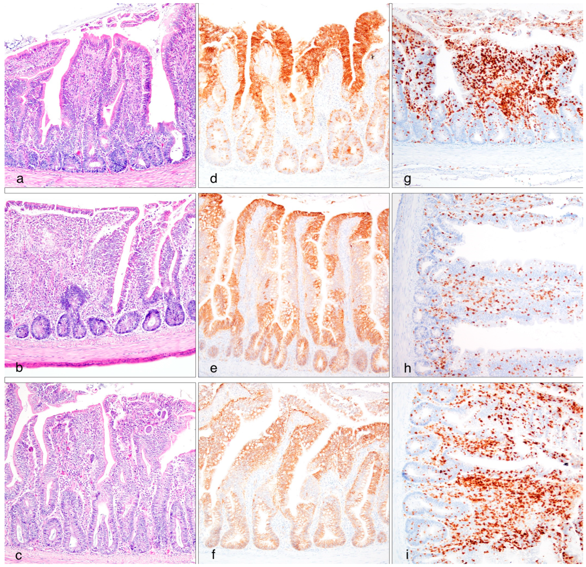
3.3. Histomorphometrical and Immunohistochemical Analysis

Table 4 and Figure 1 present data on the morphological characteristics of the duodenum, the jejunum, and the ileum. The gut segments evaluated include the villus height (VH), crypt depth (CD), and the ratio of villus height to crypt depth (VH/CD), which are indicative of intestinal health and function.
In the duodenum, the measurements of VH across the different treatment groups was relatively consistent, with minimal variation observed among the groups. Significant differences were observed in CD among the treatment groups. Crypt depth is an important indicator of epithelial cell turnover and gut health. In this study, the CD varied notably across the groups, with group CI showing a significantly higher value compared to all other groups, while groups PN, PI, and SI showed intermediate values. The CN had the lowest value. The ratio of VH to CD is another important parameter reflecting the functional capacity of the intestine. A higher VH/CD ratio typically indicates better nutrient absorption and overall gut health. In this study, the VH/CD ratio ranged from 3.23 to 7.89 across the treatment groups. Notably, group CI had the lowest ratio (3.23), while group CN had the highest (7.89).
Moving to the jejunum, the data reveal significant differences among the groups for VH. Broilers in the uninfected groups exhibited the highest villus heights, followed by the SI and PI groups. In contrast, the CI group had significantly lower villus height compared to all other groups. As for CD, the PI and SI groups had the deepest crypts. In contrast, the CN and PN groups exhibited significantly shallower crypts, and the CI group had intermediate values of CD. The villus height to crypt depth ratio tended to be higher in the uninfected groups compared to the infected groups, as expected.
Lastly, in the ileum, the CN, PN, and PI groups exhibited similar and significantly higher villus heights compared to those in the CI group, while the SI group gave intermediate values. In terms of crypt depth, the differences among the groups are also notable. The CN and PN groups had significantly shallower crypts compared to the PI and SI groups. The VH/CD ratio varied among the groups, with the CN, PN, and PI groups exhibiting higher ratios above 2, while CI and PI groups had lower ratios below 2.
Regarding the intensity and distribution pattern of Claudin-3 expression, as well as lymphocytic infiltration, no statistically significant differences were observed among the treatment groups.

3.4. Antioxidant Status Analysis

Table 5 presents the results of antioxidant analysis, specifically measuring malondialdehyde (MDA) levels and protein carbonyls, across different body parts (breast, thigh, intestine) of broilers on days 1 and 5 post sample collection.
For MDA levels in the breast and thigh muscles, there were no significant differences observed among the treatment groups on both day 1 and day 5. Similarly, in the intestine, MDA levels showed no significant variation among the treatment groups on day 1.
Regarding protein carbonyls, which are markers of protein oxidation, the analysis of breast and thigh muscles on day 1 post-sampling revealed no significant differences among the treatment groups. Similarly, there were no significant differences in thigh muscle protein carbonyls on day 1 post-sampling.

4. Discussion

Phytobiotics, also known as botanicals or phytogenics, have a rich history dating back centuries. Historically, various cultures worldwide have used plants and plant extracts for medicinal and health purposes in both humans and animals [29]. Ancient civilizations, such as those in Iran, Egypt, and India, documented the use of herbs and plant extracts for promoting health and treating diseases in livestock [30,31].
In recent decades, there has been a resurgence of interest in phytobiotics within the animal industry. This renewed focus is driven by several factors, including concerns about antibiotic resistance, consumer demand for natural and organic products, and stricter regulations regarding the use of antibiotics in animal feed [32,33].
The use of phytobiotics in animal feed aims to improve overall animal health, enhance feed efficiency, and boost performance [33]. Phytobiotics exert their effects through various mechanisms, including antimicrobial, antiparasitic, and anti-inflammatory properties [34,35,36].
In poultry production, phytobiotics are incorporated into feed formulations or administered directly to animals as dietary supplements. They have been shown to improve FI, nutrient absorption, gut health, and immune responses, leading to enhanced growth rates, FCR, and overall productivity [36,37]. The previous research aligns with the results of our experiment, where the group administered with the phytobiotic mixture exhibited superior overall performance parameters compared to the control negative group.
Moreover, phytobiotics have emerged as valuable tools in the management of broiler production, particularly in the face of challenges such as coccidiosis [38]. By enhancing the bird’s immune system and promoting gut health, phytobiotics can contribute to improved performance parameters in broilers, such as enhanced growth rates, improved FCR, and reduced mortality rates, even under the stress of coccidial challenge [38,39,40]. In our research, clear distinctions emerged among the infected groups, highlighting the superior performance of the phytobiotic mixture group compared to the control group. Remarkably, the group receiving the phytobiotic mixture exhibited outcomes surpassing or matching those of the salinomycin-supplemented group.
The observed pattern extends to OPG and CLS, where the PI group exhibited comparable OPG values to the uninfected groups on day seven and had lower numerical values compared to the SI group throughout the acute phase of the infection. Oikeh [41] investigated the effects of diet with lignocellulose produced from fresh spruce trees on OPG values in broiler chickens. The study found that there was a notable linear decrease in oocyst output with increasing levels of diet dilution at days 7, 8, and 9 pi. Additionally, the day post-infection significantly influenced OPG, with the highest values observed at day 7 pi compared to other days pi. Notably, in our experiment, the SI group exhibited higher lesions with scores significantly exceeding those of the uninfected groups. In contrast, the PI group showed no statistical difference from the uninfected groups. This phenomenon may be attributed to the protective properties of phytobiotics, as previously mentioned. Their antimicrobial, antiparasitic, and anti-inflammatory attributes contribute to alleviating the effects of Eimeria in the gut, thereby reducing lesions. Indeed, similar findings have been reported by other researchers, where phytobiotic-supplemented groups demonstrated milder lesion scores [9,42]. These factors collectively contribute to the anticoccidial index, with noteworthy results observed in the PI group, which exhibited an index comparable to the uninfected groups and significantly outperformed the SI group, characterized by a partially effective index. This phenomenon is not unprecedented; a study by Han et al. [43] showcased superior results with a polyherbal mixture compared to the coccidiostat Nicarbazin. Similarly, Chang et al. [44] administered garlic essential oil and diclazuril to separate infected groups with Eimeria tenella, where the garlic essential oil demonstrated comparable anticoccidial effects to diclazuril, albeit with less significant results.
Coccidiosis can significantly affect the histomorphometry of the duodenum, jejunum, and ileum in broilers. This condition often leads to villous atrophy, crypt hyperplasia, and inflammatory cell infiltration in the intestinal mucosa, resulting in reduced nutrient absorption and compromised intestinal integrity [45]. In our study, the phytobiotic mixture demonstrated enhanced efficacy in the ileum, as evidenced by the PI group exhibiting values comparable to those of the uninfected control groups. The ileum, the final segment of the small intestine, plays a crucial role in nutrient absorption. It absorbs nutrients such as vitamin B12, bile acids, and remaining nutrients not absorbed in the earlier segments. Additionally, it facilitates the absorption of water and electrolytes before passing the remaining contents to the large intestine [46]. Eimeria tenella is a parasite that predominantly targets the ileum in poultry, causing extensive damage to the intestinal lining. Its invasion leads to hemorrhage, inflammation, and necrosis, resulting in bloody diarrhea, decreased nutrient absorption, and compromised bird health. Our experiment yielded promising results, showcasing the remarkable efficacy of the phytobiotic mixture against Eimeria tenella. As a result, we observed significant improvements in the intestinal morphometry of the ileum, highlighting the potential of this treatment approach in poultry management.
Phytobiotics offer a natural solution to enhance the antioxidant status of chicken meat. These bioactive compounds, including essential oils, tannins, flavonoids, and saponins, possess inherent antioxidant properties [47]. Additionally, essential oils such as thymol and carvacrol, commonly present in phytobiotics, demonstrate significant antioxidant activity by inhibiting lipid peroxidation and protecting cellular membranes against oxidative damage [48]. In our study, there were no significant differences between the groups. This lack of effect could potentially be attributed to the coccidial challenge, which may have damaged the intestinal lining of the broilers, impairing the absorption of the bioactive compounds present in the phytobiotics. If these compounds were not absorbed adequately, their potential antioxidant effects may not have been realized systemically [1]. Another plausible explanation is related to the concentration and dosage of the phytobiotics provided in the diet. The effectiveness of phytobiotics often depends on their quantity, and it is possible that the amount included in the feed was insufficient to exert a measurable antioxidant effect under the conditions of this experiment [49]. These factors highlight the need for further investigation to better understand the mechanisms behind these results and optimize the application of phytobiotics in poultry nutrition.
In conclusion, the use of a specific blend of phytobiotics containing essential oils, saponins and tannins in poultry production holds great promise for enhancing animal health, performance, and productivity, particularly in the face of challenges such as coccidiosis. Our research findings align with previous studies, demonstrating the efficacy of phytobiotics in improving growth rates, feed efficiency, and immune responses while mitigating the detrimental effects of coccidial infection on gut health and performance parameters. The observed improvements in intestinal morphometry and antioxidant status underscore the potential of phytobiotics as natural alternatives to traditional antimicrobials and coccidiostats in poultry management. Overall, these findings contribute to the growing body of evidence supporting the use of phytobiotics as valuable tools for promoting sustainable and eco-friendly poultry production practices.

Author Contributions

Conceptualization, D.G., I.G., V.T., E.P. and J.B.-N.; validation, D.G., I.P., G.S. and I.S.; formal analysis, D.G., I.P., T.M., M.V., J.B.-N. and I.G.; investigation, D.G., T.M., E.P., V.T. and I.G.; resources, J.B.-N., V.T. and I.G.; data curation, D.G., I.P. and G.S.; writing—original draft preparation, D.G., J.B.-N., V.T. and I.G.; writing—review and editing, I.P., T.M., I.S., J.R., M.V. and D.B.; visualization, I.S., V.T. and I.G.; supervision, D.G., V.T. and I.G.; project administration and funding acquisition, I.G. All authors have read and agreed to the published version of the manuscript.

Funding

This research was funded by PATENT CO. DOO under research project nr74036 of the Research Committee of the Aristotle University of Thessaloniki.

Institutional Review Board Statement

All animals received humane care, whereby the procedures described herein cohered to the principles and guidelines of the EU regulations and were approved by the Ethical Committee of Animal Welfare of the Aristotle University of Thessaloniki and local Veterinary Authorities (authorization code 420364 (1913)–11/07/2023).

Informed Consent Statement

Not applicable.

Data Availability Statement

The data presented in this study are available on request from the corresponding author. The data are not publicly available due to privacy restrictions.

Conflicts of Interest

Authors Jog Raj, Marko Vasiljevic, and Jasna Bošnjak-Neumuller were employed by the company PATENT CO. DOO (Mišićevo, Serbia). These authors were not involved in the analysis of the results or the experimental design, and there were no influences on any aspects of the study or its presented data. The remaining authors declare that the research was conducted in the absence of any commercial or financial relationships that could be construed as a potential conflict of interest.

References

  1. Blake, D.P.; Knox, J.; Dehaeck, B.; Huntington, B.; Rathinam, T.; Ravipati, V.; Ayoade, S.; Gilbert, W.; Adebambo, A.O.; Jatau, I.D.; et al. Re-Calculating the Cost of Coccidiosis in Chickens. Vet. Res. 2020, 51, 115. [Google Scholar] [CrossRef] [PubMed]
  2. Aarthi, S.; Dhinakar Raj, G.; Raman, M.; Gomathinayagam, S.; Kumanan, K. Molecular Prevalence and Preponderance of Eimeria Spp. among Chickens in Tamil Nadu, India. Parasitol. Res. 2010, 107, 1013–1017. [Google Scholar] [CrossRef]
  3. Taylor, M.A.; Coop, R.L.; Wall, R.L. Veterinary Parasitology, 4th ed.; Wiley-Blackwell: Hoboken, NJ, USA, 2015; pp. 1–1006. [Google Scholar] [CrossRef]
  4. Snyder, R.P.; Guerin, M.T.; Hargis, B.M.; Kruth, P.S.; Page, G.; Rejman, E.; Rotolo, J.L.; Sears, W.; Zeldenrust, E.G.; Whale, J.; et al. Restoration of Anticoccidial Sensitivity to a Commercial Broiler Chicken Facility in Canada. Poult. Sci. 2021, 100, 663–674. [Google Scholar] [CrossRef]
  5. Mesa-Pineda, C.; Navarro-Ruíz, J.L.; López-Osorio, S.; Chaparro-Gutiérrez, J.J.; Gómez-Osorio, L.M. Chicken Coccidiosis: From the Parasite Lifecycle to Control of the Disease. Front. Vet. Sci. 2021, 8, 787653. [Google Scholar] [CrossRef]
  6. Jamil, M.; Aleem, M.T.; Shaukat, A.; Khan, A.; Mohsin, M.; Rehman, T.U.; Abbas, R.Z.; Saleemi, M.K.; Khatoon, A.; Babar, W.; et al. Medicinal Plants as an Alternative to Control Poultry Parasitic Diseases. Life 2022, 12, 449. [Google Scholar] [CrossRef]
  7. Abd El-Ghany, W.A. Phytobiotics in Poultry Industry as Growth Promoters, Antimicrobials and Immunomodulators-A Review. Sci. Publ. J. World Poult. Res. 2020, 10, 571–579. [Google Scholar] [CrossRef]
  8. Abdelli, N.; Solà-Oriol, D.; Pérez, J.F. Phytogenic Feed Additives in Poultry: Achievements, Prospective and Challenges. Animals 2021, 11, 3471. [Google Scholar] [CrossRef]
  9. Tsiouris, V.; Giannenas, I.; Bonos, E.; Papadopoulos, E.; Stylianaki, I.; Sidiropoulou, E.; Lazari, D.; Tzora, A.; Ganguly, B.; Georgopoulou, I. Efficacy of a Dietary Polyherbal Formula on the Performance and Gut Health in Broiler Chicks after Experimental Infection with Eimeria spp. Pathogens 2021, 10, 524. [Google Scholar] [CrossRef]
  10. Bozkurt, M.; Ege, G.; Aysul, N.; Akşit, H.; Tüzün, A.E.; Küçükyllmaz, K.; Borum, A.E.; Uygun, M.; Akşit, D.; Aypak, S.; et al. Effect of Anticoccidial Monensin with Oregano Essential Oil on Broilers Experimentally Challenged with Mixed Eimeria spp. Poult. Sci. 2016, 95, 1858–1868. [Google Scholar] [CrossRef]
  11. Mączka, W.; Twardawska, M.; Grabarczyk, M.; Wińska, K. Carvacrol—A Natural Phenolic Compound with Antimicrobial Properties. Antibiotics 2023, 12, 824. [Google Scholar] [CrossRef]
  12. Felici, M.; Tugnoli, B.; Ghiselli, F.; Massi, P.; Tosi, G.; Fiorentini, L.; Piva, A.; Grilli, E. In Vitro Anticoccidial Activity of Thymol, Carvacrol, and Saponins. Poult. Sci. 2020, 99, 5350–5355. [Google Scholar] [CrossRef]
  13. Sidiropoulou, E.; Marugán-Hernández, V.; Skoufos, I.; Giannenas, I.; Bonos, E.; Aguiar-Martins, K.; Lazari, D.; Papagrigoriou, T.; Fotou, K.; Grigoriadou, K.; et al. In Vitro Antioxidant, Antimicrobial, Anticoccidial, and Anti-Inflammatory Study of Essential Oils of Oregano, Thyme, and Sage from Epirus, Greece. Life 2022, 12, 1783. [Google Scholar] [CrossRef] [PubMed]
  14. Hikal, W.M.; Tkachenko, K.G.; Ahl, H.A.H.S.-A.; Sany, H.; Sabra, A.S.; Baeshen, R.S.; Bratovcic, A.; Hikal, W.M.; Tkachenko, K.G.; Ahl, H.A.H.S.-A.; et al. Chemical Composition and Biological Significance of Thymol as Antiparasitic. Open J. Ecol. 2021, 11, 240–266. [Google Scholar] [CrossRef]
  15. Lee, J.W.; Kim, D.H.; Kim, Y.B.; Jeong, S.B.; Oh, S.T.; Cho, S.Y.; Lee, K.W. Dietary Encapsulated Essential Oils Improve Production Performance of Coccidiosis-Vaccine-Challenged Broiler Chickens. Animals 2020, 10, 481. [Google Scholar] [CrossRef]
  16. Masyita, A.; Mustika Sari, R.; Dwi Astuti, A.; Yasir, B.; Rahma Rumata, N.; Emran, T.B.; Nainu, F.; Simal-Gandara, J. Terpenes and Terpenoids as Main Bioactive Compounds of Essential Oils, Their Roles in Human Health and Potential Application as Natural Food Preservatives. Food Chem. X 2022, 13, 100217. [Google Scholar] [CrossRef] [PubMed]
  17. Sahak, M.K.A.; Kabir, N.; Abbas, G.; Draman, S.; Hashim, N.H.; Hasan Adli, D.S. The Role of Nigella sativa and Its Active Constituents in Learning and Memory. Evid.-Based Complement. Altern. Med. 2016, 2016, 6075679. [Google Scholar] [CrossRef] [PubMed]
  18. Alfaro, D.M.; Silva, A.V.F.; Borges, S.A.; Maiorka, F.A.; Vargas, S.; Santin, E. Use of Yucca Schidigera Extract in Broiler Diets and Its Effects on Performance Results Obtained with Different Coccidiosis Control Methods. J. Appl. Poult. Res. 2007, 16, 248–254. [Google Scholar] [CrossRef]
  19. Tonda, R.M.; Rubach, J.K.; Lumpkins, B.S.; Mathis, G.F.; Poss, M.J. Effects of Tannic Acid Extract on Performance and Intestinal Health of Broiler Chickens Following Coccidiosis Vaccination and/or a Mixed-Species Eimeria Challenge. Poult. Sci. 2018, 97, 3031–3042. [Google Scholar] [CrossRef] [PubMed]
  20. Marzo, F.; Urdaneta, E.; Santidrián, S. Liver Proteolytic Activity in Tannic Acid-Fed Birds. Poult. Sci. 2002, 81, 92–94. [Google Scholar] [CrossRef] [PubMed]
  21. Pyrka, I.; Mantzouridou, F.T.; Nenadis, N. Optimization of Olive Leaves’ Thin Layer, Intermittent near-Infrared-Drying. Innov. Food Sci. Emerg. Technol. 2023, 84, 103264. [Google Scholar] [CrossRef]
  22. Velkers, F.C.; Blake, D.P.; Graat, E.A.M.; Vernooij, J.C.M.; Bouma, A.; de Jong, M.C.M.; Stegeman, J.A. Quantification of Eimeria Acervulina in Faeces of Broilers: Comparison of McMaster Oocyst Counts from 24 h Faecal Collections and Single Droppings to Real-Time PCR from Cloacal Swabs. Vet. Parasitol. 2010, 169, 1–7. [Google Scholar] [CrossRef] [PubMed]
  23. De Pablos, L.M.; Dos Santos, M.F.B.; Montero, E.; Garcia-Granados, A.; Parra, A.; Osuna, A. Anticoccidial Activity of Maslinic Acid against Infection with Eimeria Tenella in Chickens. Parasitol. Res. 2010, 107, 601–604. [Google Scholar] [CrossRef]
  24. Johnson, J.; Reid, W.M. Anticoccidial Drugs: Lesion Scoring Techniques in Battery and Floor-Pen Experiments with Chickens. Exp. Parasitol. 1970, 28, 30–36. [Google Scholar] [CrossRef] [PubMed]
  25. Gava, M.S.; de Moraes, L.B.; Carvalho, D.; Chitolina, G.Z.; Fallavena, L.C.B.; Moraes, H.L.S.; Herpich, J.; Salle, C.T.P. Determining the Best Sectioning Method and Intestinal Segment for Morphometric Analysis in Broilers. Rev. Bras. Cienc. Avic./Braz. J. Poult. Sci. 2015, 17, 145–150. [Google Scholar] [CrossRef]
  26. Chieh Sung, W.; Haryono, M. Quality Changes after Oven-Drying and Frozen Storage of Bluestripe Herring (Herklotsichthys quadrimaculatus). J. Food Nutr. Res. 2017, 5, 935–940. [Google Scholar] [CrossRef]
  27. Vyncke, W. Evaluation of the Direct Thiobarbituric Acid Extraction Method for Determining Oxidative Rancidity in Mackerel (Scomber scombrus L.). Fette Seifen Anstrichm. 1975, 77, 239–240. [Google Scholar] [CrossRef]
  28. Charan, J.; Kantharia, N. How to Calculate Sample Size in Animal Studies? J. Pharmacol. Pharmacother. 2013, 4, 303–306. [Google Scholar] [CrossRef]
  29. Grover, J.K.; Yadav, S.; Vats, V. Medicinal Plants of India with Anti-Diabetic Potential. J. Ethnopharmacol. 2002, 81, 81–100. [Google Scholar] [CrossRef]
  30. Tadjbakhsh, H. Traditional Methods Used for Controlling Animal Diseases in Iran. Rev. Sci. Tech. 1994, 13, 599–614. [Google Scholar] [CrossRef] [PubMed]
  31. Kikusato, M. Phytobiotics to Improve Health and Production of Broiler Chickens: Functions beyond the Antioxidant Activity. Anim. Biosci. 2021, 34, 345–353. [Google Scholar] [CrossRef]
  32. Pandey, S.; Kim, E.S.; Cho, J.H.; Song, M.; Doo, H.; Kim, S.; Keum, G.B.; Kwak, J.; Ryu, S.; Choi, Y.; et al. Cutting-Edge Knowledge on the Roles of Phytobiotics and Their Proposed Modes of Action in Swine. Front. Vet. Sci. 2023, 10, 1265689. [Google Scholar] [CrossRef] [PubMed]
  33. Pandian, S.C.; Krishna Murthy, G.T.; Rao, A.V. Antimicrobial Activity of Selected Phytobiotics Individually and in Combination against Gram Positive and Gram Negative Bacteria. J. Entomol. Zool. Stud. 2021, 9, 2255–2260. [Google Scholar]
  34. Skoufos, I.; Bonos, E.; Anastasiou, I.; Tsinas, A.; Tzora, A. Effects of Phytobiotics in Healthy or Disease Challenged Animals. Feed. Addit. Aromat. Plants Herbs Anim. Nutr. Health 2020, 311–337. [Google Scholar] [CrossRef]
  35. Mohiti-Asli, M.; Ghanaatparast-Rashti, M. Dietary Oregano Essential Oil Alleviates Experimentally Induced Coccidiosis in Broilers. Prev. Vet. Med. 2015, 120, 195–202. [Google Scholar] [CrossRef] [PubMed]
  36. Bortoluzzi, C.; Lumpkins, B.; Mathis, G.F.; França, M.; King, W.D.; Graugnard, D.E.; Dawson, K.A.; Applegate, T.J. Zinc Source Modulates Intestinal Inflammation and Intestinal Integrity of Broiler Chickens Challenged with Coccidia and Clostridium Perfringens. Poult. Sci. 2019, 98, 2211–2219. [Google Scholar] [CrossRef] [PubMed]
  37. Lee, Y.; Lu, M.; Lillehoj, H.S. Coccidiosis: Recent Progress in Host Immunity and Alternatives to Antibiotic Strategies. Vaccines 2022, 10, 215. [Google Scholar] [CrossRef]
  38. Pop, L.M.; Varga, E.; Coroian, M.; Nedisan, M.E.; Mircean, V.; Dumitrache, M.O.; Farczádi, L.; Fülöp, I.; Croitoru, M.D.; Fazakas, M.; et al. Efficacy of a Commercial Herbal Formula in Chicken Experimental Coccidiosis. Parasit. Vectors 2019, 12, 343. [Google Scholar] [CrossRef]
  39. Giannenas, I.A.; Florou-Paneri, P.; Botsoglou, N.A.; Christaki, E.; Spais, A.B. Effect of Supplementing Feed with Oregano and/or α-Tocopheryl Acetate on Growth of Broiler Chickens and Oxidative Stability of Meat. J. Anim. Feed Sci. 2005, 14, 521–535. [Google Scholar] [CrossRef]
  40. Aljedaie, M.M.; Al-Malki, E.S. Anticoccidial Activities of Salvadora Persica(Arak), Zingiber Officinale (Ginger) and Curcuma Longa (Turmeric) Extracts on the Control of Chicken Coccidiosis. J. King Saud Univ. Sci. 2020, 32, 2810–2817. [Google Scholar] [CrossRef]
  41. Oikeh, I.; Sakkas, P.; Taylor, J.; Giannenas, I.; Blake, D.P.; Kyriazakis, I. Effects of Reducing Growth Rate via Diet Dilution on Bone Mineralization, Performance and Carcass Yield of Coccidia-Infected Broilers. Poult. Sci. 2019, 98, 5477–5487. [Google Scholar] [CrossRef]
  42. Villar-Patiño, G.; del C. Camacho-Rea, M.; Olvera-García, M.E.; Baltazar-Vázquez, J.C.; Gómez-Verduzco, G.; Téllez, G.; Labastida, A.; Ramírez-Pérez, A.H. Effect of an Alliaceae Encapsulated Extract on Growth Performance, Gut Health, and Intestinal Microbiota in Broiler Chickens Challenged with Eimeria spp. Animals 2023, 13, 3884. [Google Scholar] [CrossRef]
  43. Han, M.; Hu, W.; Chen, T.; Guo, H.; Zhu, J.; Chen, F. Anticoccidial Activity of Natural Plants Extracts Mixture against Eimeria Tenella: An in Vitro and in Vivo Study. Front. Vet. Sci. 2022, 9, 1066543. [Google Scholar] [CrossRef] [PubMed]
  44. Chang, L.-Y.; Di, K.-Q.; Xu, J.; Chen, Y.-F.; Xi, J.-Z.; Wang, D.-H.; Hao, E.-Y.; Xu, L.-J.; Chen, H.; Zhou, R.-Y. Effect of Natural Garlic Essential Oil on Chickens with Artificially Infected Eimeria Tenella. Vet. Parasitol. 2021, 300, 109614. [Google Scholar] [CrossRef]
  45. Nabian, S.; Arabkhazaeli, F.; Seifouri, P.; Farahani, A. Morphometric Analysis of the Intestine in Experimental Coccidiosis in Broilers Treated with Anticoccidial Drugs. Iran J. Parasitol. 2018, 13, 493–499. [Google Scholar]
  46. Kiela, P.R.; Ghishan, F.K. Physiology of Intestinal Absorption and Secretion. Best Pract. Res. Clin. Gastroenterol. 2016, 30, 145. [Google Scholar] [CrossRef] [PubMed]
  47. Prakash, B.; Kumar, A.; Singh, P.P.; Songachan, L.S. Antimicrobial and Antioxidant Properties of Phytochemicals: Current Status and Future Perspective. Funct. Preserv. Prop. Phytochem. 2020, 1–45. [Google Scholar] [CrossRef]
  48. Chodkowska, K.A.; Abramowicz-Pindor, P.A.; Tuśnio, A.; Gawin, K.; Taciak, M.; Barszcz, M. Effect of Phytobiotic Composition on Production Parameters, Oxidative Stress Markers and Myokine Levels in Blood and Pectoral Muscle of Broiler Chickens. Animals 2022, 12, 2625. [Google Scholar] [CrossRef] [PubMed]
  49. Urban, J.; Kareem, K.Y.; Matuszewski, A.; Bień, D.; Ciborowska, P.; Lutostański, K.; Michalczuk, M. Enhancing Broiler Chicken Health and Performance: The Impact of Phytobiotics on Growth, Gut Microbiota, Antioxidants, and Immunity. Phytochem. Rev. 2024, 1–15. [Google Scholar] [CrossRef]
Figure 1. Left column: HE. Small intestine. (a) Villus shortening and surface and glandular epithelial hyperplasia, (b) villus blunting and fusion, surface and glandular epithelial hyperplasia, lymphoplasmacytic infiltrate in the lamina propria, (c) multiple apicomplexan coccidian forms in various developmental stages in the mucosa. Central column. (df) The immunohistochemical expression of Claudin-3 (on apical and basal regions and the pericellular borders of the epithelial cells) showed continuousity or differences in the spatial distribution, resulting in epithelial gaps. Right column. (gi) CD3+ positive cells. Magnification 10×.
Poultry 04 00004 g001
Table 1. Total phenolic content (mg GAE/g Feed) of the phytobiotic mixture and basal diet.
Samples
Phytobiotic MixtureBasal Diet
TPC359.1313.18
Table 2. Effect of a natural phytobiotic mixture on weekly performance arameters in broiler chickens before and after coccidial challenge (day 14) and over the entire experiment duration.
Age of ChickensParametersGroupsSEMp
CNPNCIPISI
Day 1BW44.342.544.143.644.70.40.322
Day 7BW120.1126.1121.4121.9123.02.00.909
BWG75.883.677.378.378.32.00.799
FI867.7857.7825.3836.7796.815.80.675
FCR1.171.041.111.071.030.030.636
Day 14BW320.8327.2326.3309.1336.05.60.651
BWG200.8201.1204.8187.2213.05.60.706
FI27142668.82770.82600.52892.256.50.577
FCR1.361.331.371.401.360.030.958
Day 21BW719.7 ab744.9 a637.7 bc624.8 c731.0 a11.9<0.001
BWG398.8 a417.8 a311.4 b315.7 b395.0 a8.1<0.001
FI5750.3 ab5926.2 a5356.8 bc5008.7 c5786.8 ab70.0<0.001
FCR1.45 b1.43 b1.73 a1.59 ab1.47 b0.030.006
Day 28BW1326.5 a1332.8 a1093.8 c1140.17 bc1263.8 ab21.0<0.001
BWG606.8 a587.9 a456.1 b515.4 ab532.8 ab13.0<0.001
Day 35BW2019.0 ab2066.9 a1721.4 c1843.8 bc1919.4 abc27.1<0.001
BWG692.5 ab734.08 a627.67 b703.58 ab655.67 ab12.00.041
FI21,038 a20,151.5 bc20,558.3 ab20,272.2 bc19,776.7 c107.9<0.001
FCR1.62 b1.53 b1.91 a1.66 b1.66 b0.03<0.001
Days 1–14BWG276.5284.7282.2265.5291.35.50.661
FI3581.73526.53596.23437.23689.052.60.747
FCR1.301.251.281.301.270.020.858
Days 14–35BWG1698.2 ab1739.8 a1395.2 c1534.7 b1583.4 ab24.6<0.001
FI26,788.3 a26,077.6 ab25,915.2 bc25,280.8 c25,563.5 bc120.8<0.001
FCR1.58 b1.50 b1.87 a1.65 b1.62 b0.03<0.001
Days 1–35BWG1974.7 ab2024.4 a1677.3 c1800.2 bc1874.8 abc27.0<0.001
FI30,370.0 a29,604.2 ab29,511.3 abc28,718.0 c29,252.5 bc133.2<0.001
FCR1.54 b1.47 b1.77 a1.60 b1.56 b0.02<0.001
BW: body weight. BWG: body weight gain. FI: feed intake. FCR: feed conversion ratio. SEM: standard error of mean. a,b,c Different letters denote significant (p ≤ 0.05) differences between treatments.
Table 3. Effect of a natural phytobiotic mixture after a coccidial challenge on oocysts per gram, coccidial lesion scores, and anticoccidial index post infection.
Days (Post Infection)CNPNCIPISISEMp-Value
OPG40 b0 b1900 a300 b200 b152.3<0.001
50 b0 b32000 a1850 b2050 b2343.4<0.001
60 b0 b150,500 a11,500 b14,500 b10,884.1<0.001
70 c0 c304,000 a21,000 bc27,500 b21,905.6<0.001
CLS0.0 a0.0 a3.46 b3.13 b5.17 c <0.001
ACI21200 a200 a93.36 c188.45 ab136.91 b10.1<0.001
OPG: oocysts per gram. CLS: coccidial lesion scores. ACI: anticoccidial index. SEM: standard error of mean. a,b,c Different letters denote significant (p ≤ 0.05) differences between treatments.
Table 4. Effect of a natural phytobiotic mixture after a coccidial challenge on the histomorphometry of the duodenum, jejunum, and ileum.
Gut Segment GroupsSEMp-Value
CNPNCIPISI
DuodenumVillus Height695.6742.2611.1648.0690.621.80.404
Crypt Depth88.2 c150.2 ab189.0 a148.2 ab126.2 ab8.0<0.001
VH/CD7.894.943.234.375.47
JejunumVillus Height558.2 a571.4 a436.1 b524.1 ab531.9 ab14.50.015
Crypt Depth97.4 c107.9 c134.4 b167.3 a152.7 ab5.9<0.001
VH/CD5.735.33.243.133.48
IleumVillus Height222.7 a232.2 a163.3 b232.7 a178.1 ab17.40.003
Crypt Depth81.9 c91.3 c100.0 bc106.3 b141.4 a4.6<0.001
VH/CD2.722.541.632.191.26
SEM: standard error of mean. a,b,c Different letters denote significant (p ≤ 0.05) differences between treatments.
Table 5. Effect of a natural phytobiotic mixture on antioxidant markers (malondialdehyde and protein carbonyls) in breast, thigh, and intestine tissues following a coccidial challenge, one and five days post-sampling.
Antioxidant AnalysisBody PartDay
(Post-Sampling)
GroupsSEMp-Value
CNPNCIPISI
MDAbreast17.15510.0597.3876.0987.0380.7470.527
517.78221.51217.9516.16517.53510.529
thigh113.45914.44912.07511.28213.4730.7720.722
526.13227.32125.53823.95326.841.3550.952
intestine157.28781.18159.86352.89871.4485.140.399
Protein Carbonylsbreast14.54.44.54.553.970.570.897
thigh13.993.943.893.843.910.1580.782
MDA: malondialdehyde. SEM: standard error of mean.
Disclaimer/Publisher’s Note: The statements, opinions and data contained in all publications are solely those of the individual author(s) and contributor(s) and not of MDPI and/or the editor(s). MDPI and/or the editor(s) disclaim responsibility for any injury to people or property resulting from any ideas, methods, instructions or products referred to in the content.

