Abstract
GSH (glutathione) is crucial for the removal and detoxification of carcinogens in healthy cells, while in cancer cells, GSH is associated with cancer expansion and increased resistance to drugs. O2•− acts as a secondary messenger and plays a major role in the cell signalling pathways of normal and cancer cells. Herein, the levels of O2•− and GSH were measured in MRC-5 and HCT-116 cells after incubation with BAL (Bifidobacterium animalis spp. lactis) and BAL/EALS (ethyl acetate extract of Laetiporus sulphureus) in co-culture systems, and for the first time, sensitivity was compared between these cell lines. The O2•− and GSH parameters were measured spectrophotometrically after 12 and 24 h. The levels of the O2•− were slightly increased in the MRC-5 cells after the effect of BAL and BAL/EALS (10 µg/mL), while the highest concentration of O2•− was recorded in treatment with BAL/EALS (50 µg/mL). On the other hand, the GSH values were elevated already after 12 h of incubation, and then further increased after 24 h in the MRC-5 cells. In the HCT-116 cells, the concentration of O2•− was not enhanced at 12 and 24 h of incubation compared to that of the control. The GSH level also remained relatively low. We observed a positive dose-dependent effect on the GSH levels in the MRC-5 and a negative dose-dependent effect in the HCT-116 cells. Generally, high GSH levels in the MRC-5 after 12 and 24 h indicate a strong reaction to oxidative stress and more sensitivity compared with the HCT-116 cells, where GSH stayed at a low concentration.
1. Introduction
Oxidative stress might induce genome instability and change the proliferation of healthy cells, resulting in cancer [1]. Oxidative stress can be caused by accumulated super-oxide anion radicals (O2•−), whose reactive nature is due to the presence of extra unpaired electrons. Compared to healthy cells, cancer cells have aberrant levels of O2•−. These cells have a higher O2•− set point than normal cells do, which supports their growth, proliferation, metastasis, and survival. However, low or extreme levels of O2•− lead to instability and cancer suppression, which is the main mechanism of conventional anticancer drugs [2,3]. One of the main defence mechanisms in normal cells against O2•− and reactive oxygen species (ROS), in general, is glutathione (GSH). GSH neutralizes ROS in several ways, by being included in the regeneration of enzymatic and non-enzymatic antioxidants or by direct neutralization. Although in healthy cells it is crucial for the regulation of oxidative stress, elevated GSH levels in cancer cells are usually associated with their progression, as well as increased resistance to treatment [2].
In this study, for the first time, we examined the levels of O2•− and GSH in MRC-5 and HCT-116 cells and compared their sensitivity after incubation with BAL and BAL/EALS treatments in co-culture systems.
2. Materials and Methods
The probiotic species Bifidobacterium animalis spp. lactis (strain BB-12) (BAL) was obtained in the Microbiology Laboratory, Institute for Information Technologies, University of Kragujevac, Serbia. The detailed preparation of the BAL suspension has been described by Muruzović et al. [4].
Colorectal cancer cells (HCT-116) and healthy human lung fibroblast cells (MRC-5) were obtained from ATCC (Manassas, VA, USA). The cell lines were cultured in standard Dulbecco’s modified Eagle’s minimal essential medium (DMEM) supplemented with 10% Foetal Bovine Serum (FBS) and antibiotics (100 U penicillin and 100 U/mL streptomycin).
The modified co-culture system was formed in 50 mL test tubes. A total of 40 µL of BAL diluted suspension was inoculated into 40 mL of sterile Mueller–Hinton soft agar (0.7%, w/v). Detailed instructions have been described in a study by Arsenijevic et al. [5].
Laetiporus sulphureus was gathered from the Šumadija area, Serbia (43°54′00.32″ N, 20°52′02.90″ E, Adžine Livade; altitude: 629 m). The identification and classification of the mushroom were performed with standard keys from the Mycological society “Šumadija” (Kragujevac, Serbia). Ethyl acetate solvent was used for extraction [5,6]. Ethyl acetate extract of L. sulphureus (EALS) was applied in two concentrations, 10 and 50 µg/mL.
GSH (reduced form of glutathione) was assessed by measuring the oxidation of the reduced form of GSH using sulfuric reagent DTNB (5,5′-dithiobis-(2-nitrobenzoic acid)). The levels of the O2•− were measured by an NBT assay. The NBT assay is based on the reduction of nitro-blue tetrazolium to nitro-blue formazan in the presence of O2•− [7]. The O2•− and GSH levels were measured spectrophotometrically after 12 and 24 h.
For statistical analysis, ANOVA (SPSS for Windows, version 17, 2008, Chicago, IL, USA) was used. A statistically significant difference was p < 0.05 *.
3. Results and Discussion
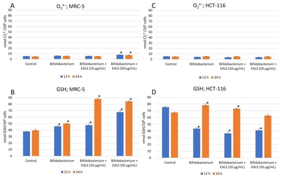
We detected a slightly elevated level of O2•− in the MRC-5 cells after incubation with the BAL and BAL/EALS (10 µg/mL) treatments, while a significant increase in O2•− was only observed in the treatment with BAL/EALS (50 µg/mL) (Figure 1A). However, the concentration of GSH was significantly elevated in all the treatments compared to that of the control. We noticed the positive dose-dependent effects of the treatments on the GSH parameters in the MRC-5 cell line (Figure 1B). When it comes to the HCT-116 cells, the O2•− levels remained almost unchanged, while the treatments induced a dose-dependent decrease in GSH (Figure 1C, D). Increased concentrations of GSH in the MRC-5 cells indicate the occurrence of oxidative stress and greater sensitivity of these cells to treatment. On the other hand, the HCT-116 cells showed greater resistance to the tested treatments, which can be concluded based on relatively low values of O2•− and GSH compared to those of the MRC-5 cells.

Figure 1.
Levels of O2•− and GSH in MRC-5 (A,B) and HCT-116 cells (C,D) after the treatment with BAL and BAL/EALS (10 and 50 µg/mL). * Statistical significance shows a difference between the control and treatments at 12 and 24 h of incubation.
4. Conclusions
The results of our study indicate the strong sensitivity of MRC-5 cells to the applied treatments compared to that of the HCT-116 cells that show resistance. This can be concluded from the high GSH values in the MRC-5 cells that activated the defence mechanism against oxidative stress.
Author Contributions
Conceptualization, D.A.; methodology, D.A. and K.P.; software, D.A.; validation, D.Š.; formal analysis, D.A. and D.Š.; investigation, D.A.; resources, K.M.; data curation, D.Š. and D.A.; writing—original draft preparation, D.A.; writing—review and editing, D.Š. and M.J.; visualization, D.A.; supervision, D.Š. and M.J. All authors have read and agreed to the published version of the manuscript.
Funding
The research was supported by the Ministry of Education, Science and Technological Development of the Republic of Serbia (agreement nos. 451-03-47/2023-01/200122, 175103, and 451-03-68/2023-14/200124).
Institutional Review Board Statement
Not applicable.
Informed Consent Statement
Not applicable.
Data Availability Statement
Data are contained within the article.
Conflicts of Interest
The authors declare no conflict of interest. The funders had no role in the design of the study; in the collection, analyses, or interpretation of data; in the writing of the manuscript; or in the decision to publish the results.
References
- Gašparović, Č.A.; Milković, L.; Rodrigues, C.; Mlinarić, M.; Soveral, G. Peroxiporins Are Induced upon Oxidative Stress Insult and Are Associated with Oxidative Stress Resistance in Colon Cancer Cell Lines. Antioxidants 2021, 10, 1856. [Google Scholar] [CrossRef] [PubMed]
- Kennedy, L.; Sandhu, J.K.; Harper, M.E.; Cuperlovic-Culf, M. Role of Glutathione in Cancer: From Mechanisms to Therapies. Biomolecules 2020, 10, 1429. [Google Scholar] [CrossRef] [PubMed]
- Sahoo, M.B.; Banik, K.B.; Borah, P.; Jain, A. Reactive Oxygen Species (ROS): Key Components in Cancer Therapies. Anti-Cancer Agents Med. Chem. 2022, 22, 215–222. [Google Scholar] [CrossRef] [PubMed]
- Muruzović, Ž.M.; Mladenović, G.K.; Stefanović, D.O.; Vasić, M.S.; Čomić, R.L.J. Extracts of Agrimonia eupatoria L. as sources of biologically active compounds and evaluation of their antioxidant, antimicrobial, and antibiofilm activities. J. Food Drug Anal. 2016, 24, 539–547. [Google Scholar] [CrossRef] [PubMed]
- Arsenijević, D.; Jovanović, M.; Pecić, K.; Grujović, M.; Marković, K.; Šeklić, D. Effects of Laetiporus sulphureus on Viability of HeLa Cells in Co-Culture System with Saccharomyces boulardii. Biol. Life Sci. Forum 2022, 18, 69. [Google Scholar]
- Jovanović, M.M.; Marković, K.G.; Grujović, M.Ž.; Pavić, J.; Mitić, M.; Nikolić, J.; Šeklić, D. Anticancer assessment and antibiofilm potential of Laetiporus sulphureus mushroom originated from Serbia. Food Sci. Nutr. 2023, 11, 6393–6402. [Google Scholar] [CrossRef] [PubMed]
- Šeklić, D.S.; Stanković, M.S.; Milutinović, M.G.; Topuzović, M.D.; Štajn, A.Š.; Marković, S.D. Cytotoxic, antimigratory, pro-and antioxidative activities of extracts from medicinal mushrooms on colon cancer cell lines. Arch. Biol. Sci. 2016, 68, 93–105. [Google Scholar] [CrossRef]
Disclaimer/Publisher’s Note: The statements, opinions and data contained in all publications are solely those of the individual author(s) and contributor(s) and not of MDPI and/or the editor(s). MDPI and/or the editor(s) disclaim responsibility for any injury to people or property resulting from any ideas, methods, instructions or products referred to in the content. |
© 2023 by the authors. Licensee MDPI, Basel, Switzerland. This article is an open access article distributed under the terms and conditions of the Creative Commons Attribution (CC BY) license (https://creativecommons.org/licenses/by/4.0/).