Proceeding Paper

Nicotiana benthamiana γ-Thionin Synthesis Is Induced in Response to Foreign Nucleus-Targeted Proteins †

by
Ekaterina Sheshukova
1,
Natalia Ershova
1,
Fedor Lipskerov
1 and
Tatiana Komarova
1,2,*
1
Vavilov Institute of General Genetics Russian Academy of Sciences, 119991 Moscow, Russia
2
Belozersky Institute of Physico-Chemical Biology, Lomonosov Moscow State University, 119991 Moscow, Russia
*
Author to whom correspondence should be addressed.
Presented at the 2nd International Electronic Conference on Plant Sciences—10th Anniversary of Journal Plants, 1–15 December 2021; Available online: https://iecps2021.sciforum.net/.
Biol. Life Sci. Forum 2022, 11(1), 11; https://doi.org/10.3390/IECPS2021-12006
Published: 30 March 2022

Abstract

:
Pathogenic and symbiotic bacteria secrete protein factors—nucleomodulins—to affect the host cell nucleus. During evolution, plants have acquired a great variety of defense mechanisms, including the synthesis of such antimicrobial peptides (AMPs) as defensins. We have demonstrated that the transient production of a foreign protein containing nuclear localization signal (NLS) in Nicotiana benthamiana leaves leads to the increased expression of the γ-thionin (NbγThio) that belongs to the defensin group of AMPs. We hypothesized that NbγThio is induced by the nucleomodulins of pathogenic bacteria and, in particular, in response to their NLSs. We used artificial nuclear proteins based on green fluorescent protein (GFP) fused with the human prothymosin α NLS or VirE3 NLS from Agrobacterium tumefaciens as mimetics of bacterial effectors. We demonstrated that the super-production of these NLS-containing reporters in the transient expression system in N. benthamiana leaves resulted in the increase in the NbγThio mRNA level. We isolated the NbγThio gene promoter (PrγThio) and created an expression vector (PrγThio-GUS) directing GUS synthesis in agroinfiltrated leaves. The co-expression of PrγThio-GUS with 35S-GFP:NLS variants led to the significant stimulation of GUS synthesis. We concluded that NbγThio gene expression is activated in response to bacterial nucleus-targeted proteins in the cell and is regulated both at the level of transcription and post-transcription stages.

1. Introduction

Many plant bacterial pathogens can affect the plant cell nucleus by means of the nucleomodulins—the effectors that reprogram the nucleus of the host cell [1]. Most of the nucleomodulins enter the host cell nucleus via the mechanism that is based on nuclear localization signal (NLS) recognition [2]. Plants have numerous lines of defense, among which are different antimicrobial peptides, including defensins. Plant γ-thionins, or defensins, are relatively small proteins that possess antifungal or antibacterial activities [3]. These proteins are allocated to a separate group of cis-thionins according to their primary and secondary structure and amino acid composition: they usually have a signal peptide and contain several cysteine residues forming disulfide bonds stabilizing the αβ (CSαβ) motif [4,5]. The mechanism of γ-thionins’ antibacterial effect is still not completely clear, but their amphipathic helix and disulfide bonds are believed to play an important role [4]. Here, we propose a model system in which leaf mesophyll cells perceive the artificial NLS-containing protein as a bacterial effector and, hence, as a signal of bacterial invasion. In response to the bacterial pathogen attack, the plant cell switches on defense mechanisms, including the induction of γ-thionin synthesis. We suggested that the induction of γ-thionin is performed via the activation of its transcription in response to NLS-containing foreign proteins.

2. Methods

2.1. Agroinfiltration

Genetic material was delivered into the cells of the fully expanded N. benthamiana leaves using the agroinfiltration approach. The Agrobacterium tumefaciens strain GV3101 was transformed with individual binary vectors and grown at 28 °C in LB medium supplemented with 50 mg/L rifampicin, 25 mg/L gentamycin and 50 mg/L carbenicillin/kanamycin. Agrobacterium overnight culture was diluted in 10 mM MES (pH 5.5) buffer supplemented with 10 mM MgSO4 and adjusted to a final OD600 of 0.1. Agroinfiltration was performed using a 2 mL syringe, after which the plants were grown under standard greenhouse conditions at 24 °C with a 16 h/8 h light/dark photoperiod.

2.2. GFP and mRFP Imaging

GFP and mRFP fluorescence was detected using an AxioVert 200M light fluorescence microscope (Carl Zeiss AG, Oberkochen, Germany) equipped with a Plan-Neofluar 100x NA 1.3 objective (Carl Zeiss AG, Oberkochen, Germany) and an ORCA-II ERG2 digital camera (Hamamatsu Photonics K.K., Hamamatsu City, Japan). The excitation and emission wavelength for GFP or mRFP were 488 nm and 584 nm, respectively, and the detection window was 493–607 nm.

2.3. GUS Activity Measurement

Plant material from infiltrated areas was harvested and ground in GUS extraction buffer (50 mM sodium phosphate, pH 7.0, 10 mM β-mercaptoethanol, 10 mM EDTA, 0.1% Triton X-100). The GUS enzymatic activity in plant extracts was estimated with the substrate 4-methylumbelliferyl-β-D-glucuronide (MUG). The fluorescence of the MUG cleavage product was analyzed with a Turner Quantech fluorimeter (Thermo Scientific Quantech, Philadelphia, PA, USA) using an NB455 (narrowband) excitation filter and an NB520 emission filter. All measurements were performed according to the previously described standard protocol [6].

2.4. Quantitative Real-Time PCR (qRT-PCR) Analysis of Transcript Concentrations

Total RNA was extracted from plant tissues using TriReagent (Molecular Research Center, Inc., Montgomery Rd Cincinnati, Cincinnati, OH, USA) according to the manufacturer’s instructions. The synthesis of the first strand, followed by a real-time qPCR, was performed as described in [7]. The real-time quantitative PCR was carried out using the iCycler iQ real-time PCR detection system (Bio-Rad, Laboratories, Inc., Hercules, CA, USA). Target genes were detected using sequence-specific primers for 18S rRNA (for normalization) and NbγThio and Eva Green master mix (Syntol, Moscow, Russia), according to the manufacturer’s instructions. The results of the RT-qPCR were evaluated using the Pfaffl algorithm [8].

3. Results

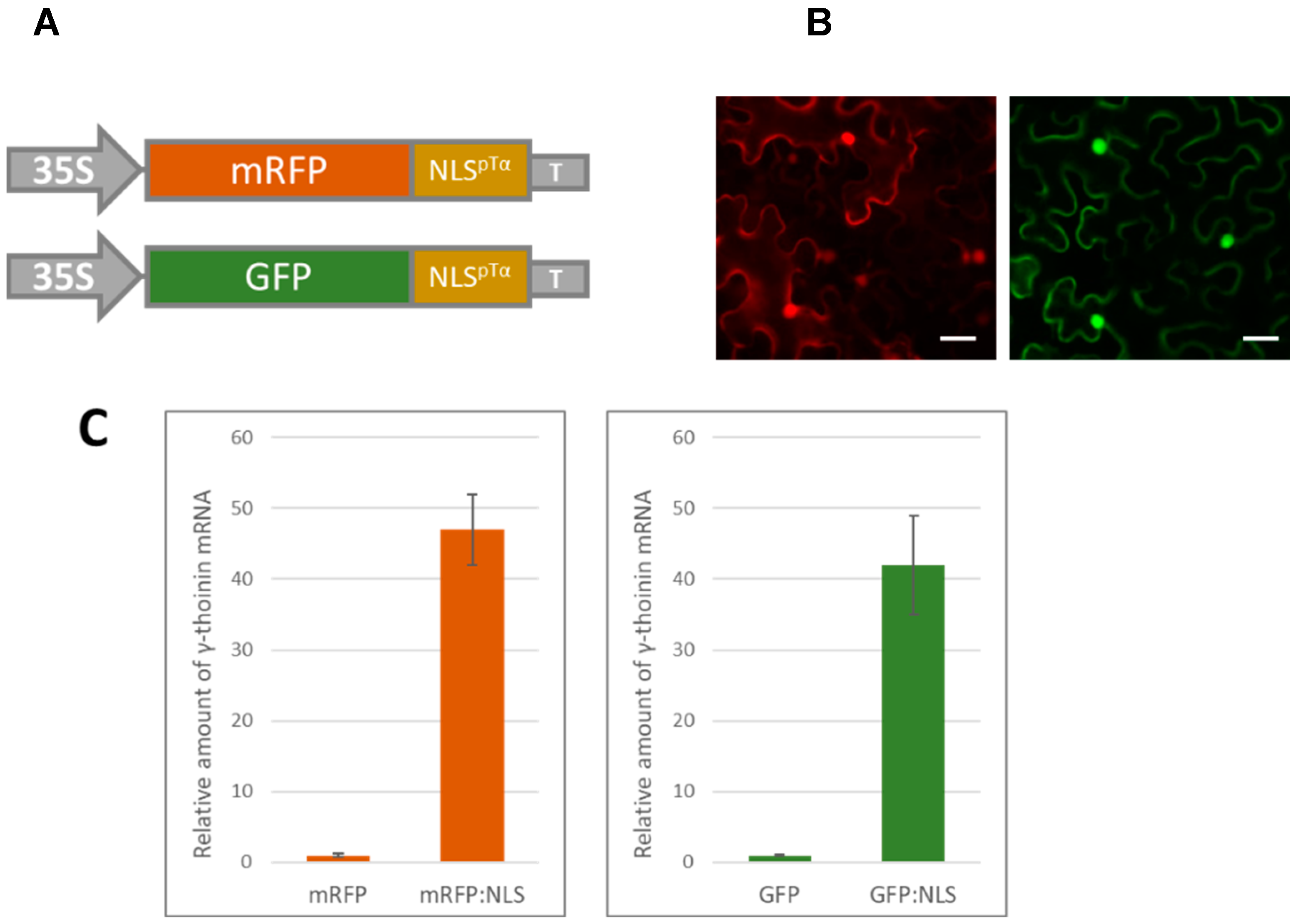
3.1. A Massive Synthesis of mRFP Fused with a Nuclear Localization Signal Stimulates γ-Thionin mRNA Accumulation

We selected a nuclear localization signal from human prothymosin α (pTα) (NLSpTα) to obtain an artificial model of NLS-containing reporter proteins based on an mRFP or GFP sequence. We created genetic constructs encoding mRFP:NLSpTα and GFP:NLSpTα fusion proteins (Figure 1A). Using an agrobacterium-mediated delivery of the genetic material for the transient expression, we have shown that NLSpTα effectively targets mRFP and GFP to the nucleus (Figure 1B).
We hypothesize that the super-production of a foreign nuclear protein can be recognized by the cell as a signal for the induction of pathogenesis-related (PR) gene expression, especially those that are activated in response to bacterial infection because NLS-containing proteins could be perceived by the plant cell as bacterial nucleomodulins, protein factors that are delivered to the nucleus to interfere with its functioning [9,10]. We demonstrated that the expression of an mRFP:NLSpTα-encoding construct in the leaves drastically stimulated the accumulation of N. benthamiana γ-thionin mRNA (Acc. FR686584.1) (Figure 1C, left). To exclude the effect of the reporter protein, we also checked the γ-thionin response to GFP:NLSpTα and assessed the level of γ-thionin mRNA in leaves 3 days after agroinfiltration with 35S-GFP:NLSpTα. We observed a comparable increase in γ-thionin expression in response both to mRFP:NLSpTα and GFP:NLSpTα (Figure 1C).

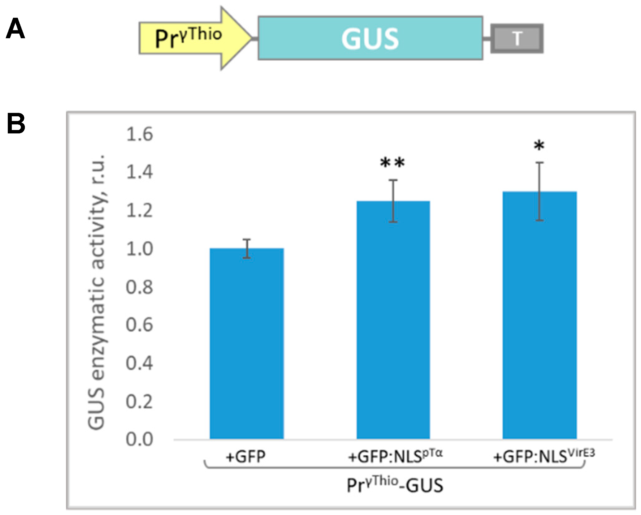
3.2. Isolation of the γ-Thionin Promoter (PrγThio) and Assessment of Its Sensitivity to the Intensive Accumulation of the Model Nuclear Protein

To identify the γ-thionin promoter (PrγThio), we used the “chromosome walking” approach and isolated the 1142-nucleotide sequence upstream of the γ-thionin gene, which we regarded as PrγThio (EMBL Acc. ERA1901858). In the next step, we created plant expression vectors containing a reporter gene encoding Escherichia coli β-glucuronidase (GUS) under the control of PrγThio (Figure 2). Here, we used GFP fused with NLSpTα or A. tumefaciens virulence protein E3 [11] NLS (NLSVirE3) to model the entrance of bacterial NLS-containing nucleomodulins. In accordance with our hypothesis regarding the stimulatory role of the foreign NLS-containing protein for γ-thionin, the co-expression of PrγThio-GUS with 35S-GFP:NLSpTα or 35S-GFP:NLSVirE3 should result in increased GUS accumulation. We extracted GUS from agroinfiltrated leaves at 3 dpi and assessed its enzymatic activity, which reflects PrγThio-GUS expression. Both GFP-NLSpTα and GFP:NLSVirE3 synthesis activated PrγThio and stimulated GUS production by about 20% (Figure 2), indicating that PrγThio is sensitive to the foreign nuclear proteins. However, the effect of NLS-containing reporter proteins on PrγThio-mediated mRNA accumulation is not as pronounced as was demonstrated for endogenous γ-thionin mRNA (Figure 1C). Thus, we suggested that γ-thionin gene expression is regulated both at the level of transcription and post-transcriptional stages.

4. Conclusions

We concluded that N. benthamiana γ-thionin expression is stimulated and PrγThio activity increases in response to the production of the foreign NLS-containing protein in the cell.

Author Contributions

E.S. and T.K. designed the research; E.S., T.K., N.E. and F.L. performed the research; E.S. and T.K. analyzed the data and wrote the paper. All authors have read and agreed to the published version of the manuscript.

Funding

This research was funded by the Russian Science Foundation (project No. 19-74-20031).

Institutional Review Board Statement

Not applicable.

Informed Consent Statement

Not applicable.

Data Availability Statement

Not applicable.

Conflicts of Interest

The authors declare no conflict of interest. The funders had no role in the design of the study; in the collection, analyses, or interpretation of data; in the writing of the manuscript, or in the decision to publish the results.

References

  1. Hanford, H.E.; Von Dwingelo, J.; Abu Kwaik, Y. Bacterial Nucleomodulins: A Coevolutionary Adaptation to the Eukaryotic Command Center. PLoS Pathog. 2021, 17, e1009184. [Google Scholar] [CrossRef]
  2. Le, L.H.M.; Ying, L.; Ferrero, R.L. Nuclear Trafficking of Bacterial Effector Proteins. Cell. Microbiol. 2021, 23, e13320. [Google Scholar] [CrossRef] [PubMed]
  3. Pelegrini, P.B.; Franco, O.L. Plant γ-Thionins: Novel Insights on the Mechanism of Action of a Multi-Functional Class of Defense Proteins. Int. J. Biochem. Cell Biol. 2005, 37, 2239–2253. [Google Scholar] [CrossRef] [PubMed]
  4. Sathoff, A.E.; Samac, D.A. Antibacterial Activity of Plant Defensins. Mol. Plant-Microbe Interact. 2019, 32, 507–514. [Google Scholar] [CrossRef] [PubMed]
  5. Höng, K.; Austerlitz, T.; Bohlmann, T.; Bohlmann, H. The Thionin Family of Antimicrobial Peptides. PLoS ONE 2021, 16, e0254549. [Google Scholar] [CrossRef] [PubMed]
  6. Jefferson, R.A.; Kavanagh, T.A.; Bevan, M.W. GUS Fusions: Beta-Glucuronidase as a Sensitive and Versatile Gene Fusion Marker in Higher Plants. EMBO J. 1987, 6, 3901–3907. [Google Scholar] [CrossRef] [PubMed]
  7. Dorokhov, Y.L.; Komarova, T.V.; Petrunia, I.V.; Frolova, O.Y.; Pozdyshev, D.V.; Gleba, Y.Y. Airborne Signals from a Wounded Leaf Facilitate Viral Spreading and Induce Antibacterial Resistance in Neighboring Plants. PLoS Pathog. 2012, 8, e1002640. [Google Scholar] [CrossRef] [PubMed] [Green Version]
  8. Pfaffl, M.W. A New Mathematical Model for Relative Quantification in Real-Time RT–PCR. Nucleic Acids Res. 2001, 29, e45. [Google Scholar] [CrossRef] [PubMed]
  9. Bierne, H.; Cossart, P. When Bacteria Target the Nucleus: The Emerging Family of Nucleomodulins. Cell. Microbiol. 2012, 14, 622–633. [Google Scholar] [CrossRef] [PubMed]
  10. Bierne, H.; Pourpre, R. Bacterial Factors Targeting the Nucleus: The Growing Family of Nucleomodulins. Toxins 2020, 12, 220. [Google Scholar] [CrossRef] [PubMed] [Green Version]
  11. Lacroix, B.; Vaidya, M.; Tzfira, T.; Citovsky, V. The VirE3 Protein of Agrobacterium Mimics a Host Cell Function Required for Plant Genetic Transformation. EMBO J. 2005, 24, 428–437. [Google Scholar] [CrossRef] [PubMed] [Green Version]
Figure 1. Foreign NLS-containing protein stimulates γ-thionin mRNA accumulation in N. benthamiana leaves: (A) schematic representation of genetic constructs encoding mRFP:NLSpTα and GFP:NLSpTα; (B) fluorescent images of epidermal cells of N. benthamiana leaves agroinfiltrated with 35S-mRFP:NLSpTα (left) or 35S-GFP:NLSpTα (right), scale bar = 20 μm; (C) relative amount of γ-thionin mRNA in leaves 3 days after agroinfiltration with 35S-mRFP or 35S-mRFP:NLSpTα (left) and 35S-GFP or 35S-GFP:NLSpTα (right) and quantified via qRT-PCR. p < 0.001 (Student’s t-test) for statistical significance of the difference compared to the control (mRFP or GFP).
Blsf 11 00011 g001
Figure 2. An artificial nuclear protein based on a GFP:NLSpTα or A. tumefaciens VirE3 (GFP:NLSVirE3) stimulates γ-thionin promoter (PrγThio)-directed GUS synthesis (PrγThio-GUS). (A) Schematic representation of the 1142 nt PrγThio-based vector encoding GUS; (B) comparative analysis of GUS activity in leaves 3 days after co-agroinfiltration with PrγThio-GUS and 35S-based vectors encoding GFP, GFP:NLSpTα, or GFP:NLSVirE3. The fluorescence detected for the combination of PrγThio-GUS and 35S-GFP is taken as 1. Standard error bars are indicated. * p < 0.05, ** p < 0.01 (Student’s t-test).
Blsf 11 00011 g002
Publisher’s Note: MDPI stays neutral with regard to jurisdictional claims in published maps and institutional affiliations.

