Case Report

Navigating Rarity: Pathological Challenges and Diagnostic Ambiguity in Rare Gliomas—A Case Series with a Focus on Personalized Treatment and Quality of Life

1
Department of Neurosurgery, BKH Günzburg at Ulm University, Lindenallee 2, 89312 Günzburg, Germany
2
Department of Neuropathology, University Clinic Heidelberg, 69120 Heidelberg, Germany
3
Department of Neuroradiology, BKH Günzburg at Ulm University, Lindenallee 2, 89312 Günzburg, Germany
4
Department of Neuropathology, BKH Günzburg at Ulm University, Lindenallee 2, 89312 Günzburg, Germany
5
Department of Neurology, University and Rehabilitation Clinic, Oberer Eselsberg 45, 89081 Ulm, Germany
*
Author to whom correspondence should be addressed.
Submission received: 3 May 2025 / Revised: 4 June 2025 / Accepted: 8 June 2025 / Published: 10 June 2025

Simple Summary

This case series explores four patients with rare gliomas treated at the University Hospital, Ulm, highlighting the challenges of diagnosis and treatment despite advancements in molecular classification per WHO 2021 guidelines. Each case demonstrates the importance of personalized care and ongoing diagnostic reevaluation. One patient experienced a 12-year evolution from pleomorphic xanthoastrocytoma to a tumor with PATZ1 fusion, achieving excellent outcomes after multiple surgeries. Another case showed long-term survival and improved quality of life in a young woman with astroblastoma. A third patient survived over 20 years, with 10 years therapy-free, after transformation from oligodendroglioma to glioblastoma. The fourth case involved a misdiagnosis corrected through molecular testing, revealing a rare astrocytoma in a patient with neurofibromatosis type 1. These cases emphasize the value of precise diagnostics, individualized treatments, and multidisciplinary approaches in managing rare gliomas while addressing the psychological burden of shifting diagnoses.

Abstract

Gliomas are incurable, heterogeneous brain tumors, with rare forms often constituting diagnostic and treatment challenges. Molecular diagnostics, mainly implemented through the World Health Organization (WHO) 2021 guidelines, have refined the classification, but highlight difficulties in diagnosing rare gliomas remain. This case series analyzes four patients with rare gliomas treated at the University Hospital, Ulm, between 2002 and 2024. Patients were selected based on unique histopathological features and long-term clinical follow-up. Clinical records, imaging, and histological data were reviewed. Molecular diagnostics followed WHO 2021 guidelines. Quality of life was assessed using standardized tools including the EQ-5D-5L, EQ VAS, the Distress Thermometer, and the Montreal Cognitive Assessment (MoCA). In the first case, a 51-year-old male’s diagnosis evolved from pleomorphic xanthoastrocytoma to a high-grade glioma with pleomorphic and pseudopapillary features, later identified as a neuroepithelial tumor with a PATZ1 fusion over 12 years. Despite multiple recurrences, extensive surgical interventions led to excellent outcomes. The second case involved a young female with long-term survival of astroblastoma, demonstrating significant improvements in both longevity and quality of life through personalized care. The third case involved a patient with oligodendroglioma, later transforming into glioblastoma, emphasizing the importance of continuous diagnostic reevaluation and adaptive treatment strategies, contributing to prolonged survival and quality of life improvements. Remarkably, the patient has achieved over 20 years of survival, including 10 years of being both therapy- and progression-free. The fourth case presents a young woman with neurofibromatosis type 1, initially misdiagnosed with glioblastoma based on histopathological findings. Subsequent molecular diagnostics revealed a subependymal giant cell astrocytoma-like astrocytoma, highlighting the critical role of early advanced diagnostic techniques. These cases underscore the importance of precise molecular diagnostics, individualized treatments, and ongoing diagnostic reevaluation to optimize outcomes. They also address the psychological impact of evolving diagnoses, stressing the need for comprehensive patient support. Even in complex cases, extensive surgical interventions can yield favorable results, reinforcing the value of adaptive, multidisciplinary strategies based on evolving tumor characteristics.

1. Introduction

Gliomas are among the most complex and heterogeneous brain tumors, encompassing a spectrum from slow-growing low-grade lesions to highly aggressive high-grade malignancies [1]. While significant research has focused on more common glioma subtypes [1,2,3], rare gliomas remain poorly understood due to their low incidence, diagnostic ambiguity, and the lack of standardized treatment guidelines [4].
These tumors frequently exhibit unique clinical, radiological, and molecular features that challenge even experienced clinicians and pathologists. Although the 2021 World Health Organization (WHO) classification system and the updates from the cIMPACT-NOW consortium have introduced integrated biomolecular diagnostics to improve tumor classification, rare gliomas often continue to defy conventional categorization [4,5,6].
Molecular tools such as DNA methylation profiling and next-generation sequencing (NGS) have helped identify novel entities, yet diagnostic uncertainty remains high, particularly for tumors with overlapping or atypical features [7,8,9]. Despite advances in imaging and molecular diagnostics, diagnosing gliomas remains a complex challenge, particularly for rare forms that often exhibit atypical features and overlapping characteristics with other tumor types. Importantly, rare gliomas are typically incurable, shifting the therapeutic focus toward symptom control, functional preservation, and quality of life (QoL) [10]. QoL is a multidimensional construct that includes physical, psychological, cognitive, and social aspects, as well as disease- and treatment-related symptoms [11,12,13]. In neuro-oncology, QoL has become a vital outcome measure alongside survival metrics, particularly in the management of rare and incurable tumors [10,12,14,15]. As a result, QoL has become an important outcome measure in clinical trials, alongside traditional metrics like overall survival, progression-free survival, and treatment response. Recent molecular profiling studies have revealed a far greater genetic and epigenetic diversity in central nervous system (CNS) tumors than previously appreciated through histopathological methods alone. These insights are particularly relevant for rare gliomas, which often display morphological features that hinder accurate classification based solely on histology [8,16]. Furthermore, the rarity of specific glioma variants means that there are often limited clinical data to guide treatment protocols, making it imperative for clinicians to consider the unique presentation and evolving nature of each case [9].
In this case series, we present detailed clinical, imaging, and molecular data from four patients with rare gliomas treated at our institution. These cases exemplify the diagnostic and therapeutic challenges posed by rare gliomas and highlight the critical role of precise classification, personalized treatment, and longitudinal quality-of-life assessment in optimizing patient outcomes.

2. Methodology

This retrospective case series analyzes four patients with rare gliomas treated at the Neuro-Oncology Center of the University Hospital, Ulm, between 2002 and 2024. Cases were selected based on diagnostic complexity, rare histopathological and molecular profiles, distinct treatment responses, and comprehensive clinical documentation. A multidisciplinary team, including neurosurgery, neuropathology, neurology, and radiotherapy, ensured coordinated and individualized care, supported by regular tumor board discussions.
Inclusion criteria comprised: (1) a confirmed glioma diagnosis with rare or ambiguous features; (2) treatment and longitudinal follow-up at our institution; (3) availability of detailed histological and molecular diagnostic data; and (4) informed patient consent. Clinical, imaging, and histopathological data—including demographics, treatment histories, and follow-up results—were extracted from medical records.
Histological examinations were conducted in accordance with the WHO 2021 classification system, following integrated histomolecular diagnostic approaches [5,6,17]. Tumor classification was supported by DNA methylation profiling and next-generation sequencing (NGS), performed either at the Department of Neuropathology of the University Hospital, Ulm, or the University Clinic, Heidelberg.
Quality of life (QoL) was assessed at regular intervals using validated tools: the European Quality of Life 5 Dimensions 5-Level Version (EQ-5D-5L), the EQ Visual Analog Scale (EQ VAS) [18], the Distress Thermometer (0–10 scale) [19], and the Montreal Cognitive Assessment (MoCA) [20]. Due to the clinical heterogeneity of the cases, follow-up schedules were individualized to each patient’s disease course and treatment trajectory.
Ethical approval for this study was obtained from the Ethics Committee at Ulm University (approval code: 381/15, date: 19 February 2016). All patients provided written informed consent for the use of their anonymized data in research and publication.

3. Case Series

3.1. Case 1: Pleomorphic Xanthoastrocytoma to High-Grade Glioma with Pleomorphic and Pseudopapillary Features (HPAP) to Neuroepithelial Tumor with PATZ1 Fusion

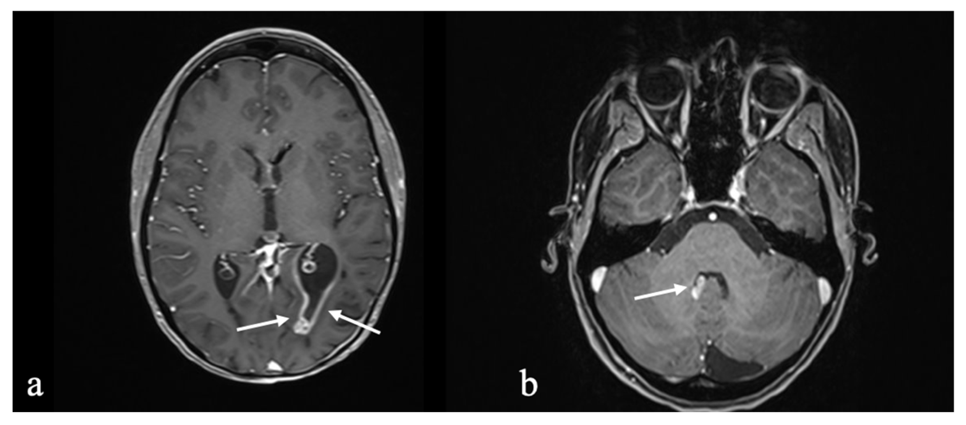
3.1.1. Initial Diagnosis

A 51-year-old healthy male presented in 2012 with mild left-sided hemiparesis, which he first noticed during a skiing holiday. His medical history was unremarkable, and magnetic resonance imaging (MRI) demonstrated a sizeable intra-axial tumor mass in the right frontal lobe. In 2012, the patient underwent a right frontal craniotomy with gross total resection (GTR) of the tumor, initially diagnosed as pleomorphic xanthoastrocytoma (PXA), CNS WHO grade 2. Follow-up visits and MRIs were conducted every 3–6 months, with no adjuvant treatment.

3.1.2. Molecular Profiling and Treatment Timeline

Following initial tumor control, recurrences were observed in 2014 and 2015, leading to further surgeries. Between 2016 and 2021, the patient experienced further tumor progression, leading to reclassification in 2021 as anaplastic pleomorphic xanthoastrocytoma (APXA), CNS WHO grade 3. Due to rapid progression and multifocal growth, advanced diagnostic techniques, including DNA-methylation profiling and next-generation sequencing (NGS), with the Heidelberg Tumor classifier version 12.5, were employed. Based on the tumor classifier, the brain tumor could not be assigned to a preexisting class of brain tumors. Therefore, it was diagnosed as glioma, not elsewhere classified (NEC). Considering the histopathologic features and the epigenetic molecular profile, the integrated analysis ultimately described the tumor as a high-grade glioma with pleomorphic and pseudopapillary features (HPAP), moving away from the initial diagnosis of PXA. Its molecular markers are summarized in Table 1. The patient underwent several interventions, including CyberKnife therapy in 2019 (18 Gy) and six cycles of temozolomide chemotherapy, initially maintaining a Karnofsky performance score (KPS) of 90%. However, in October 2022, he experienced multifocal tumor progression, necessitating another extensive surgery, which was the sixth operation in total. The samples taken during the last surgery (Figure 1) were analyzed molecularly, and the results could not be correlated with those in previous surgeries. As a result of further developments of the Heidelberg brain tumor classifier (version 12.7) and integrating data of RNA sequencing, at this time, the diagnosis of a neuroepithelial tumor with PATZ1 fusion was established (Table 1). The tissue received further work-up; however, no rationale for targeted therapy could be found for this patient. The patient’s condition improved significantly following this procedure due to massive tumor mass reduction. He recovered completely from left-sided hemiparesis, allowing him to return to work within three months after surgery. Six months after the most recent surgery, three small recurrences adherent to the dura were detected, which were again treated with CyberKnife therapy in September 2023 (1 × 20 Gy right temporal and parietal) and March 2024 (5 × 6 Gy right high parietal) due to the lack of available chemotherapy options. Conventional radiation therapy was deemed too toxic due to the large irradiation volume.

3.1.3. Long-Term Outcome and Life Quality

After the most recent surgery in 2022, the patient experienced significant clinical improvement due to substantial tumor mass reduction. In this case, radical surgical therapy, including resection of the infiltrated dura, was an individual decision because radio- and chemotherapy led to progression. Despite multiple recurrences, extended tumor resections, and complex tumor reclassifications, the patient has maintained a long-term survival and excellent functional recovery, returning to work three months after his last surgery with a current Karnofsky performance status (KPS) scale of 90%.
The patient’s QoL was regularly monitored using the distress questionnaire. Throughout the disease, the patient’s perceived burden increased from an initial 2/10 (reported twice in 2015) to 6/10 (in 2021 and 2022) during a period marked by significant tumor progression, neurological deterioration, and subsequent tumor resection. However, during the most recent assessment in December 2024, the distress level had decreased to 1/10. At this point, the patient’s MRI indicated tumor control, and he can maintain normal daily functioning. In the EQ-5D-5L questionnaire completed in December 2024, the patient reported no problems in the dimensions of mobility, self-care, or anxiety. Only mild issues (2/5) were noted in the daily activities and pain domains, and overall health status was rated at 90% on the EQ Visual Analog Scale (VAS).
A summary of the clinical, diagnostic, therapeutic, and quality-of-life (QoL) data of all four patients included in this case series is presented in Table 2.

3.2. Case 2: Astroblastoma

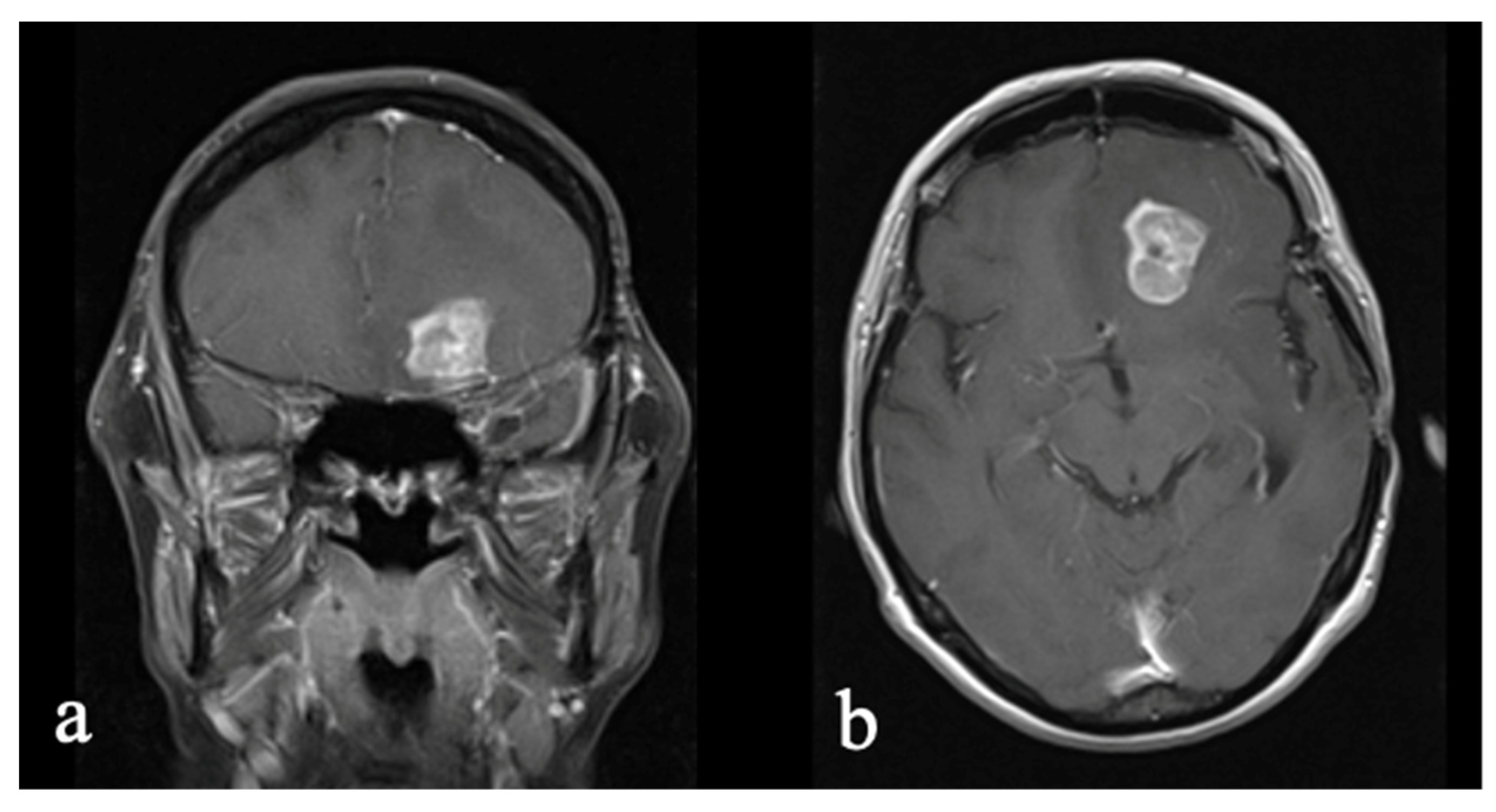
3.2.1. Initial Diagnosis

This case presents the long-term course of a now 30-year-old woman diagnosed with an astroblastoma at the age of 4 years. The initial symptom was a visual disorder in the left eye, detected during a routine school enrollment exam. Referral to an ophthalmologist led to imaging for suspected left optic nerve involvement. Subsequently, the patient accidentally hit a wall, resulting in headaches, nausea, and vomiting. For urgent further evaluation, an emergency computer tomography (CT) scan and MRI revealed a 5 × 5 × 6 cm heterogeneous, contrast-enhancing, well-demarcated mass with a basal cystic component. At this time, the patient exhibited no neurological symptoms, although she did have blurred vision in her left eye, prompting a GTR.

3.2.2. Molecular Profiling and Treatment Timeline

Initially, a necrotic malignant tumor was identified; three weeks later, a malignant papillary meningioma was considered a differential diagnosis. However, after a positive glial fibrillary acidic protein (GFAP) staining, an astroblastoma with papillary features was deemed more likely. One month later, the diagnosis of an astroblastoma with rhabdoid transformation was confirmed. At that time, there was no WHO classification for this tumor, but it was estimated to be at least WHO grade 3.
One year later, the patient underwent radiochemotherapy according to glioblastoma treatment protocols. She remained tumor-free for 15 years, after which a recurrence occurred in the same area, with the spinal axis showing no evidence of tumor spread. Neurologically, the patient reported a tendency to fall to the left side and mild right hemiparesis, rated 4/5 on the Medical Research Council (MRC) scale. Again, a gross total resection (GTR) was performed, complemented by adjuvant temozolomide chemotherapy, and the hemiparesis fully regressed.
Nineteen months later, a marginal dural lesion raised suspicion for recurrence. Following resection, histochemical analysis revealed a granulating, scarring lesion with no evidence of regression. Only six months later, in November 2018, MRI showed a new contrast enhancement. After resection, histopathological analysis confirmed a relapse. In January 2021, progression into the fourth ventricle was detected and treated with proton irradiation. Two years later, an MRI revealed four progressive intraventricular metastases, two of which were resected via endoscopic surgery. Nonresectable metastases are shown in Figure 2. EPIC array and next-generation sequencing confirmed the diagnosis of astroblastoma, MN1-altered (Table 3). No neurological deficits occurred. Since 2016, astroblastomas have been classified as part of “other gliomas” following changes to the WHO guidelines.

3.2.3. Long-Term Outcome and Life Quality

The patient has lived with the diagnosis for over 25 years, undergoing multiple surgeries and treatments. Despite multiple recurrences, her long-term survival is remarkable. QoL and cognitive functions were assessed via the EQ-5D-5L questionnaire, EQ-VAS, and MoCA before and after her last surgery in July 2024. The patient experienced a mild decline in MoCA scores, from 27/30 to 24/30 points, particularly regarding memory retention. In terms of QoL, she reported no impairments in daily activities, including pain and anxiety.
In conclusion, the patient showed minimal cognitive decline and reported no impairments in daily activities despite multiple surgeries and treatments, indicating a high quality of life. Despite experiencing severe wound-healing disorders and multiple revision and reconstructive surgeries after the latest tumor resection, the patient reports no burden in the current distress monitoring, either in her daily life or physically or emotionally (zero on a scale from zero to 10), confirmed in the EQ-5D-5L questionnaire and EQ VAS, where she ranks her current healthiness as a maximum.

3.3. Case 3: Oligodendroglioma to Glioblastoma

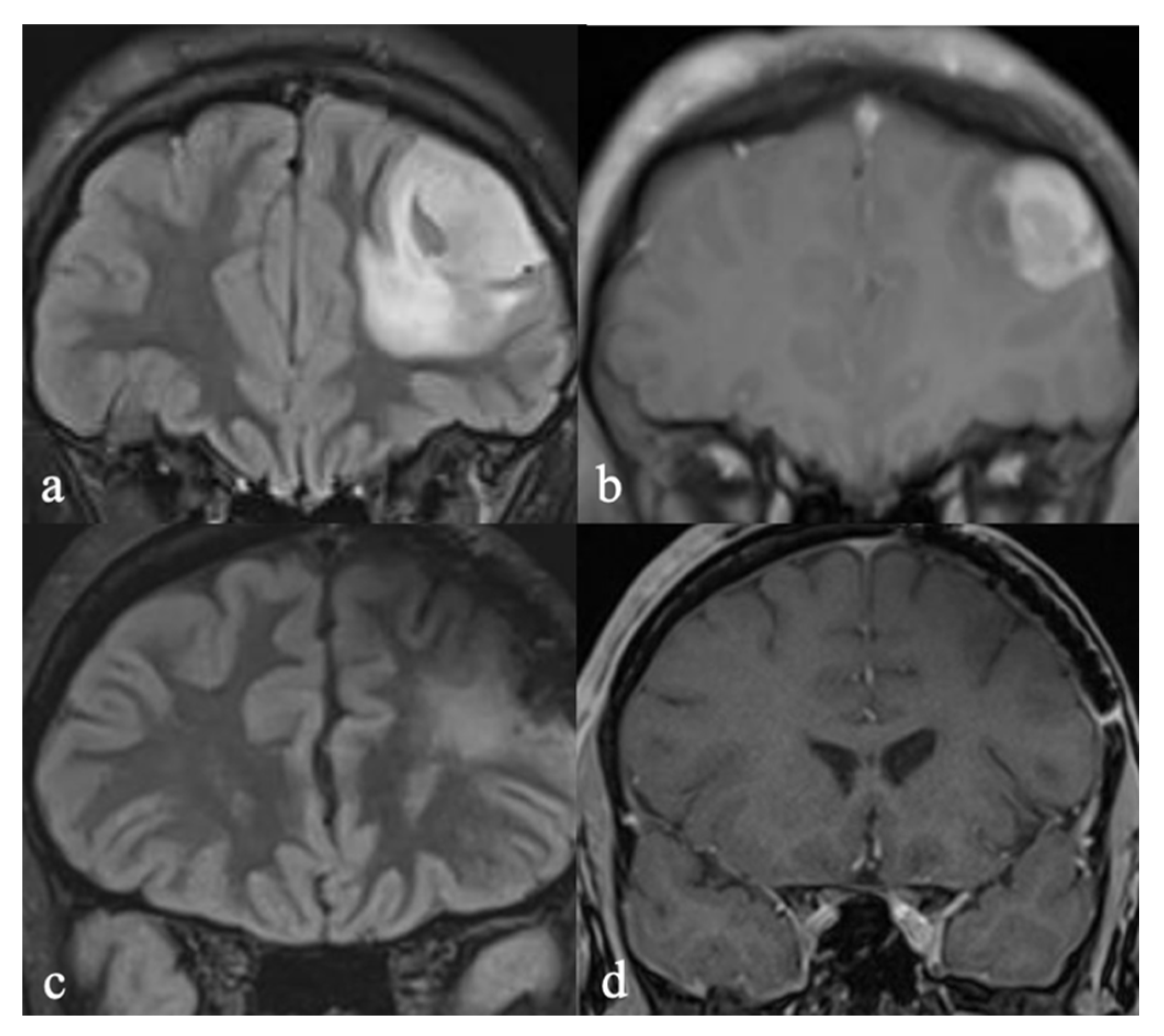
3.3.1. Initial Diagnosis

In 2002, a 40-year-old woman underwent surgery for a left temporal lobe glioma. After histopathological analysis, the tumor was diagnosed and classified as an anaplastic oligodendroglioma, WHO grade 3. She received adjuvant radiochemotherapy with temozolomide according to the NOA-04 study design [21]. After a progression-free interval of nearly nine years, multiple resections and adjuvant chemotherapy with temozolomide were performed when the tumor recurred.

3.3.2. Molecular Profiling and Treatment Timeline

In 2014, the histopathological diagnosis changed from oligodendroglioma to glioblastoma multiforme with oligodendroglial components, WHO grade 4 with IDH mutation, MGMT 11% methylated—a diagnosis that no longer exists under the latest WHO classification [5,17]. The diagnosis was rightly abandoned. This example illustrates that the patient’s tumor-free survival for over 10 years reflects a distinctly different clinical behavior between what was formerly labeled as GBM with oligodendroglial components and oligodendroglioma grade 4. Figure 3 shows the recurrent tumor in 2014, and Figure 4 the current follow-up image.

3.3.3. Long-Term Outcome and Life Quality

Ten years later, and still therapy-free, the patient remains clinically stable with no neurological deficits and in excellent overall condition (KPS 90%). In the MRI follow-ups conducted every three months, there are no indications of a recurrence. Clinical suspicion and long-term survival indicate that the tumor diagnosis is not a glioblastoma. However, given the patient’s presently stable condition, there is no indication for another tissue sampling or re-evaluation of the old tissue. This patient has survived for a substantial period (over 20 years) since her initial surgery, remaining stable and therapy-free in recent years despite the tumor’s supposed aggressive transformation. The patient reports a low level of distress in the current distress scale, rated at three out of 10. She denies experiencing any emotional or practical stressors. Regarding her physical condition, she primarily mentions headaches and scars as sources of discomfort. Additionally, she suffers from sleep disturbances, dry skin, and dry mucous membranes. These issues are also confirmed in the EQ-5D-5L questionnaire, where the patient indicates no problems related to mobility or daily activities. The pain, which she describes as moderate, is her only complaint. After completing the VAS assessment, the patient rates her overall health at 70% on a scale from zero to 100%.

3.4. Case 4: Glioblastoma to Low-Grade, SEGA-like Astrocytoma

3.4.1. Initial Diagnosis

A 24-year-old woman with a genetically proven diagnosis of neurofibromatosis 1 (NF1) underwent routine MRI scans of the head and spine. As an incidental finding, an intracranial mass, most likely with intracerebral fractions, in the direct neighborhood of the meninges, was observed in the left frontal lobe (Figure 5a,b). Based on the MR imaging findings, the most likely diagnosis was assessed as a meningioma. Since the tumor showed a tendency of growth, the decision for resection was made. A complete macroscopic resection was achieved (Figure 5c,d).

3.4.2. Molecular Profiling and Treatment Timeline

Neuropathological work-up led to the diagnosis of glioblastoma (GBM), IDH-wildtype, MGMT promoter unmethylated, based on histopathology and immunohistochemistry. As a postoperative complication, the patient developed an epidural abscess, leading to abscess evacuation and intravenous antibiotic therapy for one month. Hereafter, she received glioblastoma-adapted radiochemotherapy, according to Stupp [22], with a cumulative dose of 60 Gray (Gy), followed by six cycles of temozolomide. Initially, this treatment was combined with tumor-treating fields (TTFs), which she later ceased.
PET-MRI at the end of treatment indicated a complete tumor response (CR). Subsequent MRI scans within the following two years showed a stable postoperative situation with ongoing CR. At the same time, clinical-neurological findings remained stable and unremarkable despite a chronic pain syndrome, including dermatome L5 on the left side, and a depressive syndrome with a sleeping disorder. In addition, our patient was diagnosed with neurocognitive disorder, including reduced concentration and resilience. She could not return to the primary employment market to elaborate on her desired career then. Facing the long-term stable follow-up imaging results as illustrated in Figure 4, the concurrent diagnosis of NF1, which is not typically associated with an enhanced incidence of glioblastoma and the young patient age, we opted for reevaluation of neuropathological diagnosis, making use of NGS and methylome analysis. In November 2023, we came to the final diagnosis of a subependymal giant cell astrocytoma (SEGA)-like astrocytoma without evidence of malignancy was reached. However, the tumor was unclassifiable by its molecular profile, using the brain tumor classifier V12.8. By adding t-distributed stochastic neighbor embedding (t-SNE), a proximity to pilocytic astrocytoma was described. SEGA-like astrocytomas are a distinct phenotype and rare brain tumors, which are closely associated with the tuberous sclerosis complex and NF1. They account for around 2% of pediatric brain tumors [23], typically occurring in the first or second decade of life. They are classified as primary brain tumors [24]. Their localizations can vary, but characteristic anatomical sites of manifestations are the lateral ventricle wall surrounding the Monro’s septum and the third ventricle [25]. In rare cases, they have been found in patients without tuberous sclerosis or NF1 clinical features [26,27].

3.4.3. Long-Term Outcome and Life Quality

The patient’s quality of life had been impacted in her history by multiple neurofibromas, with a maximum of pain and dysesthesia in dermatoma L5 on the left side. After primary glioblastoma diagnosis, she reported anxiety, problems within her partnership, and a deterioration of her abilities to handle her personal life. After the termination of primary radiochemotherapy, her QoL was regularly monitored using the distress questionnaire. At that time, she reported high levels of distress, indicated by seven out of 10. While being formerly independent, she partially moved back to her parents’ house. Plans for her professional career had to be abandoned. She was diagnosed with neurocognitive disorders. Therefore, she was deemed unable to start an apprenticeship. While including tumor treating fields in the primary treatment plan, she later discontinued this treatment due to social stigmatization and constraints in leisure activities. After correcting the diagnosis and providing extensive information to the patient, she presented as ambivalent. Within the next 6 months to 2.5 years, the distress decreased to five out of 10. In the EORTC QLQ-C30, she reported severe fatigue and concentration disorder, ongoing light to moderate depressive symptoms, and moderate anxiety. Furthermore, she described difficulties in walking longer distances, an ongoing severe pain syndrome despite extensive specialized treatment including opioids and adjuvant analgetic and antidepressive therapy, moderate sleeping disorders resulting in slight restrictions of her everyday life, but mild to pronounced restrictions of her activities of special personal interest. These issues are confirmed in the EQ-5D-5L questionnaire. The patient reports a low level of distress in the current distress scale, rated at three out of 10. She suffers from sleep disturbances, dry skin, and dry mucous membranes. These issues are confirmed in the EQ-5D-5L questionnaire, where the patient indicates no problems related to mobility or daily activities. The pain, as her major complaint, is described as moderate. Considering her single-item rating, she rated her subjective global health state as 75 out of 100 in the end. Her KPS was 80.

4. Discussion

4.1. Diagnostic Ambiguity

Diagnosing rare gliomas remains a significant challenge due to their variable and often overlapping histopathological and molecular characteristics [4,7,9,28]. These tumors frequently undergo reclassification with new diagnostic tools and molecular profiling techniques, such as DNA methylation and next-generation sequencing (NGS), which are becoming increasingly available. However, even advanced diagnostics may yield inconclusive results, necessitating continuous reevaluation. This ambiguity can lead to delayed or inappropriate treatment, as seen in cases of misdiagnosis or evolving tumor identities.
The evolution of the patient’s diagnosis from PXA to HPAP and ultimately to a neuroepithelial tumor with PATZ1 fusion in Case 1 underscores the complexities and challenges inherent in diagnosing rare gliomas. Initially diagnosed as PXA, this tumor type is characterized as a rare astrocytic glioma, typically classified as CNS WHO grade 2 [29,30]. PXAs generally have a favorable prognosis after surgical resection. These tumors account for less than 1% of astrocytic neoplasms. They are occasionally linked to neurofibromatosis type 1 [4], which was not the case in our instance. As the patient experienced multiple recurrences, it became evident that the tumor exhibited atypical behavior characterized by rapid and multifocal progression. The subsequent identification of the tumor as HPAP marked a significant shift; HPAP represents a distinct and recently defined entity with unique epigenetic characteristics. Pratt et al. recently established the identity of a high-grade glioma with pleomorphic and pseudopapillary features (HPAP) in 31 patients out of a 14,000-sample collection [31]. The authors postulated that overall survival in patients with HPAP is better than in patients with GBM but worse than in patients with PXA [31]. Our experience underlines this aspect, probably due to the less invasive pattern and superficial spread adjacent to the dura. Interestingly, in some cases, PXA is described as attached to the dura, especially in tumor recurrence [29,30]. The latest analysis, utilizing 850 k methylation profiling and NGS, revealed the tumor’s classification as a neuroepithelial tumor with PATZ1 fusion. Clinical data on PATZ1-fused tumors indicate a better prognosis compared to typical GBM despite the frequent occurrence of relapses [16]. Furthermore, it is postulated that PATZ1 fusions are crucial in initiating tumor development [16]. This progression highlights the importance of using advanced molecular techniques in diagnosing gliomas, especially those that do not conform to established reference classes. The change in tumor classification in Case 1 emphasizes the necessity for continuous re-evaluation, as initial histopathological assessments may not fully capture the complexity of rare gliomas.
Astroblastoma is a rare primary brain tumor, estimated to represent between 0.45% and 2.8% of all primary brain gliomas [32]. In Case 2, the initial diagnosis of astroblastoma was particularly challenging due to its rare occurrence [33,34,35]. In 2000, there was no WHO classification for this type of glioma, and it was easily mistaken for an ependymoma because of its MRI characteristics and immunohistochemical positivity for GFAP. Assuming that it was a high-grade glial tumor, radiochemotherapy was administered according to the glioblastoma treatment guidelines [35]. Currently, there is limited information available about astroblastomas. GTR appears to be the most critical therapy for achieving progression-free survival, with radiochemotherapy often added in many cases [36]. Since 2016, astroblastomas have been classified as “other gliomas” in the WHO guidelines, incorporating histopathological and molecular pathological criteria.
In Case 3, the initial histopathological diagnosis was that of anaplastic oligodendroglioma, CNS WHO grade 3. The patient received treatment consistent with the NOA-04 trial protocol [21] and achieved progression-free survival (PFS) of nearly nine years. This PFS substantially exceeds the reported median PFS of 52 months for this diagnosis in the NOA-04 study, representing an exceptionally favorable outcome. Several positive prognostic factors likely contributed to this extended PFS, including the diagnosis of anaplastic oligodendroglioma associated with the presence of a 1p/19q codeletion, age under 50 years, and complete tumor resection [21]. An extended resection is critical in achieving longer PFS [21]. Despite this, the tumor recurred, requiring multiple surgeries and chemotherapies. Over time, the diagnosis was revised to glioblastoma multiforme with oligodendroglial components, CNS WHO grade 4, characterized by MGMT promoter methylation of 11% and an IDH mutation. However, this specific diagnosis no longer exists under the updated WHO classification [5]. GBMs are generally associated with a poor prognosis [37,38] particularly in cases with an unmethylated MGMT promoter [39]. Remarkably, this patient has achieved a long-term survival of over 20 years and progression- and therapy-free survival of 10 years, which challenges the likelihood of the GBM diagnosis and highlights the need for reconsideration of the tumor’s actual biological behavior.
In Case 4, the initial imaging findings were misinterpreted as a meningioma. At progression, surgery was performed. The histopathological diagnosis of glioblastoma, IDH wild type, CNS WHO Grade 4, was given but revised later. Notably, patients with NF1 have an increased risk of developing CNS tumors. Most commonly, among an extensive series of NF1 cases with CNS tumors, a broad spectrum has been found. The most common findings were low-grade astrocytoma (9/45, 20%), followed by glioblastoma (7/45, 16%), high-grade astrocytoma with piloid features (4/45), pilocytic astrocytoma (4/45, 9%), high-grade astrocytoma. More frequently, gliosarcoma, ganglioglioma, embryonal tumors, and diffuse midline gliomas were diagnosed (each 1/45, 2%). In a further 9%, the diagnosis could not be established [40]. Of interest is the work of Romo and colleagues, including the central neuropathologic core laboratory and molecular diagnostics, which have shown that NF1-associated tumors are IDH 1/2 wildtype [40].
The new WHO classification led to a more detailed molecular characterization of these rare tumor entities [5]. Case 2 illustrates how identifying an MN1 alteration supported the diagnosis of MN1-altered astroblastoma in line with the WHO classification. However, this more precise diagnosis did not lead to a change in clinical management. In Case 3, the current diagnosis remains questionable. A diagnosis of glioblastoma is unlikely due to the presence of an IDH mutation, which excludes GBM under the 2021 WHO criteria. The patient’s long-term survival of over 20 years further supports this. Based on the IDH mutation, 1p/19q codeletion, and a dominant oligodendroglial component, the most likely diagnosis is oligodendroglioma, IDH-mutant, 1p/19q-codeleted, WHO grade 3. This case highlights the need to reconsider the tumor’s biological behavior. In the event of future progression, we would recommend diagnostic reassessment, such as re-resection or tumor biopsy.
The cases presented here are exceptionally rare, with such prolonged disease courses being highly uncommon and well-documented.

4.2. Treatment Options

Based on our insights from the four cases, radical surgical therapy and resection of as much tumor volume and infiltrated dura as possible are suggested therapies to achieve the best possible outcomes. Especially in Case 1, where the tumor progress was multilocular and had a leptomeningeal attachment, broad resection of the dura with dura substitute material is recommended. What can be suggested for nonresectable tumor parts, such as interaxial eloquent regions and nonresectable tumor parts adherent to the dura, remains unclear. Embolization of the middle meningeal artery (MMA) with particles could be a treatment approach for those nonresectable tumor parts with attachment to the dura. In Cases 2 and 3, tumor treatment involved various interventions over time, including proton irradiation, temozolomide chemotherapy, and endoscopic surgery for intraventricular metastases. The treatment approach evolved with the recurrence and progression of the tumor. In Case 4, the initial diagnosis of glioblastoma led to the decision for radiochemotherapy and, therefore, overtherapy compared to the final diagnosis of SEGA-like, low-grade astrocytoma. To overcome such situations, an early referential neuropathologic exam, including molecular pathology, can enhance diagnostic certainty. According to the current therapeutic approach, adjuvant therapy could have been avoided after a GTR.

4.3. Quality of Life Considerations

In clinical practice, QoL data offer valuable insights into patients’ functioning throughout their disease journey and play a crucial role in guiding tailored treatment plans. This is especially important for patient populations with rare gliomas, where survival rates may be relatively short and curative treatment options are limited. Given the unique and often aggressive nature of these tumors, understanding QoL can help healthcare providers to address not only the physical symptoms but also the emotional and psychological challenges faced by patients [11,12,13,41]. In Cases 1–3, patients maintained a relatively high QoL and functional independence (KPS 90%) over long periods, even after aggressive tumor behavior and multiple treatments. Their resilience underscores the effectiveness of personalized care in not only extending life but also maintaining its quality. In Case 1, the patient’s QoL has been closely monitored through the distress questionnaire. Over time, the individual burden reported by the patient increased from an initial 2/10 to 6/10 on the scale, reflecting a growing emotional or physical toll as treatment progressed. With the current stable disease course and symptom reduction, a decrease in distress to 1/10 was even reported. Despite this, the patient’s overall functionality remained high, with a KPS of 90%. Despite multiple surgeries and re-openings of the same incision site, no wound infections occurred. The patient could stay independent and return to work shortly after the latest radical surgery. The combination of frequent surgeries, tumor reclassifications, and radiation therapy led to physical recovery and a reduced emotional burden, as measured by the distress scale. The patient rates his quality of life as very good (EQ VAS: 90%), reflecting a balance between functional recovery and emotional or psychological challenges, with ongoing attention needed for emotional and cognitive aspects. In Case 4, the patient was ultimately diagnosed with a SEGA-like astrocytoma after an initial misdiagnosis as glioblastoma led to overtreatment with aggressive cranial irradiation and chemotherapy, resulting in functional limitations that prevented the patient from returning to work and significantly impacting her quality of life. Earlier accurate diagnosis could have avoided unnecessary toxicities, as SEGA-like astrocytomas are often effectively managed with complete surgical resection alone [23,25]. Ensuring clear and consistent communication with patients is essential in such cases.

4.4. Mental Health

The psychological burden on patients with rare gliomas is indeed substantial and presents significant challenges in patient management [11,12,13]. These patients often experience heightened anxiety and emotional stress due to the uncertainty surrounding their diagnosis, the rarity of their condition, and the potential lack of well-established treatment protocols. Additionally, the chronic nature of gliomas, along with potential cognitive decline and neurological symptoms, can further exacerbate psychological distress [15,42]. Although it might be assumed that a tumor diagnosis changing after every surgery and histopathological evaluation would amplify the psychological burden, introducing additional uncertainty, stress, and a constant need to adapt to new prognoses and treatment plans, our collected quality-of-life data tells a different story. These findings indicate that our patients have a good quality of life despite the diagnostic fluctuations. One potential explanation is the individualized treatment and close collaboration among the care team. Up to now, due to the rareness of such events, little is known about the psychological burden and individual reactions that substantial changes in the primary diagnosis of brain tumors may trigger. The described cases highlight the importance for healthcare teams to provide consistent psychological support and clear communication to help patients navigate these shifting medical circumstances while maintaining trust in their care process.
This case series underscores not only the diagnostic ambiguity of rare gliomas but also the potential for positive outcomes through individualized care. Despite diagnostic fluctuations, long-term survival and preserved quality of life were observed, suggesting that consistent reevaluation and multidisciplinary management are essential. These findings support the integration of advanced molecular diagnostics and QoL monitoring into routine care for patients with rare gliomas.

5. Conclusions

While DNA methylation profiling and NGS are invaluable tools for identifying new tumor entities in gliomas and can lead to innovative treatment strategies, our case series highlights the need for ongoing diagnostic reevaluation, particularly in progressive tumors. In these cases, radical surgical interventions, including the resection of infiltrated dura, appear to be viable approaches. Notably, despite the extensive nature of the resections, the patients experienced excellent outcomes with favorable QoL. This manuscript contributes not only to the field of neuro-oncology but also to broader areas of medicine dealing with rare and complex diagnoses. By integrating molecular diagnostics, surgical decision-making, and patient-reported outcomes, including QoL, it offers a model for precision-based care that is applicable across multiple medical disciplines.

Author Contributions

Conceptualization, M.L., A.P. and N.G.; methodology, N.G. and M.L.; resources, R.K. (Rebecca Kassubek), B.S., F.S. and A.O.; data curation, N.G., M.L. and A.W.; writing—original draft preparation, N.G., A.P., M.L. and A.W.; writing—review and editing, R.K. (Ralph König), J.E. and C.R.W.; visualization, N.G.; supervision, C.R.W.; project administration, M.L. and N.G. All authors have read and agreed to the published version of the manuscript.

Funding

This research received no external funding.

Institutional Review Board Statement

The study was conducted according to the guidelines of the Declaration of Helsinki. Ethics Committee: Ulm University Ethics Committee, approval code: 381/15, approval date: 19 February 2016.

Informed Consent Statement

Informed consent was obtained from all subjects involved in the study.

Data Availability Statement

The data presented in this study are available on request from the corresponding author.

Conflicts of Interest

The authors declare no conflicts of interest.

Abbreviations

The following abbreviations are used in this manuscript:
ABAstroblastoma
APXAAnaplastic pleomorphic xanthoastrocytoma
CNSCentral nervous system
CTComputer tomography
CRComplete tumor response
DNADeoxyribonucleic acid
EQ-5D-5LEuropean Quality of Life 5 Dimensions 5-Level Version
EQ VASEuropean Quality Visual Analog Scale
GFAPGlial fibrillary acidic protein
GTRGross total resection
GBMGlioblastoma multiforme
GYGray
IDHIsocitrate dehydrogenase
KPSKarnofsky performance score
MGMTO (6)-methylguanine-DNA methyltransferase
MRIMagnetic resonance imaging
MoCAMontreal Cognitive Assessment
MRCMedical Research Council
NGSNext-generation sequencing
NF 1Neurofibromatosis 1
HPAPHigh-grade glioma with pleomorphic and pseudopapillary features
KPSKarnofsky performance score
PXAPleomorphic xanthoastrocytoma
QoLQuality of life
SEGASubependymal giant cell astrocytoma
TTFTumor-treating field
WHOWorld Health Organization

References

  1. Duffau, H. A Personalized Longitudinal Strategy in Low-Grade Glioma Patients: Predicting Oncological and Neural Interindividual Variability and Its Changes over Years to Think One Step Ahead. J. Pers. Med. 2022, 12, 1621. [Google Scholar] [CrossRef] [PubMed]
  2. Diaz, M.; Pan, P.C. Management of Low-Grade Gliomas. Cancer J. 2025, 31, e0760. [Google Scholar] [CrossRef]
  3. Morshed, R.A.; Young, J.S.; Hervey-Jumper, S.L.; Berger, M.S. The Management of Low-Grade Gliomas in Adults. J. Neurosurg. Sci. 2019, 63, 450–457. [Google Scholar] [CrossRef]
  4. Franceschi, E.; Frappaz, D.; Rudà, R.; Hau, P.; Preusser, M.; Houillier, C.; Lombardi, G.; Asioli, S.; Dehais, C.; Bielle, F.; et al. Rare Primary Central Nervous System Tumors in Adults: An Overview. Front. Oncol. 2020, 10, 996. [Google Scholar] [CrossRef] [PubMed]
  5. Louis, D.N.; Perry, A.; Wesseling, P.; Brat, D.J.; Cree, I.A.; Figarella-Branger, D.; Hawkins, C.; Ng, H.K.; Pfister, S.M.; Reifenberger, G.; et al. The 2021 WHO Classification of Tumors of the Central Nervous System: A Summary. Neuro Oncol. 2021, 23, 1231–1251. [Google Scholar] [CrossRef] [PubMed]
  6. Figarella-Branger, D.; Appay, R.; Metais, A.; Tauziède-Espariat, A.; Colin, C.; Rousseau, A.; Varlet, P. The 2021 WHO Classification of Tumours of the Central Nervous System. Ann. Pathol. 2022, 42, 367–382. [Google Scholar] [CrossRef]
  7. Pratt, D.; Penas-Prado, M.; Gilbert, M.R. Clinical Impact of Molecular Profiling in Rare Brain Tumors. Curr. Opin. Neurol. 2023, 36, 579–586. [Google Scholar] [CrossRef]
  8. Capper, D.; Jones, D.T.W.; Sill, M.; Hovestadt, V.; Schrimpf, D.; Sturm, D.; Koelsche, C.; Sahm, F.; Chavez, L.; Reuss, D.E.; et al. DNA Methylation-Based Classification of Central Nervous System Tumours. Nature 2018, 555, 469–474. [Google Scholar] [CrossRef]
  9. Saaid, A.; Monticelli, M.; Ricci, A.A.; Orlando, G.; Botta, C.; Zeppa, P.; Bianconi, A.; Osella-Abate, S.; Bruno, F.; Pellerino, A.; et al. Prognostic Analysis of the IDH1 G105G (Rs11554137) SNP in IDH-Wildtype Glioblastoma. Genes 2022, 13, 1439. [Google Scholar] [CrossRef]
  10. Duffau, H.; Mandonnet, E. The “Onco-Functional Balance” in Surgery for Diffuse Low-Grade Glioma: Integrating the Extent of Resection with Quality of Life. Acta Neurochir. 2013, 155, 951–957. [Google Scholar] [CrossRef]
  11. Dirven, L.; Aaronson, N.K.; Heimans, J.J.; Taphoorn, M.J.B. Health-Related Quality of Life in High-Grade Glioma Patients. Chin. J. Cancer 2014, 33, 40–45. [Google Scholar] [CrossRef] [PubMed]
  12. Coomans, M.; Dirven, L.; Aaronson, N.K.; Baumert, B.G.; van den Bent, M.; Bottomley, A.; Brandes, A.A.; Chinot, O.; Coens, C.; Gorlia, T.; et al. The Added Value of Health-Related Quality of Life as a Prognostic Indicator of Overall Survival and Progression-Free Survival in Glioma Patients: A Meta-Analysis Based on Individual Patient Data from Randomised Controlled Trials. Eur. J. Cancer 2019, 116, 190–198. [Google Scholar] [CrossRef] [PubMed]
  13. Aaronson, N.K.; Taphoorn, M.J.B.; Heimans, J.J.; Postma, T.J.; Gundy, C.M.; Beute, G.N.; Slotman, B.J.; Klein, M. Compromised Health-Related Quality of Life in Patients with Low-Grade Glioma. J. Clin. Oncol. 2011, 29, 4430–4435. [Google Scholar] [CrossRef]
  14. Duffau, H. Diffuse Low-Grade Glioma, Oncological Outcome and Quality of Life: A Surgical Perspective. Curr. Opin. Oncol. 2018, 30, 383–389. [Google Scholar] [CrossRef]
  15. Boele, F.W.; Klein, M.; Reijneveld, J.C.; Verdonck-de Leeuw, I.M.; Heimans, J.J. Symptom Management and Quality of Life in Glioma Patients. CNS Oncol. 2014, 3, 37–47. [Google Scholar] [CrossRef]
  16. Alhalabi, K.T.; Stichel, D.; Sievers, P.; Peterziel, H.; Sommerkamp, A.C.; Sturm, D.; Wittmann, A.; Sill, M.; Jäger, N.; Beck, P.; et al. PATZ1 Fusions Define a Novel Molecularly Distinct Neuroepithelial Tumor Entity with a Broad Histological Spectrum. Acta Neuropathol. 2021, 142, 841–857. [Google Scholar] [CrossRef]
  17. Smith, H.L.; Wadhwani, N.; Horbinski, C. Major Features of the 2021 WHO Classification of CNS Tumors. Neurotherapeutics 2022, 19, 1691–1704. [Google Scholar] [CrossRef] [PubMed]
  18. Borchert, K.; Jacob, C.; Wetzel, N.; Jänicke, M.; Eggers, E.; Sauer, A.; Marschner, N.; Altevers, J.; Mittendorf, T.; Greiner, W. Application Study of the EQ-5D-5L in Oncology: Linking Self-Reported Quality of Life of Patients with Advanced or Metastatic Colorectal Cancer to Clinical Data from a German Tumor Registry. Health Econ. Rev. 2020, 10, 40. [Google Scholar] [CrossRef]
  19. Ownby, K.K. Use of the Distress Thermometer in Clinical Practice. J. Adv. Pract. Oncol. 2019, 10, 175. [Google Scholar] [CrossRef]
  20. Jammula, V.; Rogers, J.L.; Vera, E.; Christ, A.; Leeper, H.E.; Acquaye, A.; Briceno, N.; Choi, A.; Grajkowska, E.; Levine, J.E.; et al. The Montreal Cognitive Assessment (MoCA) in Neuro-Oncology: A Pilot Study of Feasibility and Utility in Telehealth and in-Person Clinical Assessments. Neurooncol. Pract. 2022, 9, 429–440. [Google Scholar] [CrossRef]
  21. Wick, W.; Hartmann, C.; Engel, C.; Stoffels, M.; Felsberg, J.; Stockhammer, F.; Sabel, M.C.; Koeppen, S.; Ketter, R.; Meyermann, R.; et al. NOA-04 Randomized Phase III Trial of Sequential Radiochemotherapy of Anaplastic Glioma with Procarbazine, Lomustine, and Vincristine or Temozolomide. J. Clin. Oncol. 2009, 27, 5874–5880. [Google Scholar] [CrossRef] [PubMed]
  22. Stupp, R.; Mason, W.P.; van den Bent, M.J.; Weller, M.; Fisher, B.; Taphoorn, M.J.B.; Belanger, K.; Brandes, A.A.; Marosi, C.; Bogdahn, U.; et al. Radiotherapy plus Concomitant and Adjuvant Temozolomide for Glioblastoma. N. Engl. J. Med. 2005, 352, 987–996. [Google Scholar] [CrossRef]
  23. Jóźwiak, S.; Mandera, M.; Młynarski, W. Natural History and Current Treatment Options for Subependymal Giant Cell Astrocytoma in Tuberous Sclerosis Complex. Semin. Pediatr. Neurol. 2015, 22, 274–281. [Google Scholar] [CrossRef] [PubMed]
  24. Pucko, E.; Sulejczak, D.; Ostrowski, R.P. Subependymal Giant Cell Astrocytoma: The Molecular Landscape and Treatment Advances. Cancers 2024, 16, 3406. [Google Scholar] [CrossRef]
  25. Gao, C.; Zabielska, B.; Jiao, F.; Mei, D.; Wang, X.; Kotulska, K.; Jozwiak, S. Subependymal Giant Cell Astrocytomas in Tuberous Sclerosis Complex-Current Views on Their Pathogenesis and Management. J. Clin. Med. 2023, 12, 956. [Google Scholar] [CrossRef]
  26. Yamada, S.; Tanikawa, M.; Matsushita, Y.; Fujinami, R.; Yamada, H.; Sakomi, K.; Sakata, T.; Inagaki, H.; Yokoo, H.; Ichimura, K.; et al. SEGA-like Circumscribed Astrocytoma in a Non-NF1 Patient, Harboring Molecular Profile of GBM. A Case Report. Neuropathology 2024, 44, 190–199. [Google Scholar] [CrossRef]
  27. Movahed-Ezazi, M.; Schwartz, P.J.; Zimmerman, D.L.; Song, X. Subependymal Giant Cell Astrocytoma in a 79-Year-Old Woman without Clinical Features of Tuberous Sclerosis: A Case Report. J. Neuropathol. Exp. Neurol. 2021, 80, 199–201. [Google Scholar] [CrossRef]
  28. Soffietti, R.; Rudà, R.; Reardon, D. Rare Glial Tumors. In Handbook of Clinical Neurology; Springer: Berlin/Heidelberg, Germany, 2016; Volume 134, pp. 399–415. [Google Scholar] [CrossRef]
  29. Mallick, S.; Giridhar, P.; Benson, R.; Melgandi, W.; Kishor Rath, G. Demography, Pattern of Care, and Survival in Patients with Xanthoastrocytoma: A Systematic Review and Individual Patient Data Analysis of 325 Cases. Neurosci. Rural. Pr. 2019, 10, 430–437. [Google Scholar] [CrossRef] [PubMed]
  30. Watanabe, N.; Ishikawa, E.; Kohzuki, H.; Sakamoto, N.; Zaboronok, A.; Matsuda, M.; Shibuya, M.; Matsumura, A. Malignant Transformation of Pleomorphic Xanthoastrocytoma and Differential Diagnosis: Case Report. BMC Neurol. 2020, 20, 21. [Google Scholar] [CrossRef]
  31. Pratt, D.; Abdullaev, Z.; Papanicolau-Sengos, A.; Ketchum, C.; Panneer Selvam, P.; Chung, H.J.; Lee, I.; Raffeld, M.; Gilbert, M.R.; Armstrong, T.S.; et al. High-Grade Glioma with Pleomorphic and Pseudopapillary Features (HPAP): A Proposed Type of Circumscribed Glioma in Adults Harboring Frequent TP53 Mutations and Recurrent Monosomy 13. Acta Neuropathol. 2022, 143, 403–414. [Google Scholar] [CrossRef]
  32. Joshi, P.R.; Pandey, S.B.; Manandhar, U.; GC, S.; Sedain, G. Cerebral Astroblastoma Radiologically Mimicking Pilocytic Astrocytoma: A Case Report. Clin. Case Rep. 2022, 10, e05781. [Google Scholar] [CrossRef] [PubMed]
  33. Brat, D.J.; Hirose, Y.; Cohen, K.J.; Feuerstein, B.G.; Burger, P.C. Astroblastoma: Clinicopathologic Features and Chromosomal Abnormalities Defined by Comparative Genomic Hybridization. Brain Pathol. 2000, 10, 342. [Google Scholar] [CrossRef] [PubMed]
  34. Hirose, T.; Nobusawa, S.; Sugiyama, K.; Amatya, V.J.; Fujimoto, N.; Sasaki, A.; Mikami, Y.; Kakita, A.; Tanaka, S.; Yokoo, H. Astroblastoma: A Distinct Tumor Entity Characterized by Alterations of the X Chromosome and MN1 Rearrangement. Brain Pathol. 2018, 28, 684–694. [Google Scholar] [CrossRef]
  35. Mallick, S.; Benson, R.; Venkatesulu, B.; Melgandi, W.; Rath, G.K. Patterns of Care and Survival Outcomes in Patients with Astroblastoma: An Individual Patient Data Analysis of 152 Cases. Childs Nerv. Syst. 2017, 33, 1295–1302. [Google Scholar] [CrossRef]
  36. Merfeld, E.C.; Dahiya, S.; Perkins, S.M. Patterns of Care and Treatment Outcomes of Patients with Astroblastoma: A National Cancer Database Analysis. CNS Oncol. 2018, 7, CNS13. [Google Scholar] [CrossRef]
  37. Wen, P.Y.; Kesari, S. Malignant Gliomas in Adults. N. Engl. J. Med. 2008, 359, 492–507. [Google Scholar] [CrossRef] [PubMed]
  38. Witthayanuwat, S.; Pesee, M.; Supaadirek, C.; Supakalin, N.; Thamronganantasakul, K.; Krusun, S. Survival Analysis of Glioblastoma Multiforme. Asian Pac. J. Cancer Prev. 2018, 19, 2613–2617. [Google Scholar] [CrossRef]
  39. Häni, L.; Kopcic, M.; Branca, M.; Schütz, A.; Murek, M.; Söll, N.; Vassella, E.; Raabe, A.; Hewer, E.; Schucht, P. Quantitative Analysis of the MGMT Methylation Status of Glioblastomas in Light of the 2021 WHO Classification. Cancers 2022, 14, 3149. [Google Scholar] [CrossRef]
  40. Romo, C.G.; Piotrowski, A.F.; Campian, J.L.; Diarte, J.; Rodriguez, F.J.; Bale, T.A.; Dahiya, S.; Gutmann, D.H.; Lucas, C.H.G.; Prichett, L.; et al. Clinical, Histological, and Molecular Features of Gliomas in Adults with Neurofibromatosis Type 1. Neuro Oncol. 2023, 25, 1474–1486. [Google Scholar] [CrossRef]
  41. Klein, M.; Taphoorn, M.J.B.; Heimans, J.J.; Van der Ploeg, H.M.; Vandertop, W.P.; Smit, E.F.; Leenstra, S.; Tulleken, C.A.F.; Boogerd, W.; Belderbos, J.S.A.; et al. Neurobehavioral Status and Health-Related Quality of Life in Newly Diagnosed High-Grade Glioma Patients. J. Clin. Oncol. 2001, 19, 4037–4047. [Google Scholar] [CrossRef]
  42. Lucas, M.R. Psychosocial Implications for the Patient with a High-Grade Glioma. J. Neurosci. Nurs. 2010, 42, 104–108. [Google Scholar] [CrossRef] [PubMed]
Figure 1. Neuroimaging of Case 1. Axial contrast-enhanced MRI demonstrates three contrast-enhancing masses right temporally, right frontally, and right frontotemporally. These three tumors, along with large portions of the presumably infiltrated dura, were resected during the most recent surgery in October 2022. The excised tissue samples (I–IV) were analyzed histopathologically and by 850 k analysis, confirming the diagnosis of a neuroepithelial tumor with PATZ1 fusion.
Onco 05 00028 g001
Figure 2. Neuroimaging of Case 2. (a,b) Axial contrast enhancement MRI in 2023 shows the non-resectable metastasis in the left lateral ventricle’s posterior horn with ependymal seeding along the ventricle (a, arrows) and the metastasis in the fourth ventricle (b, arrow) after proton irradiation.
Onco 05 00028 g002
Figure 3. Coronal (a) and axial (b) contrast-enhanced MRI in 2014 show the recurrent contrast-enhancing mass of a size of 2.2 × 2.7 cm in the left frontobasal region with surrounding perifocal edema, small calcifications, and broad-based contact with the dura. There is mild compression of the frontal horn and a slight midline shift.
Onco 05 00028 g003
Figure 4. Coronal (a) and axial (b) contrast-enhanced MRI from the most recent follow-up in December 2024 shows a stable finding with no evidence of recurrent tumors in either the temporal or frontal resection cavities.
Onco 05 00028 g004
Figure 5. Neuroimaging of Case 4 (a,b). Preoperative coronal FLAIR and contrast-enhanced MRI from June 2021 show a FLAIR-hyperintense, T1 contrast-enhanced, heterogeneous solid mass without a dural tail sign. Follow-up MRI FLAIR and contrast-enhanced images (c,d) illustrate the long-term stable results, with no evidence of tumor recurrence as of July 2024, following macroscopic total tumor resection and adjuvant radiochemotherapy with temozolomide, presented in the same MRI sequence order.
Onco 05 00028 g005
Table 1. Molecular diagnosis of the latest surgery using 850 k analysis and next-generation sequencing (NGS).
Sample I, DuraMethylation grade neuroepithelial tumor with PATZ1 fusion.
Sample II: FrontotemporalNo allocation for an established reference class.
Sample III: FrontobasalNo allocation for an established reference class.
Sample IV: TemporalMethylation grade neuroepithelial tumor with PATZ1 fusion.
Histological examinationGlioma with pleomorphic and pseudopapillary features.
Molecular genetic analysisneuroepithelial tumor with PATZ1 fusion, IDH1 R132H negative, nuclear ATRX expression present, BRAF V600E negative, H3 G34R negative, and an unmethylated MGMT promoter.
Integrated DiagnosisGlioma, not classified elsewhere.
Table 2. Summary of the clinical, diagnostic, therapeutic, and quality-of-life (QoL) data of all four patients included in this case series. Despite distinct tumor types, multiple reclassifications, and individualized treatment approaches, all patients achieved long-term survival with preserved functional independence (KPS ≥ 80). Molecular profiling (DNA methylation, NGS) contributed to precise diagnosis, while QoL assessments (EQ-VAS, EQ-5D-5L, MoCA, Distress Thermometer) revealed good to excellent outcomes in all cases, including those with ongoing symptoms.
CaseDiagnosis (Initial → Final)Age */SexWHOMolecular FindingsSurvivalCurrent Neurological StatusQoL Scores (Latest)KPS
1PXA → APXA → HPAP → Neuroepithelial tumor with PATZ1 fusion51/MNECPATZ1 fusion, IDH1 wildtype, MGMT unmethylated>12 yearsNo deficits; returned to workEQ-VAS: 90%; Distress: 1/10; EQ-5D-5L: mild issues in two domains90
2Astroblastoma → MN1-altered astroblastoma4/FNot definedMN1:BEND2 fusion, MGMT unmethylated>25 yearsNo deficits; mild memory decline (MoCA 24)EQ-VAS: 100%; Distress: 0/10; EQ-5D-5L: no problems90
3Anaplastic oligodendroglioma → GBM with oligodendroglial component40/F4IDH mutant, 1p/19q codeletion, MGMT 11% methylated>20 years (10 years therapy-free)No deficitsEQ-VAS: 70%; Distress: 3/10; EQ-5D-5L: Pain: moderate problems90
4Glioblastoma → SEGA-like astrocytoma24/FNECNF1; t-SNE proximity to pilocytic astrocytoma~3 yearsChronic pain, fatigueEQ-VAS: 75%; Distress: 3/10; EQ-5D-5L: pain: moderate problems80
Abbreviations: Age *: at initial diagnosis; PXA = Pleomorphic Xanthoastrocytoma; APXA = Anaplastic PXA; HPAP = High-grade glioma with pleomorphic and pseudopapillary features; EQ-VAS = European Quality of Life Visual Analog Scale; MoCA = Montreal Cognitive Assessment; KPS = Karnofsky Performance Score.
Table 3. Molecular diagnosis of the latest surgery using EPIC array and next-generation sequencing (NGS).
WHO gradeNot defined
Histological examinationHigher-grade glioma with astroblastic rosettes.
Molecular genetic analysisAstroblastoma, fusion of MN1:BEND2, unmethylated MGMT promoter, no relevant variations in NGS
Integrated DiagnosisAstroblastoma, MN1-altered
Disclaimer/Publisher’s Note: The statements, opinions and data contained in all publications are solely those of the individual author(s) and contributor(s) and not of MDPI and/or the editor(s). MDPI and/or the editor(s) disclaim responsibility for any injury to people or property resulting from any ideas, methods, instructions or products referred to in the content.

